Université de Montréal
Biogenesis of the C. elegans germline syncytium: from
nucleation to maturation
par Rana Amini
Programme de Biologie Moléculaire Faculté de Médicine
Thèse présentée à la Faculté de Médicine en vue de l’obtention du grade de Doctorat
en Biologie Moléculaire option Biologie des System
Juillet, 2015
i
Résumé
La vie commence par la fusion des gamètes pour générer un zygote, dans lequel les constituants à la fois de l'ovocyte et des spermatozoïdes sont partagés au sein d'un syncytium. Le syncytium consiste en des cellules ou tissus dans lesquels des cellules nucléées individuelles distinctes partagent un cytoplasme commun. Alors que l’avantage du syncytium durant la fécondation est tout à fait évident, les syncytia se produisent également dans de nombreux contextes de développement différents dans les plantes, les champignons et dans le règne animal, des insectes aux humains, pour des raisons qui ne sont pas immédiatement évidentes. Par exemple, la lignée germinale de nombreuses espèces de vertébrés et d'invertébrés, des insectes aux humains, présente une structure syncytiale, suggérant que les syncytia constituent des phases conservées de développement de la lignée germinale. Malgré la prévalence commune des syncytia, ces derniers ont cependant confondu les scientifiques depuis des décennies avec des questions telles que la façon dont ils sont formés et maintenus en concurrence avec leurs homologues diploïdes, et quels sont les avantages et les inconvénients qu'ils apportent.
Cette thèse va décrire l'utilisation de la lignée germinale syncytiale de C. elegans afin d'approfondir notre compréhension de l'architecture, la fonction et le mode de formation des tissus syncytiaux. Les cellules germinales (CGs) dans la lignée germinale de C. elegans sont interconnectées les unes aux autres par l'intermédiaire de structures appelées des anneaux de CG. En utilisant l'imagerie des cellules vivantes, nous avons d'abord analysé l'architecture syncytiale de la lignée germinale au long du développement et démontré que la maturation de l'anneau de CG se produit progressivement au cours de la croissance des larves et que les anneaux de CG sont composés de myosine II, de l'anilline canonique ANI-1, et de la courte isoforme d’anilline ANI-2, qui n'a pas les domaines de liaison à l’actine et à la myosine, depuis le premier stade larvaire, L1. Parmi les composants de l'anneau de CG, ANI-2 est exprimé au cours du développement et exclusivement enrichi entre les deux CGs primordiales (CGPs) au cours de l'embryogenèse de C. elegans, indiquant qu’ANI-2 est un composant bona fide des anneaux de CG. Nous avons en outre montré que les anneaux de CG sont largement
ii
absents dans les animaux mutants pour ani-2, montrant que leur maintien repose sur l'activité d'ANI-2. Contrairement à cela, nous avons trouvé que la déplétion d’ANI-1 a augmenté à la fois le diamètre des anneaux de CG et la largeur du rachis. Fait intéressant, la déplétion d’ANI-1 dans les mutants d’ani-2 a sauvé les défauts d'anneaux de CG des gonades déficientes en ani-2, ce qui suggère que l'architecture syncytiale de la lignée germinale de C. elegans repose sur un équilibre de l'activité de ces deux protéines Anilline. En outre, nous avons montré que lors de leur entrée à l'âge adulte, les mutants ani-2 présentent de sévères défauts de multinucléation des CGs qui découlent de l'effondrement des membranes de séparation des CGs individuelles. Cette multinucléation a coïncidé avec le début de la diffusion cytoplasmique, dont le blocage réduit la multinucléation des gonades mutantes pour ani-2, suggérant que les anneaux de CG résistent au stress mécanique associé au processus de diffusion cytoplasmique. En accord avec cela, nous avons trouvé aussi que la gonade peut soutenir la déformation élastique en réponse au stress mécanique et que cette propriété repose sur la malléabilité des anneaux de CGs.
Dans une étude séparée afin de comprendre le mécanisme de formation du syncytium, nous avons suivi la dynamique de division de la cellule précurseur de la lignée germinale, P4 en deux CGP dans l’embryon de C. elegans. Nous avons démontré que les CGPs commencent la cytocinèse de manière similaire aux cellules somatiques, en formant un sillon de clivage, qui migre correctement et transforme ainsi l'anneau contractile en anneau de « midbody ring » (MBR), une structure qui relie de manière transitoire les cellules en division. Malgré cela, les CGPs, contrairement à leurs homologues somatiques, ne parviennent pas à accomplir la dernière étape de la cytocinèse, qui est la libération abscission-dépendante du MBR. Au lieu de cela, le MBR persiste à la frontière entre les CGPs en division et subit une réorganisation et une maturation pour se transformer finalement en structures en forme d'anneau qui relient les cellules en division. Nous montrons en outre que les composants du MB/MBR; UNC-59Septin, CYK-7, ZEN-4Mklp1,RHO-1RhoA sont localisés à des anneaux de CG au long du développement de la lignée germinale du stade L1 à l'âge adulte, ce qui suggère que les anneaux de CG sont dérivés des MBR.
iii
Bien qu'il reste encore beaucoup à faire pour comprendre pleinement le mécanisme précis de la formation du syncytium, le maintien, ainsi que la fonction du syncytium, nos résultats appuient un modèle dans lequel la stabilisation du MBR et la cytocinèse incomplète pourraient être une option conservée dans l’évolution pour la formation du syncytium. En outre, notre travail démontre que les régulateurs de la contractilité peuvent jouer un rôle dans la maturation et l’élasticité de l'anneau de CG au cours du développement de la lignée germinale, fournissant un ajout précieux pour une plus ample compréhension de la syncytiogenèse et de sa fonction.
Mots-clés: cytocinèse incomplète, syncytium, lignée germinale de C. elegans, anilline, abscission, « midbody », CGP, formation du syncytium
iv
Abstract
Life begins by the union of oocyte and sperm to generate a zygote, in which the constituents of both gametes are shared within a single cytoplasm in a syncytium. Syncytium is referred to cells or tissues wherein discrete single nucleated cells share a common cytoplasm. While the purpose of a syncytium in fertilization is quite evident, syncytia occur in many different developmental settings in plants, fungi and throughout the animal kingdom for reasons that are not immediately obvious. For instance, germline of many vertebrate and invertebrate species, from insects to humans exhibit syncytial structure, suggesting that syncytia are conserved phase of germline development. Despite the common prevalence of syncytia however, syncytia have confounded scientist for decades with questions such as how they are formed and maintained in competition with their diploid counterparts, and what advantages and disadvantages they bear.
This thesis will describe the use of the C. elegans syncytial germline to further our understanding of the architecture, function and mode of formation of the syncytial tissues. Germ cells (GCs) in germline of C. elegans are interconnected to one another via structures here referred to as GC rings. Using live-cell imaging, we first analyzed the germline syncytial architecture throughout development and demonstrated that GC ring maturation occurs progressively during larval growth and that the GC rings are composed of Myosin II, the canonical anillin ANI-1 and ANI-2 the short isoform of anillin that lacks the actin- and myosin- binding domains, since the first larval stage, L1. Among GC ring components, ANI-2 is developmentally expressed and exclusively enriched between the two primordial GCs (PGCs) during C. elegans embryogenesis, indicating that ANI-2 is a bona fide component of GC rings. We further showed that the GC rings are largely absent in ani-2 mutant animals, showing that their maintenance relies on the activity of ANI-2. Contrary to this, we found that ANI-1 depletion increased both the diameter of GC rings and the width of the rachis. Interestingly, depletion of ANI-1 partially rescued the GC ring defects of ani-2-deficient gonads, suggesting that the C. elegans germline syncytial architecture relies on a balance between activities of these two Anillin proteins. Moreover, we showed that adult ani-2
v
mutants exhibit severe GC multinucleation defects that arise from a collapse of the membranes separating individual GCs. This GC multinucleation initiated at the transition from L4 to adult, which coincided with the onset of oogenesis and cytoplasmic streaming in the rachis. We found that multinucleation is dependent on oogenesis, as GC multinucleation was reduced in conditions where oogenesis was absent. In consistent with this, we further found that the gonad can sustain elastic deformation in response to mechanical stress and that this property relies on malleability of GC rings provided by ANI-2.
In a separate study to understand the mechanism of syncytium formation, we monitored the dynamics of the germline founder cell (P4) cytokinesis into Z2 and Z3 during embryogenesis. We found that P4 accomplishes the first phase of cytokinesis, cytoplasmic isolation. In support
of this, we found that there is no cytoplasmic exchange of a fluorescent marker between Z2 and Z3 shortly after birth, suggesting that they are not syncytial at this stage. Interestingly however, P4 fails to complete the last phase of cytokinesis, abscission wherein the midbody-ring (MBR) is released from the cell-cell boundary and eventually disappears. Instead, the MBR connecting Z2 and Z3 remains tightly associated to the cortex throughout embryogenesis, forming a stable structure. Interestingly, we found that components of persisting MBRs are all stable constituents of GC rings of the syncytial gonad, suggesting that GC rings are derived from stabilized MBRs.
While much remains to be done to fully understand the precise mechanism of syncytium formation, maintenance and function, our findings support a model in which MBR stabilization and incomplete cytokinesis could be an evolutionary conserved feature for syncytium formation. In addition, our work demonstrates that contractility regulators may play a role in GC ring maturation and GC ring elasticity during germline development, providing a valuable addition for further understanding syncytiogenesis and its function.
Keywords: abscission, anillin, germline development, incomplete cytokinesis, midbody, syncytiogenesis
vi
Table of Contents
RÉSUMÉ ... I ABSTRACT ... IV TABLE OF CONTENTS ... VI LIST OF FIGURES AND TABLES ... XI LIST OF ABBREVIATIONS ... XIII ACKNOWLEDGEMENT ... XVIII
PREFACE ... 1
1. INTRODUCTION ... 2
1.1. Cytokinesis, the final stage of cell division, is a multi-step event ... 3
1.1.1. Stage I: Positioning of the Division Plane ... 3
1.1.1.1. Mitotic spindle microtubules ... 5
1.1.1.2. Rho GTPase signaling ... 6
1.1.2. Stage II: contractile ring assembly ... 8
1.1.3. Stage III: contractile ring ingression ... 9
1.1.4. Stage IV: The biogenesis and architecture of MB and MBR formation ... 11
1.1.4.1. Maturation of the central spindle to form the MB ... 11
Structural composition of the MB ... 11
Function of the MB ... 14
1.1.4.2. Maturation of the CR to form the MBR ... 16
MBR function ... 17
1.1.5. Stage V: Abscission a multistep event ... 18
1.1.5.1. Background on ESCRTs ... 20
1.1.5.2. I: The MB/MBR as a platform to recruit abscission machinery ... 21
1.1.5.3. II: Abscission site formation ... 22
1.1.5.4. III: ESCRT-III recruitment to the abscission sites ... 23
vii
1.2. Anillin ... 26
1.2.1. History and background ... 27
1.2.2. Anillin is a highly conserved protein ... 27
1.2.3. Interactions of anillin multidomain scaffolding protein ... 27
1.2.3.1. N-terminus ... 28
Actin- and myosin-binding domains ... 28
1.2.3.2. C-terminus ... 28
The Anillin homology domain (AHD) ... 28
The Pleckstrin-homology domain (PH) ... 29
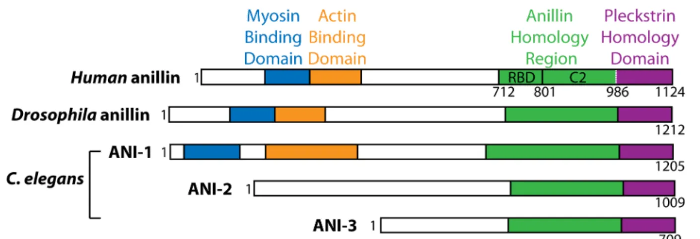
1.2.4. Anillin domain structure in animals ... 29
1.2.4.1. Drosophila ... 29
1.2.4.2. human ... 30
1.2.4.3. C. elegans ... 30
1.2.5. Functions of Anillin ... 31
1.2.5.1. Anillin as an organizer of contractility components to CR ... 32
1.2.5.2. Specification of the division plane by anillin ... 33
1.2.5.3. Anillin as an anchor of the CR and/or MBR to the membrane during late cytokinesis ... 34
1.2.5.4. Anillin as a potential regulator of abscission ... 35
1.3. Introduction to syncytial tissues ... 37
1.3.1. Historical background ... 37
1.3.2. The structural features of syncytia ... 41
1.3.2.1. Germline Syncytia ... 42
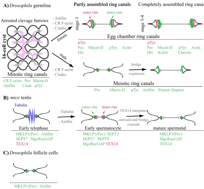
Drosophila germline ... 42
Mammalian germline ... 49
1.3.2.2. Overview of somatic syncytia ... 51
viii
1.3.3. Comparing the syncytial systems ... 54
1.3.3.1. Common features of syncytia ... 54
1.3.3.2. Dissimilarities among syncytia ... 54
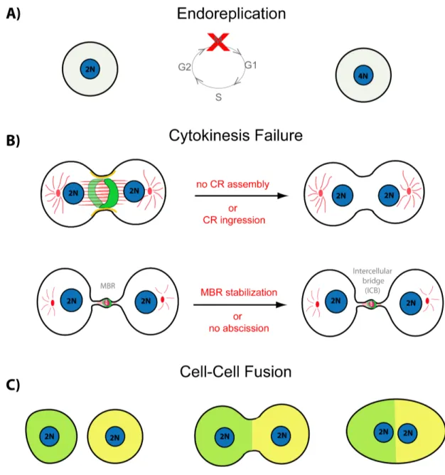
1.4. How are syncytia formed? ... 57
1.4.1. Syncytium formation through modifications in cytokinesis ... 59
1.4.1.1. Syncytium formation via failure of cytokinesis during CR assembly/formation or CR ingression ... 59
Post-natal rat liver cells ... 59
Cardiomyocytes ... 60
Megakaryocytes ... 61
Drosophila egg chambers ... 62
1.4.1.2. Syncytium formation via abscission failure ... 62
1.4.2. Syncytium formation through fusion ... 65
1.4.2.1. Myoblast fusion during muscle development in Drosophila ... 66
Finding a fusion partner: recognition, migration and adhesion ... 67
Enhancing cell membrane proximity, membrane fusion and formation of a syncytium67 1.5. The known function of syncytia ... 68
1.5.1. Synchronous behaviors ... 69
1.5.2. Cytoplasmic exchange ... 70
1.5.3. Haploid gene sharing ... 71
1.6. Benefits of syncytia in naturally diploid cells ... 72
1.6.1.Wound healing ... 73
1.6.2. Survival ... 73
1.7. C. elegans as a model to study syncytiogenesis ... 74
1.7.1. History and background ... 74
ix
1.7.2.1. Germline development in the embryo: P4 divides into Z2 and Z3 ... 75
1.7.2.2. Development of the C. elegans germline ... 78
L1 ... 78
L2 ... 78
L3 ... 79
L4 ... 79
Adult ... 79
1.7.3. C. elegans adult germline has a syncytial architecture ... 82
2. OBJECTIVES ... 84
2.1. Article 1 ... 84
2.2. Article 2 ... 85
3. RESULTS ... 86
3.1. Article 1 ... 87
C. elegans Anillin proteins regulate intercellular bridge stability and ... germline syncytial organization ... 88
3.2. Article 2 ... 135
Formation of the two C. elegans primordial germ cells occurs by incomplete ... cytokinesis ... 136
4. DISCUSSION ... 170
4.1. Syncytium biogenesis: Its all about maintaining good connections ... 172
4.2. When failure is success: syncytiogenesis of the C. elegans germline by incomplete cytokinesis ... 182
4.3. ANI-2 potential role in syncytiogenesis ... 187
4.4. GC rachis rings: to contract or not to contract ... 190
x
4.6. On the functional significance of syncytial tissues ... 195
xi
List of Figures and Tables
1. Introduction:Figure 1.1: Cytokinesis is a multistep process. ... 4
Figure 1.2: Illustrations of midbody (MB) and midbody ring (MBR) structural components. ... 13
Figure 1.3: Different steps of abscission. ... 19
Figure 1.4: Anillin structure in different organisms. ... 31
Figure 1.5: Drawings of mammalian germline bridges by nineteenth-century scientists. ... 39
Figure 1.6: Mammalian male germline of various species are syncytial. ... 40
Figure 1.7: Syncytium is a conserved phase of germline development. ... 43
Figure 1.8: Schematic overview of germline development during Drosophila oogenesis and spermatogenesis. ... 45
Figure 1.9: Follicle cells of the Drosophila ovary are syncytial. ... 53
Figure 1.10: Comparison of syncytial tissues in different organisms. ... 56
Figure 1.11: Schematic showing different mechanism of syncytium formation ... 58
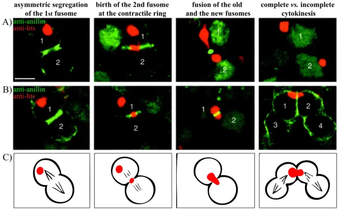
Figure 1.12: Dynamics of the fusome in the Drosophila ovary during mitosis. ... 64
Figure 1.13: Illustration of SGP and PGC morphogenetic behavior during gonadal primordium formation in the C. elegans embryo. ... 77
Figure 1.14: Illustration of germline development during C. elegans larval stages and adulthood. ... 81
Figure 1.15: 3D illustration of the adult hermaphrodite C. elegans gonad. ... 83
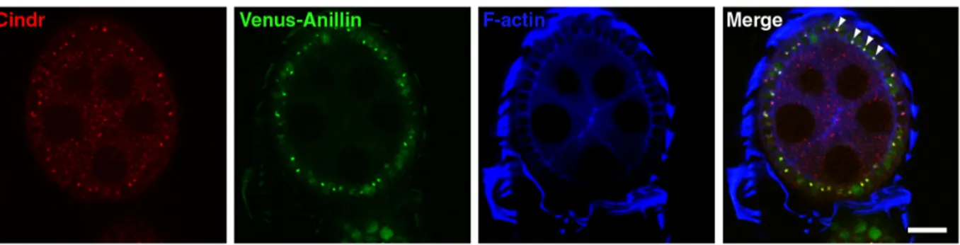
3.1. Article 1: Figure 3.1.1: ANI-2 stably accumulates at the midbody between the two primordial germ cells. ... 113
Figure 3.1.2: ANI-2 is found at the rachis bridge of all germ cells throughout larval development. ... 114
Figure 3.1.3: Germ cell rachis bridge formation arises progressively during larval development. ... 115
Figure 3.1.4: ANI-1 and NMY-2 localize to the rachis of wild-type hermaphrodites and report on rachis bridge organization. ... 117
Figure 3.1.5: Molecule diffusion in the cytoplasm of embryonic blastomeres. ... 119
Figure 3.1.6: ANI-2 is required for rachis bridge stabilization. ... 120
Figure 3.1.7: Loss of ANI-2 causes germ cell multinucleation and collapse of membrane partitions. 122 Figure 3.1.8: ANI-2 is required for rachis bridge stability. ... 124
Figure 3.1.9: Phenotypic analysis of ani-2 mutants. ... 126
xii
Figure 3.1.11: Cytoplasmic streaming in the rachis may be responsible for germline disorganization in
ani-2 mutants ... 130
Figure 3.1.12: ANI-2 permits elastic deformation of the adult hermaphrodite gonad and rachis bridges. ... 132
3.2. Article 2: Figure 3.2.1: Contractility regulators are assembled properly to the division plane of PGC division in the C. elegans embryo. ... 152
Figure 3.2.2: Assay to quantify CR diameter upon furrow initiation. ... 154
Figure 3.2.3: Cytoplasmic isolation occurs during PGC division in the C. elegans embryo. ... 156
Figure 3.2.4: MBR of PGCs is not released... 158
Figure 3.2.5: MBR trajectory in soma vs. PGCs. ... 160
Figure 3.2.6: MB/MBR is stabilized in PGCs. ... 162
Figure 3.2.7: Cytoskeletal proteins are enriched at GC rachis rings throughout larval development. . 164
Figure 3.2.8: Centralspindlin protein ZEN-4 is enriched at GC rachis rings throughout larval development. ... 166
Figure 3.2.9: A model for germline syncytium nucleation during PGC division in the C. elegans embryo. ... 168
4. Discussion: Figure 4.1.1: Organization of the C. elegans hermaphrodite gonad. ... 180
Figure 4.1.2: ANI-2 localization in control and ANI-1-depleted animals ... 181
Tables: Table 3.1.1: C. elegans strains used in this study ... 134
xiii
List of abbreviations
ACT-4: ACTin-4ACT-5: ACTin-5
AHD: Anillin homology domain AKAP4: A-kinase anchor proteins
ALG-2: Argonaute (plant)-Like Gene ALIX: ALG-2–interacting protein X (ALIX)
ANI-1: ANIllin-1 ANI-2: ANIllin-2 ANI-3: ANIllin-3
ANK: N-terminal ankyrin repeats AP: anteroposterior axis
APC: Anaphase promoting complex ARF6: ADP-ribosylation factor 6 C-terminal: Carboxy-terminal C. elegans: Caenorhabditis elegans
Cdc42: Cell division control protein 42 homolog CENP-E: Centromere-associated protein E CEP55: Centrosomal protein of 55 kDa
CHMP-4B: charged multivesicular body (MVB) protein 4B CHMP-4C: charged multivesicular body (MVB) protein 4C CPC: chromosomal passenger complex
CR: Contractile ring
CYK-4: CYtoKinesis defect-4 CYK-7: CYtoKinesis defect-7
DIC: differential interference contrast
DMYPT: Drosophila Myosin Phosphatase Targeting Protein DNA: Deoxyribonucleic acid
DTC: distal tip cell
xiv
ECT-2: epithelial cell transforming 2
ESCRT: endosomal sorting complex required for transport F-actin: filamentous actin
FCM: fusion-competent myoblasts fGSCs: female germline stem cells fli-1: Drosophila FLIghtless
GAP: GTP-ase activating protein GC: germ cell
GDP: Guanosine triphosphatase GEF: GDP-GTP Exchange
GEF-H1: GDP-GTP Exchange-H1 GFP: green fluorescent protein
GnRH: gonadotropin-releasing hormone GSCs: germline stem cells
GTP: Guanosine-5'-triphosphate (GTP GTPase: guanine triphosphatase
hGH: human growth hormone him-4: High Incidence of Males HP: human parthenogenetic
HSF2: Heat shock transcription factors HSFs: Heat shock transcription factors Hts: hu-li tai shao
ICB: intercellular bridge Ig: immunoglobulin
IGF: insulin-like growth factor 1 INCENP: inner centromeric protein kDa: kilo Dalton
KIF4: Kinesin-4 LET-21: LEThal-21 LET-502: LEThal-502 MB: midbody
xv
MBR: midbody ring
MDCK: Madin–Darby canine kidney cells MEL-11: Maternal Effect LethaL-11
MgcRacGAP: male germ cell Rac GTPase-activating protein Mid1p: mating pheromone-induced death protein 1
Mid2p: mating pheromone-induced death protein 2 MKLP-1: Mitotic Kinesin-Like Protein 1
MLC: myosin light chain
MRLC: myosin II regulatory light chain Mucin-D: Mucin- Drosophila
MYPT: myosin phosphatase-targeting subunit N-terminal: amino-terminal
NLS: nuclear localization sequences NMY-II: Non-muscle myosin II
PAR-4: abnormal embryonic PARtitioning of cytoplasm Pav-Klp: pavarotti, a Drosophila kinesin-like protein PGC: primordial germ cell
PH: pleckstrin-homology
PH domain: Pleckstrin homology domain PIP: Phosphatidylinositol phosphate
PIP2: Phosphatidylinositol 4,5-bisphosphate PLC: phospholipase C
PRC1: protein regulator of cytokinesis 1
pTyr proteins: phosphotyrosine-containing protein(s) Rac1: ras-related C3 botulinum toxin substrate 1 RacGAP1: Rac GTPase-activating protein RBD: RhoA-binding domain
RC: ring canal
RFP: red fluorescence protein Rho-1: Rho family GTPase Rho-1
xvi
RNAi: RNA interference ROCK: Rho-dependent kinase
S. pombe: Schizosaccharomyces pombe S2: Schneider 2 cells
SDS-PAGE: sodium dodecyl sulphate polyacrylamide gel electrophoresis SEPT: septin
SGP: somatic gonad precursor SH3: Src homology
SIM: structured illumination microscopy SPh: somatic gonad primordium
TEX14: testis-expressed gene 14 TSG101: tumor susceptibility gene 101 TZ: transition zone
UNC-45: UNCoordinated-45 UNC-59: UNCoordinated-59
ZEN-4: Zygotic epidermal ENclosure defective-4 α-spectrin: alpha-spectrin
δ Tubulin: gamma-tubulin
WASp: Wiskott-Aldrich syndrome protein SCAR/WAVE:
xvii
xviii
Acknowledgement
First and foremost, I would like to thank my advisor, Jean-Claude Labbé. Jean-Claude has been and, I sincerely hope, will continue to be a great mentor. I am especially grateful to Jean-Claude for teaching me the essence of critical thinking, scientific integrity and for all the insightful discussions that improved my scientific outlook. I have been amazingly fortunate to have an advisor who gave me the freedom to explore on my own, while being a source of constructive input, encouragement, unconditional support and understanding throughout my PhD.
Moreover, I owe a profound thank you to Nicolas Chartier, a great friend and a former colleague, who in addition to patiently walking me through his project before leaving the Labbé laboratory provided indispensible source of scientific motivation and inspiration with his passion during the course of my PhD. Merci Kiki!
Thanks to all the Labbé lab members, past and present, particularly Abby Gerhold, for reading the two manuscripts and providing very useful suggestions and perceptive inputs and more importantly for kindly and patiently sharing her time and knowledge with me. I would also like to thank Alexia Rabilotta-Faure for her advice, support, problem-solving skills and friendship over these years. Thanks to Patrick Narbonne for helping me understand the C. elegans genetics and for his advice. I would also like to thank members of Amy and Paul Maddox laboratories, in particular Benjamin Lacroix and Joel Ryan, for being generous with their time and patient with their explanations that have certainly enriched my ideas. My special thanks goes to my former colleagues and office mates: Anne-Marie Laduceure, Rajesh Ranjan Vincent Hyenne, Ramraj Velmurugan, Ken Lam and Laura Benkemoun, I couldn’t ask for better colleagues and friends. The majority of my thesis relied on having access to the imaging facility in IRIC, so I would like to thank Christian Charbonneau for being very patient and helpful throughout the years.
xix
Dr. Jean-François Côté and Dr. Alisa Piekny for their helpful advice on my research project.
Thanks to my amazing mom and my sister Mitra for their unconditional love and support throughout my life. Thanks to my best friends Gloria Assaker and Sanaz Rassouli for always being there for me. And thanks to the rest of my family and friends near and far. You all made this time all the more pleasant.
Finally, I would like to dedicate this thesis to my lovely grandparents, Baya Azarpour and Papa Anousheh, the greatest mentors and friends, source of inspiration who passed away during the course of my PhD.
Preface
Cytokinesis failure has been proposed to be the most frequently used mode of syncytiogenesis across the animal kingdom. Surprisingly, while accidental failures of cytokinesis in normal tissues cause pathological disorders such as cancer, regulated cytokinesis failure is a physiological element of syncytial tissues. Hence, to be most effective in understanding cytokinesis failure and its influence on syncytium formation, we must first obtain an exhaustive description of how cytokinesis normally progresses. Accordingly, in the following pages, I first provide a summary of what is currently known about cytokinesis and one of its key regulators; anillin. Then, I will describe different types of syncytia and their structure, followed by two chapters on mechanisms of syncytium formation and its function. Lastly, I will introduce the system we used to study syncytiogenesis; the C. elegans germline. Importantly, the story of syncytiogenesis and its exact roles and benefits is not complete; there are many unknowns and confusing results, but if we could only figure out how syncytia pull it off, maybe we could gain some insights on potential underlying mechanism of cytokinesis failure and their associated disorders.
2
1. Introduction
1.1. Cytokinesis, the final stage of cell division, is a
multi-step event
Cytokinesis is the final and irreversible stage of animal cell division during which one cell is physically separated into two distinct cells (Glotzer, 1997a; Glotzer, 1997b). To ensure the faithful propagation of the genome, cytokinesis must be spatiotemporally coordinated with chromosome segregation. This coordination is partly achieved by a tight regulation of a well-orchestrated chain of events involving the establishment of the division plane, furrow ingression through contraction of an actomyosin ring, formation of the midbody (MB) and midbody ring (MBR) and cell separation in a process called abscission (Figure 1.1).
The proper execution of each stage depends on its prior stage and thus defects at any step of this cascade may result in cytokinesis failure, the consequence of which could result in problems such as aneuploidy, centrosome amplification and genetic instability, which are characteristics of many cancers. Here, I will overview the different stages of cytokinesis in animal cells, as we currently understand them.
1.1.1. Stage I: Positioning of the Division Plane
To ensure that each of the two daughter cells receives a complete and single copy of the genetic and cytoplasmic materials upon completion of cytokinesis, the cleavage furrow must form in the accurate place in a dividing cell (Figure 1.1 B). This accuracy is gained by specification of the site of cleavage furrow, through assembling a structure known as the central spindle during anaphase (Reviewed in Eggert et al., 2006; Glotzer, 1997a; Glotzer, 1997b; Glotzer, 2001; Glotzer, 2005; Green et al., 2012).
4 Figure 1.1: Cytokinesis is a multistep process.
Schematic diagram of the progression through cytokinesis. (A) metaphase (B-F) Different stages of cell division. Black arrows in (B) point to cues from the central spindle that activate RhoA and astral microtubules that reinforce the localization of active RhoA the upstream regulator of the contractile ring (CR).
5
The central spindle is composed of anti-parallel microtubules, the microtubule binding and bundling protein PRC1 and a kinesin KIF4 (kinesin-4) (Glotzer, 2009a; Glotzer, 2009b; Green et al., 2012), two protein complexes; centralspindlin (composed of two molecules of MKLP1 (known as ZEN-4 in C. elegans) the motor component of the centralspindlin complex and two molecules of MgcRacGAP (known as CYK-4 in C. elegans)) (Mishima et al., 2002) and the chromosomal passenger complex (CPC) composed of the kinase Aurora B, Incenp, Borealin and Survivin (Carmena et al., 2012; Glotzer, 2009b; Ruchaud et al., 2007). Central spindle assembly and thereby specification of the division plane site are subject to both temporal regulation (coupling to cell cycle) and spatial regulation (coupling to spindle position) (Reviewed in Eggert et al., 2006; Glotzer, 1997a; Glotzer, 1997b; Glotzer, 2001; Glotzer, 2005; Green et al., 2012). Here I will only describe current understanding on the spatial regulation aspect of the first stage of cytokinesis to understand how the cleavage furrow is localized to a single, defined place at the cell cortex.
1.1.1.1. Mitotic spindle microtubules
In 1985, Ray Rappaport, a pioneer in the field of cytokinesis, performed his classic physical micromanipulation experiments that were aimed to tackle how cleavage furrow is placed in an appropriate place during division. He observed that the cleavage furrow site is determined by the position of the mitotic spindle (the microtubule-based structure that forms during mitosis) in late metaphase or early anaphase of the cell cycle and concluded that the positioning of the cleavage furrow involves continuous communication between the mitotic spindle and the cell cortex (Pollard, 2004; Rappaport, 1985).
The precise mechanism by which microtubules position the division plane between the segregated chromosomes is still elusive, however different models have been proposed to explain this. One well-established model is that the mitotic spindle contributes to establishment of cleavage furrow positioning. But which part of the mitotic spindle functions in this process? The answer to this question is a subject of perhaps one of the oldest debates in
6
the field. While some experiments are in favor of “the astral stimulation hypothesis” which postulates that the non-spindle astral microtubules that radiate from centrosomes to the cell periphery, are essential for determining the cleavage furrow site (Rappaport, 1961; Rappaport and Rappaport, 1985), others contradicted this view by proposing the second hypothesis, “the central spindle hypothesis”, arguing that midzone microtubules are key regulators of this process (Bonaccorsi et al., 1998; Cao and Wang, 1996).
While abundant evidence from many species supports the latter hypothesis, a third hypothesis, “the astral relaxation hypothesis” postulates that astral microtubules generate a negative signal that enhances cortical relaxation in their immediate vicinity close to the pole (White, 1985; Wolpert, 1960). In addition, Antony Hyman’s group using laser micro-dissection, spatially separated aster and midzone microtubules and showed that the furrow is first positioned by an astral signal and subsequently by a second midzone-derived signal, suggesting that both arrays send signals to the cortex (Bringmann and Hyman, 2005). Perhaps the simplest explanation for these contradictory results is that different factors and mechanisms or a combination of different mechanisms are likely to determine the cleavage furrow site depending on the cell types, sizes and organism. Alternatively, the critical determinant for the specification of cleavage furrow positioning may not be evolutionarily conserved. Nevertheless, in all cases, regardless of the exact source, it is evident that the positional cue for the site of division plane comes from the microtubule-rich mitotic spindle, either through a traditionally believed model of “direct microtubule/cortical contact” or through the novel “diffusion-based” mechanism of transport along microtubules (Canman, 2009; von Dassow et al., 2009).
1.1.1.2. Rho GTPase signaling
Clues to the molecular basis of cleavage furrow positioning may be found in proteins that accumulate at the equator in a microtubule-dependent manner and their function to deliver signal(s) from microtubules to specify the site of the presumptive furrow. One such molecule
7
is the small GTPase RhoA that has been shown to contribute to specifying the plane of cell division in animal cells (Bement et al., 2005; Nishimura and Yonemura, 2006; Piekny et al., 2005; Yonemura et al., 2004; Yuce et al., 2005). Members of the Rho family guanosine triphosphatases or GTPases (including RhoA, Rac1, and Cdc42) (Piekny et al., 2005) spatiotemporally regulate various distinct cellular processes, including cytokinesis. Rho GTPases are activated by RhoGEF (guanine nucleotide exchange factors or GEFs) that catalyze the exchange of GDP for GTP (Rossman et al., 2005) and are inactivated by GTPase activating proteins (GAPs) which promote GTP hydrolysis (Tcherkezian and Lamarche-Vane, 2007). It has been shown that both GEFs (e.g. ECT-2/Pebble) (Yuce et al., 2005) and GAPs (e.g. MgcRacGAP) respectively control RhoA activation and inactivation, and as such help cells progress through cytokinesis (Bement et al., 2005; Nishimura and Yonemura, 2006; Yonemura et al., 2004; Yuce et al., 2005). For example, disruption of the RhoGEF ECT2 leads to a failure in cytokinesis (Tatsumoto et al., 1999).
Previous work proposed that the proper localization and activation of the RhoGEF ECT2 (Pebble in Drosophila melanogaster, and LET-21 or ECT-2 in nematodes) to the presumptive site of division depends on the centralspindlin complex (Nishimura and Yonemura, 2006; Somers and Saint, 2003). The activated RhoGEF ECT2 then triggers the localized activation and accumulation of RhoA at the site of cell division (Kimura et al., 2000; Prokopenko et al., 1999; Tatsumoto et al., 1999). Contrary to this, others suggested that ECT-2 only functions to restrict the already-active RhoA to a tightly focused region at the equatorial cortex, while another GEF, GEF-H1, is required for RhoA activation (Birkenfeld et al., 2007). Besides ECT-2, Anillin has been also shown to stabilize restricted RhoA localization at the furrow, suggesting that there is feedback between downstream components of cytokinesis and RhoA (Piekny and Glotzer, 2008; Zhao and Fang, 2005).
While RhoA is activated at the equatorial cortex, its activation is inhibited by astral microtubules at the polar cortex (Werner et al., 2007). Active RhoA at the equatorial cortex in turn promotes assembly and contraction of the actomyosin ring and thereby helps to initiate
8
cytokinetic ring ingression (Bement et al., 2006; Piekny et al., 2005). However, there is also compelling evidence that in Rat1A cell lines of embryonic rat fibroblasts, RhoA is dispensable for specification of the cleavage furrow positioning and that RhoA inactivation does not abrogate successful cytokinesis (Yoshizaki et al., 2004). In addition, it was found that RhoA requirement during cytokinesis might depend on the degree of cell adhesion in mammalian NIH 3T3 isolates (O'Connell et al., 1999). Altogether, these data suggest that cytokinesis may proceed by a RhoA-independent mechanism in some cell types.
1.1.2. Stage II: contractile ring assembly
In animal cells, attachment of the contractile ring (CR) to the plasma membrane creates a cleavage furrow that ultimately partitions the dividing cell into two (Figure 1.1 C). Accumulation of active RhoA at the equatorial cortex (Bement et al., 2005) promotes the recruitment of actin and myosin and thus assembly of the actomyosin-based contractile ring (CR) beneath the plasma membrane via two regulatory pathways. First, active RhoA stimulates polymerization of unbranched F-actin filaments through recruiting and activating an actin nucleator, formin (Diaphonous in Drosophila, CYK-1 in C. elegans) (Castrillon and Wasserman, 1994; Piekny et al., 2005; Watanabe et al., 1999). On the other hand, RhoA also indirectly promotes non-muscle myosin II (NMY-II) activity by activating kinases such as Citron kinase (Shandala et al., 2004) and Rho kinase (ROCK), which act on myosin either by phosphorylation of the myosin light chain (MLC) or by inhibiting the myosin phosphatase-targeting subunit (MYPT) (Amano et al., 1996; Matsumura, 2005). In addition to this pathway, several other mechanisms have been also shown to function in actin and myosin assembly at the cortex (Yumura et al., 2008; Zhou and Wang, 2008). For instance during pseudocleavage in the C. elegans embryo, signaling by astral microtubules, through an unknown mechanism functions in asymmetric CR assembly by locally inhibiting myosin recruitment to the posterior pole, a region with high microtubule densities (Werner et al., 2007).
9
The CR is composed of a parallel array of actin filaments (Maupin and Pollard, 1986) and myosin, two essential CR components. Besides these two components, the CR also contains other proteins assembled in an ordered fashion, including septin filaments (Eggert et al., 2006; Estey et al., 2010; Joo et al., 2007; Maddox et al., 2007), the scaffold protein anillin (Field and Alberts, 1995; Piekny and Maddox, 2010) and actin crosslinking proteins (Reichl et al., 2008). Among these, anillin plays an important scaffolding role by binding to membrane, actin, myosin, RhoA and CYK-4/MgcRacGAP and thus links the equatorial cortex to the signals coming from the mitotic spindle and to the CR (D'Avino, 2009; Hickson and O'Farrell, 2008a; Piekny and Maddox, 2010; Sun et al., 2015). I will further expand on anillin in Section 1.2.
1.1.3. Stage III: contractile ring ingression
Once the CR is assembled, it initiates constriction (Figure 1.1 D), which progressively draws the plasma membrane inward until it closes the gap between the two dividing cells, forming two separated cells. In addition to cytokinesis, CR ingression has been shown to function in several other processes, including wound closure (Mandato and Bement, 2001), epithelial morphogenetic movements (Bloor and Kiehart, 2002) and epithelial cell delamination (Rosenblatt et al., 2001), suggesting that the CR has been adapted to serve different functions. Despite the importance of CR ingression during cytokinesis, the exact mechanisms generating tension to draw the constriction of the CR remain elusive. The limitation to the current knowledge is in part due to the lack of high-resolution images that depict the ingression and rare measurements of tension in vivo. Nevertheless, several different models have been proposed to explain actomyosin contractility, on the basis of ultrastructural studies and biophysical considerations.
A classic model for CR constriction is the “sliding filament” model (Schroeder, 1972), which is based on the mechanism of muscle contraction and assumes that the interactions of bipolar myosin motor filaments with actin filaments generate the contractile force required to invaginate the plasma membrane furrow between the segregating cells during cytokinesis
10
(Mierzwa and Gerlich, 2014; Salmon, 1989). Consistent with this concept, several ultrastructural studies reported presence of a layer of actin filaments arranged circumferentially beneath the plasma membrane (Arnold, 1969; Maupin and Pollard, 1986; Schroeder, 1972; Selman and Perry, 1970), resembling the organization of actin in striated muscle cells. In parallel to this, several other studies reported presence of filamentous myosin at the CR (Vale et al., 2009; Yumura et al., 2008; Zhou and Wang, 2008). In addition, evidence supporting a force-generating role for myosin in this model came from studies showing that mutations that disturb myosin filament assembly result in CRs that are unable to contract (Egelhoff et al., 1993). Despite these, over the years, several other ultrastructural studies revealed principal discrepancies between CR structure in dividing cells and muscle sarcomeres. For instance, while myosin was proposed to drive the major force-generating element during CR constriction, experimental evidence showed that the presence or proper function of myosin is not needed for cytokinesis in some cells and/or under specific conditions (Lord et al., 2005; Ma et al., 2012; Mendes Pinto et al., 2013; Neujahr et al., 1997; Zang et al., 1997). Another major difference is that the CR in animal cells gets disassembled and releases material during constriction and, as such, its volume decreases over time. Despite this, the function of the CR is not affected (Schroeder, 1972). Altogether, these dissimilarities suggest the existence of alternative force-generating mechanisms that would not require muscle-like filament organizations.
Several other mechanisms have been proposed to explain how force is generated during CR constriction. For many years, the leading theory was the “purse-string” model, wherein the alignment of actin filaments with the division plane is an essential element. In this model, the bipolar myosin filaments walk along antiparallel actin filaments using their motor activity, drawing the F-actin strands together in a purse-string-like fashion (Satterwhite and Pollard, 1992). Because the plasma membrane is anchored to the F-actin filaments, constriction of the actomyosin ring pulls the membrane inside. However, several experimental evidences have challenged this model, mainly based on lack of a highly organized structure of concentric actin filaments in mammalian NRK cells, Swiss 3T3 cells and Dictyostelium cells (Fishkind and
11
Wang, 1993; Reichl et al., 2008). Another model proposes that randomly distributed myosin filaments within homogenous bundles of actin, generate the force needed for CR constriction (Carlsson, 2006). Altogether, these findings highlight the importance of new efforts to investigate force-generating mechanism underlying CR ingression during cytokinesis, perhaps by examining the exact arrangement of the filaments in CR and their function.
1.1.4. Stage IV: The biogenesis and architecture of MB and MBR
formation
1.1.4.1. Maturation of the central spindle to form the MB
Constriction of the CR squeezes the cytoplasm at the center of the central spindle, generating an intercellular bridge (ICB) that transiently interconnects the two daughter cells (Glotzer, 2005; Green et al., 2012). The CR constricts until it reaches the central spindle and compacts its antiparallel microtubule into a single large bundle, forming a structure called midbody (MB) (Figure 1.1 E). The MB was first described in 1891 by Walther Flemming using light microscopy and histochemical methods, as a specialized structure conjoining the divided daughter cells in the lung epithelium of the salamander larva (Chen et al., 2013; Paweletz, 2001). He speculated that these structures are derived from spindle midzone between the segregating chromosomes and described them as densely stained bodies of 1-1.5 µm in size and named them “Zwischenkörper” (“Zwischen” and “körper” mean “between” and “body”, respectively). Despite this, the MB has been only sporadically studied since its discovery and, as such, remained a mysterious structure for more than 100 years. In fact, the MB was so much ignored that some scientists considered it a “remnant”, “scar”, and even “cell garbage can” (Schicktanz and Schweda, 2009). But this was changed by advanced microscopy techniques and methods to dissect protein functions.
Structural composition of the MB
The MB is derived from the central spindle and, as such, its most prominent structural component is a densely packed array of anti-parallel microtubules (Figure 1.2 A). Along the
12
microtubules, the MB contains microtubule interacting proteins that co-localize to the central spindle (e.g., the centralspindlin complex) (Figure 1.2 A). For instance, MKLP-1, is one of the earliest centralspindlin components that was found to be associated with the MB (Sellitto and Kuriyama, 1988). Functional proteomic and comparative genomic approaches showed that MBs contain many different components, which are not only cytoskeleton-related proteins but also proteins involved in other pathways, such as secretory, or, membrane-associated, lipid rafts and vesicle trafficking (Skop et al., 2004). However, this is certainly an underestimation of the structural complexity of the MB, given the fact that its structural composition changes during cytokinesis progression.
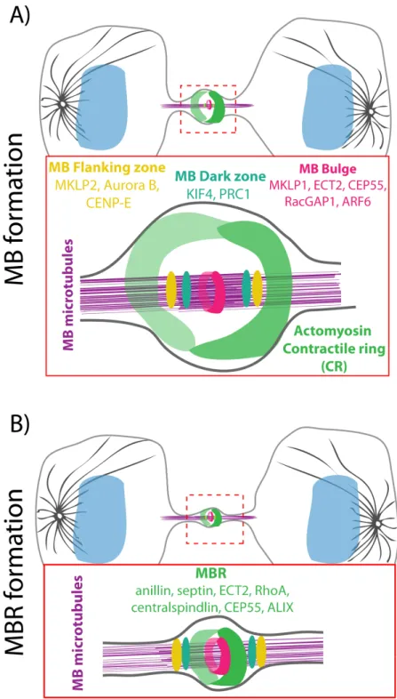
Central spindle proteins relocate to different regions of the MB (Figure 1.2 A): 1) Bulge, a region that is surrounded by the plasma membrane and develops at the center of the MB during central spindle maturation and microtubule compaction. Proteins including ECT-2 and MKLP-1 the motor component of the centralspindlin complex, centrosomal protein of 55 kDa (CEP55), ARF6 and RacGAP1 are released from the MB microtubules and re-localized to the bulge, which has a ring-like structure and wraps tightly around the MB (Elia et al., 2011; Hu et al., 2012). 2) Dark zone, a narrow region on the microtubule bundles at the center of the MB. PRC1 and KIF4 remain associated with the microtubules in this zone (Elia et al., 2011; Hu et al., 2012). 3) Flanking regions, two bands on microtubules at the periphery of the dark zone. CENPE, MKLP2, and Aurora B localize with microtubules at these regions (Hu et al., 2012). This re-localization pattern suggests that different regions probably serve distinct functions in the MB formation as well as abscission. In addition to this concept, the Mitchison group proposed that the MB core, wherein the antiparallel microtubules overlap, provides architectural integrity to the midzone while the flanking regions function to position abscission sites (Hu et al., 2012). Before proceeding, it is important to mention that despite attempts to map the central spindle proteins to different subregions of the MB, researchers are still confounded by the exact molecular composition and biophysical nature of the MB.
13
Figure 1.2: Illustrations of midbody (MB) and midbody ring (MBR) structural components.
14
Another interesting question is how these components are targeted to the MBs and how ultimately the MBs are formed. In almost all systems, central spindle is essential for the MB formation (Matuliene and Kuriyama, 2002) and several of the proteins that localize to the central spindle play key roles in the MB formation (Matuliene and Kuriyama, 2002; Matuliene and Kuriyama, 2004). In fact, it has been shown that the centralspindlin complex besides being important for stage I of cytokinesis (Positioning of the division plane), is also necessary for the MB formation, and ultimately for completion of cytokinesis (Matuliene and Kuriyama, 2002; Matuliene and Kuriyama, 2004). Thus, it is not surprising to assume that the central spindle directly orchestrates re-localization of its components to form the MB.
Moreover, conversion of the central spindle to the MB is positively correlated with ingression of the CR, as either blocking CR ingression (Straight et al., 2003) or actin depolymerization (Hu et al., 2012), perturbs the MB formation. The CR ingression has been shown to contribute to the MB formation, perhaps through directing the re-localization of the MB components to different zones of the MB. It has been reported that without CR ingression, proteins that are normally partitioned into three distinct subregions of the MB; PRC1 and KIF4, Aurora B and MKLP-1, remain co-localized at the center of microtubule bundles, suggesting that furrow ingression is required for relocation of central spindle components in order to form the MB (Hu et al., 2012).
Function of the MB
Originally, the MB, together with its associated membranes and its compact microtubules had been proposed to serve as a diffusion barrier to limit cytoplasmic exchange between the dividing daughter cells, a process that is also known as cytoplasmic solation (Green et al., 2013). Indeed, monitoring the diffusion of fluorescent probes between the two dividing daughter cells upon photoactivation confirmed that the dividing cells undergo cytoplasmic isolation in different systems (Guizetti et al., 2011; Sanger et al., 1985; Steigemann et al., 2009). Currently, however, there is no consensus in the field regarding the timing of cytoplasmic isolation. While previous work in HeLa cells showed that cytoplasmic isolation occurs ∼60 min after the completion of CR constriction and coincides with
15
endosomal sorting complex required for transport (ESCRT)–mediated scission (more details in Section 1.1.5.) (Guizetti et al., 2011; Steignemann et al., 2009), recent work in the C. elegans 1-cell stage embryo, revealed that the cytoplasmic isolation occurs upon completion of furrowing in an ESCRT-independent manner (Green et al., 2013).
Despite these unequivocal evidences and despite the compact appearance of MB microtubules, some other work suggest that this barrier might function selectively, as some proteins can still diffuse through the MB and transverse from one dividing daughter cell to the other, while others are not (Chen et al., 2013; Guizetti et al., 2011; Sanger et al., 1985; Schmidt and Nichols, 2004; Steigemann et al., 2009). Perhaps, the simplest way to explain this selective barrier would be that there is a space between the MB and the plasma membrane at the interface of the dividing cells that only allows proteins with a certain size to pass. Another possibility is that the microtubule-based MB is not capable of closing the bridge between the dividing cells. In fact, whether MB microtubules are required for cytoplasmic isolation during cytokinesis is a controversial subject. Recently, Karen Oegema group showed that cytoplasmic isolation occurs in the absence of MB microtubules in the C. elegans 1-cell stage embryo, suggesting the microtubules are not essential to block cytoplasmic diffusion between the dividing daughter cells (Green et al., 2013). In consistent with this, it has been shown that the germline stem cells (GSCs) of the Drosophila testis accomplish cytoplasmic isolation hours after microtubule disassembly, suggesting that a diffusion barrier is formed in the absence of MB microtubules (Lenhart and DiNardo, 2015). Undoubtedly, further studies are required to more thoroughly parse the MB structure and its role as a barrier, particularly in cells within different tissues and at carefully defined times during cytokinesis, to determine the function of the MB as a barrier and timing of cytoplasmic isolation.
Another widely accepted role of the MB and its microtubule bundles is its role as a platform to bring together a large number of abscission-related components (Schiel and Prekeris, 2013). Given that MBs are composed of various proteins with several distinct roles, it is not surprising that they would function as such a binding platform for abscission regulators. Recent progress on the molecular mechanism of abscission showed that the MB and its
16
microtubules serve as platform to coordinate the cytoskeleton and plasma membrane rearrangements and recruit functional complexes including microtubule severing enzyme spastin and the endosomal sorting complex required for transport (ESCRT) needed for abscission (more details in Section 1.1.5.). Despite this, work in the C. elegans embryo has provided convincing evidence that depletion of the MB microtubules does not affect abscission. Similar to this, it has been reported that the MB microtubules in HeLa cells are not directly required for abscission (Guizetti et al., 2011), suggesting that at least in some cell types, the MB microtubules are not critical for abscission. Moreover, domain analysis of three MB components in HeLa cells, namely MKLP1, KIF4 and PRC1 showed that they can still localize to the MB even when they lack their microtubule-interacting regions, suggesting that microtubules are not even essential for the MB assembly (Hu et al., 2012).
Besides functioning as a diffusion barrier and/or as a platform several other distinct non-cytokinetic roles have been attributed to the MB including signaling events (Skop et al., 2004), communication with centrosomes (Piel et al., 2001), polarity specification (Pollarolo et al., 2011), dorso-ventral axis formation in the C. elegans embryo (singh and Pohl, 2014), cell fate determination (Ettinger et al., 2011; Kuo et al., 2011) and cell fate specification (Dubreuil et al., 2007).
1.1.4.2. Maturation of the CR to form the MBR
As the constriction nears completion, the CR becomes progressively tighter until it reaches a diameter of ∼1 µm (Mullins and Biesele, 1977) and subsequently transforms itself into the midbody ring (MBR), a dense structure that forms around the center of the MB (Figure 1.2 B). The MBR contains no or very few microtubules while several CR components, including myosin (Green et al., 2013), anillin (El Amine et al., 2013; Fields and Alberts, 1995; Hickson and O’Farrell, 2008a; Hu et al., 2012; Kechad et al., 2012; Straight et al., 2005) septin (Green et al., 2013), Citron kinase (Hu et al., 2012; Madaule et al., 1998), and RhoA (Hu et al., 2012) localize to it. Work using Drosophila S2 cells showed that the CR-to-MBR transition, requires the scaffolding protein, anillin (Kechad et al., 2012). Interestingly, a recent paper revealed that this anillin-dependent CR-to-MBR transformation occurs via opposing
17
mechanisms of membrane removal from the nascent MBR and anillin maintenance at the mature MBR (El Amine et al., 2013). On one hand, septin acts on the C-terminus of anillin to locally remove membrane from the nascent MBR through internalization, shedding and extrusion. On the other hand, Citron kinase acts on N-terminus of anillin to maintain anillin at the mature MBR through acting on, suggesting that the removal of membrane is coordinated with the CR disassembly, a process that is coupled to the formation of the MBR (El Amine et al., 2013).
MBR function
Most of the actin filaments that are found in the CR are disassembled following its complete constriction (Guizetti et al., 2011). While this is essential to allow for abscission, it could weaken the stability of the furrow and thereby its link to the membrane, leading the furrow to retract. To prevent this, the MBR has been proposed to mechanically stabilize the furrow and tether it to the plasma membrane at the division plane. This model has been confirmed by recent studies from Gilles Hickson’s group (El Amine et al., 2013). They demonstrated that to prevent furrow regression during abscission, the MBR acts as an anchor to the plasma membrane. This anchor then persists until the complete separation of the two dividing cells. Cortical anchoring of the MBR depends on anillin, which localizes to the MBR. Anillin being a scaffolding protein interacts with both membrane (Sun et al., 2015) and the plasma membrane–associated septins (D’Avino et al., 2008; Oegema et al., 2000) and thereby tethers the MBR and its adjacent regions to the cell cortex.
Alternatively, the MBR has been proposed to serve as the platform that brings together the abscission-related components to specify the place and timing of abscission (Green et al., 2012; Steigemann and Gerlich, 2009). As described in Section 1.1.4.1 Functions of MB, a recent study from Karen Oegema’s group provided evidence supporting this model by demonstrating that abscission-related events including membrane shedding, ESCRT machinery recruitment and MB/MBR release all occur normally in the absence of MB microtubules (Green et al., 2013). Another evidence supporting the microtubule-independent abscission recruitment model comes from a recent study from DiNardo’s group wherein they
18
showed that the ESCRT-III components are delivered to the abscission site in GSCs of the Drosophila testis in the absence of microtubules (Lenhart and DiNardo, 2015), suggesting that in the absence of MB microtubules, the MBR is sufficient to orchestrate abscission and abscission-related events.
1.1.5. Stage V: Abscission a multistep event
CR constriction dynamically narrows the cell to the point where abscission can take place (Figure 1.1 F). Abscission represents the final stage of cytokinesis and is referred to the severing of membrane and the MB connecting the two dividing daughter cells at the end of cytokinesis. The small size of the MB (diameter of ~1 µm), its transient nature and the unsynchronized timing of abscission are only a few of the obstacles in the field of abscission research. Thus, it is not surprising that abscission is the least well-understood stage of cytokinesis. Nevertheless, advent of sophisticated super-resolution live and ultrastructural imaging techniques in combination with high-throughput genomics screening and proteomics analysis of the late MB/MBR enabled scientists to study and monitor the structural and molecular dynamics of different steps of abscission (Figure 1.3).
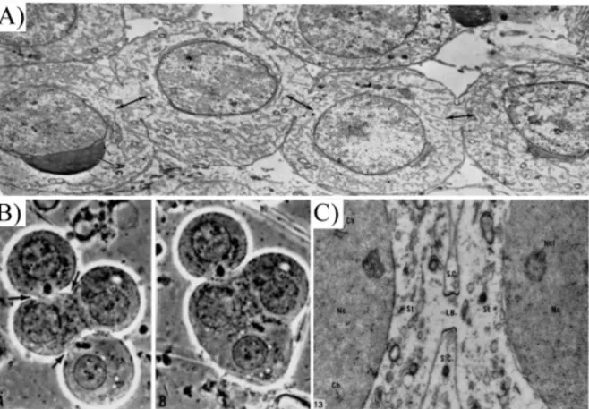
19 Figure 1.3: Different steps of abscission.
(A-C) Transmission electron micrographs of ICBs of HeLa cells at different stages of abscission. (A) Early-stage ICB appears short with bundles of straight microtubules. (B) Mid-stage, elongated ICB. Microtubule bundles appear compressed at either end, where the ICB has a reduced diameter. (C) Late-stage ICB with rippled, electron-dense cortex at a constriction zone. Microtubules at the constriction zone appear curved and highly compressed. (D) Schematics of abscission. Complete ingression of the cleavage furrow is followed by disassembly of cortical F-actin. Fusion of vesicles correlates with gradual narrowing of the ICB on both sides of the MB. Abscission proceeds by assembly and constriction of 17 nm filaments adjacent to the MB and simultaneous disassembly of the microtubules lateral to the MB. (Mierzwa and Gerlich, 2014).
20
To ensure that abscission undergoes properly, several events must occur in a temporally and spatially concerted manner. The first step is to prime the cell for assembly of the abscission machinery. Next, microtubule bundles, the MB/MBR and other cellular materials are removed during abscission. During this step, the plasma membrane and MB/MBR are budding away from the cytoplasm of the dividing cells, a process that results in the membrane and MB/MBR release also known as shedding. The final stage is sealing of the plasma membrane between the dividing daughter cells that ultimately results in their physical separation. Over the past few years, based on the topology of the membrane at the boundary of the two dividing daughter cells, different models have been proposed to explain the mechanisms of membrane deformation and severing including the mechanical rupture model (Burton and Taylor, 1997; Steigemann and Gerlich, 2009), the Golgi and endocytosis vesicle-mediated model (Gromley et al., 2005) and the ESCRT-mediated membrane fission model (Carlton and Martin-Serrano, 2007; Elia et al., 2011; Guizetti et al., 2011; Steigemann and Gerlich, 2009). Below I will give a concise overview of the sequence of main events occurring during abscission with a focus on the ESCRT-mediated plasma membrane fission model (Figure 1.3). Before proceeding however, for the sake of clarity, I will first give a brief overview of the ESCRT machinery and its function.
1.1.5.1. Background on ESCRTs
Although the endosomal sorting complex required for transport (ESCRT) complex has been only discovered in 2001, it is evolutionarily conserved from Archaea to animals (Wollert et al., 2009b). ESCRTs were named initially for their role in sorting membrane proteins from
endosomes to lysosomes (Katzmann et al., 2001) but ESCRTs are also known for their role in
membrane remodeling, constriction, scission and fission events during different cellular processes. One example is the ESCRT-dependent biogenesis of viral buddings and membrane severing that function in the release of viruses including HIV-1 from the plasma membrane of the infected cells (von Schwedler et al., 2003). Interestingly, extensive research during the past few years has led to discovering novel functions for ESCRT machinery from plasma membrane wound repair (Jimenez et al., 2014) to membrane scission of axons and dendrites
21
during neuron pruning (Issman-Zecharya and Schuldiner, 2014; Loncle et al., 2015; Zhang et al., 2014a). It worth mentioning that most, if not all, of both classic and novel ESCRT-dependent functions involve membrane severing. Based on the similar topology of the budding virus and the membrane at the ICB prior to abscission, it has been proposed that the membrane and MB/MBR breakage might be analogous to virus release from the infected cell and thus abscission may require similar ESCRT-dependent fission strategies.
The ESCRT machinery is comprised of five protein components, including ESCRT-0, ESCRT-I, ESCRT-II, ESCRT-III, and vacuolar protein sorting 4 (Vps4) and several other ESCRT-associated proteins such as the apoptosis-linked gene 2-interacting protein X (ALIX) (Wollert et al., 2009b). It has been shown that depletion of any of these ESCRT or ESCRT-related components result in cytokinesis failure (Carlton et al., 2008; Carlton and Martin-Serrano, 2007). Altogether, these features make ESCRTs attractive candidates to mediate abscission.
1.1.5.2. I: The MB/MBR as a platform to recruit abscission machinery
Prior to abscission, the MB and the MBR play a critical role as an anchorage to provide mechanical stability to the bridge and thus prevent furrow regression and subsequently abscission failure. However, the main function of MB/MBR during abscission is presumably to create the preconditions for abscission by performing as an assembly platform for the abscission-relevant machinery. In parallel with this, high-resolution structured illumination
microscopy (SIM) of Madin–Darby canine kidney cells (MDCK) revealed that CEP55 (Zhao
et al., 2006), the ESCRT-I subunit tumor-susceptibility gene 101 (TSG101) and the charged
multivesicular body (MVB) protein 4B (CHMP4B) an ESCRT-III related protein are all sequentially assembled into ring-like structures at the center of MB (Elia et al., 2011). Currently, the model for ESCRT recruitment to the MB/MBR is that ALIX and TSG101 loading to the MB/MBR takes place by their binding to CEP55 (Mierzwa and Gerlich, 2014).
22
In addition, it was shown that prior to abscission, different types of vesicles including the recycling endosomes accumulate along microtubules with the highest concentration near the MB, wherein they play crucial role in abscission (Fielding et al., 2005; Wilson et al., 2005). These results indicate that the MB may serve as an anchoring scaffold for molecules and complexes that facilitate vesicle accumulation at or near MB during abscission. Despite this, the membrane-fission event during abscission does not occur at the MB (Elia et al., 2011; Guizetti et al., 2011), further confirming the notion that the MB rather than being an abscission site, functions as a platform for initiation of abscission by recruiting proteins necessary for abscission.
1.1.5.3. II: Abscission site formation
For a proper abscission to take place, a functional abscission site must be first formed. Approximately 10-20 minutes before membrane fission, in HeLa cells for instance, the cortex adjacent to the MB (0.95 ± 0.41 µm away from the center of MB) undergoes a secondary ingression and forms two narrow wave-like structures termed cortical secondary constriction zones (Figure 1.3 C), one at each side of the MB (Elia et al., 2011; Guizetti et al., 2011; Mullins and Biesele, 1977). Interestingly, at this time, ESCRT-IIIis redistributed from the MB to these two constriction sites (Figure 1.3 C, cartoon) (Elia et al., 2011). The narrow rippled constriction zones have been previously observed and proposed to mature into abscission sites (Mullins and Biesele, 1977). This concept is supported by studies depicting that in human cells, proteins believed to drive abscission, including ESCRTs and microtubule severing enzyme spastin, are recruited to these sites only after the MB formation (Elia et al., 2011; Guizetti et al., 2011; Morita et al., 2007). In addition, these rippled zones are composed of tightly compressed bundles of microtubules as well as spiral-shaped filaments of 17 nm diameter (Figure 1.3 C, cartoon). Based on septin ability to form filamentous structures (Cao et al., 2009) and their role in cytokinesis, Schiel and Prekeris, proposed that these 17 nm filaments might be composed of septins (Schiel and Prekeris, 2011), however studies from Gerlich laboratory suggest that ESCRT-III could be a more attractive candidate. These 17 nm