HAL Id: dumas-01758320
https://dumas.ccsd.cnrs.fr/dumas-01758320
Submitted on 4 Apr 2018HAL is a multi-disciplinary open access archive for the deposit and dissemination of sci-entific research documents, whether they are pub-lished or not. The documents may come from teaching and research institutions in France or abroad, or from public or private research centers.
L’archive ouverte pluridisciplinaire HAL, est destinée au dépôt et à la diffusion de documents scientifiques de niveau recherche, publiés ou non, émanant des établissements d’enseignement et de recherche français ou étrangers, des laboratoires publics ou privés.
KRAS, NRAS, BRAF et EGFR comme facteur prédictif
de réponse au traitement par cetuximab chez les patients
atteints de carcinomes épidermoïdes cutanés inopérables
Alexandra Picard
To cite this version:
Alexandra Picard. Évaluation du statut mutationnel des gènes HRAS, KRAS, NRAS, BRAF et EGFR comme facteur prédictif de réponse au traitement par cetuximab chez les patients atteints de carcinomes épidermoïdes cutanés inopérables. Médecine humaine et pathologie. 2016. �dumas-01758320�
FACULTÉ DE MÉDECINE
THESE D’EXERCICE DE MEDECINE
POUR LE
DIPLÔME d’ETAT DE DOCTEUR EN MEDECINE
PAR
Alexandra PICARD
Née le 25 juin 1987 à Nice
SPECIALITE : DERMATOLOGIE-VENEROLOGIE
EVALUATION DU STATUT MUTATIONNEL HRAS, KRAS, NRAS, BRAF
ET EGFR COMME FACTEUR PREDICTIF DE REPONSE AU TRAITEMENT
PAR CETUXIMAB CHEZ LES PATIENTS ATTEINTS DE CARCINOMES
EPIDERMOÏDES CUTANES INOPERABLES
SOUTENUE ET PRESENTEE PUBLIQUEMENT
A NICE LE 30 MARS 2016
Devant le jury composé de :
Monsieur le Professeur Jean-Philippe LACOUR Président du jury Monsieur le Docteur Henri MONTAUDIE Directeur de thèse Madame le Professeur Florence PEDEUTOUR Assesseur
Monsieur le Professeur Thierry PASSERON Assesseur Monsieur le Docteur Frédéric PEYRADE Assesseur
UNIVERSITÉ NICE-SOPHIA ANTIPOLIS
FACULTÉ DE MÉDECINE
_________
Liste des professeurs au 1er septembre 2015 à la Faculté de Médecine de Nice
Doyen M. BAQUÉ Patrick
Vice-Doyen
M. BOILEAU Pascal
Assesseurs M. ESNAULT Vincent
M. CARLES Michel
Mme BREUIL Véronique
M. MARTY Pierre Conservateur de la bibliothèque
Mme DE LEMOS Annelyse Directrice administrative des services
Mme CALLEA Isabelle
Doyens Honoraires M. AYRAUD Noël
M. RAMPAL Patrick Professeurs Honoraires M. BENCHIMOL Daniel
M. BALAS Daniel M. BATT MichelM. BLAIVE Bruno M. BOQUET Patrice M. BOURGEON André M. BOUTTÉ Patrick M. BRUNETON Jean-Noël Mme BUSSIERE Françoise M. CAMOUS Jean-Pierre M. CHATEL Marcel M. COUSSEMENT Alain M. DARCOURT Guy M. DELLAMONICA Pierre M. DELMONT Jean M. DEMARD François M. DOLISI Claude M . FRANCO Alain M. FREYCHET Pierre M. GÉRARD Jean-Pierre M. GILLET Jean-Yves M. GRELLIER Patrick M. HARTER Michel M. INGLESAKIS Jean-André M. LALANNE Claude-Michel M. LAMBERT Jean-Claude M. LAZDUNSKI Michel M. LEFEBVRE Jean-Claude M. LE BAS Pierre M. LE FICHOUX Yves Mme LEBRETON Elisabeth M. LOUBIERE Robert M. MARIANI Roger M. MASSEYEFF René M. MATTEI Mathieu M. MOUIEL Jean
Mme MYQUEL Martine M. OLLIER Amédée M. ORTONNE Jean-Paul M. SAUTRON Jean Baptiste M. SCHNEIDER Maurice M. SERRES Jean-Jacques M. TOUBOL Jacques M. TRAN Dinh Khiem
M VAN OBBERGHEN Emmanuel M. ZIEGLER Gérard
M.C.A. Honoraire Mlle ALLINE Madeleine
M.C.U. Honoraires M. ARNOLD Jacques
M. BASTERIS Bernard
Mlle CHICHMANIAN Rose-Marie
Mme DONZEAU Michèle
M. EMILIOZZI Roméo M. FRANKEN Philippe M. GASTAUD Marcel M.GIRARD-PIPAU Fernand M. GIUDICELLI Jean M. MAGNÉ Jacques
Mme MEMRAN Nadine
M. MENGUAL Raymond M. POIRÉE Jean-Claude
PROFESSEURS CLASSE EXCEPTIONNELLE
M. AMIEL Jean Urologie (52.04)
M. BENCHIMOL Daniel Chirurgie Générale (53.02)
M. BOILEAU Pascal Chirurgie Orthopédique et Traumatologique (50.02) M. DARCOURT Jacques Biophysique et Médecine Nucléaire (43.01)
M. DESNUELLE Claude Biologie Cellulaire (44.03) Mme EULLER-ZIEGLER Liana Rhumatologie (50.01)
M. FENICHEL Patrick Biologie du Développement et de la Reproduction (54.05) M. FUZIBET Jean-Gabriel Médecine Interne (53.01)
M. GASTAUD Pierre Ophtalmologie (55.02)
M. GILSON Éric Biologie Cellulaire (44.03)
M. GRIMAUD Dominique Anesthésiologie et Réanimation Chirurgicale (48.01) M. HASSEN KHODJA Reda Chirurgie Vasculaire (51.04)
M. HÉBUTERNE Xavier Nutrition (44.04)
M. HOFMAN Paul Anatomie et Cytologie Pathologiques (42.03) M. LACOUR Jean-Philippe Dermato-Vénéréologie (50.03)
M. MARTY Pierre Parasitologie et Mycologie (45.02)
M. MICHIELS Jean-François Anatomie et Cytologie Pathologiques (42.03) M. MOUROUX Jérôme Chirurgie Thoracique et Cardiovasculaire (51.03)
M. PAQUIS Philippe Neurochirurgie (49.02)
M. PRINGUEY Dominique Psychiatrie d'Adultes (49.03)
M. QUATREHOMME Gérald Médecine Légale et Droit de la Santé (46.03) M. M.ROBERT Philippe Psychiatrie d’Adultes (49.03)
M. SANTINI Joseph O.R.L. (55.01)
M. THYSS Antoine Cancérologie, Radiothérapie (47.02) .
PROFESSEURS PREMIERE CLASSE
Mme ASKENAZY-GITTARD Florence Pédopsychiatrie (49.04)
M. BAQUÉ Patrick Anatomie - Chirurgie Générale (42.01)
M. BÉRARD Étienne Pédiatrie (54.01)
M. BERNARDIN Gilles Réanimation Médicale (48.02)
M. BONGAIN André Gynécologie-Obstétrique (54.03)
M. CASTILLO Laurent O.R.L. (55.01)
Mme CRENESSE Dominique Physiologie (44.02)
M. DE PERETTI Fernand Anatomie-Chirurgie Orthopédique (42.01) M. DRICI Milou-Daniel Pharmacologie Clinique (48.03)
M. ESNAULT Vincent Néphrologie (52-03)
M. FERRARI Émile Cardiologie (51.02)
M. FERRERO Jean-Marc Cancérologie ; Radiothérapie (47.02)
M. GIBELIN Pierre Cardiologie (51.02)
M. GUGENHEIM Jean Chirurgie Digestive (52.02)
Mme ICHAI Carole Anesthésiologie et Réanimation Chirurgicale (48.01)
M. LONJON Michel Neurochirurgie (49.02)
M. MARQUETTE Charles-Hugo Pneumologie (51.01)
M. MOUNIER Nicolas Cancérologie, Radiothérapie (47.02) M. PADOVANI Bernard Radiologie et Imagerie Médicale (43.02)
Mme PAQUIS Véronique Génétique (47.04)
M. PRADIER Christian Épidémiologie, Économie de la Santé et Prévention (46.01) M. RAUCOULES-AIMÉ Marc Anesthésie et Réanimation Chirurgicale (48.01) Mme RAYNAUD Dominique Hématologie (47.01)
M. ROSENTHAL Éric Médecine Interne (53.01)
M. SCHNEIDER Stéphane Nutrition (44.04)
M. STACCINI Pascal Biostatistiques et Informatique Médicale (46.04)
M. TRAN Albert Hépato Gastro-entérologie (52.01)
PROFESSEURS DEUXIEME CLASSE
M. ALBERTINI Marc Pédiatrie (54.01)
Mme BAILLIF Stéphanie Ophtalmologie (55.02)
M. BAHADORAN Philippe Cytologie et Histologie (42.02) M. BARRANGER Emmanuel Gynécologie Obstétrique (54.03) M. BENIZRI Emmanuel Chirurgie Générale (53.02)
M. BENOIT Michel Psychiatrie (49.03)
Mme BLANC-PEDEUTOUR Florence Cancérologie – Génétique (47.02)
M. BREAUD Jean Chirurgie Infantile (54-02)
Mlle BREUIL Véronique Rhumatologie (50.01) M. CANIVET Bertrand Médecine Interne (53.01)
M. CARLES Michel Anesthésiologie Réanimation (48.01) M. CASSUTO Jill-Patrice Hématologie et Transfusion (47.01) M. CHEVALLIER Patrick Radiologie et Imagerie Médicale (43.02) Mme CHINETTI Giulia Biochimie-Biologie Moléculaire (44.01) M. DELOTTE Jérôme Gynécologie-obstétrique (54.03) M. DUMONTIER Christian Chirurgie plastique
M. FONTAINE Denys Neurochirurgie (49.02)
M. FOURNIER Jean-Paul Thérapeutique (48-04)
M. FREDENRICH Alexandre Endocrinologie, Diabète et Maladies métaboliques (54.04) Mlle GIORDANENGO Valérie Bactériologie-Virologie (45.01)
M. GUÉRIN Olivier Gériatrie (48.04)
M. HANNOUN-LEVI Jean-Michel Cancérologie ; Radiothérapie (47.02)
PROFESSEURS DEUXIEME CLASSE (suite)
M. IANNELLI Antonio Chirurgie Digestive (52.02) M JEAN BAPTISTE Elixène Chirurgie vasculaire (51.04)
M. JOURDAN Jacques Chirurgie Thoracique et Cardiovasculaire (51.03) M. LEVRAUT Jacques Anesthésiologie et Réanimation Chirurgicale (48.01) M. PASSERON Thierry Dermato-Vénéréologie (50-03)
M. PICHE Thierry Gastro-entérologie (52.01)
M. ROGER Pierre-Marie Maladies Infectieuses ; Maladies Tropicales (45.03)
M. ROHRLICH Pierre Pédiatrie (54.01)
M. RUIMY Raymond Bactériologie-virologie (45.01)
Mme SACCONI Sabrina Neurologie (49.01)
M. SADOUL Jean-Louis Endocrinologie, Diabète et Maladies Métaboliques (54.04) M. TROJANI Christophe Chirurgie Orthopédique et Traumatologique (50.02)
M. VENISSAC Nicolas Chirurgie Thoracique et Cardiovasculaire (51.03)
PROFESSEUR DES UNIVERSITÉS
M. HOFLIGER Philippe Médecine Générale
PROFESSEURS AGRÉGÉS
Mme LANDI Rebecca Anglais
Mme ROSE Patricia Anglais
MAITRES DE CONFÉRENCES DES UNIVERSITÉS - PRATICIENS HOSPITALIERS
Mme ALUNNI Véronique Médecine Légale et Droit de la Santé (46.03) M. AMBROSETTI Damien Cytologie et Histologie (42.02)
Mme BANNWARTH Sylvie Génétique (47.04)
M. BENOLIEL José Biophysique et Médecine Nucléaire (43.01) Mme BERNARD-POMIER Ghislaine Immunologie (47.03)
Mme BUREL-VANDENBOS Fanny Anatomie et Cytologie pathologiques (42.03)
M. DOGLIO Alain Bactériologie-Virologie (45.01)
M FAVRE Guillaume Néphrologie (52.03)
M. FOSSE Thierry Bactériologie-Virologie-Hygiène (45.01) M. GARRAFFO Rodolphe Pharmacologie Fondamentale
(48.03)
Mme GIOVANNINI-CHAMI Lisa Pédiatrie (54.01)
Mme HINAULT Charlotte Biochimie et biologie moléculaire (44.01) Mme LEGROS Laurence Hématologie et Transfusion (47.01) Mme MAGNIÉ Marie-Noëlle Physiologie (44.02)
Mme MOCERI Pamela Cardiologie (51.02)
Mme MUSSO-LASSALLE Sandra Anatomie et Cytologie pathologiques (42.03) M. NAÏMI Mourad Biochimie et Biologie moléculaire (44.01) M. PHILIP Patrick Cytologie et Histologie (42.02)
Mme POMARES Christelle Parasitologie et mycologie (45.02)
M. ROUX Christian Rhumatologie (50.01)
M. TESTA Jean Épidémiologie Économie de la Santé et Prévention (46.01) M. TOULON Pierre Hématologie et Transfusion (47.01)
PROFESSEURS ASSOCIÉS
M COYNE John Anatomie et Cytologie (42.03)
M. GARDON Gilles Médecine Générale
Mme PACZESNY Sophie Hématologie (47.01) Mme POURRAT Isabelle Médecine Générale
MAITRES DE CONFÉRENCES ASSOCIÉS
M BALDIN Jean-Luc Médecine Générale
M. DARMON David Médecine Générale
Mme MONNIER Brigitte Médecine Générale
M. PAPA Michel Médecine Générale
PROFESSEURS CONVENTIONNÉS DE L’UNIVERSITÉ
M. BROCKER Patrice Médecine Interne Option Gériatrie
M. CHEVALLIER Daniel Urologie
Mme FOURNIER-MEHOUAS Manuella Médecine Physique et Réadaptation M. JAMBOU Patrick Coordination prélèvements d’organes M. QUARANTA Jean-François Santé Publique
Remerciements
A Monsieur Le Professeur LACOUR, Président du Jury :
Votre sens clinique inégalable, votre expérience et votre respect envers les étudiants font de vous un très grand Professeur. Je suis très fière d’être votre élève et j’espère encore pouvoir travailler à vos côtés.
A Monsieur le Professeur PASSERON :
Vos connaissances tant sur le plan clinique que fondamental m’impressionneront toujours. Je
vous remercie pour votre dynamisme, votre gentillesse et votre partage de savoir. C’est un vrai plaisir de pouvoir travailler avec vous, nous avons beaucoup de chance.
A Madame le Professeur PEDEUTOUR :
Je tiens sincèrement à vous remercier pour votre aide apportée dans ce projet. Merci également d’avoir accepté de siéger dans ce jury de thèse et de nous avoir fait partager votre expertise reconnue dans ce domaine.J’espère que nous aurons d’autres occasions de collaborer ensemble à l’avenir.
A Monsieur le Docteur PEYRADE :
Je vous remercie d’avoir accepté de siéger dans ce jury de thèse. Vous avez su me donner goût
à la cancérologie durant mon premier semestre passé à vos côtés. Merci également pour votre bonne humeur, mais aussi pour votre partage de connaissances dans ce domaine complexe qu’est la cancérologie.
J’ai eu énormément de chance que tu encadres ce travail. Je te remercie pour ta constante
disponibilité, ta patience, ton sérieux et la confiance que tu m’as accordée. J’ai beaucoup apprécié travailler à tes côtés. Merci pour tout ce que tu m’as transmis. Tu as le profil d’un
« grand » Monsieur et je suis sûre que tu iras loin.
A tous les autres médecins que j’ai pu rencontrer au cours de mon internat :
Professeur Bahadoran : Je vous remercie de m’avoir fait confiance dans plusieurs projets.
Merci d’avoir partagé avec moi vos connaissances dans le domaine de la dermoscopie et de l’imagerie cutanée. Je tiens à vous signaler mon plus grand respect.
Professeur Thyss : Je vous remercie de m’avoir accueillie plusieurs fois au sein du CAL. Vous
m’avez transmis votre expérience à maintes reprises et avait toujours été disponible pour des
questions diverses. Soyez assuré de mon profond respect.
Docteur Damien Giacchero : Damien, tu es l’un des médecins les plus brillants que je
connaisse. Tu as un très grand sens clinique et une pédagogie hors norme. J’ai eu de la chance
de pouvoir profiter de tes connaissances au sein de la dermatologie mais aussi de la cancérologie.
Docteur Lauris Gastaud : Je me rappellerai toujours de ce premier semestre au CAL où tu as
su me transmettre ta passion pour la cancérologie et j’ai toujours été impressionnée par tant d’humanité et gentillesse envers tes patients.
Docteur Florence Le Duff : ta grande expertise dans le domaine du laser m’a beaucoup aidé
Docteur Sylvie Lagrange : Je te remercie pour toutes ces heures de pédagogie avec nous qui
parfois (souvent) te mettent grandement en retard mais tu n’hésites jamais à nous transmettre ton savoir. Tu es une vraie passionnée. Nous avons beaucoup de chance.
Docteur Christine Chiaverini pour ce partage de connaissances dans ce domaine si complexe
qu’est la dermato-pédiatrie
Docteur Laura Sillard : Ma première chef de clinique en dermatologie, ta patience et ton
savoir m’ont permis d’évoluer et je t’en remercie.
Docteur Emeline Castela : je crois que si je dois remercier quelqu’un en particulier c’est bien
toi… Tu as été mon interne lorsque je suis passée dans le service de dermatologie en 4ème année. Tu m’as transmis cette merveilleuse passion et tu m’as aiguillé dans ce choix. Merci
Emeline.
Docteur Marine Cavalié : Merci Marine pour toutes ses heures à papoter (oui ma batterie s’en
souvient !) de tout et de rien mais surtout pour nous épauler quotidiennement dans nos diagnostics et prises en charge ! Je te souhaite une bonne continuation et je n’en doute pas une belle carrière.
Docteur Feriel Boukari : ma marraine de la dermato! Merci pour tout ce que tu m’as apporté
et transmis durant mon internat, mais merci surtout d’être tout simplement toi : drôle, intelligente, belle et gentille à la fois… J’ai de la chance de te connaître !
Ma famille :
A mes parents : je vous dois tout, je suis consciente des sacrifices que vous avez faits pour
moi. Je vous dédie cette thèse.
A ma mère : Tu tiens à la fois ton rôle de maman mais aussi de confidente. Tu as toujours su
m’aider à prendre les bonnes décisions tout en respectant mes choix (chose qui parfois n’est pas
toujours facile, je sais !).Tu sais m’apaiser, trouver les mots, et me guider à la fois. Tu es tout pour moi, la maman rêvée. Je t’aime.
A mon père : Merci mon papa pour tous ces moments passés et à venir, aussi bien de jeu lorsque
j’étais petite, d’enseignement mais aussi sportifs. Ta patience, ta douceur et ta bienveillance envers moi font de moi ta petite kikou. Je t’aime.
A ma tante : Merci ma tatie pour cette complicité que nous avons su développer. Tu as toujours
été là pour moi. Je regrette que tu sois si loin de moi, mais ne t’inquiète pas tu es très près de mon cœur. Je t’aime.
A mon tonton Marco, Tonton Gilou, les meilleurs tontons du monde !
A ma mamie : merci d’avoir été là pour moi, je n’oublierai jamais tous ces samedis passés à
tes côtés.
A ma néno, à mon papy : je ne vous oublierai jamais, vous êtes à jamais gravés dans mon
cœur. Vous me manquez tellement.
A Stéphanie : ma sœur de cœur ; j’ai eu cette chance de grandir à tes côtés et d’avoir cette
formidable complicité. Je te souhaite beaucoup de bonheur dans cette nouvelle vie que tu mèneras, j’en suis sûre, comme une reine.
A Eléonore : ma petite chérie, ma princesse, tu es un être exceptionnel. Reste comme tu es.
A Sébastien et Andréa ; un merveilleux couple qui rayonne par tant de bonheur et d’amour
A Jean-Jacques, Annick
A Suzanne : merci pour tous ces bons moments passés et à venir
A mes amis médecins :
Merci à ma troupe chérie : sans vous cet internat aurait été beaucoup plus fade !
Adeline : tu pétilles par tant de charme et d’humour. Tu es parfaite ! (débite, c’est à dire ?)
Aurélie : Une très belle complicité s’est installée. Merci pour ton écoute et tes conseils ! Un
big up à ton Ludo !
Céline : madame SMUR tu es au top … et merci à Vincent d’éblouir ta vie !
Juju : ma super coloc de Marseille, merci pour tout ma juju ! De très bons souvenirs avec toi !
Je t’ai dit que j’adorais Bordeaux ? Si tu trouves le couteau en mousse un jour je te le l’achète…
Merci à JB de te faire sourire et de si bien s’occuper de toi
Marine : Tu sais qu’on s’est quand même connues en gériatrie et tu es gériatre, tu ne trouves
pas ça bizarre toi ?! Je te surkiffe ma beauté et je suis tellement heureuse que tu aies trouvé ton petit homme parfait Thomas ! Le brut de pomme te remercie…
Raph : CARTON CLASSE (tu vas en avoir plein dans la soirée c’est sûr) et carton authenticité
aussi ;) Merci pour tout. Merci à ma belle Fanny de te rendre si heureux.
Romain : c’est grâce à toi que j’ai pu connaître tout ce merveilleux groupe, merci pour la
confiance que tu m’as apportée pour le bureau mais aussi ta gentillesse constante, ton
dévouement pour les autres et ton originalité. Tu es inégalable !
Sonia(ch) : je ne te remercierai jamais assez pour tous ces fous rires, ce fameux 30 février aussi
… et j’en passe … notre mascotte on t’adore ! Ne pars pas trop vite à Paris please…
Merci à mes co-internes de dermatologie : Helène (ma binôme d’amour !), Sophie (I love
bordeaux), Aurélie, PM (le fuego), Adrien, Charline, Laura, Azam, Huda, Ahmad, Catherine, Davide (l’italiano au cœur pur) Laura T (la marseillaise !), et enfin Alicia (une merveilleuse
découverte, tu es une fille formidable, aie confiance en toi) mais aussi Yona, Katerina.
Merci à toute l’équipe de dermatologie, à nos super infirmières, aides-soignants et
secrétaires. Merci pour votre bonne humeur et votre gentillesse, vous êtes un peu nos mamans ! : Danièle (le pilier des consultations), les deux Christelle, Anne, Elsa (le rayon de soleil de l’hospit !), Catherine, Carole ( tu nous manques) Sandrine N (juste pour un coup, désolée c’était trop tentant), Stéphanie (I repeat) , Valérie, Mickaël, Agnès, Sandra Serre ( notre
maman à tous, merci pour tout ce que tu fais pour le service et pour nous…), Isabelle G (notre deuxième maman !), Cathy, Christine, Sandrine de l’accueil, Sandra Soussan, Olivia (la douceur incarnée), Stéphanie lombard (désolée mais toutes ces longues phrases incompréhensibles !), Magali, Jojo ( on te regrette !), Vilma (merci de ta bienveillance pendant
ces longues nuits de garde), Julie et enfin madame Valin qui chaque jour prend soin de nous ! Je n’oublie pas les filles du laser qui nous chouchoutent si bien aussi : Isabelle, Nathalie, Audrey, Julie !
A l’équipe 1 du C3M : j’ai passé une inoubliable année de master grâce à vous, merci pour
votre accueil, votre gentillesse et tous ces fous rires :
Robert et Corine. Steph, Mickael (Armageddon, chifoumi et j’en passe), Ali (allez allez allez),
Robinou, Jeff, (mes sauveurs), Justine (merci pour ta bienveillance envers moi), Céline (dispatch ?!!), Karine (ma voisine de paillasse !), Claire, Marina (merci pour les cours de bachata), Nadia , Emilie , Najla , Greg (merci pour ton aide, je te souhaite une belle carrière médicale), Tanesha
A Amine également.
Merci à mes co-internes que j’ai pu croiser au cours de mon internat ou pendant mes études :
Charline, Clem (ma jolie Clem, brillante et gentille à la fois, on ne se quitte plus depuis 5 ans ! Une jolie amitié depuis ce premier semestre), Cyrilou, Alexander (le boss), Audrey (merci pour ces bons moments à Marseille et les moments à venir !), Hannah (ma pneumologue !), Aurore, Faustine, Ophélie, Romain J, Mélissa, Elodie, Léa, Claire M, Morgane P, Delphine O et Valentine (les cours de DESC sont tout de suite beaucoup plus fun grâce à vous !)
Mention spéciale à Delphine N : ton amitié perdure depuis cette deuxième année de médecine.
Nous avons traversé beaucoup d’épreuves ensemble et je suis fière de notre complicité. Merci pour cette merveilleuse amitié qui je pense n’en est qu’à ses débuts …
Merci à mes fidèles amis du lycée :
Je vous dois mes plus beaux souvenirs de mon adolescence, plus de 10 ans après vous êtes toujours auprès de moi.
Jean-Charles (ça te fait bizarre hein quand je t’appelle comme ça !) : mais qui parle ??? le
fameux carton classe, le « jté cassé »…j’ai tellement de souvenirs à tes côtés. Toutes ces fêtes passées dans la petite salle du vieux, tous ces verres au Juke, tous ces moments passés au château ou à Matisse. Je t’aime tu sais canaille ! Je suis ravie que Faustine fasse briller ta vie.
Mathias : mon fillot, je sais que cette distance à Lyon ne nous éloignera pas, reste entier comme
tu es et ne nous oublie pas !
Romain : on a vécu tellement de choses ! Toutes ces soirées, ce merveilleux voyage à la réunion
grâce à toi et à diane que je n’oublierai jamais ! Et ce petit bijou si exceptionnel qu’est Nora !
Je suis très fière de vous et de notre amitié.
Helio : près de 15 ans d’amitié fidèle, des tonnes de balades, fous rires, goûter, soirées….Je te
remercie d’être toujours à mes côtés et de toujours être là pour moi. J’ai énormément confiance en toi et ce n’est pas la distance qui nous éloignera, on le sait ! Je t’adore.
Nelly et Chris : merci pour tous ces moments mythiques
Nicolas (Timon) et Camille
Noémie L : Une très belle personne.
A la famille Tuzzeo-Venditti-Jayne
Ludi : que dire ? On se connait depuis la maternelle… On a grandi ensemble… et maintenant tu es mariée et tu as un trésor nommé Logan. Je suis très fière de notre amitié et je sais que rien ne nous séparera. Merci beaucoup Jeremy de la rendre si heureuse
Gwendo et Nicolas : vous êtes si adorables ! Merci
Les parents Tuzzeo : merci pour toutes ces merveilleuses balades, sorties et autres, vous êtes formidables.
Je remercie aussi un être si cher à mes yeux qu’il serait impossible de l’oublier :
Marc-Olivier, mon chéri, mon amour. Je n’aurais pu imaginer qu’un soir de garde ma vie serait
chamboulée. Chaque jour passé avec toi est un trésor qu’il me tarde de découvrir toujours un peu plus. Tu n’as fait qu’embellir ma vie par tant de complicité, de douceur et de passion. Je suis déjà si fière de notre chemin parcouru. La vie à deux est si merveilleuse à tes côtés… Je t’aime….
Table des matières
Introduction ... 21 1. Epidémiologie ... 21 2. Etiologies ... 21 3. Histopathologie ... 22 4. Clinique ... 22 5. Pronostic (cf. tableau 1)(2) ... 24 Facteurs pronostiques cliniques ... 24 Facteurs histo‐pronostiques ... 24 6. Traitement des CEC ... 25 7. Rationnel des anti-EGFR dans les CEC ... 26 Etude réalisée ... 33 RESUME ... 36 ABSTRACT ... 37 BACKGROUND ... 39 MATERIALS AND METHODS ... 41 RESULTS ... 45 DISCUSSION ... 53 CONCLUSION ... 55 REFERENCES ... 57 Serment d’Hippocrate ... 60Introduction
1. Epidémiologie
Les carcinomes épidermoïdes cutanés (CEC) représentent environ 20 à 30 % des cancers cutanés non mélanocytaires. Les CEC atteignent plus souvent les patients âgés ou immunodéprimés. L’incidence des CEC est en constante augmentation, en France, celle-ci est estimée à environ 30 pour 10000 personnes par an et la moyenne d’âge au diagnostic est à 74
ans chez les hommes et 77 ans chez les femmes (1). L’incidence des CEC chez l’homme est 2 à 3 fois celle de la femme.
Cinquante à 60 % des CEC surviennent sur la tête et le cou, le reste étant disposé préférentiellement sur les zones à haute exposition solaire : mains et avant-bras, région supérieure du tronc et jambes.
2. Etiologies
Les facteurs de risques des CEC sont : l’irradiation par les rayons Ultra-Violets ou les radiations ionisantes, la PUVAthérapie, certaines génodermatoses comme le xeroderma pigmentosum, l’albinisme oculo-cutané, certaines formes d’épidermolyses bulleuses héréditaires , les infections à HPV ( Human Papilloma Virus), différentes substances chimiques dont l’arsenic et les hydrocarbures, le tabac, l’immunodépression, les dermatoses ulcérantes ou cicatricielles
chroniques notamment les cicatrices de brûlures ou les lésions de lichen scléreux.
Des mutations du gène suppresseur de tumeur p53 ont été observées dans un pourcentage élevé de CEC chez l’Homme. Ces mutations résultent souvent de pontage interpyrimidiniques
secondaires aux rayons UVB. Le rôle des rayons UV dans le développement des cancers cutanés est double. Premièrement, ils induisent des mutations de l’ADN cellulaire et
deuxièmement ils agissent sur le système immunitaire cutané induisant une immunodépression locale relative qui empêche l’élimination des clones tumoraux.
3. Histopathologie
Par définition, un CEC est une prolifération de kératinocytes malins. L’image histologique est celle d’une prolifération irrégulière et anarchique de cellules malpighiennes avec différentiation
cornée variable. La tumeur est formée de lobules ou cordons mal limités constitués de couches cellulaires plus ou moins concentriques, s’ordonnant parfois autour de petites masses de
kératine appelées globes cornés. Les cellules sont riches en mitoses et en monstruosités nucléaires et cytoplasmiques. Lorsque la lésion est limitée à l’épiderme il s’agit d’un CEC in
situ ou maladie de Bowen. Les CEC invasifs franchissent la membrane basale et envahissent plus ou moins profondément le derme voire l’hypoderme au sein d’un stroma inflammatoire ou plus rarement scléreux.
Tous les intermédiaires peuvent se voir entre des formes très différenciées riches en globes cornés et les formes indifférenciées ou anaplasiques voire sarcomatoïde. Le degré de différenciation est exprimé selon une échelle à 3 niveaux : bien, moyennement ou peu différencié. Lorsque les cellules sont très indifférenciées elles deviennent fusiformes et sont alors difficiles à différencier de celles des fibrosarcomes ou des mélanomes. Les techniques modernes d’immunomarquage permettent de montrer que ces cellules sont d’origine épithéliale en utilisant des marqueurs de filaments intermédiaires de kératine ou d’autres marqueurs
antigéniques kératinocytaires.
4. Clinique
Les CEC invasifs se présentent le plus souvent comme une tumeur d’évolution plus ou moins
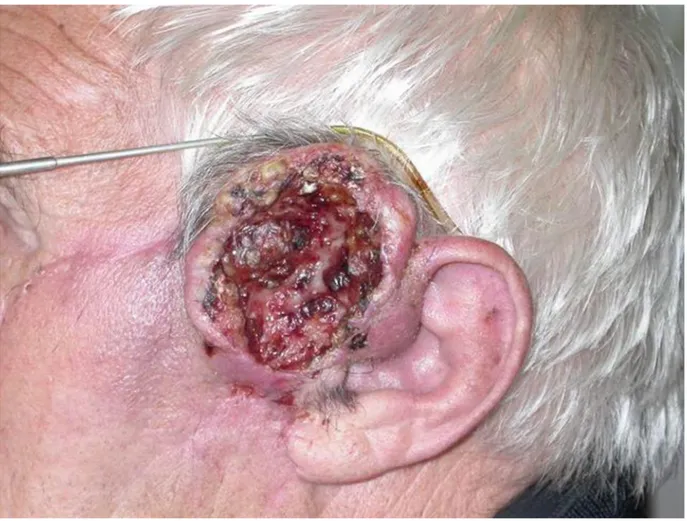
l’infiltration. La forme ulcéro‐bourgeonnante, la plus fréquente, est une lésion saillante, infiltrante, à surface irrégulière, siège d’une ulcération à fond bourgeonnant, saignotant et à
bords indurés (Figure 1). Les localisations les plus fréquentes sont les zones photo‐exposées : le visage, le cuir chevelu, le dos des mains et les avant‐bras.
Les CEC se développent sur une lésion préexistante comme une kératose actinique, une maladie de Bowen, une ulcération cutanée chronique ou encore sur des lésions dysplasiques virales, de radiodermites ou cicatricielles chroniques.
Figure 1 : Carcinome épidermoïde cutané de forme ulcéro-végétante, de localisation pré-tragienne gauche.
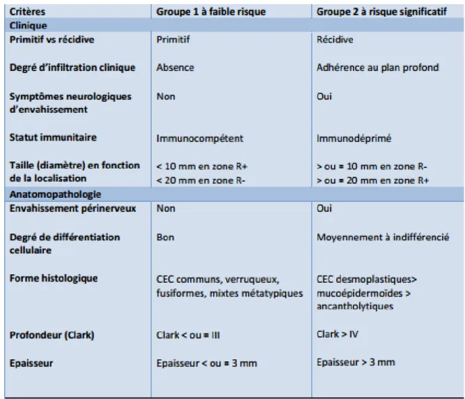
5. Pronostic (cf. tableau 1)(2)
Facteurs pronostiques cliniques
- La localisation de la tumeur : les récidives et les métastases sont plus fréquentes dans les
CEC des zones péri‐orificielles de la face, des zones génitales et sur les lésions cutanées
chroniques comme les ulcères chroniques et les cicatrices.
- La taille de la tumeur : la plupart des études retiennent un seuil de 2 cm comme étant
significatif de développer des récidives et des métastases.
- La récidive locale : le risque de récidive locale dépend de l’extension de la tumeur qui
conditionne la qualité de l’exérèse initiale.
- L’immunosuppression augmente le risque de récidives et de métastases. Il s’agit à la fois d’un facteur de risque et d’un facteur pronostique. Il a été démontré que l’agressivité des CEC chez les greffés d’organes est supérieure à celle de la population immunocompétente.
Facteurs histo‐pronostiques
- L’épaisseur tumorale et la profondeur de l’invasion : elles ont la meilleure valeur
prédictive positive de récidive et de métastases.
- L’invasion périnerveuse.
- Le faible degré de différenciation tumorale est un facteur de mauvais pronostic.
- Le type histologique : les CEC acantholytiques, muco‐épidermoïdes et desmoplastiques
sont de moins bons pronostics que les formes communes, verruqueuses, mixtes
Tableau 1 : Classification des Carcinomes épidermoïdes cutanés établie par la société française de dermatologie (2009)
6. Traitement des CEC
La chirurgie est le traitement de première intention des tumeurs primitives :
•elle permet un contrôle histologique de la pièce d’exérèse ;
Les marges d’exérèse (marges de sécurité) sont standardisées et varient de 4–5mm à 1cm en
fonction du type de tumeur et des critères de gravité définis plus haut. Elles seront déterminées par rapport aux limites cliniques de la lésion. La marge profonde sera dans la majorité des cas hypodermique.
En cas d’exérèse chirurgicale incomplète (exérèse passant en tissu tumoral à l’analyse
histologique de la pièce opératoire), la reprise chirurgicale est indispensable.
D’autres méthodes sont réservées à des malades inopérables ou pour certaines localisations,
après une biopsie de confirmation diagnostique :
•radiothérapie (électronthérapie ou curiethérapie)
•chimiothérapie de « réduction tumorale » pouvant être justifiée dans les CEC de grande taille pour réduire la masse tumorale avant l’intervention ou pour les CEC inopérables. Elle est avant tout fondée sur l’utilisation de sels de platine ;
•les thérapies ciblées anti-EGFR (hors Autorisation de Mise sur le Marché)(3)
7. Rationnel des anti-EGFR dans les CEC
Le pronostic est excellent pour la majorité des patients ayant un CEC mais devient médiocre à des stades localement avancés ou métastatiques, impossibles à traiter chirurgicalement ou par radiothérapie (1). Peu d’options thérapeutiques sont disponibles pour ces tumeurs. Les chimiothérapies conventionnelles systémiques telles que les combinaisons à base de Cisplatine ont une efficacité démontrée mais leur toxicités empêchent souvent leur utilisation chez les patients âgés (4). Le récepteur à l’EGF (Epidermal Growth Factor, EGFR), un récepteur transmembranaire tyrosine kinase de type 1, est très fortement exprimé dans de très nombreuses tumeurs épithéliales, incluant les carcinomes épidermoïdes de la tête et du cou ainsi que les CEC (5-7). L’EGFR est activé par un ligand permettant la dimérisation du récepteur activant
successivement de multiples voies de signalisation impliquée dans la survie et la prolifération cellulaire(8).
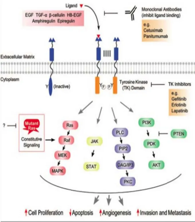
Comme illustré sur la Figure 2, l’activation intracellulaire fait intervenir les voies de signalisation suivantes :
- la voie Ras/Raf/Mek/MAPKinase (prolifération cellulaire),
- la voie JAK‐STAT (transcription de certains gènes de l’oncogénèse),
- la voie PI3K‐AKT (prolifération et migration cellulaire),
Figure 2 : Voies de signalisation cellulaires activées par EGFR et conséquence de son inhibition par Cranmer et al. (6)
Le cetuximab est un anticorps inhibant de manière compétitive l’EGFR. Il a déjà été approuvé
dans le traitement des carcinomes épidermoïdes de la tête et du cou et des cancers colo-rectaux (CCR).
La détermination du statut mutationnel RAS de type sauvage (exons 2, 3 et 4 du gène KRAS et du gène NRAS) est désormais impérative avant l’instauration d’un traitement par cetuximab dans les CCR. Le statut mutationnel du gène KRAS sur l’exon 2 de type sauvage était déjà nécessaire pour l’initiation du traitement par cetuximab mais de nouvelles données montrent que d’autres gènes RAS de type sauvage jouent un rôle dans l’activité du cetuximab. (9-12)Une
survie globale (SG) et une survie sans progression (SSP) plus courtes ainsi que des taux de réponses objectives inférieurs ont été observés chez les patients porteurs des mutations RAS (exons 2, 3 et 4 de KRAS et NRAS) qui ont reçu du cetuximab en association avec une chimiothérapie de type FOLFOX4 (5 fluoro-uracile/oxaliplatine, cycle de 4 semaines) par rapport à ceux porteurs de ces mutations ayant uniquement reçu le protocole FOLFOX4.(11) De tels résultats indiquent que la mutation KRAS pourrait servir de marqueur prédictif de l’efficacité des anti-EGFR.
Dans une étude publiée de phase II , le cetuximab en monothérapie a démontré une efficacité clinique chez les patients naïfs de tout traitement, atteints de carcinomes épidermoïdes cutanés non résécables ou localement évolués avec un taux de contrôle global de la maladie de 69 % et un taux de réponse de 28%. (13)
Plusieurs séries rétrospectives de cas rapportés ont décrit l’efficacité du cetuximab chez les
patients naïfs de tout traitement ou pré-traités, atteints de carcinomes épidermoïdes cutanés localement évolués ou métastatiques. (14, 15)
Cependant, à notre connaissance, aucune étude n’a pour l’instant évalué le rôle de biomarqueurs pour prédire l’efficacité du cetuximab dans les CEC localement avancés ou métastatiques, d’où l’intérêt de notre projet.
Ainsi et par analogie avec les données existantes pour les CCR nous avons voulu évaluer si le statut mutationnel des gènes HRAS, KRAS, NRAS, BRAF et EGFR pouvait être un facteur prédictif de réponse au traitement par cetuximab chez les patients atteints de carcinomes epidermoïdes cutanés inopérables.
BIBLIOGRAPHIE
1. Alam M, Ratner D. Cutaneous squamous-cell carcinoma. The New England journal of medicine. 2001;344(13):975-83.
2. Jambusaria-Pahlajani A, Kanetsky PA, Karia PS, Hwang WT, Gelfand JM, Whalen FM, et al. Evaluation of AJCC tumor staging for cutaneous squamous cell carcinoma and a proposed alternative tumor staging system. JAMA dermatology. 2013;149(4):402-10.
3. Reigneau M, Robert C, Routier E, Mamelle G, Moya-Plana A, Tomasic G, et al. Efficacy of neoadjuvant cetuximab alone or with platinum salt for the treatment of unresectable advanced nonmetastatic cutaneous squamous cell carcinomas. The British journal of dermatology. 2015;173(2):527-34.
4. Sadek H, Azli N, Wendling JL, Cvitkovic E, Rahal M, Mamelle G, et al. Treatment of advanced squamous cell carcinoma of the skin with cisplatin, 5-fluorouracil, and bleomycin. Cancer.
1990;66(8):1692-6.
5. Fogarty GB, Conus NM, Chu J, McArthur G. Characterization of the expression and activation of the epidermal growth factor receptor in squamous cell carcinoma of the skin. The British journal of dermatology. 2007;156(1):92-8.
6. Maubec E, Duvillard P, Velasco V, Crickx B, Avril MF. Immunohistochemical analysis of EGFR and HER-2 in patients with metastatic squamous cell carcinoma of the skin. Anticancer research. 2005;25(2B):1205-10.
7. Herbst RS, Arquette M, Shin DM, Dicke K, Vokes EE, Azarnia N, et al. Phase II multicenter study of the epidermal growth factor receptor antibody cetuximab and cisplatin for recurrent and refractory squamous cell carcinoma of the head and neck. Journal of clinical oncology : official journal of the American Society of Clinical Oncology. 2005;23(24):5578-87.
8. Cranmer LD, Engelhardt C, Morgan SS. Treatment of unresectable and metastatic cutaneous squamous cell carcinoma. The oncologist. 2010;15(12):1320-8.
9. De Roock W, Claes B, Bernasconi D, De Schutter J, Biesmans B, Fountzilas G, et al. Effects of KRAS, BRAF, NRAS, and PIK3CA mutations on the efficacy of cetuximab plus chemotherapy in chemotherapy-refractory metastatic colorectal cancer: a retrospective consortium analysis. The Lancet Oncology. 2010;11(8):753-62.
10. Karapetis CS, Khambata-Ford S, Jonker DJ, O'Callaghan CJ, Tu D, Tebbutt NC, et al. K-ras mutations and benefit from cetuximab in advanced colorectal cancer. The New England journal of medicine. 2008;359(17):1757-65.
11. Lievre A, Bachet JB, Boige V, Cayre A, Le Corre D, Buc E, et al. KRAS mutations as an
independent prognostic factor in patients with advanced colorectal cancer treated with cetuximab. Journal of clinical oncology : official journal of the American Society of Clinical Oncology.
2008;26(3):374-9.
12. Linardou H, Briasoulis E, Dahabreh IJ, Mountzios G, Papadimitriou C, Papadopoulos S, et al. All about KRAS for clinical oncology practice: gene profile, clinical implications and laboratory recommendations for somatic mutational testing in colorectal cancer. Cancer treatment reviews. 2011;37(3):221-33.
13. Maubec E, Petrow P, Scheer-Senyarich I, Duvillard P, Lacroix L, Gelly J, et al. Phase II study of cetuximab as first-line single-drug therapy in patients with unresectable squamous cell carcinoma of the skin. Journal of clinical oncology : official journal of the American Society of Clinical Oncology. 2011;29(25):3419-26.
14. Giacchero D, Barriere J, Benezery K, Guillot B, Dutriaux C, Mortier L, et al. Efficacy of
cetuximab for unresectable or advanced cutaneous squamous cell carcinoma--a report of eight cases. Clinical oncology. 2011;23(10):716-8.
15. Bauman JE, Eaton KD, Martins RG. Treatment of recurrent squamous cell carcinoma of the skin with cetuximab. Archives of dermatology. 2007;143(7):889-92.
Etude réalisée
Oncogenic mutations in advanced cutaneous squamous cell
cacinomas and correlation with response to cetuximab treatment
Alexandra Picard1, Florence Pedeutour2, Fréderic Peyrade3, Laurence Saudes4, Valérie
Duranton-Tanneur2, Emmanuel Chamorey5 Nathalie Cardot-Leccia6, Anne Sudaka7, Gilles Poissonnet8, Jean-François Michiels6, Jean-Philippe Lacour1, Thierry Passeron1,9, Henri Montaudié1
1Department of Dermatology, University Hospital of Nice, France,
2Laboratory of Solid Tumors Genetics, Nice University Hospital and Institute of Research on
Cancer and Aging (IRCAN), Faculty of Medicine, Nice, France,
3Medical Oncology Department, Antoine Lacassagne Center, Nice, France, 4Medical Oncology Department, Cannes Hospital, France,
5Biostatistics unit, Antoine Lacassagne Center, Nice, France, 6-Department of Pathology, University Hospital of Nice, France,
7Oncopharmacology and Pathology Departments, Antoine Lacassagne Center, Nice, France, 8_Department of Head and Neck Surgery Institut Universitaire de la Face et du Cou (IUFC),
Antoine Lacassagne Center, Nice, France,
9INSERM, U1065, Centre Méditerranéen de Médecine Moléculaire, Team 12, Nice, France,
Key words: cutaneous squamous cell carcinoma, oncogenic mutations, cetuximab
Figures: 3
Tables: 3
Correspondance : picard.a@chu-nice.fr
Address: 151 route de Saint Antoine de Ginestiere, Hôpital Archet 2, 06200 Nice, France
ABBREVIATIONS
cSCC: cutaneous Squamous Cell Carcinoma: CR: Complete Response
DCR: Disease Control Rate
EGFR: Epidermal Growth Factor Receptor mCRC: metastatic ColoRectal Cancer: PFS: Progression-free-survival
PR: Partial Response OS: Overall Survival SD: Stable Disease
What’s already known about this topic?
Cetuximab has been recently proposed in advanced cutaneous squamous cell carcinomas (cSCC) when surgery or radiotherapy could not be used. KRAS and NRAS mutations are independent predictive markers of response to cetuximab therapy in colo-rectal cancer but there is so far no available data in cSCC.
What does this study add?
The incidence of HRAS, KRAS, NRAS, BRAF and EGFR mutations is very low in cSCC and the mutational status of those genes is not predictive of resistance to cetuximab.
These results show that the detection of mutations before initiating a cetuximab treatment for advanced cSCC is not necessary.
RESUME
Introduction Le traitement par cetuximab a été récemment proposé dans les carcinomes
épidermoïdes cutanés (CEC) localement avancés lorsque la chirurgie ou la radiothérapie ne peuvent pas être réalisées. Cependant, son efficacité est inconstante. Des biomarqueurs pourraient aider à sélectionner les patients potentiellement répondeurs au traitement par cetuximab.
Objectifs Déterminer l’incidence des mutations des gènes EGFR, HRAS, KRAS, NRAS et BRAF
chez les patients atteints de CEC non résécables ou métastatiques traités par cetuximab en monothérapie et déterminer l’impact de ces mutations sur la réponse au cetuximab.
Matériel et méthodes Une étude rétrospective multicentrique a été conduite entre 2008 et 2014.
Tous les patients avec un diagnostic de CEC non résécable ou métastatique traité par cetuximab en monothérapie et confirmé histologiquement ont été inclus.
Résultats Trente et un échantillons de CEC provenant de 31 patients ont été analysés.
Seulement 2 échantillons mutés RAS (6.5 %) ont été identifiés. Le premier avait une mutation
NRAS (c.35G>A) dans le codon 12 résultant d’une substitution p.G12D. Le second échantillon
présentait une mutation HRAS (c.38G>T) dans le codon 13 résultant d’une substitution p.G13V. Nous n’avons pas identifié de mutations dans les gènes KRAS, BRAF et EGFR, dans
les loci investigués. De manière surprenante, les deux patients avec les mutations NRAS et
HRAS ont eu une réponse partielle et complète au cetuximab, respectivement.
Discussion Cette étude démontre que l’incidence des mutations HRAS, KRAS, NRAS, BRAF et EGFR est très faible dans les CEC et que le statut mutationnel de ces gènes n’est pas prédictif
d’une mauvaise réponse au cetuximab.
Conclusion Ces résultats montrent que la détection des mutations HRAS, KRAS, NRAS, BRAF
ABSTRACT
Background Cetuximab has been recently proposed for advanced cutaneous squamous cell
carcinoma (cSCC) when surgery or radiotherapy could not be used. However, its efficacy is inconstant. Molecular markers could help select patients most likely to respond to cetuximab therapy.
Objectives To seek for somatic mutations of the EGFR, KRAS, NRAS, HRAS and BRAF genes
in patients with unresectable or metastatic cSCC treated with cetuximab; to correlate the response to treatment to this mutational status.
Material and methods A multicentric and retrospective study was conducted from 2008 to 2014.
All patients with a diagnosis of unresectable or metastatic cSCC, histologically confirmed and treated with single-agent cetuximab were included.
Results Thirty-one samples of cSCC from 31 patients were analyzed. Only 2 RAS mutated
samples (6.5%) were identified. The first one harbored a NRAS point mutation (c.35G>A) in codon 12 resulting in a p.G12D substitution. The second sample presented a HRAS point mutation (c.38G>T) in codon 13 resulting in a p.G13V substitution. We did not find any mutation of KRAS, BRAF and EGFR genes at the investigated loci. Surprisingly, the two patients with NRAS and HRAS mutations showed a partial and complete response to cetuximab, respectively.
Discussion This study demonstrates that the incidence of HRAS, KRAS, NRAS, BRAF and EGFR mutations is very low in cSCC and that the mutational status of those genes is not
predictive of a lack of response to cetuximab.
Conclusion These results show that the detection of mutations before initiating a cetuximab
BACKGROUND
Non-melanoma skin cancers are the most common cancers worldwide. Cutaneous squamous cell carcinoma (cSCC) represents approximately 20 % to 30 % of them. In France, the incidence is estimated to be at least 30 per 100,000 persons per year and has increased by 10% in recent years. The median age at diagnosis of cSCC is 70 years.(1) cSCC has a good prognosis when it
is diagnosed at an early stage. The long-term overall survival rate is below 20% for patients with local lymph node metastases of cSCC and below 10% for those with distant metastases.(1)
The treatment of unresectable or metastatic cSCC is challenging. Cisplatin-based, combination chemotherapy is the most commonly used treatment, with an overall response rate of up 80%.(2,
3) However, the available data are limited and the side effects are a strong limitation of this
approach in elderly patients.
The epidermal growth factor receptor (EGFR) is a membrane-bound tyrosine kinase receptor that is highly expressed in normal epidermal keratinocytes and in many epithelial tumors, including cSCC. (4, 5) Cetuximab is a human-mouse chimeric monoclonal antibody that competitively inhibits the EGFR. It is approved by the Food and Drug Administration and the
European Medicines Agency for the treatment of wild-type-KRAS metastatic colorectal cancer
(6) and for advanced or metastatic head and neck SCC combined with radiotherapy and with
cisplatin/5-FU.(7-9) However, the response rate to cetuximab in unselected metastatic colorectal cancer (mCRC) is lower than 30%, underlining the need for identifying a predicting biomarker of its efficacy.(10) A lack of response to the anti-EGFR therapy has been demonstrated in mCRC
harboring KRAS or NRAS mutations. KRAS and NRAS mutations are independently associated with lower progression free survival (PFS) and overall survival in mCRC.(11, 12) Cetuximab
has been recently proposed in advanced cSCC when surgery or radiotherapy could not be used.(13, 14) However, there is presentlyno available data concerning oncogenic mutations in cSCC related to the response to cetuximab therapy.
We studied somatic mutations of the EGFR, KRAS, NRAS, HRAS and BRAF genes in patients with unresectable or metastatic cSCC treated with cetuximab in order to correlate the response to treatment to the mutational status.
MATERIALS AND METHODS
Study design and objectives
We conducted a retrospective multicentric study in three French centers (Departement of Dermatology, Nice Hospital, Department of Oncology, Centre- Antoine- Lacassagne, Nice and Department of Oncology, Cannes Hospital, Cannes). Our main objective was to evaluate the incidence of KRAS, NRAS, HRAS, BRAF and EGFR mutations in unresectable or metastatic cSCC and to correlate the cetuximab response to the mutational status.
Our secondary objectives were to assess; (1) the disease control rate (DCR) defined as the percentage of patients who have achieved complete response (CR), partial response (PR) or stable disease (SD) at week 6 ; (2) the response rate at week 6 of treatment with cetuximab; (3) the progression-free survival (PFS), defined as the number of days between the first dose of cetuximab and the earliest day of progression, start of another anticancer treatment or death; (4) the overall survival (OS), defined as the time between the first infusion of chemotherapy and the latest news.
Patients
Patients with cSCC confirmed by pathological evaluation,
cSCC locally advanced surgically unresectable or metastatic with documented progression,
Treated with single-agent cetuximab between 2008 and 2014,
Criteria of unresectability were determined by a multidisciplinary committee composed of dermatologists, surgeons and radiation therapists. They were based on the impossibility to achieve a complete resection and on the risk of critical cosmetic or functional outcomes,
Patients with a recurrent primary cSCC having previously undergone surgery or radiotherapy could be included,
A sample of tumor had to be accessible before treatment,
All patients signed written informed consent (IRB waived).
Treatment
Cetuximab was administered as an intravenous infusion at an initial dose of 400 mg/m2, followed by weekly 1-hour infusions of 250 mg/m2. Patients received pretreatment with an antihistamine drug. In case of infusion reactions or dermatologic toxicity, dose modifications
were planned. Cetuximab could be continued as long as the response or the stabilization persisted. After 3 months of weekly treatment, cetuximab was administred every 15 days.
Adverse events
Adverse events and grades were recorded according to National Cancer Institute Criteria, version 4.0.
Assessment
Tumor response was assessed at week 6 clinically or radiologically according to RECIST criteria.(15)
Samples
Formalin-fixed and paraffin-embedded tumor samples form surgical resection of primitive tumor were obtained. The samples were collected after informed consent of all patients according to the ethical rules of our institutions.
Genetic analysis
Genomic DNA was extracted from formalin-fixed paraffin-embedded (FFPE) tissue containing more than 50% neoplastic cells as determined by a pathologist. DNA extraction was performed using the automated Maxwell 16 Instrument with the Maxwell 16 FFPE Plus LEV DNA Purification Kit (Promega, Madisson, WI, USA), following the manufacturer’s instructions.
Genotyping of codons 12 and 13 of the exon 2 of KRAS was performed by allelic discrimination. The design of sequences of the TaqMan probes was kindly provided by Pr Laurent-Puig (16). The seven hot-spot mutations (c.34G > C (p.G12R); c.35G > C (p.G12C); c.34G > A (p.G12S); c.35G > A (p.G12D); c.35G > C (p.G12A); c.35G > T (p.G12V); c.38G > A(p.G13D)) located in codons 12 and 13 of exon 2 of the KRAS gene were screened. Specific probes for each mutated and non-mutated allele were labeled using the fluorescence reporter dyes FAM and VIC at their 5’-end, respectively. Briefly, reactions were performed in 20 µL comprising 10 ng
of DNA, specific primers and probes, and TaqMan Universal PCR Master Mix (Applied Biosystems, Foster City, CA, USA). Runs were performed using a 7500 Fast Applied Biosystems Real Time PCR (Applied Biosystems) at the following cycling conditions: 95°C for 15 min; 40 cycles, 95°C for 15 s; and 60°C for 1 min. Data were analyzed using SDS2.0 software (Applied Biosystems).
The genotyping of the exons 3 and 4 of KRAS and the exons 2 and 3 of NRAS gene was performed by pyrosequencing method. Primers chosen for PCR amplification were designed using Pyromark Assay Design2.0 software from Qiagen (Hilden, Germany). The primers used were: KRAS 3F 5’- AATTGATGGAGAAACCTGTCTCTT-3’, KRAS 3R 5’biotin-
TCCTCATGTACTGGTCCCTCATT-3’, KRAS 4F 5’-
AGGCTCAGGACTTAGCAAGAAGTT-3’ KRAS 4R
5’biotin-AGTTATGATTTTGCAGAAAACAGA-3’, NRAS 2F
5’biotin-TCTGGATTAGCTGGATTGTCAGT-3’, NRAS 3F
5’-TTTGTTGGACATACTGGATACAGC-3’, NRAS 3R
5’biotin-CGCAAATGACTTGCTATTATTGA-3’. The cycling conditions consisted of initial denaturing at 95°C for 15 min, followed by 42 amplification cycles of 95 °C for 20 s, 53 °C for 30 s, 72 °C for 20 s and a final extension step at 72 °C for 5 min. The pyrosequencing reaction was performed according to the manufacturer's recommendations using a Q24 Pyromark (Qiagen). The pyrosequencing primers were 5'- CTTGGATATTCTCGACAC-3' for KRAS
exon 3, 5’-AATTCCTTTTATTGAAACAT-3’ for KRAS exon 4,
5’-GTGGTGGTTGGAGCA-3’ for NRAS exon 2 and 5’-GGACATACTGGATACAGCT-3’ for NRAS exon 3.
The genotyping of exons 2 and 3 of HRAS gene was performed by Sanger sequencing using the following primers: HRAS 2F 5’-CAGGAGACCCTGTAGGAGGA-3’, HRAS 2R 5’-
CCTATCCTGGCTGTGTCCTG-3’, HRAS 3F 5’- AGAGGCTGGCTGTGTGAACT-3’, HRAS 3R 5’- TGGTGTTGTTGATGGCAAAC-3’.
Statistical analysis
Categorical values at baseline were expressed as counts and percentages whereas continuous values were expressed as medians. Overall survival (OS) and progression-free survival (PFS) were estimated by the Kaplan-Meier method. All statistical analyses were performed with α=5% in bilateral hypothesis using R 3.2.2 Software for windows.
RESULTS
Patient Characteristics
A total of 31 patients were included. The median age was 86 years (48-96 years). Patient characteristics are summarized in Table 1. Seventy one percent of tumors were located on head and neck. Thirty-nine percent of patients had a local disease, 42% lymph node disease and 19 % distant metastases. Twenty-three percent of patients had not received therapy before cetuximab.
Somatic mutations of EGFR, BRAF, HRAS, KRAS, NRAS and correlation with response to cetuximab
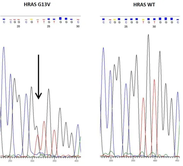
Two mutated samples (6.5%) were identified. One harbored a NRAS point mutation (c.35G>A) in codon 12 resulting in a p.G12D substitution. The second sample presented a HRAS point mutation (c.38G>T) in codon 13 resulting in a p.G13V substitution (Fig. 1). We did not find any mutation of KRAS, BRAF and EGFR genes at the investigated loci. Surprisingly, the two patients with NRAS and HRAS mutations showed a PR and CR to cetuximab, respectively. Those patients were two women with poorly differentiated cSCC. Both had a past history of cancer in CR (breast and cervical cancer for the first, head and neck SCC for the second, cf. Table 2).
Response to cetuximab treatment
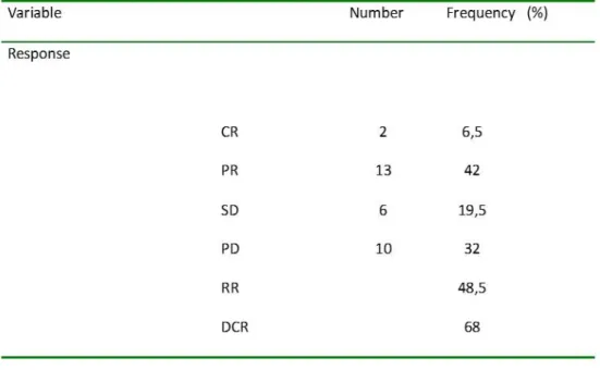
The mean duration of follow up was 19 months (1-36 months). At week 6, the DCR was 68%. Two patients (6.5%) achieved a CR, thirteen patients (42%) a PR, six (19.5%) a stable disease and ten (32%) progressive disease (PD) The objective response rate was 48.5 % (Table 3,
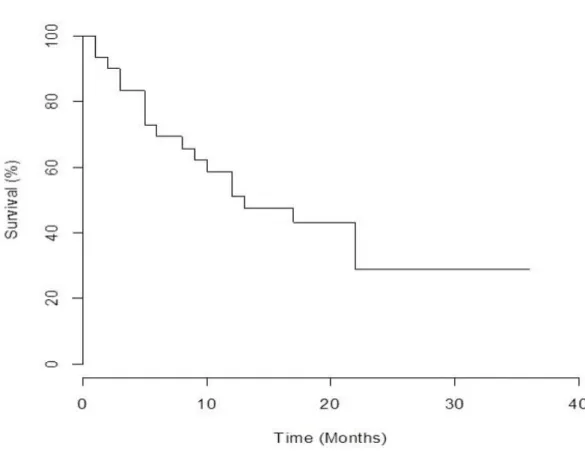
Fig.2). For patients with local disease, the DCR was 75 % (two CR (17%), six PR (50%), and one SD (8%)). For the patients with metastatic or lymph node disease, the DCR was 63 % with no complete response, seven PR (37%) and five SD (26 %). The median OS was 13 months (1-36 months), and the median PFS was 9 months (0-(1-36 months). At 18 months and (1-36 months 43.2% and 28.8% of patients were still alive, respectively (Fig.3).
Safety
Fifty-two percent of patients developed cetuximab-induced grade 1-2 folliculitis. One patient had a grade 3 folliculitis. She received 4 cetuximab infusions followed by CR. All patients could continue cetuximab treatment without dose reduction. No related-death to treatment occurred.
Table 1: Clinical characteristics.
Abbreviations: AJCC: American Joint Committee on Cancer, ECOG: Eastern Cooperative Oncology Group, PS: Performans status
Figure 1: Direct sequencing (Sanger method) of DNA showing a heterozygous HRAS G13V
point—mutation (left panel). Right panel shows the wild-type (WT) sequence
HRAS exons 2 and 3 were amplified using the following primers: for exon 2 HRAS_2F (5'- CAGGAGACCCTGTAGGAGGA-3') and HRAS_2R (5'- CCTATCCTGGCTGTGTCCTG-3' and for exon 3 HRAS_3F 5’- AGAGGCTGGCTGTGTGAACT-3’ and HRAS_3R 5’-
TGGTGTTGTTGATGGCAAAC -3’. Briefly, 100 ng of DNA was amplified with the thermal cycling profile of 94° C for 10 min, then 42 cycles of 94° C for 30 sec, 62° C for 45 sec, 72° C for 45 sec, with a final extension at 72° C for 10 min. Direct sequencing was performed using the above primers using the Big-Dye DyeDeoxy terminator cycle sequencing kit (Applied Biosystems, Foster City, CA). Sequencing reactions were carried out on the ABI Prism 3100 Genetic Analyzer (Applied Biosystems, Foster City, CA).
Table 2: Clinical characteristics, results of detection of HRAS, NRAS, KRAS, BRAF and EGFR mutations and
response to Cetuximab, in a series of 31 patients affected by unresectable or metastatic cutaneous squamous cell carcinoma.
Abbréviations : F (Female), M (Male), PR (Partial response), CR (Complete Response), SD (Stable Disease), PD (Progressive Disease), wt (wild type), AJCC ( American Joint Committee Cancer)
Table 3: Response and disease control rate at week 6.
CR (Complete Response), PR (Partial Response), SD (Stable Disease), PD (Progressive Disease), DCR (Disease Control Rate), RR (Response Rate)
Figure 2: Example of patient showing a partial response to cetuximab.
A 88-year-old male with a well differentiated local tumor of the head experienced a partial response at week 6.
DISCUSSION
Our results clearly show that NRAS, HRAS, KRAS, BRAF and EGFR gene mutations are rare events in cSCC. Although having extended the search of somatic mutations to NRAS, HRAS,
KRAS, BRAF and EGFR genes, our results confirm the very low frequency of these mutations
in cSCC. Data from the literature have showed a high level of EGFR expression by immunohistochemistry in cSCC that was not associated with cetuximab efficacy. Therefore, we dit not screen over expression of EGFR. EGFR gene was reported to be altered in 1% of the
samples of cSCC listed in the COSMIC database (19) and the overall incidence of EGFR mutations in cSCC was also found to be low (2.5% to 5%) in several studies. (20-22) No EGFR mutations were found in our study. There is a wide variation in the reported incidence of RAS mutations in cSCC as it ranges from 3% up to 30%.(13, 23) Our results are in accordance with the largest study reported so far (13) showing a low rate of mutations. Indeed, we identified only one
case of HRAS mutation (1/31, 3%), and one case of NRAS mutation (1/31, 3%). HRAS mutation was confirmed by next generation sequencing, using the Ion AmpliSeq Cancer Hotspot Panel (v2) and we did not find other mutations in this tumor (data not shown).
As reported in other studies, no mutation, in our cohort, was found in KRAS or BRAF genes.(13,
24-26) The HRAS gene was found to be mutated in 8 to 9 % of patients with cSCC.(20, 24) A phase
II study assessing the efficacy of cetuximab for treating unresectable cSCC in 36 patients found only one HRAS mutation.(13) The mutation in NRAS p.G12D identified in one of our patient has never been described in cSCC but was reported in melanoma and in colorectal cancer.(27)
Interestingly, the mutation in HRAS p.G13V has been reported in cSCC treated by BRAF inhibitors.(28) Our patient had never been treated by targeted therapy or chemotherapy. In
metastatic colorectal cancer KRAS mutations are strongly associated with lack of cetuximab benefit while no correlation between the mutation status and response to treatment could be
In contrast, the only two patients in our study harboring NRAS or HRAS mutations showed PR and CR response to cetuximab. Overall dependence on EGFR signaling for growth and survival ,and subsequent EGFR inhibition effect , may not be the same in cSCC and mCRC Interestingly, comparative genomic hybridation of the HRAS mutated tumor showed a gain of chromosome 7 and two copies of chromosome 11, which are located on EGFR and HRAS genes, respectively ( data not shown). Thus, the efficacy of cetuximab could be explained by an overexpression of EGFR. Moreover, these HRAS and NRAS mutations may not be hotspot activating mutation with oncogenic potential for cSCC tumors. However, even without a strong explanation, NRAS and HRAS mutations seem to have different effects in advanced cSCC.
In our study that included unresectable and metastatic cSCC, single-agent cetuximab showed a 68 % DCR. These data are consistent with those from the literature, especially compared to two prospective phase II studies. Indeed, Maubec et al., in a clinical trial including 36 treatment-naïve patients with unresectable cSCC treated with cetuximab reported comparable results, with a DCR of 69% at 6 weeks (13) Moreover, Foote et al. published, data using panitumumab, another
monoclonal anti-EGFR antibody and reported also a DCR at 6 weeks of 69%.(30) Compared to cisplatin chemotherapy, cetuximab is generally well tolerated and may be considered as a therapeutic option especially in elderly patients. As expected, the patient with grade 3 rash folliculitis had a CR following four injections. The efficacy observed in our study is consistent with previous data and encourages the use of EGFR inhibitors for unresectable or metastatic cSCC.
Cetuximab sensitivity is related to KRAS and NRAS status in mCRC. The main objective of our study was to correlate the response to cetuximab treatment to these mutations in cSCC. We focused our research on these mutations and we didn’t perform a full genome sequencing.
However, some other molecular determinants of response to cetuximab may be implicated in cSCC. Maubec et al. reported that combined Fc�RIIa-131H/H and/or Fc�RIIIa-158V/V
polymorphisms were not associated with the response to cetuximab.(13) Several recent studies evaluated recurrent genomic alterations in metastatic cSCC. Li et al. performed targeting sequencing of 504 genes in 29 patients with metastatic cSCC. They found previously identified
recurrently altered genes such as TP53, CDKN2A, NOTCH1/2 and also a wide spectrum of oncogenic mutations affecting RAS/RTK/PI3Kpathways. This suggest additional therapeutic targets for this tumors.(31) Moreover, another study in 39 aggressive cSCC, identified three novel candidates tumor suppressors genes with putative links to cancer or differentiation, NOTCH2,
PARD3 and RASA1, which were identified as possible drivers in cSCC.(26) The mutational spectrum of cSCC seems dominated by tumor suppressor genes, as in head and neck squamous cell carcinomas.
CONCLUSION
Our study confirms the efficacy of cetuximab as a single-agent in advanced cSCC. The incidence of HRAS, KRAS, NRAS, BRAF and EGFR mutations is very low in cSCC and the mutational status of those genes is not predictive of response to cetuximab. Conversely to mCRC, seeking for mutations in these genes not of interest before initiating a cetuximab therapy for cSCC.
ACKNOLEDEGMENTS
Marc Ettaiche for statistical analysis, Sophie Gimet, Nicolas Weinebreck, Maxime Benechetrit for their contribution to the study.