Science Arts & Métiers (SAM)
is an open access repository that collects the work of Arts et Métiers Institute of
Technology researchers and makes it freely available over the web where possible.
This is an author-deposited version published in:
https://sam.ensam.eu
Handle ID: .
http://hdl.handle.net/10985/19056
To cite this version :
Georges MJAESS, Chris LABAKI, Aya KARAM, Ziad BAKOUNY, Aren Joe BIZDIKIAN, Joeffroy
OTAYEK, Fares YARED, Waffa SKALLI, Ismat GHANEM, Ayman ASSI - How does the variation
of the 3D orientation of the acetabulum during walking influence hip kinematics? - Gait & Posture
- Vol. 65, p.136-138 - 2018
Any correspondence concerning this service should be sent to the repository
Administrator :
archiveouverte@ensam.eu
O 061
—How does the variation of the 3D orientation of the acetabulum
during walking in
fluence hip kinematics?
G. Mjaess
a, C. Labaki
a, A. Karam
a, Z. Bakouny
a, A.J. Bizdikian
a, J. Otayek
a, F. Yared
a, W. Skalli
b,
I. Ghanem
a, A. Assi
a,b,⁎aFaculty of Medicine–University of Saint-Joseph, Laboratory of Biomechanics and Medical Imaging, Beirut, Lebanon bArts et Métiers ParisTech, Institut de Biomécanique Humaine Georges Charpak, Paris, France
1. Introduction
Acetabular cup orientation is crucial for total hip arthroplasty (THA), and its malpositioning could lead to impingement and disloca-tion [1]. Acetabular cup orientation currently relies on static 3D hip
parameters [2] and was shown to be related to changes in pelvic po-sitioning [3]. While pelvic position varies during walking, it is still unknown how dynamic variation of hip orientation during gait could influence hip kinematics.
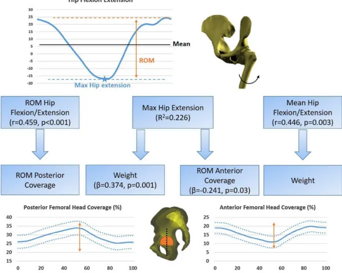
Fig. 1. 3D hip skeletal parameters computed during the gait cycle.
E-mail address: gmjaess@gmail.com (A. Assi).
2. Research question
How do dynamic variations of 3D skeletal hip parameters during gait influence hip kinematics in asymptomatic subjects?
3. Methods
77 asymptomatic subjects (age: 28 ± 10years [18–38], 32F) un-derwent 3D gait analysis [4], with additional markers on thighs and shanks, from which the means, maxima, minima, and ROM of hip ki-nematics were extracted. Then, subjects underwent full-body biplanar X-rays with markers still in place. Full body 3D reconstructions were obtained for the spine, pelvis and lower limbs. The 3D bones were re-gistered at each frame of the gait cycle [5]. A new technique developed for this study, utilizingfinite element modelling, was used to reduce soft tissue artefacts. Then, 3D hip skeletal parameters (acetabular or-ientations, coverage of the femoral head by the acetabulum and sector angles) were computed during the gait cycle, using the 3D registered bones, at each time frame (Fig. 1): means, maxima, minima, and ROM were extracted. In order to assess the influence of demographic (age, sex, weight, and height) and dynamic variations of 3D skeletal hip parameters on hip kinematics during gait, a univariate analysis (Pear-son’s correlation test) followed by a multivariate analysis (stepwise
multiple linear regression) were computed; the dependent variables were hip kinematics, while the independent variables were demo-graphic and hip dynamic 3D skeletal parameters.
4. Results
ROM hip flexion/extension was determined by
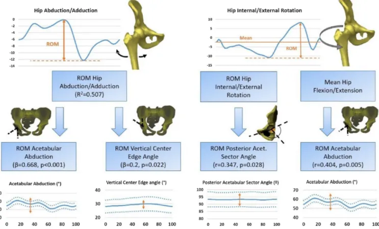
ROM_Posterior_Coverage (r = 0.459, p < 0.001). Maximum hip ex-tension was determined (R2 = 0.226) by weight (β = 0.374, p = 0.001), and ROM_Anterior_Coverage (β = −0.241, p = 0.03). Mean hip flexion/extension was determined by weight (r = 0.446, p = 0.003). ROM hip abduction/adduction was determined (R2 = 0.507) by ROM_Acetabular_Abduction (β = 0.668, p < 0.001) and ROM_Vertical_Center_Edge_Angle (β = 0.2, p = 0.022). ROM hip
internal/external rotation was determined by
ROM_Posterior_Acetabular_Sector_Angle (r = 0.347, p = 0.028). Mean
hip internal/external rotation was determined by
ROM_Acetabular_Abduction (r = 0.404, p = 0.005). 5. Discussion
This is thefirst study to evaluate the effect of variation of 3D ske-letal hip parameters during gait on hip kinematics (Figs. 2 and 3). A
larger ROM of the posterior coverage and a smaller ROM of the anterior coverage of the femoral head by the acetabulum during gait seem to be related to a larger mobility of the hip in the sagittal plane. A larger ROM of the acetabular abduction during gait seem to be related to a larger mobility of the hip in both the frontal and axial planes. Planning acetabular cup orientation in THA should take into consideration these dynamic variations of the acetabulum during gait in order to ensure a full mobility of the hip during walking.
References [1] Teeter, 2018. [2] Bendaya, 2016. [3] Kanawade, 2014. [4] Davis, 1990. [5] Söderkvist, 1993.
Fig. 3. Determinants of hip abduction/adduction and hip internal/external rotation during gait among demographic parameters and 3D hip radiological parameters computed during gait.