HAL Id: inserm-02167741
https://www.hal.inserm.fr/inserm-02167741
Submitted on 28 Jun 2019HAL is a multi-disciplinary open access
archive for the deposit and dissemination of sci-entific research documents, whether they are pub-lished or not. The documents may come from teaching and research institutions in France or abroad, or from public or private research centers.
L’archive ouverte pluridisciplinaire HAL, est destinée au dépôt et à la diffusion de documents scientifiques de niveau recherche, publiés ou non, émanant des établissements d’enseignement et de recherche français ou étrangers, des laboratoires publics ou privés.
Interleukin-22 binding protein (IL-22BP) is
constitutively expressed by a subset of conventional
dendritic cells and is strongly induced by retinoic acid
Jcj Martin, G Bériou, M Heslan, Christian Chauvin, L Utriainen, A
Aumeunier, C Scott, A Mowat, V Cerovic, S Houston, et al.
To cite this version:
Jcj Martin, G Bériou, M Heslan, Christian Chauvin, L Utriainen, et al.. Interleukin-22 binding pro-tein (IL-22BP) is constitutively expressed by a subset of conventional dendritic cells and is strongly induced by retinoic acid. Mucosal Immunology, Nature Pub. Group, 2014, 7 (1), pp.101-113. �10.1038/mi.2013.28�. �inserm-02167741�
Interleukin-22 binding protein (IL-22BP) is constitutively
expressed by a subset of conventional dendritic cells and is
strongly induced by retinoic acid
JCJ Martin1,2,3,6, G Bériou1,2, M Heslan1,2, C Chauvin1,2, L Utriainen4, A Aumeunier4, CL Scott4, A Mowat4, V Cerovic4, SA Houston4, M Leboeuf5, FX Hubert1,2,6, C Hémont1,2,3,6, M Merad5, S Milling4, and R Josien1,2,3,6
1INSERM Center of Research in Transplantation and Immunology, UMR1064, Nantes, F - 44000, France
2CHU Nantes, Institut de Transplantation Urologie Néphrologie (ITUN), Nantes, F-44000, France 3CHU Nantes, Laboratoire d’immunologie, Nantes, F-44000, France
4Centre for Immunobiology, Institute of Infection, Immunity and Inflammation, College of Medical, Veterinary and Life Sciences, University of Glasgow, Glasgow, G12 8TA, UK
5Department of Gene and Cell medicine and the Department of Medicine, Mount Sinai School of Medicine, New York 10029, USA
6Université de Nantes, Faculté de Médecine, Nantes, F-44000, France
Abstract
IL-22 is mainly produced at barrier surfaces by T cells and innate lymphoid cells and is crucial to maintain epithelial integrity. However, dysregulated IL-22 action leads to deleterious
inflammation and is involved in diseases such as psoriasis, intestinal inflammation and cancer. IL-22BP is a soluble inhibitory IL-22 receptor and may represent a crucial regulator of IL-22. We show both in rats and mice that, in the steady state, the main source of IL-22BP is constituted by a subset of conventional dendritic cells (DC) in lymphoid and non lymphoid tissues. In mouse intestine, IL-22BP was specifically expressed in lamina propria CD103+CD11b+ DC. In humans, IL-22BP was expressed in immature monocyte-derived DC (MDDC) and strongly induced by retinoic acid (RA) but dramatically reduced upon maturation. Our data suggest that a subset of immature DC may actively participate in the regulation of IL-22 activity in the gut by producing high levels of IL-22BP.
Users may view, print, copy, and download text and data-mine the content in such documents, for the purposes of academic research, subject always to the full Conditions of use:http://www.nature.com/authors/editorial_policies/license.html#terms
Address correspondence: Régis Josien, MD, PhD, INSERM U1064 - ITUN, CHU Nantes Hôtel Dieu, 30 boulevard Jean Monnet,
HHS Public Access
Author manuscript
Mucosal Immunol. Author manuscript; available in PMC 2015 January 12.
Published in final edited form as:
Mucosal Immunol. 2014 January ; 7(1): 101–113. doi:10.1038/mi.2013.28.
Author Manuscript
Author Manuscript
Author Manuscript
Introduction
Interleukin 22 (IL-22) is a member of the IL-10 cytokine family1. IL-22 signals through the class 2 cytokine receptor family member (CRF2) IL-22 receptor (IL-22R) consisting of IL-10Rβ1 and IL-22R1 chains. IL-22R1 expression is restricted to epithelial cells,
hepatocytes and acinar cells of the pancreas2-4, while IL-10Rβ1 expression is ubiquitous. In contrast, IL-22 is produced by a broad variety of immune cells. Innate sources of IL-22 are principally innate lymphoid cells (ILCs)1, mainly Lymphoid Tissue inducer (LTi)-like cells and the recently described Innate Lymphoid Cells 22 (ILC22), which shares characteristics with LTi and NK cells, and specialize in IL-22 production. Adaptive IL-22 sources are exclusively T lymphocytes1.
The IL-22/IL22R expression pattern makes IL-22 an important cytokine mediating the crosstalk between leucocytes and epithelia, particularly at barrier surfaces. Indeed, IL-22 is critical in reinforcing innate immune defences of epithelial cells in gut and lung infectious models5,6. IL-22 is also important in the promotion of tissue repair3,7-9. Nevertheless, numerous studies have also demonstrated that IL-22 can be a potent inducer of pathological inflammation. Indeed, IL-22 can promote tissue inflammation and self-destruction10-13, and is involved in the pathophysiology of several immune-mediated inflammatory diseases, such as psoriasis and rheumatoid arthritis. These paradoxical effects of IL-22 are dependent on the context of IL-22 production13, as IL-22 can synergistically act with other inflammatory cytokines including IL-17 and TNFα13,14. A tight regulation of the IL-22/IL-22R axis appears therefore critical in maintaining the beneficial effects of IL-22 and avoiding deleterious inflammatory effects.
IL-22 is the only member of the IL-10 cytokine family to have a soluble secreted receptor, called IL-22 binding protein (IL-22BP, also known as IL22RA2)15-17. IL-22BP specifically binds to IL-2218 and is also a CRF2 member. Among the CRF2 members, IL-22BP shares the highest structural homology with the IL-22R1 chain. Nevertheless, IL-22BP exhibits a much higher affinity for IL-22 than the transmembrane IL-22R19. Crystallization
experiments revealed the molecular basis of the affinity differences between the IL-22/ IL22BP and IL-22/IL-22R1 complexes20. IL-22BP inhibits IL-22 biological effects in
vitro15-17 and in vivo8 and is constitutively expressed in secondary lymphoid organs (SLO), breast and epithelial tissues such as gut, lung and skin15. Taken together, these results suggest that IL-22BP acts as a natural regulator of IL-22, preventing exaggerated effects of the cytokine. Understanding IL-22BP biology may allow a better assessment of IL-22 paradoxical effects mentioned above. However, very little is known regarding IL-22BP cellular sources, regulation and properties in vivo.
Dendritic cells (DCs) are rare cells of hematopoietic origin, widely distributed in the majority of tissues and specialized in capture, processing and presentation of antigens to naïve T cells21. Depending on the type of DC and its activation state, the DC/T cell interaction leads to the initiation of an immune response or to T cell tolerance. DCs are classified as conventional, plasmacytoid or inflammatory DCs22. Conventional DCs (cDCs) include non-lymphoid resident tissue DCs that migrate to draining lymph nodes, and lymphoid resident DCs. Both are equipped with various pathogen recognition receptors
Author Manuscript
Author Manuscript
Author Manuscript
(PRRs) allowing them to sense the environment and recognize pathogen-associated
molecular patterns23. Before encountering a pathogen, DCs are present in an immature state and specialize in sampling the environment. After pathogenic antigen capture and
recognition, cDCs undergo maturation and express high levels of MHC and co-stimulatory molecules, allowing them to activate naïve T cells. By this process, cDCs are able to elicit specific adaptive immune responses making them the necessary link between innate and adaptive immunity. In the absence of maturation signals, cDCs present antigens in an immature state leading to T cell tolerance. cDCs therefore also contribute to the maintenance of peripheral tolerance to self-antigens.
In this study we show that the constitutive production of IL-22BP in lymphoid and gut tissues results from a production by a subset of cDCs at an immature state. We identify retinoic acid as a potent inducer of IL-22BP expression in DCs, and show that IL-22BP expression is dramatically down regulated upon DC maturation.
Our results suggest a new role for cDCs in the maintenance of tissue homeostasis by participating in the IL-22 mediated dialogue between immune and epithelial cells.
Results
Tissue expression pattern of IL-22BP in the rat
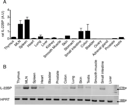
We first assessed the expression of IL-22BP mRNA in rat tissues and organs. The highest levels of IL-22BP were found in SLO, i.e. spleen and mesenteric, axillary and cervical lymph nodes (Figure 1A and Supplementary Figure 1A). Lower levels of expression were found in thymus and several epithelial tissues including gut, lungs, skin and testis. This expression pattern was in agreement with those reported in human and mouse15,17,24. However, although we confirmed that IL-22BP mRNA was strongly expressed in LN in mouse, it was expressed at very low levels in spleen (Supplementary Figure 1B), suggesting species or strain specific variation in pattern of IL-22BP expression.
Three isoforms of IL-22BP exist in human15-17 but only isoform 2 efficiently binds and inhibits IL-2215,16. The physiological roles of the other isoforms remain undetermined. Interestingly, only the human isoform 2 counterpart was identified in mouse24, suggesting that IL-22 regulation by IL-22BP is an evolutionary conserved process. Only one band, corresponding to human isoform 2, was detected in rat tissues (Figure 1B). The absence in the rat il22ra2 gene of a sequence corresponding to human exon 4a16 suggested that isoform 1 did not exist in rat. In agreement with this observation, human isoform 1 was shown to be the result of a long terminal repeat insertion in the ape lineage25. Finally, rat counterpart of human isoform 3 was not detected in these samples.
IL-22BP is highly expressed in rat spleen by a subset of resident cDCs in the steady state
High expression of il22ra2 gene in SLO suggested a hematopoietic origin of IL-22BP cellular sources. To test this hypothesis, different populations of hematopoietic cells were isolated from rat spleen by cell sorting, including the three subsets of DCs we have previously described (plasmacytoid DCs (pDCs), CD172α+ CD4+ and CD172α- CD4- cDCs)26,27. qPCR analysis revealed high IL-22BP expression by the CD172α+ CD4+ cDC
Author Manuscript
Author Manuscript
Author Manuscript
subset (referred thereafter as CD4+ spDCs) whereas other populations only showed low levels of expression (Figure 2A). Moreover, CD4+ spDCs presented a 5-fold higher
expression of IL-22BP than whole spleen (Figure 2B) strongly suggesting that these cells are the major source of IL-22BP in spleen in the steady state.
IL-22BP protein expression was evaluated by immunofluorescence staining of sorted DCs. IL-22BP staining was detected in virtually all CD4+ spDCs, whereas fluorescence observed in CD4- spDCs was not different from that obtained after staining with secondary antibody only (Figure 3).
Finally, we did not detect any expression of the IL-22RA1 transmembrane receptor in the various splenic hematopoietic cell subsets, including CD4+ spDCs (Figure 2C).
Taken together, our data suggest a role for CD4+ spDCs in the regulation of IL-22 biological effects in the steady state via IL-22BP secretion.
A subset of intestinal DC expresses high levels of IL-22BP in the steady state
We hypothesized that cDCs might also be a constitutive source of IL-22BP in epithelial tissues, and particularly in the gut were significant levels of IL-22BP expression were detected (Figure 1). We first analyzed IL-22BP expression by migrating intestinal rat cDC subsets. For this purpose, intestinal lymph DCs (ilDCs) were obtained by lymph collection after thoracic duct cannulation of mesenteric lymphadenectomized rats28. MHCIIhi CD103+ lymph DC subsets were then sorted into three subsets based on their differential expression of CD172α and CD11b, as previously described28,29. High levels of IL-22BP mRNA expression were found in the CD172αhigh ilDC subset (referred thereafter as CD172αhigh ilDC). This expression was about 50-fold higher than IL-22BP expression in small intestine and colon (Figure 4A). High expression of IL-22BP was also observed in CD172+ CD4+ DC from rat MLN (Figure 4B and Supplementary Figure 2) and Peyer Patches (PP) (data not shown). In addition, in vitro DC depletion abolished IL-22BP expression in rat MLN (Figure 4C).
Interestingly, detailed phenotypical, functional and transcriptomic analysis of CD172αhigh ilDCs vs. CD172+ CD4+ spDCs indicated that they have very strong similarities (R. Josien and S. Milling, unpublished observations) and most likely represent the same DC subset. This suggests that high IL-22BP expression is an intrinsic property of this subset of cDCs. Of note, IL-22BP expression was 5-fold higher in CD172αhigh ilDCs than in CD4+ spDCs (Figure 4A). Thus, even if IL-22BP expression is a characteristic of these cells, factors present in the gut environment are also likely to promote higher levels of IL-22BP expression.
To determine whether such a functional specialization of cDCs subsets regarding IL-22BP expression also holds true in mice, we first analyzed data from mouse cDNA microarrays obtained from the Immunological Genome Project (available from the ImmGen website:
www.immgen.org). High levels of IL-22BP expression in steady state were observed in one small intestinal lamina propria (LP) DC subset, namely CD103+ DCs. We confirmed IL-22BP expression by the LP CD103+ CD11b+ subset by qPCR (Figure 4D). In contrast,
Author Manuscript
Author Manuscript
Author Manuscript
only low levels could be detected in CD103+ CD8α+ LPDCs and intestinal LP macrophages. In MLN, MHCIIhigh CD103+ DCs which are known to be migrating DCs30, were sorted in CD11b+ and CD11b- DCs; IL-22BP expression was much stronger in CD11b+ DCs as compared to CD11b- (Figure 4E). Moreover, IL-22BP expression was also significantly diminished in the colon of mice deficient for Flt3L, which are known to lack CD103+ DC (Figure 4F). Unexpectedly, SI from Flt3L−/− mice expressed similar levels of IL-22BP mRNA as compared to wild type mice.
Finally, no IL-22BP expression was found among the different populations of human peripheral blood cells including polymorphonuclear neutrophils, eosinophils and basophils, lymphocytes, monocytes, as well as BDCA1+ and BDCA3+ cDCs and plasmacytoid DCs (data not shown).
Taken together, our results highly support the hypothesis that natural production of IL-22BP results mainly from a constitutive expression by a subset of cDCs both in secondary
lymphoid organs and in epithelial tissues.
Rat CD4+ spDCs express a counterpart of the human short isoform of IL-22BP
PCR experiments revealed the presence of 2 transcripts in rat CD4+ spDCs (Figure 5), the longest and most intense one corresponding to the expected 678 bp mRNA, as further confirmed by sequencing. Translation of the nucleotide sequence obtained after sequencing gave a putative protein of 229 AA (data not shown), showing 72% identity with the human isoform 2 of IL-22BP, i.e the isoform inhibiting IL-22 effects. Interestingly, sequencing of the PCR product corresponding to the shorter band identified a shorter isoform of rat IL-22BP. Like in human, the sequence of this isoform presented a complete excision of exon 5. Alignment of the rat short isoform with its human counterpart revealed 80% identity (Supplementary Figure 3A). Translation of the sequence gave a putative protein of 135 AA, showing 73% identity with human isoform 3 (Supplementary Figure 3B). Expression of this shorter isoform probably results from an alternative mRNA splicing and its physiological significance is unknown. Two important residues for IL-22 binding, Y65 and R117, which are the rat counterparts of human Y67 and R119, are conserved in this short isoform. These 2 residues have been demonstrated to be independently crucial for IL-22 binding20, suggesting that the rat IL-22BP short isoform may also be able to bind IL-22, although probably less efficiently.
IL-22BP is expressed in human monocyte-derived dendritic cells
To study in more detail the regulation of IL-22BP expression in DCs, we analyzed its expression in monocyte-derived dendritic cells (MDDCs). As shown in Figure 6A & B. IL-22BP mRNA expression was strongly induced during DC differentiation. IL-22BP expression at the protein level was confirmed by IFI experiments (Figure 6C). Moreover, no clear IL-22BP induction was detected when monocytes were differentiated in other
conditions than GM-CSF and IL-4-supplemented media (Supplementary Figure 4). Finally, we found that both IL-22BP isoforms 1 and 2 were expressed by MDDCs (Figure 6D) as further confirmed by sequencing (data not shown). Isoform 3 was not detected in human MDDC.
Author Manuscript
Author Manuscript
Author Manuscript
Retinoic acid is a potent inducer of IL-22BP expression by DCs
Since IL-22BP expression was 5-fold higher in rat intestinal DCs than in splenic DCs (Figure 4A), we hypothesized that inducers of IL-22BP expression could be present in the gut environment. We thus analyzed IL-22BP expression by MDDC cultured with various molecules present in high amounts in the gut.
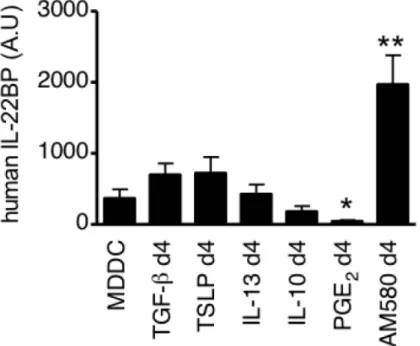
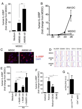
When added during the last 2 days of DC culture, TGF-β, TSLP, IL-13 and IL-10 had no effect on IL-22BP mRNA expression (Figure 7). In contrast, a strong upregulation (8 fold) of IL-22BP expression was induced by AM580, an agonist of retinoic acid receptor alpha (RARα) (Figure 7), suggesting that retinoic acid can enhance IL-22BP expression in DC. The effects of the RARα agonist AM580 were even stronger when added on day 0 of MDDC differentiation. AM-DC (AM580 added on d0) exhibited a strong upregulation of IL-22BP mRNA and protein expression (Figure 8A-C). Interestingly, AM-DC not only upregulated IL-22BP expression, but also acquired a different phenotype characterized by CD103 and CD1d expression, and loss of CD1a expression (Figure 8D), confirming previously published results31,32. The same results were obtained when retinoic acid itself was added in the culture (Figure 8E and data not shown). Moreover, experiments using selective inhibitors of retinoic acid nuclear receptor revealed that retinoic acid effects on IL-22BP, CD103 and CD1d expression induction by MDDC mostly involved RARα signalling (Figure 8E and data not shown).
Retinoic acid is the active metabolite of dietary vitamin A. Vitamin A is first oxidized into retinal by several enzymes including alcohol dehydrogenase, then retinal is oxidized into RA by specific dehydrogenases including retinal dehydrogenase 2 (RALDH2)33. GM-CSF and IL-4 induce the expression of RALDH2 in human MDDC34 early during differentiation (Supplementary Figure 5A&B), which endows them with the capacity to metabolize retinal into RA. IL-22BP expression was upregulated by addition of retinal during DC
differentiation. This induction was significantly diminished by a RALDH2 reversible inhibitor, DEAB (Figure 8F). These findings implied that specific metabolism of retinal by human MDDC allows RA production, which in turn induces IL-22BP expression, probably in an autocrine fashion.
Interestingly, addition of PGE2, which was recently described as a RALDH2 inhibitor34, abolished the expression of IL-22BP in MDDC (Figure 7). This suggests that constitutive expression of IL-22BP by MDDC could be due to low levels of RA production, from retinal present in the serum added in the culture media. However, MDDC differentiated in serum free media or in serum supplemented media with the RARα antagonist also expressed IL-22BP (data not shown and Supplementary Figure 5C), indicating that IL-22BP production is not totally dependent on DC ability to metabolize RA.
RALDH2 expression in MDCC peaked after 20h and progressively decreased until day 6 (Supplementary Figure 5B) which could suggest that RA exerts its effects on MDDC at early stages of the differentiation. We therefore examined the effect of delayed addition of the RARα inhibitor on MDDC differentiation in the presence of RA. CD103 induction was inhibited when the RARα inhibitor was added at day 0, 1, 2 and 3 of the culture but not later
Author Manuscript
Author Manuscript
Author Manuscript
(Supplementary Figure 5D). The effects on CD1d were even more pronounced since inhibition was only observed when the inhibitor was added at the beginning of the culture. This indicated that RA effects on the AM-DC phenotype occur early during the
differentiation. This likely reflects the effects of RA on blood pre-DC entering the gut mucosa, which enter a specific differentiation program and consequently display unique functional properties, as confirmed by previous studies. In contrast, IL-22BP expression was inhibited even when the RARα inhibitor was added at day 5 of the culture (Supplementary Figure 5E) indicating that RA-induced IL-22BP expression required continuous signaling through RAR.
Altogether, these data identify RA as a potent inducer of IL-22BP expression in DCs. This suggests that the higher expression of IL-22BP by rat CD172a+ ilDCs as compared to CD4+ spDCs is due to the ability of the former cells to produce RA. In agreement with this,
aldh1a2 gene expression (encoding for RALDH2) was detected in ilDCs and not in spleen
cDCs (Figure 8G). Thus, the higher levels of IL-22BP expression of rat intestinal DCs compared to their splenic counterpart are likely to be the consequence of a permanent exposure to RA, explained notably by their ability to produce it.
IL-22BP is down-regulated when DCs undergo maturation
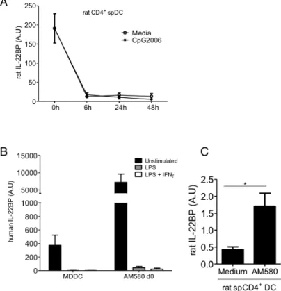
IL-22BP expression appears to be a constitutive property of a subset of cDC at an immature state in SLO and epithelial tissues. We then assessed whether IL-22BP expression was modulated upon DC maturation. Spontaneous maturation, which occurs when DCs are cultured without any stimulators, induced a rapid and dramatic down-regulation of IL-22BP expression in rat CD4+ spDCs and MLN-DCs (Figure 9A and Supplementary Figure 6). This down-regulation persisted for up to 48 hours. Stimulation by CpG ODN2006, a ligand for TLR9, did not affect this down-regulation of IL-22BP expression (Figure 9A). Similar results were obtained after stimulation of DCs by other TLR ligands (including ligands for TLRs 1-2, 2-6, 3, 4 and 7/8) (data not shown).
In human MDDC and AM-DC, LPS and LPS + IFNγ-induced maturation was associated with a dramatic decrease of IL-22BP expression (Figure 9B). Similar results were obtained with ligands of TLR3 (polyI:C) or TLR5 (flagellin) (data not shown). Altogether, these data indicate that constitutive expression of IL-22BP by DC at an immature state is lost upon DC maturation. This is concordant with previous studies describing a down-regulation of IL-22BP expression in inflammatory conditions, notably in mouse DSS induced colitis8,19. Interestingly, AM580 could partly restore IL-22BP expression by rat CD4+ spDCs after spontaneous maturation (Figure 9C), again supporting a role for RA as a positive regulator of IL-22BP expression. The incomplete restoration of IL-22BP expression in mature DCs probably reflects that RARα signaling is not sufficient to fully counteract the maturation-induced down regulation of IL-22BP. Similar observations of RA and maturation impacts on IL-22BP expression could be made in mouse bone marrow-derived DCs (Supplementary Figure 7), even if IL-22BP expression levels were low in this model.
Author Manuscript
Author Manuscript
Author Manuscript
Discussion
Since its first description in 2000, the actions of IL-22 in mediating the cross-talk between the immune and epithelial systems were extensively described. By its ability to reinforce innate immunity of epithelial cells, notably by inducing antimicrobial peptides production, IL-22 has been demonstrated to be crucial in the resolution of Enterobacteriaceae gut and lung infections5,6. Combined with the capacity of IL-22 to promote tissue repair in several inflammatory conditions3,7-9, IL-22 properties seem necessary in the maintenance of tissue homeostasis. However, excessive actions of IL-22 may be particularly deleterious for epithelial tissues10-12, suggesting that IL-22 needs to be tightly regulated to prevent unwarranted tissue damage and dysregulated epithelial cell proliferation. This regulation may be partly achieved by IL-22BP, given its specific inhibitory properties on IL-22 actions8,15-17.
Our study strongly suggests that the natural production of IL-22BP, observed in secondary lymphoid organs and epithelial tissues, may be principally achieved by a subset of cDCs. These data extend the recent report by Huber et al. which showed in mice that IL-22BP-producing cells in colon were mainly MHCII+ CD11c+ cDCs35. IL-22BP production by rat spCD4+ cDCs was observed at an immature state, whereas maturation induces a dramatic down-regulation of IL-22BP expression. Consistent with previous studies8,19, these results suggest that IL-22BP production is prominent in the steady state, but not in inflammatory conditions, which lead to DC maturation. Nevertheless, IL-22BP up-regulation has been observed in several infectious models, particularly in acute polymicrobial peritonitis, and after orally administration of Toxoplasma gondii36,37. Interestingly, IL-22 was shown to be detrimental during these processes, and led to uncontrolled deleterious inflammation. This suggests that control of IL-22 effects requires a tight regulation of IL-22BP production by cDCs. cDCs may produce high levels of IL-22BP at physiological state, when IL-22 actions are not desired, then, depending on the stimuli, IL-22BP production could be further enhanced or stopped. In our study, we could not induce an up-regulation of IL-22BP expression by stimulating spCD4+ cDCs with several TLR ligands. In addition, inflammasome activation was recently shown to play an important role in the
down-regulation of IL-22BP expression in mouse colon during inflammation35. Moreover, in vitro spontaneous maturation of cDCs and MDDC also down-regulated IL-22BP expression. Spontaneous maturation of mouse BMDC was shown to result from the disruption of E-cadherin-mediated adhesion of DCs within each other, inducing the activation of the β-catenin signalling pathway38. This process is characterized by a specific transcriptional profile and supposed to favour the generation of tolerogenic DCs in vivo. However, during the early phase of maturation, spontaneous and TLRs matured DCs exhibit a similar
expression profile. Because IL-22BP is rapidly and equally down-regulated in spontaneously and TLRs-matured DCs (Figure 9A), it is likely that the il22ra2 gene belongs to this
common program of maturated DCs. Consequently, it remains to determine which cells can produce IL-22BP under inflammatory conditions.
The significance of the high levels of IL-22BP in SLO remains elusive. Indeed, only low levels of IL-22R1 expression are found in SLO. Moreover, IL-22 is not likely to act on immune cells in SLO since they do not express IL-22R1 at all2 (Figure 2C). One possible
Author Manuscript
Author Manuscript
Author Manuscript
explanation could be that the constitutive expression of IL-22BP by resident cDCs, which are highly represented in SLO, reflects ontogeny-related specializations similar to epithelial tissue cDCs. Indeed, high IL-22BP expression in rat CD4+ spDCs and CD172α+ iLDCs might reflect a specific gene signature of this particular subset, similar to other genes such as SIRPα or DCIR1 for instance. DCs subsets are classified on the basis of distinct cell-surface markers and display a specific gene signature explaining some functional specializations39. Although IL-22R1 is not expressed by normal immune cells, aberrant expression by transformed lymphocytes has been described40. Actions of IL-22 on these cells contributed to the pathogenesis of mantle cell leukemia and anaplastic large cells lymphoma40,41. Therefore, high IL-22BP production in SLO could be necessary to prevent IL-22 actions on tumor cells thus limiting the installation of these malignancies.
Strikingly, IL-22BP expression was about 5-fold higher in the rat intestinal CD172αhigh iLDCs compared to their splenic counterparts, suggesting that the gut environment is likely to favour IL-22BP production by these cells. We found that RA was a potent inducer of IL-22BP expression in vitro in human MDDC. Moreover, MDDC can oxidize retinal into RA, due to their expression of RALDH234. De novo production of RA could further enhance IL-22BP expression by these cells, probably in an autocrine fashion.
Interestingly, in mouse, a subset of intestinal DCs expressing CD103 constitutively expresses RALDH2. This subset mainly resides in the small intestine lamina propria (SI-LP) and constitutively migrates toward draining lymph nodes30. Mouse intestinal CD103+ DCs derive from a DC precursor continuously seeding the SI-LP from the circulation42. Differentiation of this precursor toward the CD103+ DC subset is likely due to
environmental factors present in the SI-LP43, including RA itself.
Our data suggest that, in mouse, the CD103+ CD11b+ subset of cDCs represent a constitutive source of IL-22BP in the gut. Notably, although Huber et al. reported much lower expression of IL-22BP in small intestine vs. colon35, we found similar expression in both segments. Our finding that IL-22BP expression was strongly reduced in large intestine from Flt3L−/− mice suggests that Flt3L-dependent cDCs are indeed a major source of IL-22BP in large intestine which is consistent with data from Huber et al. The apparent normal expression of IL-22BP mRNA we found in SI from Flt3L-deficient mice (Figure 4F) was more surprising and suggests that IL-22BP could be expressed by Flt3L-independent DCs or by other cells than DCs in SI, either constitutively or due to compensation process. These hypotheses need to be tested in vivo.Rat CD172αhigh ilDCs, which express very high levels of IL-22BP, display similar properties to the mouse CD103+ DC subset, including CD103 expression, constitutive migration from the intestinal mucosa towards the draining mesenteric lymph nodes, and constitutive expression of RALDH2 (Figure 8G). Therefore, the CD172αhigh ilDC subset is likely to be the rat counterpart of the mouse CD103+ CD11b+ DCs. Further study will be required to identify IL-22BP protein-producing cells in the LP as well as in SLO as we did not succeeded in staining tissue with commercially available mAbs. In fact, there is no information in the current literature, including in the recent paper from Huber et al., about IL-22BP protein expression in vivo.
Author Manuscript
Author Manuscript
Author Manuscript
Maintenance of gut homeostasis is complex and implicates the establishment of equilibrium between the host and the commensal microbiota, while maintaining the ability to fight against invading pathogens. The gut immune system plays an important role in this equilibrium and needs to be tightly regulated to avoid unbalanced reactions leading to inflammatory bowel diseases (IBD). Interestingly, the CD103+ DC subset seems to actively participate in maintenance of the gut homeostasis due to its unique functional properties, conserved between mice and humans42. These properties include the ability of imprinting gut homing receptors on naive lymphocytes, the induction of CD4+ naïve T cells
differentiation into FOX-P3+ regulatory T cells and the differentiation of IgA-secreting B cells. All these properties result from the ability of these cells to efficiently produce RA from retinal44.
Regulation of inflammatory cytokine production is also crucial in the maintenance of gut homeostasis. High IL-22 production by LTi cells was observed in the mouse fetus and before weaning45. This high production of IL-22, concomitant with IL-17, is thought to be necessary to promote a local pro-inflammatory milieu at birth enabling the containment of the colonizing microbiota and the selection of appropriate flora46. After birth, down-modulation of IL-22 production is observed for both LTi and ILC22 cells, following the integration of negative signals from the commensal flora45. Interestingly, intestinal DCs appear to be involved in this regulation by responding to microbiota-induced IL-25 produced by epithelial cells45. IL-25-responding DCs could further moderate IL-22
production by ILCs, in a contact-dependent way. However, whether these DCs corresponded to the CD103+ subset was not analyzed in this study. Our results further suggests that CD103+ DCs could also regulate IL-22 actions by constitutively producing high levels of its natural soluble inhibitory receptor, reinforcing the importance of DCs in intestinal immune regulation44.
Only two studies addressed the role of IL-22BP in vivo. In a mouse model of acute DSS colitis, Sugimoto et al. demonstrated that beneficial effects of IL-22 on epithelium regeneration were abolished in the presence of IL-22BP8. This study was the first to demonstrate effects of IL-22BP on IL-22 actions described in vivo. However, IL-22BP was shown to be down-regulated during the acute phase of colitis, when IL-22 is highly produced. Thus, the model remained artificial as IL-22BP overexpression was induced by a local gene-delivery system. Moreover, neither the cellular source of native IL-22BP nor its protein expression in vivo were addressed in this study. A second study was published during the reviewing process of our manuscript by RA Flavell and coll. In this paper, the authors used a model of IL-22BP−/− mice35. They confirmed the coordinated regulation of IL-22 and IL-22BP during DSS colitis and showed that IL-22BP is crucial in the control of IL-22 proliferative effects on epithelial cells. This control was particularly important to limit the tumorigenesis in a chronic colitis-associated colon cancer model. Importantly, the authors demonstrated that the increased tumorigenesis in IL-22BP−/− mice was directly linked to the lack of IL-22 control, leading to prolonged deleterious actions of the cytokine. Thus, the authors showed for the first time the need for a tight regulation exerted by IL-22BP on IL-22 in vivo. However, IL-22BP−/− mice did not show any phenotype in the steady state thus questioning the significance of constitutive IL-22BP expression in
Author Manuscript
Author Manuscript
Author Manuscript
lymphoid tissues and intestine. Moreover, the authors also suggested in their study that DCs represent the major source of IL-22BP in mouse colon, a finding we confirmed here and further detailed by showing that only a subset of cDCs was expressing IL-22BP.
These data indicate that a tight, timely and local regulation of IL-22 actions, likely by IL-22BP-expressing DCs, is critical in inflammatory conditions. Nonetheless, IL-22 also plays a crucial role in the protection of the host from gut pathogens5,6. During infection, strong up-regulation of IL-22 is observed. IL-23 seems to be the main inducer of IL-22 production, principally by ILCs, which constitutively express IL-23R47. Interestingly, mouse CD103+ CD11b+ SI-LP DCs were recently shown to mediate rapid IL-22 production by ILC upon TLR5 triggering, through production of IL-2347. CD103+ CD11b+ DCs also promote the differentiation of Th17 cells, which are known to produce IL-2214, after TLR5 stimulation48. Interestingly, TLR5 expression is a unique feature of these cells and also appears to be RA dependent49. Combined with our data, this strongly suggests that the same subset is also the major source of IL-22BP in the intestine in the steady state, therefore reinforcing the central role of this DC subset in the control of IL-22. Finally, this subset of mouse CD103+ DC was recently proposed to be the source of IL-23 in the thymus after injury. Again, IL-23 induced IL-22 production by ILCs. This IL-22 production was crucial for thymic recovery after injury but not for thymic development50. IL-22BP expression by these cells was not explored but the low levels of IL-22BP expression in the thymus compared to SLO and gut suggest that the supposed IL-22BP production by CD103+ thymic DC would probably be less important.
In conclusion, our study identified a subset of immature cDCs as a constitutive source of IL-22BP in the steady state in SLO and gut. Together with the recent report from Huber et al, these data strongly suggest that DC-produced IL-22BP plays an important role in controlling IL-22 effect on epithelial cells. Moreover, our study suggests that the regulation of IL-22 by IL-22BP in the gut could be enhanced by RA. An unsolved crucial point concerns the constitutive secretion of IL-22BP by DCs which has never been demonstrated so far. The lack of an accurate and sensitive enough assay impeded our measurement of IL-22BP levels in culture supernatants. A better understanding of IL-22BP protein production and regulation by DCs, and its importance in IL-22 regulation in the gut is necessary to establish the relationship between the observed beneficial and deleterious effects of the cytokine. It will also be important to confirm these data in human. This could be of importance in understanding the role played by IL-22 in IBD, which still remains ambiguous today8,9,51.
Methods
AnimalsSprague Dawley (SPD) rats were obtained from the Centre d’Elevage Janvier (Le Genest-St Isle, France) and were used when 6-10 weeks old. All animal experiments were performed under specific pathogen-free conditions in accordance with the European Union Guidelines. All animal studies were conducted according to the guidelines of the French Agriculture Ministry. The studies were approved by the Veterinary Departmental Services committee (# E.44011).
Author Manuscript
Author Manuscript
Author Manuscript
Reagents
Cells were cultured in complete RPMI 1640 (Invitrogen, Carlsbad, CA). Recombinant human GM-CSF, IL-4 and IL-10 were from CellGenix (Fribourg-en-Brisgau, Germany), recombinant human TGF-β and IL-13 were from Peprotech (Neuilly-sur-Seine, France), recombinant IFN-γ was from R&D Systems Europe (Lille, France). PGE2, RA, AM580, retinal, DEAB and LPS were from Sigma Aldrich (St Louis, MI). CpG ODN2006 was from Eurofins MWG Operon (Ebersberg, Germany). Selective retinoic acid nuclear receptor inhibitors BMS 195314 (RARα inhibitor), UVI 3003 (RXR inhibitor) and CD 2665 (RARβ/γ inhibitor) were from Tocris bioscience (Lille, France).
Cell sorting
Rat splenic cells—Rat conventional and plasmacytoid DCs subsets were isolated as previously described26,52,53. For lymphocytes and monocytes, cells were first separated by Ficoll-Paque Plus (GE HealthCare Life Sciences, Uppsala, Sweden) gradient centrifugation. For lymphocytes, cells were stained with TCRαβ-biotin (clone R7.3), CD45R-PE (clone HIS24), CD45RA-FITC (clone OX33), NKR-P1-Alexa647 (clone 3-2-3) mAbs, and then with streptavidin-PE-Cy7. Live TCRαβ+ (T cells), TCRαβ− CD45R+ CD45RA+ NKR-P1− (B cells) and TCRαβ− CD45R− CD45RA− NKRP1+ (NK cells) were Facs-sorted. For monocytes, cells were stained with MHC II-PE (clone OX6), CD172α-biotin (clone OX41), CD103-Alexa647 (clone OX62) and CD11b/c-Alexa488 (clone OX42) mAbs, and then with streptavidin PE-Cy7. Live CMH-II+ CD103− CD172α+ CD11b/c+ (monocytes) were Facs-sorted.
Rat intestinal lymph DCs—Lymph was collected from male animals using previously-described methods28,29. Briefly, MLNX was performed on 5-6-week-old animals. At least five weeks later, a cannula (3Fr, Harvard Apparatus, UK) was inserted into the thoracic duct at laparotomy. Lymph was collected for up to 48 hours on ice. RBCs were lysed from lymph, and the single cell suspensions were enriched for DC using magnetic beads specific for CD103 (Miltenyi Biotec, Paris, France) according to manufacturer’s protocol. Enriched cells were stained with antibodies for MHCII (OX-6, BD Biosciences), CD103 (OX-62, in house), CD11b (OX-42, BD Biosciences) and CD172a (OX-41, in house), and the three L-DC subsets were flow sorted using BD FACS Aria Cell sorter. RNA was isolated from sorted cells using Qiagen RNeasy Mini Kit according to manufacturers’ instructions. Complementary DNA (cDNA) was reverse-transcribed from RNA using Superscript First-Strand Synthesis System for RT-PCR (Invitrogen).
Rat MLN cDCs—MLN were harvested from adult SPD rats, dilacerated using 26G needles and digested for 25 min with Collagenase D, in the presence of DNAse I. Cells were collected and low density cells were prepared using a 14,5% Nycodenz gradient. Cells were then stained with anti-TCRαβ (clone R7/3), anti-CD45R (clone HIS24), anti-CD103 (clone OX62) and anti-CD4 (clone W3/25) and sorted on a FACS Aria into TCRαβ- CD45R- CD103+ CD4+/int/− cDCs. Purity was routinely >95%.
Mouse lamina propria cells—Small intestines were flushed with HBSS 2% FCS and the Peyer’s patches excised. The intestines were opened longitudinally and cut into 0.5cm
Author Manuscript
Author Manuscript
Author Manuscript
segments, which were incubated twice in HBSS with 2mM EDTA at 37°C while shaking for twenty minutes. Supernatants were discarded and the tissue digested with 1mg/ml of collagenase VIII (Sigma-Aldrich) at 37°C with shaking for 15 minutes. Cells were passed through a 40μm cell strainer and stained for flow cytometry. DC (Live CD45+ MHCII+ CD11c+ F4/80− CD103+ CD11b+/-) and Macrophages (Live CD45+ MHCII+ F4/80+ CD11b+) were sorted using a FACSAria I (purity was routinely >95%).
Mouse MLN CD103+ cDCs—MLN were digested using liberase (0.4 Wunsch Units/mL; Roche, Meylan, France) and DNAse (50 μg/mL; Roche) for 45 min at +37°C. Single cell suspensions were stained for flow cytometry with MHCII IA/IE (clone M5/114.15.2) antibody from ebioscience (San Diego, CA) and CD11b (clone M1/70), B220 (clone RA3-6B2), CD103 (clone 2E7), CD11c (clone N418) antibodies from Biolegend (London, UK).
Human cell preparation
Human blood was obtained from healthy donors upon informed consent in accordance with our Institutional Review Board. Peripheral blood mononuclear cells were separated by density gradient centrifugation over Ficoll (PAA, Pasching, Austria). CD14+ monocytes were isolated by positive selection (Miltenyi Biotec) and differentiated into DCs with 1,000 U/ml GM-CSF and 200 U/ml IL-4 for 6 days. When indicated, cells were treated with AM580 or RA (100 nM), IL-10 (50 ng/ml), IL-13 (10 ng/ml), TGF-β (10 ng/ml), PGE2 (10 ng/ml), retinal (100 nM), DEAB (150 μM), LPS (1 μg/ml), IFN-γ (50 ng/ml), BMS 195314 (5 μmol/L), UVI 3003 (5 μmol/L) or CD 2665 (5 μmol/L).
Flow cytometry analysis
Human MDDC were stained with HLA-DR-APC (clone L243 (G46-6)), CD209-PE (clone DCN46), CD1d-PE (clone CD1d42) and CD103-FITC (cloneBer-ACT8), all from BD Biosciences, or isotype-matched control antibodies. Cells were analyzed on a BD
FACSCanto II flow cytometer (BD Biosciences). Data were analyzed using FlowJo software (Treestar, Ashland, OR).
Real-time quantitative RT-PCR
Total RNA was isolated using Trizol reagent (Invitrogen) or Qiagen RNeasy Mini Kit according to manufacturers’ instructions. Reverse transcription was performed using Murine Moloney Leukemia Virus Reverse Transcriptase (Invitrogen) or Superscript First-Strand Synthesis System for RT-PCR (Invitrogen), following manufacturer’s instructions. For rat IL-22BP, IL-22RA1, ALDH1A2, mouse IL-22BP and human ALDH1A2 gene expression, Power Sybr® Green 2× reagent was used (Applied Biosystems, Foster City, CA). Real-time PCR was performed using the Viia™ 7 Real Time PCR system (Applied Biosystems). Primers (Eurofins MWG Operon, Ebersberg, Germany) are summarized in supplementary Table 1. For human IL-22BP gene expression, TaqMan® Fast Advanced Master Mix 2× reagent was used (Applied Biosystems). Primers and probes were from Applied Biosystems. Real-time PCR was performed using the StepOne Plus system (Applied Biosystems). For both human and rat relative expression was normalized from HPRT and calculated using the
Author Manuscript
Author Manuscript
Author Manuscript
2-ΔΔCt method. For mouse, relative expression was normalized from GAPDH. Results were expressed in arbitrary units (A.U).
Whole IL-22BP cDNA amplification
In order to identify IL-22BP isoforms, whole cDNA amplification was performed in both human and rat samples. Primers used for rat and human IL-22BP are summarized in supplementary Table 2. Amplification was performed using HERCULASE II Fusion Enzyme (Agilent, Santa Clara, CA).
Indirect Immunofluorescence
Cells were let to adhere on poly-L-Lysine pre-coated slides for 30 min (Sigma Aldrich). Fixation was performed for 5 min in −20°C pre-cold acetone. After rehydration, a 30 min step saturation was made with PBS/BSA1%/Serum10%. Primary antibody (anti-IL-22BP from R&D Systems for human MDDC or anti-rat IL-22BP from Santa Cruz Biotechnology Inc. for rat cDCs) was incubated at RT for 2 hours. Secondary antibodies were then incubated for 1 hour at RT.
Statistical analysis
Data are represented as means ± SEM. Statistical analysis was performed with GraphPad Prism® Software (GraphPad Software, San Diego, CA, USA). Means comparisons were performed using the Mann & Whitney U-test or the Kruskal-Wallis test with Dunn’s post test. P-values <0.05 were considered statistically significant.
Supplementary Material
Refer to Web version on PubMed Central for supplementary material.
Acknowledgments
The authors want to thank Pr JC Renauld (Université Catholique de Louvain, Belgium) for critically reading the manuscript.
References
1. Sonnenberg GF, Fouser LA, Artis D. Border patrol: regulation of immunity, inflammation and tissue homeostasis at barrier surfaces by IL-22. Nat. Immunol. 2011; 12:383–390. [PubMed: 21502992]
2. Wolk K, et al. IL-22 increases the innate immunity of tissues. Immunity. 2004; 21:241–254. [PubMed: 15308104]
3. Radaeva S, Sun R, Pan H-N, Hong F, Gao B. Interleukin 22 (IL-22) plays a protective role in T cell-mediated murine hepatitis: IL-22 is a survival factor for hepatocytes via STAT3 activation. Hepatology. 2004; 39:1332–1342. [PubMed: 15122762]
4. Aggarwal S, Xie MH, Maruoka M, Foster J, Gurney AL. Acinar cells of the pancreas are a target of interleukin-22. J. Interferon Cytokine Res. 2001; 21:1047–1053. [PubMed: 11798462]
5. Zheng Y, et al. Interleukin-22 mediates early host defense against attaching and effacing bacterial pathogens. Nat. Med. 2008; 14:282–289. [PubMed: 18264109]
6. Aujla SJ, et al. IL-22 mediates mucosal host defense against Gram-negative bacterial pneumonia. Nat. Med. 2008; 14:275–281. [PubMed: 18264110]
Author Manuscript
Author Manuscript
Author Manuscript
7. Simonian PL, et al. γδ T cells protect against lung fibrosis via IL-22. J. Exp. Med. 2010; 207:2239– 2253. [PubMed: 20855496]
8. Sugimoto K, et al. IL-22 ameliorates intestinal inflammation in a mouse model of ulcerative colitis. J. Clin. Invest. 2008; 118:534–544. [PubMed: 18172556]
9. Zenewicz LA, et al. Innate and adaptive interleukin-22 protects mice from inflammatory bowel disease. Immunity. 2008; 29:947–957. [PubMed: 19100701]
10. Zheng Y, et al. Interleukin-22, a T(H)17 cytokine, mediates IL-23-induced dermal inflammation and acanthosis. Nature. 2007; 445:648–651. [PubMed: 17187052]
11. Geboes L, et al. Proinflammatory role of the Th17 cytokine interleukin-22 in collagen-induced arthritis in C57BL/6 mice. Arthritis Rheum. 2009; 60:390–395. [PubMed: 19180498]
12. Muñoz M, et al. Interleukin (IL)-23 mediates Toxoplasma gondii-induced immunopathology in the gut via matrixmetalloproteinase-2 and IL-22 but independent of IL-17. J. Exp. Med. 2009; 206:3047–3059. [PubMed: 19995958]
13. Sonnenberg GF, et al. Pathological versus protective functions of IL-22 in airway inflammation are regulated by IL-17A. J. Exp. Med. 2010; 207:1293–1305. [PubMed: 20498020]
14. Liang SC, et al. Interleukin (IL)-22 and IL-17 are coexpressed by Th17 cells and cooperatively enhance expression of antimicrobial peptides. J. Exp. Med. 2006; 203:2271–2279. [PubMed: 16982811]
15. Dumoutier L, Lejeune D, Colau D, Renauld JC. Cloning and characterization of IL-22 binding protein, a natural antagonist of IL-10-related T cell-derived inducible factor/IL-22. J. Immunol. 2001; 166:7090–7095. [PubMed: 11390453]
16. Kotenko SV, et al. Identification, cloning, and characterization of a novel soluble receptor that binds IL-22 and neutralizes its activity. J. Immunol. 2001; 166:7096–7103. [PubMed: 11390454] 17. Xu W, et al. A soluble class II cytokine receptor, IL-22RA2, is a naturally occurring IL-22
antagonist. Proc. Natl. Acad. Sci. U.S.A. 2001; 98:9511–9516. [PubMed: 11481447] 18. Logsdon NJ, Jones BC, Josephson K, Cook J, Walter MR. Comparison of interleukin-22 and
interleukin-10 soluble receptor complexes. J. Interferon Cytokine Res. 2002; 22:1099–1112. [PubMed: 12513909]
19. Wolk K, et al. IL-22 induces lipopolysaccharide-binding protein in hepatocytes: a potential systemic role of IL-22 in Crohn’s disease. J. Immunol. 2007; 178:5973–5981. [PubMed: 17442982]
20. De Moura PR, et al. Crystal structure of a soluble decoy receptor IL-22BP bound to interleukin-22. FEBS Lett. 2009; 583:1072–1077. [PubMed: 19285080]
21. Banchereau J, Steinman RM. Dendritic cells and the control of immunity. Nature. 1998; 392:245– 252. [PubMed: 9521319]
22. Shortman K, Naik SH. Steady-state and inflammatory dendritic-cell development. Nat. Rev. Immunol. 2007; 7:19–30. [PubMed: 17170756]
23. Villadangos JA, Schnorrer P. Intrinsic and cooperative antigen-presenting functions of dendritic-cell subsets in vivo. Nat. Rev. Immunol. 2007; 7:543–555. [PubMed: 17589544]
24. Weiss B, et al. Cloning of murine IL-22 receptor alpha 2 and comparison with its human counterpart. Genes Immun. 2004; 5:330–336. [PubMed: 15201862]
25. Piriyapongsa J, Polavarapu N, Borodovsky M, McDonald J. Exonization of the LTR transposable elements in human genome. BMC Genomics. 2007; 8:291. [PubMed: 17725822]
26. Trinité B, Voisine C, Yagita H, Josien R. A subset of cytolytic dendritic cells in rat. J. Immunol. 2000; 165:4202–4208. [PubMed: 11035052]
27. Turnbull E, MacPherson G. Immunobiology of dendritic cells in the rat. Immunol. Rev. 2001; 184:58–68. [PubMed: 12086321]
28. Milling S, MacPherson G. Isolation of rat intestinal lymph DC. Methods Mol. Biol. 2010; 595:281–297. [PubMed: 19941120]
29. Milling SWF, Jenkins C, MacPherson G. Collection of lymph-borne dendritic cells in the rat. Nat Protoc. 2006; 1:2263–2270. [PubMed: 17406466]
30. Milling S, Yrlid U, Cerovic V, MacPherson G. Subsets of migrating intestinal dendritic cells. Immunol. Rev. 2010; 234:259–267. [PubMed: 20193024]
Author Manuscript
Author Manuscript
Author Manuscript
31. Iliev ID, et al. Human intestinal epithelial cells promote the differentiation of tolerogenic dendritic cells. Gut. 2009; 58:1481–1489. [PubMed: 19570762]
32. Szatmari I, et al. PPARgamma controls CD1d expression by turning on retinoic acid synthesis in developing human dendritic cells. J. Exp. Med. 2006; 203:2351–2362. [PubMed: 16982809] 33. Iwata M, Yokota A. Retinoic acid production by intestinal dendritic cells. Vitam. Horm. 2011;
86:127–152. [PubMed: 21419270]
34. Stock A, Booth S, Cerundolo V. Prostaglandin E2 suppresses the differentiation of retinoic acid-producing dendritic cells in mice and humans. J. Exp. Med. 2011; 208:761–773. [PubMed: 21444662]
35. Huber S, et al. IL-22BP is regulated by the inflammasome and modulates tumorigenesis in the intestine. Nature. 2012; 491:259–263. [PubMed: 23075849]
36. Weber GF, et al. Inhibition of interleukin-22 attenuates bacterial load and organ failure during acute polymicrobial sepsis. Infect. Immun. 2007; 75:1690–1697. [PubMed: 17261606] 37. Wilson MS, et al. Redundant and pathogenic roles for IL-22 in mycobacterial, protozoan, and
helminth infections. J. Immunol. 2010; 184:4378–4390. [PubMed: 20220096]
38. Jiang A, et al. Disruption of E-cadherin-mediated adhesion induces a functionally distinct pathway of dendritic cell maturation. Immunity. 2007; 27:610–624. [PubMed: 17936032]
39. Merad M, Sathe P, Helft J, Miller J, Mortha A. The dendritic cell lineage: ontogeny and function of dendritic cells and their subsets in the steady state and the inflamed setting. Annu. Rev. Immunol. 2013; 31:563–604. [PubMed: 23516985]
40. Gelebart P, Zak Z, Dien-Bard J, Anand M, Lai R. Interleukin 22 signaling promotes cell growth in mantle cell lymphoma. Transl Oncol. 2011; 4:9–19. [PubMed: 21286373]
41. Savan R, et al. A novel role for IL-22R1 as a driver of inflammation. Blood. 2011; 117:575–584. [PubMed: 20971950]
42. Jaensson E, et al. Small intestinal CD103+ dendritic cells display unique functional properties that are conserved between mice and humans. J. Exp. Med. 2008; 205:2139–2149. [PubMed: 18710932]
43. Scott CL, Aumeunier AM, Mowat AM. Intestinal CD103+ dendritic cells: master regulators of tolerance? Trends Immunol. 2011; 32:412–419. [PubMed: 21816673]
44. Agace WW, Persson EK. How vitamin A metabolizing dendritic cells are generated in the gut mucosa. Trends Immunol. 2011 doi:10.1016/j.it.2011.10.001.
45. Sawa S, et al. RORγt(+) innate lymphoid cells regulate intestinal homeostasis by integrating negative signals from the symbiotic microbiota. Nat. Immunol. 2011; 12:320–326. [PubMed: 21336274]
46. Eberl G. Development and evolution of RORγt(+) cells in a microbe’s world. Immunol. Rev. 2012; 245:177–188. [PubMed: 22168420]
47. Kinnebrew MA, et al. Interleukin 23 production by intestinal CD103(+)CD11b(+) dendritic cells in response to bacterial flagellin enhances mucosal innate immune defense. Immunity. 2012; 36:276– 287. [PubMed: 22306017]
48. Uematsu S, et al. Regulation of humoral and cellular gut immunity by lamina propria dendritic cells expressing Toll-like receptor 5. Nat. Immunol. 2008; 9:769–776. [PubMed: 18516037] 49. Cho H-Y, et al. All-trans retinoic acid induces TLR-5 expression and cell differentiation and
promotes flagellin-mediated cell functions in human THP-1 cells. Immunol. Lett. 2011; 136:97– 107. [PubMed: 21237205]
50. Dudakov JA, et al. Interleukin-22 drives endogenous thymic regeneration in mice. Science. 2012; 336:91–95. [PubMed: 22383805]
51. Brand S, et al. IL-22 is increased in active Crohn’s disease and promotes proinflammatory gene expression and intestinal epithelial cell migration. Am. J. Physiol. Gastrointest. Liver Physiol. 2006; 290:G827–838. [PubMed: 16537974]
52. Anjubault T, et al. Constitutive Expression of TNF-Related Activation-Induced Cytokine (TRANCE)/Receptor Activating NF-κB Ligand (RANK)-L by Rat Plasmacytoid Dendritic Cells. PLoS ONE. 2012; 7:e33713. [PubMed: 22428075]
Author Manuscript
Author Manuscript
Author Manuscript
53. Hubert F-X, Voisine C, Louvet C, Heslan M, Josien R. Rat plasmacytoid dendritic cells are an abundant subset of MHC class II+ CD4+CD11b-OX62- and type I IFN- producing cells that exhibit selective expression of Toll-like receptors 7 and 9 and strong responsiveness to CpG. J. Immunol. 2004; 172:7485–7494. [PubMed: 15187127]
Author Manuscript
Author Manuscript
Author Manuscript
Figure 1. Tissue expression pattern of IL-22BP in the rat
(A) IL-22BP gene expression was analyzed by RT-qPCR. Bars represent the mean ± SEM
ratio of IL-22BP gene to HPRT expression as determined by the 2-ΔΔCt method of relative quantification from 2 experiments. (B) IL-22BP gene expression was analyzed by RT-PCR using primers allowing amplification of whole il22ra2 mRNA. Values indicate expected molecular mass of PCR products. Data are representative of two independent experiments.
Author Manuscript
Author Manuscript
Author Manuscript
Figure 2. IL-22BP is highly expressed in the spleen by a subset of resident cDCs
(A) The different populations of splenic hematopoietic cells were isolated by cell sorting.
IL-22BP gene expression was analyzed by RT-qPCR and normalized relative to HPRT expression (n=4 for DC; n=5 for B cells; n=3 for T cells, NK cells and monocytes). (B) IL-22BP gene expression was compared by RT-qPCR for DCs (n=3) in spleen, MLN and thymus (n=2). In A and B bars represent mean ± SEM of IL-22BP gene to HPRT expression as determined by the 2-ΔΔCt method of relative quantification. (C) Hematopoietic cells were isolated by cell sorting. IL-22R1 gene expression was analyzed by RT-qPCR with several tissues being used as positive controls (n=3 for cell samples; n=2 for tissue samples). Bars represent mean ± SEM ratio of IL-22R1 gene to HPRT expression as determined by the 2−ΔΔCt method of relative quantification. * p<0.05.
Author Manuscript
Author Manuscript
Author Manuscript
Figure 3. IL-22BP staining on sorted subsets of rat splenic conventional DC
The two subsets of rat conventional spleen DCs were isolated by cell sorting and let to adhere on poly-L-Lysine pre-coated slides for 30 min in the presence of Brefeldin A and monensin. Slides were then stained with a goat anti-rat IL-22BP pAb followed by a donkey anti-goat IgG-Alexa fluor 568. Controls were performed using the secondary antibody alone. Data are representative of two independent experiments.
Author Manuscript
Author Manuscript
Author Manuscript
Figure 4. A subset of intestinal DC expresses high levels of IL-22BP in the steady state
(A) Constitutively migrating intestinal lymph DCs (ilDCs) subsets were obtained by lymph
collection after thoracic duct cannulation of mesenteric lymphadenectomized rat, and cell sorting. IL-22BP gene expression was analyzed by RT-qPCR for the three subsets of ilDCs and compared to intestinal tissues and its CD4+ DC spleen counterpart (n=3 for ilDCs; n=2 for tissues). (B) The 3 subpopulations of rat MLN-DCs were isolated by cell sorting and analyzed for IL-22BP expression by RT-qPCR (n=3). (C) Total DCs were depleted from total MLN cells by FACS-depletion of CD103+ cells. IL-22BP gene expression was analyzed by RT-qPCR (n=3). (D) Mouse CD8+ and CD11b+ CD103+ DC subsets and macrophages were isolated from intestinal lamina propria by cell sorting. IL-22BP gene expression was analyzed by RT-qPCR (n=3 for DC and small intestine tissue; n=2 for macrophages). LP,lamina propria; SI, Small Intestine; Mø, Macrophages. (E) Mouse MLN CD103+ DCs were separated into CD11b+ and CD11b− by cell sorting and analyzed for IL-22BP expression by qPCR (n=3). (F) IL-22BP gene expression was analyzed by RT-qPCR in small intestine and colon of wt (n=3) and Flt3L−/− (n=3) mice. In panels A, B, C (rat), E and F (mouse) bars represent mean ± SEM ratio of IL-22BP gene to HPRT expression as determined by the 2-ΔΔCt method of relative quantification. In panel D (mouse), bars represent mean ± SEM ratio of IL-22BP gene to GAPDH expression as determined by the 2−ΔΔCt method of relative quantification.. * p<0.05.
Author Manuscript
Author Manuscript
Author Manuscript
Figure 5. Rat CD4+ DCs express a counterpart of the human short isoform of IL-22BP
Spleen CD4+ cDC were isolated by cell sorting. IL-22BP gene expression was analyzed by RT-PCR using primers allowing amplification of whole il22ra2 mRNA.
Author Manuscript
Author Manuscript
Author Manuscript
Figure 6. IL-22BP is expressed in human monocyte-derived dendritic cells
Human monocytes from peripheral blood of healthy donors were differentiated into DCs in complete medium with GM-CSF and IL-4 for 6 days. (A) On day 6, IL-22BP gene
expression was analyzed by RT-qPCR. Each point represents the ratio of IL-22BP gene to HPRT expression, as determined by the 2−ΔΔCt method of relative quantification, for an individual healthy donor (n=14). (B) Cells were collected at the indicated times during MDDC differentiation and IL-22BP gene expression analyzed by RT-qPCR. Each point represents the ratio of IL-22BP gene to HPRT expression as determined by the 2−ΔΔCt method of relative quantification. Data are representative of two independent experiments.
(C) Monocytes and MDDC were let to adhere on poly-L-Lysine pre-coated slides for 30 min
in the presence of Brefeldin A and monensin, and then stained with a mouse IgG1 anti-human IL-22BP as primary antibody followed by goat anti-mouse IgG1-Alexa fluor 568. Controls were performed using the secondary antibody alone. Data are representative of four independent experiments. (D) IL-22BP gene expression was analyzed by RT-PCR using primers allowing amplification of whole il22ra2 mRNA. Data are representative of three independent experiments. *** p<0.001.
Author Manuscript
Author Manuscript
Author Manuscript
Figure 7. Retinoic acid is a potent inducer of IL-22BP expression by DCs
Human monocytes from peripheral blood of healthy donors were differentiated into DCs in complete medium with GM-CSF and IL-4 for 6 days. When indicated, ligands were added at day 4 of culture. On day 6, IL-22BP gene expression was analyzed by RT-qPCR. Bars represent mean ± SEM ratio of IL-22BP gene to HPRT expression as determined by the 2−ΔΔCt method of relative quantification from 7 independent experiments ** p<0.01; * p<0.05.
Author Manuscript
Author Manuscript
Author Manuscript
Figure 8. AM580-induced differentiation of MDDC enhances IL-22BP expression
(A) MDDC were differentiated in the absence or presence of AM580 added at day 0 or day
4 of culture. IL-22BP gene expression was analyzed by RT-qPCR. Bars represent mean ± SEM of fold change compared to GM-CSF/IL-4 derived MDDC, of IL-22BP gene to HPRT, expression as determined by the 2−ΔΔCt method of relative quantification (n=4). (B) Cells were harvested at the indicated times during MDDC differentiation in the presence or the absence of AM580 added at day 0. IL-22BP gene expression was analyzed by RT-qPCR. Each point represents mean ± SEM of fold change compared to GM-CSF/IL-4 derived MDDC, of IL-22BP gene to HPRT expression as determined by the 2−ΔΔCt method of relative quantification (n=4). (C) MDDC and AM580-diferentiated MDCC were stained for IL-22BP as described in figure 6. (D) MDDC were differentiated in the absence or presence of AM580 added at day 0 or day 4 of culture. Cell surface markers were analyzed by flow cytometry. Red histograms, isotype control staining; blue histrograms, antibody staining. Data are representative of at least four independent experiments. (E) MDDC were differentiated in the absence or presence of RA and specific inhibitors of RA nuclear receptors added at d0. IL-22BP gene expression was analyzed by RT-qPCR. Bars represent mean ± SEM of fold change compared to GM-CSF/IL-4 derived MDDC, of IL-22BP gene to HPRT, expression as determined by the 2−ΔΔCt method of relative quantification (n=4).
(F) MDDC were differentiated in the presence or not of retinal and/or DEAB, a selective
inhibitor of RALDH2. IL-22BP gene expression was analyzed by RT-qPCR. Bars represent mean ± SEM of fold change compared to GM-CSF/IL-4 derived MDDC, of IL-22BP gene to HPRT, expression as determined by the 2−ΔΔCt method of relative quantification (n=3).
(G) RALDH2 gene expression was analyzed by RT-qPCR in rat CD172+ CD4+ spDCs and
Author Manuscript
Author Manuscript
Author Manuscript
CD172high ilDCs. Bars represent mean ± SEM of IL-22BP gene to HPRT, expression as determined by the 2−ΔΔCt method of relative quantification (n=3). * p<0.05.
Author Manuscript
Author Manuscript
Author Manuscript
Figure 9. IL-22BP is down-regulated when DCs undergo maturation
(A) Rat spleen CD4+ DC were isolated by cell sorting and cultured for the indicated times in complete medium, in the absence or the presence of the TLR9 ligand CpG ODN2006. IL-22BP gene expression was analyzed by RT-qPCR (n=3). (B) LPS or LPS + IFNγ was added at day 6 on human MDDC. IL-22BP gene expression was analyzed by RT-qPCR after 24h (n=2). (C) Rat spleen CD4+ cDC were isolated by cell sorting and cultured for 24 hours in the absence or the presence of AM580. IL-22BP gene expression was analyzed by RT-qPCR (n=3). Data are presented as mean ± SEM of ratios of IL-22BP gene to HPRT expression as determined by the 2−ΔΔCt method of relative quantification (Rat, A and C; Human, B).