CROSS TALK BETWEEN FANCONI ANEMIA
AND UNC5A SIGNALING PATHWAY
Thèse
FENG FEI HUANG
Doctorat en biologie cellulaire et moléculaire
Philosophiae doctor (Ph.D.)
Québec, Canada
RÉSUMÉ
L’anémie de Fanconi (AF) est une maladie infantile multigénique et complexe. Les enfants atteints d’AF souffrent d’une insuffisance médullaire progressive potentiellement mortelle. En plus du phénotype hématologique, les enfants souffrant d’AF présentent de nombreuses malformations congénitales incluant le système nerveux central et une prédisposition accrue aux cancers particulièrement de type leucémique. Plusieurs gènes associés à la maladie ont été identifiés mais leur fonction dans l’étiologie de la maladie demeure inconnue. La présence d’une mutation dans l’un des gènes Fanconi entraine une perte progressive des cellules souches hématopoïétiques (CSH) menant à un épuisement médullaire et favorisant l’apparition de leucémies. Les protéines Fanconi forment trois complexes protéiques distincts qui participent de manière séquentielle dans une voie de signalisation en réponse aux dommages à l’ADN. La protéine Fanconi Anemia de groupe C, ou FANCC, est une composante du complexe majeur de cette voie Fanconi. Outre son rôle dans la voie Fanconi et dans les mécanismes de signalisation en réponse aux dommages à l’ADN, FANCC est connue pour son implication dans la mort cellulaire programmée, la détoxification des radicaux oxygénés et la réponse aux cytokines. Afin d’identifier la fonction de la protéine FANCC dans les mécanismes de développement, nous avons procédé à un criblage d’une banque d’ADNc et identifié certains partenaires biochimiques de FANCC tel le récepteur de la Netrine-1, uncoordinated-5A (UNC5A). Puisque le récepteur UNC5A a une fonction de signal de survie cellulaire et est impliqué dans les mécanismes de croissance neuronale, nous avons étudié le rôle de l’interaction
FANCC-UNC5A dans les mécanismes de différenciation neuronale. Nos résultats indiquent que FANCC régule la fonction pro-apoptotique de UNC5A. Lorsque FANCC est surexprimée, les cellules retardent leur entrée en apoptose tandis qu’en absence de FANCC, UNC5A favorise l’entrée en apoptose. De plus, nos résultats indiquent que FANCC conjointement à UNC5A promeut la neurogénèse; FANCC et UNC5A colocalisent dans les neurites cellulaires. Globalement, nos résultats suggèrent que FANCC par le biais de UNC5A joue un rôle important dans la mort cellulaire et la croissance axonale. Ainsi, une dérégulation de l’interaction FANCC-UNC5A chez les patients souffrent de FA pourrait expliquer certains aspects cliniques notamment les anomalies de développement.
ABSTRACT
Fanconi anemia (FA) is a recessive syndrome characterized by diverse clinical symptoms including progressive bone marrow failure, various congenital abnormalities, chromosomal instability and predisposition to malignancies. Studies of the canonical FA pathway have focused on the mechanism of repair of DNA cross-linking damage. However, some data suggest that FA proteins may have other functions besides DNA damage signaling events, and these functions may explain some of the disease phenotypes such as defects in hematopoiesis and congenital malformations. For instance, FANCC, which is predominantly located in the cytoplasm, has multifunctional roles and is an anti-apoptotic regulator. In addition to its function as a repulsive mediator in neural development, UNC5A, the receptor for the axon guidance molecule Netrin-1, has also been proposed to be a “dependence receptor” that triggers apoptosis in the absence of its ligand. Here, we identified a novel interaction of UNC5A with FANCC and showed that FANCC positively regulates UNC5A-mediated apoptosis. Under conditions of FANCC overexpression, apoptosis is decreased, whereas the absence of a functional FANCC protein increases UNC5A-mediated apoptosis. Furthermore, FANCC and UNC5A function as a complex in neurogenesis; they co-localize at synapses formed by neurites, and FANCC is required for the promotion of neuronal outgrowth by UNC5A. Based on these findings, we propose that FANCC plays a key role in tissue morphogenesis by either delaying UNC5A-mediated apoptosis or positively impacting the expression of UNC5A. Under FANCC-deficient conditions, dysregulation of the UNC5A signal pathway can lead to developmental defects such as those seen in FA patients.
TABLE OF CONTENTS
RÉSUMÉ ... iii
ABSTRACT ... v
TABLE OF CONTENTS ... vii
LIST OF TABLES ... xi
LIST OF FIGURES ... xiii
ABBREVIATIONS ... xv ACKNOWLEDGEMENT ... xix FOREWORD ... xxi CHAPTER 1: INTRODUCTION ... 1 1.1 Fanconi anemia ... 1 1.1.1 Definition ... 1 1.1.2 Clinical features ... 1 1.1.2.1 Developmental abnormalities ... 1 1.1.2.2 Hematologic manifestations ... 3 1.1.2.3 Neurological defects ... 4 1.1.2.4 Neoplasia predisposition ... 5
1.1.3 Diagnosis and treatments of FA ... 6
1.1.3.1 Diagnosis ... 6
1.1.3.2 Treatment ... 7
1.2 Molecular basis of FA ... 9
1.2.1 FA genes ... 9
1.2.2 FA pathway ...11
1.3 Apoptosis and FA... 13
1.4 FANCC protein ... 14
1.4.2 Posttranslational modification of FANCC protein ... 16
1.4.3 Developmental expression of the Fac gene ... 18
1.4.4 The role of FANCC in oxidative metabolism ... 19
1.4.5 The role of FANCC in cytokines and apoptosis regulation ... 20
1.4.6 The role of FANCC in transcription ... 21
1.4.7 UNC5A, the novel protein partner of FANCC ... 22
1.5 Netrin-1 signal pathway ... 23
1.5.1 Netrins protein family ... 23
1.5.2 Netrins receptors and their biological functions ... 24
1.6 UNC5H protein family ... 28
1.6.1 Structure of human UNC5H receptors ... 29
1.6.2 Unc5 Expression during murine development ... 30
1.6.3 UNC5H receptors pro-apoptotic function ... 32
1.6.4 UNC5H and p53-dependent apoptosis ... 34
1.6.5 UNC5A protein ... 35
CHAPTER 2: PROJECT HYPOTHESIS AND OBJECTIVES ... 37
2.1 Determine the role of UNC5A-FANCC interaction in apoptosis ... 37
2.2 Determine the functional role of UNC5A-FANCC interaction in developmental processes ... 39
2.3 Characterize the inter influence of FA and UNC5H signaling pathway ... 39
CHAPTER 3: RESULTS-ARTICLES I ... 41
The Fanconi anemia group C protein interacts with uncoordinated 5A and delays apoptosis ... 41
3.1 Abstract ... 42
3.2 Introduction ... 42
3.3 Materials and Methods ... 44
3.4 Results ... 51
3.5 Discussion ... 56
3.7 References ... 59
3.8 Figure Legends ... 63
CHAPTER 4: RESULTS-ARTICLE II ... 75
FANCC increases UNC5A stability and colocalize with UNC5A to neurite outgrowth ... 75
4.1 Abstract ... 76
4.2 Introduction ... 76
4.3 Materials and Methods ... 79
4.4 Results ... 82
4.5 Discussion... 87
4.6 Acknowledgment ... 89
4.7 Reference ... 90
4.8 Figure legends ... 93
CHAPTER 5: DISCUSSION AND CONCLUSIONS ... 101
5.1 The role of FANCC-UNC5A interaction during apoptosis ... 101
5.2 The role of FANCC-UNC5A interaction during development ... 102
REFERENCES ... 105
LIST OF TABLES
CHAPTER 1
Table 1.1: Congenital malformation in patients in the International FA Registry (IFAR) ... 2
Table 1.2: Fanconi anaemia complementation groups ...11
Table 1.3: FANCC multifunction through its interacting partner ... 22
Table 1.4: Alterations of netrin-1 and UNC5H receptors in cancers ... 29
CHAPTER 3 Table 3.1: Candidate clones obtained from Yeast-2-hybrid screens ... 66
LIST OF FIGURES
CHAPTER 1
Figure 1.1: Congenital abnormalities of FA ... 3
Figure 1.2: FA cellular chromatid damage after treatment of ICLs ... 7
Figure 1.3: Canonical FA pathway ... 13
Figure 1.4: FA core complex formation ... 16
Figure 1.5: Caspase mediated proteolytic modification of FANCC ... 17
Figure 1.6: In situ hybridization analysis of embryonic Fac expression in multiple tissue ... 19
Figure 1.7: Netrin function in nervous system and other developing organs and tissues ... 26
Figure 1.8: DCC and UNC5H belong to the dependence receptor family ... 28
Figure 1.9: Structure of human UNC5H ... 30
Figure 1.10: Expression patterns of Unc5 gene ... 31
Figure 1.11: Expression patterns of Unc5 gene during limb development ... 32
Figure 1.12: Pro-apoptotic function of UNC5H ... 33
Figure 1.13: The structure of UNC5A protein ... 35
CHAPTER 3 Figure 3.1: FANCC directly interacts with UNC5A in yeasts ... 68
Figure 3.2: FANCC interacts with UNC5A ... 69
Figure 3.3: FANCC interacts with UNC5A∆DD ... 70
Figure 3.4: FANCC co-localizes with UNC5A in the cytoplasm ... 71
Figure 3.5: FANCC interferes with UNC5A-mediated apoptosis ... 72
Figure 3.6: FANCC-depleted cells are sensitive to UNC5A-mediated apoptosis ... 73
Figure 3.7: Schematic representation of FANCC interaction with UNC5A and possible role in apoptosis ... 74
CHAPTER 4 Figure 4.1: FANCC307-558 increases UNC5A protein stability ... 96
Figure 4.3: Comparison of UNC5A protein and gene between FANCC -/- mouse and wild type group ... 98 Figure 4.4: FANCC and UNC5A are required for neurite outgrowth and co-localize to extremities of neurite outgrowth ... 99 Figure supplementary 1. UNC5A directly interact with FA member proteins ... 118
ABBREVIATIONS
Units of measurement M molar mM millimolar μM micromolar mg milligram μg microgram mL milliliter μL microliter kDa kilodalton MDa megadalton Abbreviations α alphaa.a. amino acid
AA aplastic anaemia
AIY interneurons integrating the receptors of the amphid sensilla
ALL Acute Lymphoid Leukemia
AML Acute Myeloid Leukemia
APS Ammonium persulfate
ATM Ataxia Telangiectasia Mutated protein kinase ATR Ataxia Telangiectasia and RAD3 related protein
β beta
BACE Beta site APP cleaving Enzyme
BACH1 BRCA1 Associated C-terminal helicase 1
BRCA Breast Cancer
BRIP1 BRCA1 Interacting Protein 1
BM Bone Marrow
BMF Bone Marrow Failure
BMT Bone Marrow Transplant
BSA Bovine serum albumin
C.elegans Caenorhabditis elegans CD34 Cluster of Differentiation 34 Cdc2 Cyclin-dependent kinase 1
CNS Central Nervous System
CO2 Carbon dioxide
C-terminal Carboxy terminal
DA9 Ventral cord motor neurons, innervate dorsal muscles DAPK Death-Associated Protein Kinase
DCC Deleted in Colorectal Cancer
DEB Diepoxybutane
DMEM Dulbecco’s Modified Eagle’s Medium DMSO Dimethyl sulfoxide
DNA Deoxyribonucleic acid
ECL Enhanced Chemiluminescence
EDTA Ethylenediaminetetraacetic acid
EGF Epidermal Growth Factor
FA Fanconi Anemia
FANC Fanconi Anemia Protein (from FANCA to FANCP) FACS Fluorescence-activated cell sorting
FAZF Fanconi Anemia Zinc Finger protein
FBS Fetal Bovine Serum
γ gamma
G1/G2 growth phase 1/growth phase 2
GM-CSF Granulocyte/Macrophage Colony Stimulating Factors GRP94 Glucose-Related Protein 94
GSTP1 Glutathione S-Transférase P1
HA Human influenza hemagglutinin
HEK 293T Human Embryonic Kidney 293T cell HeLa Human epithelial carcinoma cell line HES Hairy and Enhancer of Split
HR Recombinaison Homologue
HSC Hematopoietic Stem Cell
Hsp Heat shock protein
H.sapiens Homo sapiens
ICLs DNA interstrand crosslinking agents
IFN Interferon
IgG Immunoglobulin
IL interleukin
IFAR International FA Registry
IP Immunoprecipitation
JAK Janus Kinase
JNK c-Jun-N-terminal kinase
M Mitosis phase
MAC mitochondrial apoptosis-induced channel
MDS Myelodysplasic Syndrome
MMC Mitomycine C
mRNA messenger RNA
NAD(P)(H) Nicotinamide Adenine Dinucleotide Phosphate reduced form NBS1 Nijmegen Breakage Sydrome 1 protein
NER Nucleotide Excision Repair
NES Nuclear Export Localization Signal NHEJ non-homologuous end-joining NLS Nuclear Localization Signal
N-proximal amino proximal
NRAGE Neurotrophin receptor-interacting melanoma antigen homolog PALB2 Partner And Localizer of BRCA2
PBS Phosphate Buffered Saline
PCD programmed cell death
PCR Polymerase Chain Reaction
PEST Proline/Glutamic acid/Serine/Threonine-rich
PHD Plant Homeodomain
PHF9 PHD Finger protein 9
PKR double stranded RNA dependent protein kinase PMFS Methyl phenyl sulfoxide
pRb Retinoblastoma protein PVDF polyvinylidene difluoride RAD50/51 RecA homologue
RMN RAD50/MRE11/NBS1
RNA Ribonucleic acid
RNA pol II RNA polymerase II
ROS Reactive Oxygen Species
RPA1 Replication Protein A1
RPM rotation per minute
RT Room Temperature
S.cerevisiae Saccharomyces cerevisiae
SCC Squamous Cell Carcinoma
SD Synthetic Dropout
SDS Sodium Dodecyl Sulfate
SDS-PAGE sodium dodecylsulfate-polyacrylamide gel electrophoresis siRNA Short Interference RNA
SMACs small mitochondria derived activator of caspases
STAT-1 Signal Transduction and Activator of Transcription protein 1 TBS(T) Tris Buffered saline (tween)
TEMED N,N,N',N'-Tetramethylethylenediamine TGF Transforming Growth Factor
TGIF TGFB (transforming growth factor beta) induced factor TLE Transducin-Like Enhancer of split
TLS Translesion Synthesis
TNF Tumor Necrosis Factor
TRAIL TNF-related apoptosis-inducing ligand
Ub Ubiquitin
Unc 5 Uncoordinated-5
USP1 ubiquitin Specific Peptidase 1 WCE Whole cell extract
WT wildtype
XIAP X-chromosome-linked inhibitor of apoptosis protein X.laevis Xenopus laevis
X.tropicalis Xenopus tropicalis
XRCC9 X-ray Repair, Complementing defective in Chinese hamster, 9 YPAD Yeast extract-peptone-adenine-dextrose medium
ACKNOWLEDGEMENT
Foremost, I would like to express my sincere gratitude to my advisor, Dr. Madeleine Carreau, for her continuous support of my Ph.D. study and research. After I completed my graduation study in China, further education in the West was one of my dreams. Dr. Madeleine provided me with this opportunity and helped me realize my dream. She opened the door for me to enter the research field of molecular and cellular biology. Her patience, motivation, enthusiasm, and immense knowledge guided and helped me throughout my research and the writing of this thesis. Especially during my two pregnancies, she was so patient and continually encouraged me to achieve my Ph.D. I could not have imagined having a better advisor and mentor for my Ph.D. study.
I would also like to thank Dr. Georges Lévesque, one of the best geneticists I have known. He was always there and generously provided his help. His solid scientific knowledge always helped me with difficulties I encountered in my project.
I would like to thank my colleagues in the groups of Dr. Madeleine Carreau and Dr. Georges Lévesque: Marie-Chantal Delisle, Tagrid Kaddar, Caroline Huard, Audrey Magron, Chantal Godin, Carolina Koutras, Manel Ben Aïssa and Kevin Goggin. I express my sincere thanks for their suggestions and help during my research.
Furthermore, I would like to express special thanks to my husband, Peidong, for his love and support throughout my Ph.D. study. Also, I give thanks to and for my two babies, who are the energy for my work, especially at the end of my thesis writing.
Finally, I would like to thank my families in China for their understanding and support from so far away. I am grateful to my mom for staying healthy and coming to visit me in Canada and to my brother and sister and their families for caring about my parents and for supporting me. This thesis is also a special commemoration of my father.
FOREWORD
This thesis is based on Ph.D. research on the functional connection between Fanconi anemia, especially FANCC protein, and the UNC5A signaling pathway. In 2006, Caroline Huard identified UNC5A as a potential partner of FANCC protein in yeast 2-hybrid screening experiments. This finding led us to further investigate the possible function of UNC5A in the FA pathway and the role of FANCC in the UNC5A signaling pathway. With the help of my supervisor, Dr. Madeleine Carreau, and my colleagues, I continued and finished most parts of this project.
The thesis consists of 5 chapters: introduction, hypothesis and objectives, results, discussion and conclusion. Two articles describing a major part of my results have been published and are included as a major part of my work in the results chapters. Manel Ben Aissa, Audrey Magron, Caroline C. Huard, Chantal Godin and Georges Lévesque all contributed to my first paper. I would like to thank François Marcouiller from the Neuroscience Department for his work that forms part of my second paper.
This thesis is the memory to my Ph.D. research study, to my thousands of days of working in the lab, battling shoulder to shoulder with my colleagues and friends, the joy in the synthesis, the hope for good results and the sadness and tiredness that came with each failed attempt.
CHAPTER 1: INTRODUCTION
1.1 Fanconi anemia 1.1.1 Definition
Fanconi anemia (FA) is a rare genetic disorder characterized by multiple developmental anomalies, hematological abnormalities and predisposition to a variety of cancers. It is named for the Swiss pediatrician Guido Fanconi (1892-1979), who described a family in which three sons suffered from various physical abnormalities such as short stature, microcephaly, intensive brown skin pigmentation, very lively tendon reflexes, and a condition that resembled pernicious anemia[1] . FA is primarily an
autosomal recessive genetic disorder. This means that two mutated alleles (one from each parent) are required to cause the disease. There is a 25% risk that each subsequent child will have FA. Approximately 2% of FA cases are X-linked recessive, which means that if the mother carries one mutated FA allele there is a 50% chance that male offspring will present with FA. FA occurs equally in males and females, and it is found in all ethnic groups [2, 3]. The prevalence of FA is 1 to 5 cases per 1 million persons, and
the heterozygous carrier frequency is approximately 1 per 300 persons. The median age at diagnosis is 6.5 years for boys and 8.0 years for girls, but the overall age at diagnosis ranges from 0 to 48 years. The current median lifespan for a patient with FA is 29 years, although there are now patients living into their 30s, 40s and 50s [4].
1.1.2 Clinical features
1.1.2.1 Developmental abnormalities
Individuals with FA display diverse developmental abnormalities ranging from physically normal (approximately 25 to 40%) to abnormalities severe enough that the pregnancy results in spontaneous abortion or prenatal lethality (Table 1.1) [5-7].
Table 1.1: Congenital malformation in patients in the International FA Registry (IFAR), adapted from Kutler, D.I., et al. Blood, 2003.
Microcephaly, hydrocephalus, Bell’s palsy, CNS arterial malformations, abnormal pituitary, absent septum pellucidum/corpus callosum, hyperreflexia, neural tube defection, Arnold-Chiari malformation, Moyamoya, single ventricle
8 Central nervous system
(CNS)
Esophageal atresia, duodenal atresia, anal atresia, tracheoesophageal fistula, annular pancreas, intestinal malrotation, intestinal obstruction, duodenal web, biliary atresia, foregut duplication cyst 14
Gastrointestinal
Patent ductus arteriosis, ventricular septal defect, pulmonic or aortic stenosis, coarcation of the aorta, double aortic arch, cardiomyopathy, tetralogy of Fallot, pulmonary atresia
13 Cardio-pulmonary
Males: micropenis, penile/scrotal fusion, undescended or atrophic or absent testes, hypospadius, chordee, phimosis, azospermia
Females: bicornate uterus, aplasia or hypoplasia of vagina and uterus, atresia of vagina, hypoplasic uterus, hypoplastic/absent ovary, hypoplastic/fused labia
20 Genital
Deafness (usually conductive), abnormal or absent pinna, prominent ears, abnormally positioned ears (low set or posteriorly rotated), small or absent ear canals, absent tympanic membrane, microtia, fused ossicles
22 Ears
Ectopic, horseshoe, rotated, hypoplastic or absent, dysplastic, hydronephrosis, hydroureter, urethral stenosis, reflux
21 Kidney and urinary tract
Dysplastic or absent ulna, micrognathia, frontal bossing, spina bifida, Klippel-Feil, vertebral anomalies, absent clavicles, Sprengel’s deformity, Perthes disease, congenital hip dysplasia/dislocation, scoliosis, rib abnormalities, clubfoot, sacral agenesis (hypoplasia), leg length discrepancy, kyphosis, brachydactyly, arachnodactyly, humeral abnormality, craniosynostosis
21 Other skeletal
Thenar hypoplasia, absence or hypoplasia of radius and/or thumb, floating thumb, bifid thumb, digitalized thumb/abnormal thumb placement
50 Thumb and radius
Microophthalmia, short or almond shaped palpebral fissures, ptosis, epicanthal folds, hyper- and hypotelorism, strabismus, cataracts
23 Eyes
Intrauterine growth retardation, short stature, endocrine abnormalities 51
Growth
Café-au-lait spots, hyper- and hypopigmentation 55
Skin
Clinical manifestations Frequency(%)
Congenital malformationsc
Microcephaly, hydrocephalus, Bell’s palsy, CNS arterial malformations, abnormal pituitary, absent septum pellucidum/corpus callosum, hyperreflexia, neural tube defection, Arnold-Chiari malformation, Moyamoya, single ventricle
8 Central nervous system
(CNS)
Esophageal atresia, duodenal atresia, anal atresia, tracheoesophageal fistula, annular pancreas, intestinal malrotation, intestinal obstruction, duodenal web, biliary atresia, foregut duplication cyst 14
Gastrointestinal
Patent ductus arteriosis, ventricular septal defect, pulmonic or aortic stenosis, coarcation of the aorta, double aortic arch, cardiomyopathy, tetralogy of Fallot, pulmonary atresia
13 Cardio-pulmonary
Males: micropenis, penile/scrotal fusion, undescended or atrophic or absent testes, hypospadius, chordee, phimosis, azospermia
Females: bicornate uterus, aplasia or hypoplasia of vagina and uterus, atresia of vagina, hypoplasic uterus, hypoplastic/absent ovary, hypoplastic/fused labia
20 Genital
Deafness (usually conductive), abnormal or absent pinna, prominent ears, abnormally positioned ears (low set or posteriorly rotated), small or absent ear canals, absent tympanic membrane, microtia, fused ossicles
22 Ears
Ectopic, horseshoe, rotated, hypoplastic or absent, dysplastic, hydronephrosis, hydroureter, urethral stenosis, reflux
21 Kidney and urinary tract
Dysplastic or absent ulna, micrognathia, frontal bossing, spina bifida, Klippel-Feil, vertebral anomalies, absent clavicles, Sprengel’s deformity, Perthes disease, congenital hip dysplasia/dislocation, scoliosis, rib abnormalities, clubfoot, sacral agenesis (hypoplasia), leg length discrepancy, kyphosis, brachydactyly, arachnodactyly, humeral abnormality, craniosynostosis
21 Other skeletal
Thenar hypoplasia, absence or hypoplasia of radius and/or thumb, floating thumb, bifid thumb, digitalized thumb/abnormal thumb placement
50 Thumb and radius
Microophthalmia, short or almond shaped palpebral fissures, ptosis, epicanthal folds, hyper- and hypotelorism, strabismus, cataracts
23 Eyes
Intrauterine growth retardation, short stature, endocrine abnormalities 51
Growth
Café-au-lait spots, hyper- and hypopigmentation 55
Skin
Clinical manifestations Frequency(%)
Congenital malformations
Microcephaly, hydrocephalus, Bell’s palsy, CNS arterial malformations, abnormal pituitary, absent septum pellucidum/corpus callosum, hyperreflexia, neural tube defection, Arnold-Chiari malformation, Moyamoya, single ventricle
8 Central nervous system
(CNS)
Esophageal atresia, duodenal atresia, anal atresia, tracheoesophageal fistula, annular pancreas, intestinal malrotation, intestinal obstruction, duodenal web, biliary atresia, foregut duplication cyst 14
Gastrointestinal
Patent ductus arteriosis, ventricular septal defect, pulmonic or aortic stenosis, coarcation of the aorta, double aortic arch, cardiomyopathy, tetralogy of Fallot, pulmonary atresia
13 Cardio-pulmonary
Males: micropenis, penile/scrotal fusion, undescended or atrophic or absent testes, hypospadius, chordee, phimosis, azospermia
Females: bicornate uterus, aplasia or hypoplasia of vagina and uterus, atresia of vagina, hypoplasic uterus, hypoplastic/absent ovary, hypoplastic/fused labia
20 Genital
Deafness (usually conductive), abnormal or absent pinna, prominent ears, abnormally positioned ears (low set or posteriorly rotated), small or absent ear canals, absent tympanic membrane, microtia, fused ossicles
22 Ears
Ectopic, horseshoe, rotated, hypoplastic or absent, dysplastic, hydronephrosis, hydroureter, urethral stenosis, reflux
21 Kidney and urinary tract
Dysplastic or absent ulna, micrognathia, frontal bossing, spina bifida, Klippel-Feil, vertebral anomalies, absent clavicles, Sprengel’s deformity, Perthes disease, congenital hip dysplasia/dislocation, scoliosis, rib abnormalities, clubfoot, sacral agenesis (hypoplasia), leg length discrepancy, kyphosis, brachydactyly, arachnodactyly, humeral abnormality, craniosynostosis
21 Other skeletal
Thenar hypoplasia, absence or hypoplasia of radius and/or thumb, floating thumb, bifid thumb, digitalized thumb/abnormal thumb placement
50 Thumb and radius
Microophthalmia, short or almond shaped palpebral fissures, ptosis, epicanthal folds, hyper- and hypotelorism, strabismus, cataracts
23 Eyes
Intrauterine growth retardation, short stature, endocrine abnormalities 51
Growth
Café-au-lait spots, hyper- and hypopigmentation 55
Skin
Clinical manifestations Frequency(%)
Congenital malformationsc
Microcephaly, hydrocephalus, Bell’s palsy, CNS arterial malformations, abnormal pituitary, absent septum pellucidum/corpus callosum, hyperreflexia, neural tube defection, Arnold-Chiari malformation, Moyamoya, single ventricle
8 Central nervous system
(CNS)
Esophageal atresia, duodenal atresia, anal atresia, tracheoesophageal fistula, annular pancreas, intestinal malrotation, intestinal obstruction, duodenal web, biliary atresia, foregut duplication cyst 14
Gastrointestinal
Patent ductus arteriosis, ventricular septal defect, pulmonic or aortic stenosis, coarcation of the aorta, double aortic arch, cardiomyopathy, tetralogy of Fallot, pulmonary atresia
13 Cardio-pulmonary
Males: micropenis, penile/scrotal fusion, undescended or atrophic or absent testes, hypospadius, chordee, phimosis, azospermia
Females: bicornate uterus, aplasia or hypoplasia of vagina and uterus, atresia of vagina, hypoplasic uterus, hypoplastic/absent ovary, hypoplastic/fused labia
20 Genital
Deafness (usually conductive), abnormal or absent pinna, prominent ears, abnormally positioned ears (low set or posteriorly rotated), small or absent ear canals, absent tympanic membrane, microtia, fused ossicles
22 Ears
Ectopic, horseshoe, rotated, hypoplastic or absent, dysplastic, hydronephrosis, hydroureter, urethral stenosis, reflux
21 Kidney and urinary tract
Dysplastic or absent ulna, micrognathia, frontal bossing, spina bifida, Klippel-Feil, vertebral anomalies, absent clavicles, Sprengel’s deformity, Perthes disease, congenital hip dysplasia/dislocation, scoliosis, rib abnormalities, clubfoot, sacral agenesis (hypoplasia), leg length discrepancy, kyphosis, brachydactyly, arachnodactyly, humeral abnormality, craniosynostosis
21 Other skeletal
Thenar hypoplasia, absence or hypoplasia of radius and/or thumb, floating thumb, bifid thumb, digitalized thumb/abnormal thumb placement
50 Thumb and radius
Microophthalmia, short or almond shaped palpebral fissures, ptosis, epicanthal folds, hyper- and hypotelorism, strabismus, cataracts
23 Eyes
Intrauterine growth retardation, short stature, endocrine abnormalities 51
Growth
Café-au-lait spots, hyper- and hypopigmentation 55
Skin
Clinical manifestations Frequency(%)
Congenital malformations
Approximately half of children with FA have congenital skeletal anomalies, frequently of the thumb (hypoplastic, duplicated, or absent) and the radius of the forearm (smaller or absent) [8-10]. However, in the most severe cases, developmental
abnormalities simultaneously affect many organ systems and involve the central nervous system and the gastrointestinal system [11-13]. FA patients may present with
vertebral anomalies, anal atresia, cardiac abnormalities, tracheo-esophageal fistula, renal anomalies, and sometimes hydrocephalus. More than 50% of patients with FA have short stature associated with deficiencies in growth hormone production and hyperthyroidism (Figure 1.1) [14, 15]. Other endocrine dysfunctions are also associated
and diabetes [14-16].
The congenital malformations observed in FA probably represent the final outcome of inappropriate cell death during embryogenesis [17]. Evidence has shown that this
abnormal apoptosis is likely related to increased inactivation of p53 due to the inability of Fanconi cells to repair DNA damage that occurs during development [18].
A B C
D E
Figure 1.1: Congenital abnormalities of FA. (A) Absence of thumb. (B) Duplication
of thumb. (C) Radial deviation of hand. (D) Café-au-lait spots and hyperpigmentation. (E) Short stature and microphthalmia. Images adapted from internet.
1.1.2.2 Hematological manifestations
A common clinical manifestation in most patients with FA is life-threatening hematologic abnormalities that occur at a median age of 7 years (range: birth to 41 years)
the pool of hematopoietic stem cells [7, 21]. The FA fetus may be exposed to a stochastic
source of DNA damage in utero such as ionizing, ultraviolet radiation or ROS that drives both developmental abnormalities and the depletion of embryonic hematopoietic reserves. The depletion of hematopoietic reserves that occurs during the development of these patients may subsequently promote a rapid progression to aplastic anemia (AA) within the first decade of life, this condition may then be followed by thrombocytopenia, leucopenia, and finally progression to full-blown pancytopenia [21]. Notably, some
patients present with myelodysplastic syndrome (MDS) or acute myeloid leukemia (AML) without prior diagnosis of AA. The risk of AML occurrence is approximately 800-fold higher than that of the general population, with a median age of onset of 14 years. The cumulative incidence of MDS by age 40 years was 33% [7].
Recent reports have revealed that the most frequent chromosomal abnormalities in FA patients with MDS or AML are translocations of chromosome 1q, monosomy 7, and gains of 3q [21-23]. Deletions of 5q or 11q, rearrangements of 6p, and gains of
chromosomes 8 and 21 have also been noted by different groups [23]. In addition, AML
in FA patients rarely involves the chromosomal rearrangements (e.g., t(15;17), t(8;21), and inv(16) or t(16;16)) observed in non-FA patients with AML [15, 16, 24, 25]. The exact
cause of these hematopoietic defects is unclear, although increasing evidence suggests an underlying intolerance of FA hematopoietic cells to oxidative stress [16].
1.1.2.3 Neurological defects
Distinct neuropathology has been reported in approximately 8% of FA patients. In many cases, predominant ventricular defects were noted together with associated anomalies including unilateral microphthalmia and cell migration defects [26]. Other
neurological abnormalities associated with FA include microcephaly, hydrocephalus, Bell’s palsy, CNS, arterial malformations, abnormal pituitary, absent septum
pellucidum/corpus callosum, hyperreflexia, neural tube defects and Arnold-Chiari malformation [26]. Currently, no data are available that correlate specific neurological
lesions with particular FA complementation groups.
1.1.2.4 Neoplasia predisposition
FA patients display a significant predisposition to neoplasia, both to leukemia and to solid tumor formation. In fact, FA patients have a much greater risk of developing leukemia acute myeloid leukemia (AML) than people without FA, with a median age of 14 years at diagnosis [21, 27, 28]. Up to 34% of patients with FA have MDS [21, 27, 28], and a
significant proportion develop leukemia. Hematologic malignancy is the major cause of early death in FA patients, who have a median estimated survival of 23 years and 81% risk of death by the age of 40 years.
In addition to the extraordinarily high frequency of leukemia in FA patients [21], the
high incidence of non-hematologic malignancies in these patients is especially striking and represents the predominant clinical problem for adult FA patients. Improved management of young FA patients, which includes new transplant protocols, and more rigorous diagnostic testing in adults has resulted in improved probability that these patients will survive to an age at which the incidence of solid tumors begins to increase. Most of the non-hematologic tumors in FA patients are squamous cell carcinomas (SCC), especially of the head and neck, esophagus and anogenital regions [7, 29]. FA
patients have a 500- to 700-fold higher incidence of head and neck SCC than the general population and a 14% cumulative incidence of head and neck SCC by the age of 40 years [30]. The cited study suggests that FA is associated with increased susceptibility
to HPV-induced carcinogenesis and that SCC in FA patients is probably associated with the inactivation of p53 by HPV-associated oncoproteins rather than by direct mutagenesis. However, published studies have yielded conflicting results [31-33].
1.1.3 Diagnosis and treatment of FA 1.1.3.1 Diagnosis
FA patients may present with various congenital abnormalities and often reveal the presence of the disease prior to age 12; however, in rare cases, no symptoms are present until adulthood. It is recommended that all patients exhibiting any congenital malformation known to be associated with FA or AA, and all patients with MDS with complex cytogenetic abnormalities be tested for FA[8]. Any patient who develops SCC
of the head and neck, gastrointestinal or gynecologic system at an early age should be tested for FA. Testing for FA is essential for patients with AA and prior to stem cell transplantation because standard chemotherapy and radiation protocols may prove toxic to FA patients.
The definitive test for FA is the assessment of cellular hypersensitivity to DNA interstrand crosslinking agents (ICLs) such as diepoxybutane (DEB) and mitomycin C (MMC) [34]. Exposure of FA cells to these agents results in high levels of chromosomal
aberrations, particularly chromosomal breaks and radial formations (Figure 1.2). The level of chromosome breakage in primary lymphocytes from a patient sample is compared to that in cells from known FA patients and from normal control subjects. These tests can also be performed prenatally on cells from chorionic villi or from the amniotic fluid. The test yields occasional false positives because the presence of other genetic disorders such as Nijmegen breakage syndrome and Roberts syndrome also results in the production of aberrant chromosomes upon exposure to these DNA ICLs
[35].
FA cells contain an increased proportion of cells in G2/M phase both before and after treatment with DNA ICLs [35]. This can be used as an alternative diagnostic method for
37] because the clinical course of the disease varies depending on the subtype. FA
patients with FANCA tend to display a milder disease phenotype with later onset of bone marrow failure, whereas patients with subtypes FANCC and FANCG tend to have more severe disease and require earlier intervention. Patients with FANCD1 show predisposition to leukemia and solid tumors in early childhood [38]. In this test, FA
cellular phenotypes such as the presence of chromosomal aberrations and hypersensitivity to DNA ICLs are corrected by the transduction of cDNA from the appropriate FA complementation group. The subtyping test helps confirm the diagnosis and provides a basis for determining the optimal care for each patient.
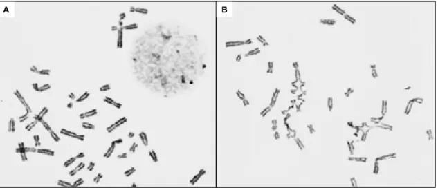
Figure 1.2: Chromatid damage after treatment of cells from FA patients with ICLs.
(A) FA lymphocytes showing spontaneous chromatid aberrations. (B) FA lymphocytes showing multiple complex chromatid exchange. Adapted from Auerbach, A.D., et al. Pediatrics, 1981.
1.1.3.2 Treatment
Bone marrow failure (BMF) BMF is the most common clinical manifestation in
FA patients and typically develops during the first decade of life [39]. Approximately
half of FA patients respond well to androgens, which stimulate the production of red blood cells and platelets and sometimes increase white cell production [27]. Long-term
androgen use has significant side effects including hirsutism and increased liver tumor incidence. Although this treatment may be effective for many years, in most patients the disease becomes refractory to androgen therapy. For these patients, hematopoietic stem-cell transplantation (HCT) may be considered and in this context, the use of androgens should be avoided because androgen treatment can adversely affect the ultimate success of a transplant. Hematopoietic growth factors (e.g., G-CSF and GM-CSF, which stimulate the production of white blood cells) are also effective in some FA patients [40].
At the present time, HCT remains the primary treatment for bone marrow failure in FA. This treatment is associated with numerous risks, and these risks are highly compounded in FA patients because of the underlying DNA repair defect. FA patients are extremely sensitive to the radiation and chemotherapy used in the transplantation procedure, and the survivors often experience multiple complications that are not routine for other transplants such as marked increased organ (pulmonary and renal) toxicity, graft-versus host disease (GVHD), immune injury, sterility, and endocrinopathies [41-45]. Histocompatible sibling donor transplants generally result in the
best outcomes for FA patients [39, 41]. Because most FA patients do not have
histocompatible siblings, some families have turned to preimplantation genetic diagnosis (PGD) [46]. While HCT is highly successful in extending the life expectancy
of FA patients, managing the long-term complications of HCT is a significant concern.
Management of cancers With successful prolongation of the survival of FA
patients after HCT, the treatment of cancers becomes more significant. Because FA patients are highly sensitive to chemotherapy and radiotherapy, treatment strategies for
FA-related cancers must differ from those for non-FA cancers. Prevention and close surveillance of cancer occurrence become more important; for instance, frequent dental evaluations and gynecologic exams of FA patients should be conducted to identify related early SCCs [47, 48]. Some FA complementation groups correlate with specific
cancer patterns; for example, patients with the FANCD1 subtype of the disease have earlier onset and increased incidence of a variety of tumors (Wilms tumor, neuroblastoma), and patients from subtype J, N, and O families are highly predisposed to breast, ovarian, and pancreatic cancers [38, 49, 50]. Specific close surveillance and
preventive treatment must be provided for patients of these FA subtypes.
Other potential therapies FA is an ideal candidate for some potential therapies.
Over 98% of FA cases are associated with identified FA genes (from FANCA to FANCP). Subtyping FA allows identification of the mutant FA gene(s) present in an individual and the generation of related retroviral or lentiviral vectors carrying the wild-type cDNA, which can be used in gene therapy. However, technical obstacles such as insufficient HSCs for ex vivo transduction, potentially inserted mutagenesis and expansion of malignant clones often must be overcome prior to successful gene therapy
[51]. Because ROS play a critical role in endogenous DNA damage, especially in FA,
antioxidants are potential therapeutic compounds for FA treatment, with the limitation that apoptosis is often induced by the excessive use of antioxidants [52-54]. Some small
molecule inhibitors, such as inhibitors of CHK1, DNA-PK, and p38 MAP kinase, may be potential therapeutic molecules with the limitation that they might enhance tumorigenesis or immune suppression [55]
1.2 Molecular basis of FA 1.2.1 FA genes
groups correspond to one of the following cloned genes: FANCA, FANCB, FANCC, FANCD1/BRCA2, FANCD2, FANCE, FANCF, FANCG, FANCI, FANCJ/BRIP1/BACH1, FANCL/PHF9, FANCM/HEF, FANCN/PALB2, FANCO/RAD51C, FANCP/SLX4, FANCQ/XPF4/ERCC4, FANCR/RAD51 and FANCS/BRCA1, FANCT/UBE2T (Table 1.2), [56-74]. The biallelic disruption of any of
these genes results in human disease, as outlined in Table 1.2. FANCA, FANCC and FANCG are the three most commonly defective genes in FA and mutations of these genes can be detected in approximately 85% of FA patients. FANCD1, FANCD2, FANCE, FANCF and FANCL account for 10%, while the remaining FA genes, FANCB, FANCI, FANCJ, FANCM, FANCN, FANCO, FANCP, FANCQ,FANCR and FANCS represent less than 5%. Some individuals with FA do not appear to have mutations in these 18 genes, indicating the possibility that novel FA genes exist [75]. FANCB is
unusual among the FA genes because it is on the X chromosome, whereas the other FA genes are located on autosomes.
FA patients with mutations in any of the FA genes present characteristic clinical features, although to various degrees, and a common cellular phenotype. This indicates that the 18 known FA genes function in the same DNA repair pathway, termed the FA pathway [76]. The discovery that the FANCD1 gene is identical to the breast cancer
susceptibility gene BRCA2 [67] and BRCA1 corresponds to FANCS [77], connects the FA
pathway and breast cancer. Identification of FANCN as PALB2, a partner and localizer of BRCA2 [78] [79], FANCJ as BRIP1, the BRCA1-interacting helicase1 [71, 80], and
recently, FANCO as RAD51C, a breast and ovarian susceptibility gene [81, 82], further
emphasize the close association of breast and ovarian tumor suppressive genes with FA and the association of both diseases with DNA repair mechanisms.
Table 1.2: FA complementation groups.
Multifunction in DNA damage repair 207
17q21 Rare
FANCS/BRCA1
DNA repair endonuclease 105
16p13.12 Rare
FANCQ/XPF4/ERCC4
Activation of HR and DSB repair 37
15q15.1 Rare
FANCR/RAD51
Holliday junction resolvase 200
16p13.3 Rare
FANCP/SLX4
Ubiquitin-Conjugating Enzyme E2T 23
1q32.1 Rare
FANCT/UBE2T
Promotes HR, RAD51 paralogue 43
17q22 Rare
FANCO/RAD51C
Mediates interaction between BRCA1 and BRCA2 during HR 130 16p12.1 Rare FANCN/PALB2 Helicase/translocase 250 14q21.3 Rare FANCM E3 ubiquitin ligase 43 2p16.1 Rare FANCL/PHF9
Interacts with BRCA1. DNA helicase, ATPase 150
17q22.3 <2%
FANCJ/BRIP1
Monoubiquitinated. Forms heterodimer with FANCD2 150 15q25-26 <2% FANCI Core complex 70 9p13 ~10% FANCG/XRCC9 Core complex 42 11p15 ~2% FANCF
Interact with FANCD2 60
6p21.3 ~2%
FANCE
Monoubiquitinated. Recruits FAN1, FANCP to chromatin, exonuclease activity
162 3p25.3
~3%
FANCD2
Recruits RAD51 and promotes HR repair 380 13q12.3 ~2% FANCD1/BRCA2 Core complex 63 9q22.3 ~10% FANCC Core complex 95 Xp22.31 ~2% FANCB Core complex 163 16q24.3 ~66% FANCA Protein function Protein MW (kDa) Locus Mutation frequency Gene (alias)
Multifunction in DNA damage repair 207
17q21 Rare
FANCS/BRCA1
DNA repair endonuclease 105
16p13.12 Rare
FANCQ/XPF4/ERCC4
Activation of HR and DSB repair 37
15q15.1 Rare
FANCR/RAD51
Holliday junction resolvase 200
16p13.3 Rare
FANCP/SLX4
Ubiquitin-Conjugating Enzyme E2T 23
1q32.1 Rare
FANCT/UBE2T
Promotes HR, RAD51 paralogue 43
17q22 Rare
FANCO/RAD51C
Mediates interaction between BRCA1 and BRCA2 during HR 130 16p12.1 Rare FANCN/PALB2 Helicase/translocase 250 14q21.3 Rare FANCM E3 ubiquitin ligase 43 2p16.1 Rare FANCL/PHF9
Interacts with BRCA1. DNA helicase, ATPase 150
17q22.3 <2%
FANCJ/BRIP1
Monoubiquitinated. Forms heterodimer with FANCD2 150 15q25-26 <2% FANCI Core complex 70 9p13 ~10% FANCG/XRCC9 Core complex 42 11p15 ~2% FANCF
Interact with FANCD2 60
6p21.3 ~2%
FANCE
Monoubiquitinated. Recruits FAN1, FANCP to chromatin, exonuclease activity
162 3p25.3
~3%
FANCD2
Recruits RAD51 and promotes HR repair 380 13q12.3 ~2% FANCD1/BRCA2 Core complex 63 9q22.3 ~10% FANCC Core complex 95 Xp22.31 ~2% FANCB Core complex 163 16q24.3 ~66% FANCA Protein function Protein MW (kDa) Locus Mutation frequency Gene (alias) 1.2.2 FA pathway
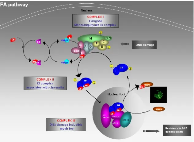
In normal cells, the FA pathway is activated during the S phase of the cell cycle and in the presence of DNA damage. In the FA pathway, FA proteins interact to form three specific complexes. Eight upstream FA proteins (FANCA, FANCB, FANCC, FANCE, FANCF, FANCG, FANCL, and FANCM) form complex I, termed the FA core complex
[68, 73, 83-91]. The primary function of the FA core complex is to monoubiquitinate the two
FA proteins FANCD2 and FANCI, components of complex II (the ID complex), through the activity of the FANCL E3 ubiquitin ligase (Figure 1.3) [68, 92-94]. Several
other FA-associated proteins are essential for the function of the core complex, including FA-associated 24-kDa protein (FAAP24)[90], FAAP100[91],
(HES1), and the recently identified FAAP20 [96]. The disease-causing mutations in these
FA-associated proteins have yet to be identified.
Homologous recombination (HR)-based repair is the major DNA repair pathway regulated by the FA proteins. In response to DNA damage, an ATR-mediated signal activates the FA core complex and monoubiquitination of the ID heterodimer. The ubiquitin-tagged ID complex move to chromatin, where it assembles nuclear DNA-repair foci. In these foci, the ID complex functionally associates with the downstream FA proteins (complex III) FANCD1, FANCN, FANCO, FANCJ and with some key HR factors such as BRCA1 and RAD51 [81, 97]. In addition to HR, the FA
pathway also mediates nucleotide incision repair (NER) and translesion synthesis (TLS). FAN1 nuclease and FANCP are recruited with ID complex and participate in the nucleolytic incision of cross-linked DNA. The TLS polymerase is essential for TLS at the step following the incision of ICL. The FA network also includes other regulatory proteins such as ubiquitin-specific peptidase 1 (USP1) and the USP1-associated protein UAF1; these function as deubiquitination enzymes of the ID complex and are required for completion of the FA pathway [98, 99].
Figure 1.3: Canonical FA pathway. DNA damage activates the FA core complex (A,
B, C, E, F, G, L, and M). The FA core complex then functions as an E3 ubiquitin ligase and monoubiquitylate the ID complex. The monoubiquitylate ID complex targets to chromatin, forms nuclear foci, and associates with other DNA repair proteins to repair DNA damage. The USP1 and UAF1 protein complex is required to deubiquitylate the ID complex and complete the FA pathway. Fengfei Huang, 2014.
1.3 Apoptosis and FA
Apoptosis is the process of programmed cell death (PCD) that may lead to characteristic cell changes (morphology) and cell death. Defective apoptotic processes have been implicated in a wide variety of diseases such as cancer. In addition, apoptosis also confers advantages during tissue development. For example, the separation of fingers and toes in a developing human embryo occurs because cells between the digits undergo apoptosis.
The environmental stress such as glucocorticoids, heat, radiation, nutrient deprivation [100], viral infection can all trigger the intracellular apoptotic signals. Two
main methods of regulation have been identified: targeting mitochondria functionality, or directly transducing the signal via adaptor proteins such as TNF-induced (tumour necrosis factor) model and the Fas-Fas ligand-mediated model. The formation of mitochondrial apoptosis-induced channel (MAC), release of small mitochondria derived activator of caspases (SMACs), Cytochrome c into the cytosol are essential process in mitochondrial regulation. Both TNF path and Fas path involve the formation of death domain (DD) and either indirect activate transcription factors or initiate caspase signaling pathway.
Significant evidence supports abnormal apoptosis of HSPCs [101, 102] occurs in the
pathogenesis of BMF and leukemia progression in FA. Increased apoptosis due to higher level of the death receptor Fas (CD95) in CD34+ cells was first reported in children with FA [102]. Subsequently, enhanced TNF-α-induced apoptosis is found in
FANCC-deficient cells and FANCC could modulates apoptotic responses to tumor necrosis factor-alpha (TNF-α) and Fas ligand [103]. Elevated expression of TNF-related
apoptosis-inducing ligand (TRAIL) at the bone marrow level has also been found in FA, which may also implicate in the pathogenesis of FA[104] and MDS[105]. In addition,
FANCD2 was shown to be a target for caspase 3 during DNA damage-induced apoptosis. These data indicate that abnormal apoptosis in the condition of FA may at least partially explain the BMF and leukemia in FA patients.
1.4 FANCC protein
FANCC gene deficiency is one of the most frequent mutations in FA patients. The gene codes for a 63-kDa protein, FANCC protein, which was the first FA protein to be identified. In addition to its critical role as part of the core complex needed to confer
cellular resistance to DNA damage, FANCC also participates widely in other non-repair pathways of oxidative metabolism, cell cycle progression and apoptosis.
1.4.1 Cellular localization of the FANCC protein
In the presence of DNA damage, the core complex is formed by the direct interaction of FANCA with FANCI, FANCB, FANCG and FANCM. FANCF interacts directly with FANCL, FANCG, and with FANCC-FANCE heterodimer. FANCM is essential for the loading of complex on chromatin (Figure 1.4) [106, 107]. Although in the
canonical FA pathway the core complex proteins cooperate and function in cell nucleus, some of these proteins can be detected in the cytoplasm. For instance, FANCC protein can be found in the nucleus but is principally located in the cytoplasm [108]. The nuclear
accumulation of both FANCC and FANCE is mutually dependent and is crucial for the function of the FA core complex [109]. The molecular chaperone glucose-related protein
94 (GRP94) has been shown to directly bind FANCC and to be involved in the regulation of the levels of the proteins in each cellular compartment [110]. In addition to
the formation of the FANCC/FANCE subcomplex, other subcomplexes, including FANCA/FANCG [87, 89] and FANCB/FANCL [69], can be found in the cytoplasm,
suggesting that FA proteins have cellular functions in addition to DNA damage repair (Figure 1.4).
Figure 1.4: FA core complex formation. (A) FANCA and FANCL first recruit FANCB
to the nucleus. (B) The FANCF-FANCG heterodimer then binds to the FANCA-FANCB-FANCL subcomplex. (C) The accumulation of FANCC in the nucleus requires FANCE. (D) The FANCC-FANCE heterodimer binds to the free nuclear complex through the interaction of FANCC with FANCF. (E) The nuclear complex is then loaded on chromatin through its interaction with FANCM. Adapted by Fengfei Huang from internet, 2014.
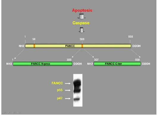
1.4.2 Posttranslational modification of FANCC protein
Posttranslational modification is critical in triggering the activity of FA proteins. FA proteins undergo multiple posttranslational modifications; some examples are the well-characterized monoubiquitination of FANCD2 and FANCI [111] and the
Unlike most other known FA proteins, FANCC function is regulated through a caspase-mediated proteolytic process [118]. Recently FANCD2 has also been shown to be
regulated through by caspase3 during apoptosis induced by DNA damage [119].
During apoptosis, FANCC undergoes proteolytic modification by a caspase that produces a predominant 47-kDa ubiquitinated protein fragment by cleavage at position 306 (LETD G) (Figure 1.5). This modification is not required for FANCC to function in DNA damage signaling but inhibits its function as a suppressor of apoptosis [118]. The
exact caspase responsible for FANCC cleavage has yet to be determined. Further studies are also required to investigate whether the C-terminal FANCC fragment has a proapoptotic function.
Figure 1.5: Caspase-mediated proteolytic modification of FANCC. FANCC is
regulated through proteolytic processing by a caspase during apoptosis. Cleavage of the protein at position 56 (53KEMD S57) and at position 306 (303LETD G307) gives rise
to a 55-kDa protein fragment and to the C-terminal 47-kDa FANCC fragment. Adapted from Brodeur, I., et al..J Biol Chem.
1.4.3 Developmental expression of the FANCC gene
The congenital abnormalities associated with FA imply that FA genes play important roles in normal development. Murine FANCC protein shares 67% amino acid identity with its human counterpart, and the function of the protein is conserved [120].
Analysis of FANCC gene expression during murine development provides clues to the function of the FANCC protein and an understanding of the basic defect in the disease. FANCC is observed initially in the mesenchyme at 8–10 days gestation, during the later stages of bone development (13–19.5 days) and later in cells of osteogenic and hematopoietic lineage. FANCC transcripts are also expressed in cells within the intramembranous cranial and facial bones. FANCC mRNA is also present in non-skeletal tissues: brain, whisker follicles, lung, kidney, gut and stomach (Figure 1.6)
[121]. The expression pattern of FANCC is consistent with the congenital defects
observed in FA patients. Interestingly, FANCC is highly expressed in rapidly dividing progenitor cells but is downregulated in differentiating cells. This is consistent with the hypothesis that structures with high rates of cell replication are the structures most likely to be affected by mutations in FA genes [122] and implies a possible role for
FANCC protein in DNA repair.
A B C
Figure 1.6: In situ hybridization analysis of embryonic FANCC expression in multiple tissues. (A) Hindlimb bud at 10 days; (B) Gut at 13 days; (C)Lung at 15 days;
(D) Vertebrae at 13 days; (E) Femur at 16 days; (F) Developing brain at 9 days [121].
Adapted from Krasnoshtein, F., et al. Hum Mol Genet, 1996.
1.4.4 The role of FANCC in oxidative metabolism
A number of studies have indicated that abnormal oxidative metabolism occurs in FA cells. FA cells show increased ROS levels and hypersensitivity to reactive oxygen species (ROS) (Table 1.3) [123]. The altered redox state of the cells is linked to specific
features of the FA cell such as reduced proliferation, reduced growth, and altered cytokine responses; these features contribute to the pathogenesis of bone marrow failure and leukemia progression [124]. Prooxidant states were found in white blood cells and in
body fluids from FA patients, which show excessive levels of luminol-dependent chemiluminescence (LDCL), 8-hydroxy-deoxyguanosine (8-OHdG), tumor necrosis factor-α (TNF-α), and reduced glutathione (GSH)/oxidized glutathione imbalance
[125-127]. All of these data indicate that FA proteins may function to mediate endogenous
oxidative metabolism. For instance, FANCC protein has been shown to interact with redox-related molecules including NADPH cytochrome-P450 reductase (RED) and glutathione S-transferase πI (GSTπI) [128, 129], implying a role of FANCC in attenuating
the redox activation of xenobiotics. Mitochondrial dysfunction, including peroxyredoxin 3 (PRDX3) cleavage and decreased peroxidase activity, has been described in FANCC-defective cells [130]. In addition, FANCC-defective lymphoblastoid
cells showed acute adenosine triphosphate (ATP) depletion and significant apoptosis after treatment with rhodamine-1,2,3 and doxycycline [131].
homeostasis regulation as shown by their association with cytochrome P450-related activities and by the presence of distorted mitochondria and decreased peroxidase activity [130, 132, 133]. FANCD2 interacts with ataxia telangiectasia mutated protein (ATM)
and forkhead box O3 (FOXO3a) in ROS, and FANCJ has been identified as a repressor of the heme oxygenase-1 gene [134-137]. Together, these data suggest that it might be
possible to design chemoprevention protocols to counteract some of the clinical implications of FA.
1.4.5 The role of FANCC in cytokine signaling and apoptosis regulation
Extensive studies have shown that FA mutant cells undergo increased apoptosis or reduced cell growth in response to stimulation by various agents such as ROS inducers, DNA-damaging agents, growth factor withdrawal, and cytokines. This implies that the FA proteins may function in pathways that regulate cell survival [138-141]. FANCC, as a
caspase-mediated target, has been reported to prevent apoptosis when overexpressed or when its cleavage is inhibited and thereby to function as a cell survival protein (Table 1.3) [101, 142]. Several clues suggest that altered cytokine regulation plays an important
role in the progression of the FA phenotype. For instance, increased TNF-α and induced Il-6 levels have been observed in FA patients, and the cytokine response genes myxovirus A (MxA), IFN response factor 1 (IRF1), p21CIP/WAF, and IFN-stimulated gene factor 3(ISGF3γ) also show high expression in FA mutant cells [143-145]. Consistent
results from an FA mouse model show that continuous injection of low doses of IFN-γ in vivo leads to BMF [146, 147], whereas TNF-α treatment leads to the clonal evolution of
leukemias [148]. Both human and mouse FA-C mutant cells have been observed to be
hypersensitive to TNF-α and IFN-γ, indicating that the cell survival function of FANCC might operate through the modulation of cytokine signaling [103, 142, 149].
activator of transcription 1 (STAT1) and subsequently to activate the IFN type II signaling cascade [150], whereas in FA-C cells STAT1 activation is defective. Other
cytokines, including IFN-α, granulocyte macrophage colony stimulating factor (GM-CSF) and stem cell factor are involved in inducing the FANCC-STAT1 interaction. An abnormal response to type I IFN was also detected in FA-C cells; this response was related to reduced phosphorylation of the Janus kinases Jak1 and Tyk2, which induces a decrease in CD4-positive cell numbers, possibly resulting in immune defects like those reported in FA patients [151-153]. FANCC may also participate in the protection of cells
against TNF-α- and IFN-γ-induced apoptosis through interaction with Hsp70. This interaction also inhibits the activity of IFN-inducible double-stranded RNA (dsRNA) and RNA dependent protein kinase (PKR) [154, 155]. Other FA proteins such as FANCA
have also been reported to be involved in cytokine signaling and in the regulation of apoptosis; these proteins have been shown to interact with IKK2 to mediate the cellular response to stressors such as dsRNA and cytokines [156-158].
1.4.6 The role of FANCC in transcription
Another important but less studied role of FANCC is transcription regulation. In the yeast two-hybrid system, FANCC has been shown to directly interact with FA zinc finger (FAZF) protein [159], a transcriptional repressor, and is plausibly hypothesized to
interfere with the transcription of critical target genes required for growth suppression in hematopoietic progenitor cells. Another transcriptional repressor identified as a FANCC protein partner is hairy enhancer of split 1 (HES1) [96]. Actually, HES1 was shown to
interact directly with several components of the FA core complex. The FA core complex was shown to regulate the transcription of HES1-responsive genes, both positively (HES1) and negatively (cyclin-dependent kinase inhibitor p21cip1/wa f 1) [96].
Table 1.3: FANCC multifunctionality through its interacting partners.
Hoshino, Wang et al. 1998, Reuter, Medhurst et al. 2003 Stress-induced chaperone
Transcription GRP94
Pang, Keeble et al. 2001, Reuter, Medhurst et al. 2003 Stress-induced chaperone
Transcription Hsp70
Hoatlin, Zhi et al. 1999, Reuter, Medhurst et al. 2003 Transcriptional repressor
Transcription FAZF
Kupfer, Yamashita et al. 1997, Reuter, Medhurst et al. 2003 Serine/threonine kinase
Cell cycle cdc2
Pang, Fagerlie et al. 2000 Cytokine response
Cell signaling STAT1
Cumming, Lightfoot et al. 2001, Reuter, Medhurst et al. 2003 Cytosolic Detoxifying
enzyme Oxidative metabolism
GSTP1
Kruyt, Hoshino et al. 1998, Reuter, Medhurst et al. 2003 Electron transfer Oxidative metabolism RED References Specfic function Functional class Interact partner
1.4.7 UNC5A, the novel protein partner of FANCC
Intensive studies of FA protein focused on DNA repair have described a canonical FA pathway that could explain some features of the FA phenotype. However, considerable evidence suggests that FA proteins may have other functions in addition to their roles in DNA damage signaling. As a protein predominantly located in the cytoplasm, FANCC assumes multifunctional roles, especially that of an anti-apoptotic regulator.
To better understand the biological functions of FA proteins, a yeast two-hybrid screen was conducted using FANCC proteolytic fragments. One of the clones obtained was identified as UNC5A. UNC5A belongs to the UNC5 family of proteins (human UNC5A-D and rodent UNCH1-4), which are receptors of the axon guidance molecule, Netrin-1. The Netrin-1 signaling pathway primarily functions to provide migrational cues in the developing central nervous system (CNS). Recently, Netrin-1 has been shown to regulate diverse processes such as cell survival, tissue development, and guidance cues during angiogenesis in a number of non-neuronal tissues. The crosstalk between FA and the Netrin-1 signaling pathway might explain some FA phenotypes, in
congenital malformations.
1.5 Netrin-1 signaling pathway
In 1990, three Caenorhabditis elegans genes, unc5, unc6, and unc40, were found to be involved in pioneer axon guidance and cell migration. This marked a significant advancement in neural development research [160]. The unc-5, unc-6, and unc-40 genes
encode proteins that guide the circumferential migration of pioneer axons and mesodermal cells on the epidermis in C. elegans. In 1994, the vertebrate homologue of unc6, Netrin-1 (named after Sanskrit ‘netr’, meaning ‘guide’), was identified and shown to have a similar function as a chemotropic guidance cue for migrating cells and axons
[161]. Later, the chemoattractive guidance receptor for Netrin-1, Deleted in Colorectal
Cancer (DCC), and its chemorepulsive receptor, uncoordinated-5 (Unc5) family, were identified as the mammalian orthologues of unc40 and unc-5 [162, 163]. Studies of the
Netrin signal pathway have been conducted in a wide range of animal species, including invertebrates such as Caenorhabditis elegans and Drosophila melanogaster, non-mammalian vertebrates such as Xenopus laevis, and mammals including rats, mice and humans. In addition to functioning as guidance cues in the CNS, Netrin-1 and its receptors have also been found to play key roles during tissue morphogenesis, angiogenesis and tumor development outside of the nervous system.
1.5.1 The Netrin protein family
Netrins are a family of extracellular proteins. In mammals, three secreted Netrins, Netrins 1, 3 and 4, and two membrane-tethered glycophosphatidylinositol (GPI)-linked Netrins, Netrins G1 and G2, have been identified. All Netrins belong to a superfamily of laminin-related proteins [164]. The N-terminal domains of Netrins contain regions that
are highly homologous to domains V and VI of laminins; the N-terminal regions of Netrin-1, 2 and 3 show most similarity to sequences found in the laminin-γ1 chain [161,