Proximal Migration of Vena Caval Filters: Report of Two Cases With
Operative Retrieval
Jean-Olivier Defraigne, Olivier Vahdat, Hendrik Lacroix and Raymond Limet Department of Cardiovascular Surgery, University Hospital, Liège, Belgium.
ABSTACT
Operative retrieval of two proximaily migrated vena caval filters was performed in two patients, ages 42 and 45 years, respectively. In the first patient the filter was encrusted in the right ventricle, and in the second one the filter was found in the pulmonary artery. Both filters were retrieved under cardiopulmonary bypass via an incision in the right atrium and the pulmonary artery, respectively. These two observations underscore the risk of increased unwarranted indications and consequent higher complication rates of vena caval filters.
Pulmonary embolism secondary to deep venous thrombosis (DVT) of the lower extremities or pelvis is a significant cause of morbidity and mortality.1 Anticoagulation treatment is most often effective in preventing the lethal complications of DVT. Placement of a filter in the inferior vena cava (IVC) has been proposed for patients who have recurrent embolism despite adequate anticoagulation therapy or when there are contraindications to heparin.2,3 Most of the currently available filters can be placed percutaneously in the IVC with no particular difficulty. Complications can arise, however, and may occasionally be severe. Two such severe complications of IVC filter placement are reported herein.
CASE REPORTS
Case 1. A 42-year-old man sustained a fracture of the right fibula, which was treated by a plaster cast at another hospital. Two weeks later he complained of pain in the right calf. After ablation of the plaster cast, phlebograms demonstrated thrombosis of the right sural and popliteal veins. The inferior vena cavogram was normal. No other clinical findings suggestive of pulmonary embolism, such as thoracic pain or acute dyspnea, were found. There were no contraindications to heparin therapy. In spite of this, the attending surgeon decided to place a vena caval filter via the right jugular vein. However, the filter (Filcard, Filcard International, Lille, France) did not open completely and migrated into the right heart. The patient was then referred to our unit.
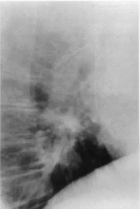
On admission, hemodynamic parameters were stable. Pulmonary x-ray examination (Fig. 1) showed that the filter was lodged in the right ventricle. Through a midline sternotomy and under cardiopulmonary bypass with induction of ventricular fibrillation, the right atrium was opened longitudinally. The filter was found to be entwined in the papillary muscles and the chordae tendineae of the right ventricle, with two of the hooks penetrating the right ventricular wall. Removal of the filter was difficult because the hooks at the end of each branch were oriented in different directions and the chordae tendineae had to be freed one at a time. After the filter was extracted, the right atrium was closed with a 4/0 continuous polypropylene suture.
The postoperative course was complicated by severe bilateral bronchopneumonia that required prolonged intubation and tracheostomy. The patient was discharged 6 weeks later with no tricuspid insufficiency on follow-up echocardiography.
Case 2. A 45-year-old man was admitted for suspected DVT in the left calf, which was confirmed by phlebography. The left iliac vein and the IVC were normal and free of any thrombus. The patient had experienced repeated episodes of DVT associated with pulmonary embolism 2 years previously. Obesity and noncompliance with treatment were the principal associated risk factors. A vena caval filter (Filcard, Filcard International) was placed. The right internal jugular vein was inadvertently punctured, and the filter was ejected into the right atrium and then migrated into the pulmonary artery. The patient was subsequently referred to our unit.
Thoracic x-ray examination (Fig. 2) showed that the filter had lodged at the level of the bifurcation of the pulmonary artery and was impacted at the origin of the left pulmonary artery. Through a midline sternotomy and
under cardiopulmonary bypass, the pulmonary artery was opened longitudinally at its left lateral aspect, the incision ending in the direction of the left pulmonary artery. The filter was retrieved fairly easily and the pulmonary artery was then closed using a 4/0 polypropylene continuous suture. This patient's postoperative course was uneventful and he was discharged 10 days after the operation on oral anticoagulation therapy.
Fig. 1. Chest x-ray film (lateral view) showing migration of vena caval filter into the right ventricle.
Fig. 2. Chest x-ray film (lateral view) showing migration of vena caval filter into the bifurcation of the
DISCUSSION
As suggested by Trousseau in 1868 and performed for the first time in 1893 by Bottini, there are several methods of filtering the IVC.4 Endocaval filter devices became available some 20 years ago.5
Although certain difficulties can be encountered when they are being inserted, essentially because of anatomic variations in the internal jugular vein and/or the vena cava, percutaneous implantation of caval filters is generally safe and rapid, and morbidity is reduced as compared with surgery. The jugular route is simpler than the femoral route, since the filter may be placed even in the presence of intracaval thrombosis. The thrombogenicity of filters is no greater than that of exocaval clips, and their efficacy in preventing pulmonary embolism is identical.5-8 Over time, the materials used for the fabrication of these filters have improved. Currently available filters are made of titanium. The diameter of the introducers is now smaller and the shape of the hooks has been changed to facilitate their placement, decrease the risk of migration or aberrant placement (e.g., in one of the affluents of the IVC), and reduce the risk of perforation. However, the multiplicity of available filters attests to the fact that none is completely devoid of complications.9
Incorrect positioning of the filter followed by migration, whether distal or proximal, is one potential complication.8 Accidental release of the filter has been described in the right renal vein, in the common iliac vein, in the suprarenal IVC, or even in the hepatic veins.9-11 Retroperitoneal positioning with irritation of the genitofemoral nerve has been reported by Adye et al.11 In addition, lateral or posterior tilting may occur.5,6 These possible mispositionings underscore the need for phlebographic control to exactly locate the filter. Distal migration is usually associated with few clinical consequences, although entrapment of emboli is thought to be less efficient. In certain instances migration of the filter toward the iliac vein has been treated simply by placing a second filter above the first.8
Proximal migration is a much more serious occurrence.12-21 A 0.9% incidence and an associated 0.6% mortality rate have been reported for the Mobin-Uddin filter.5 In their review of the English literature from 1970 to 1992, Becker et al.22 found a mortality rate of 0.12% attributable to proximal migration. This percentage is probably low since all complications have not been reported in the literature.
Several causes of proximal migration have been recognized, the most important being technical errors, defects in material, and anatomic variations in the IVC or the right atrium.18-20 The filter should be inserted with rigorous technique, by an operator experienced in percutaneous insertion of filters. The filter must be correctly loaded and carefully locked into the introducer. Insertion of the guidewire must be carefully monitored using fluoroscopy.19 Clotting within the introducer or the filter must be avoided as this may cause the struts of the filter to stick together, leading to incomplete opening of the filter, thereby facilitating its proximal migration. During all of these manipulations the introducer must be rinsed by frequent injections of saline solution and heparin.6,19 When resistance is encountered, care must be taken to avoid forcing the filter through the right atrium into the IVC, which can result in the premature release of the filter at that time.
Other causes of filter migration include defects in materials, which can be responsible for incomplete opening of the filter, inadequate hooking to the caval wall, loss of elasticity of the filter, or interlocking of the hooks as a result of manufacturing defects.19 The guidewire and the introducer should not be removed before the filter has been shown to be stabilized in the IVC with the struts completely deployed.18,19-21 In our two patients migration was associated with technical errors during positioning of the filter. In the first patient two of the struts were intertwined, leading to incomplete opening of the filter, and in the second patient the filter was released during passage within the right atrium.
Migration of the filter can also be due to anatomic variations in the IVC. When the diameter of the IVC is > 28 mm, the hooks may not enter the caval wall. This potential complication must be eliminated by routinely obtaining vena cavograms during implantation.19-21
Glock and Roux19 reported two cases of migration of Anthéor filters to the left pulmonary artery. In one patient the filter was wedged into the pulmonary valve and resulted in severe valvular insufficiency complicated by right-heart failure. Villard and Pelissier18 reported seven cases of proximal migration including four to the right atrium, one to the right ventricle, and two to the termination of the IVC in the right atrium. Five asymptomatic patients underwent surgery to avoid subsequent complications. Problems associated with aberrant filters include arrhythmia, right-heart failure due to tricuspid or pulmonary insufficiency, and perforation associated with pericardial effusion.18-21 Pouillaud et al.21 reported a fatal case in which the filter became encrusted at the junction between the IVC and the right atrium; this case was further complicated by perforation of the IVC. In
view of the severity of potential complications, we believe that once a filter has migrated to the heart cavity, it must be removed. In both of the patients described herein, clotting was found within the filter, and in the first case the filter was encrusted in the right ventricular wall with the hooks penetrating the myocardium. Six cases of migration of the Kimray-Greenfield filter have been reported.8,12-15 In one case of intraventricular migration, no removal was attempted, whereas in five other cases of migration into the right atrium, the devices were removed (three surgically, one of which was under cardiopulmonary bypass,15 one with the Greenfield apparatus,23 and one by creating a lasso using suture material, which was passed around the filter13). When the filter has migrated into the right atrium, it is theoretically possible to remove it without cardiopulmonary bypass, but the risk of injury to the coronary sinus or the tricuspid valve is high. In both cases reported herein, it was impossible to retrieve the filters percutaneously because of their location in the tricuspid valve and the bifurcation of the pulmonary artery, respectively.
However, in high-risk patients the benefits of surgical removal of the filter must be discussed,12 particularly in the absence of hemopericardium or thrombosis within the filter, as shown on echocardiography, or when it is hooked into the eustachian valve.14 For Gelbfish and Ascer16 surgical ablation of the filter is indicated in cases of intractable arrhythmia, valvular dysfunction, and signs of tamponade. In their series of two cases of atrial and one case of ventricular migration, none of the patients required surgery.16 One patient died 2 months later for no apparent reason, although the clinical picture was one of massive pulmonary embolism. The two other patients are alive and well 50 months later. Friedell et al.20 opted for conservative treatment in a case of migration into the pulmonary artery in a 76-year-old patient. This decision was prompted by the general status of the patient and by the hypothesis that thrombosis does not occur in the pulmonary artery any more frequently than it does in the IVC because the velocity of blood flow is higher in the pulmonary artery than in the IVC. Nonetheless, in the absence of major contraindications to surgery, we believe that all filters migrating to the intracardiac cavities should be removed.
Interruption of caval flow is classically indicated in patients with DVT complicated by pulmonary embolism who are at high risk for bleeding while on anticoagulation therapy (recent trauma or surgical intervention with a history of cerebral hemorrhage or gastroduodenal ulcer) and in those in whom allergy to heparin
(thrombocytopenia) develops.1,2 Placement of a vena caval filter can also be considered in cases of recurrent embolism in spite of adequate anticoagulation therapy or in the presence of previous major pulmonary
embolism.1-2 Demonstration of free-floating iliofemoral clots or thrombus in the IVC, embolism compromising > 50% of the pulmonary vascular bed, and prevention of embolic recurrence after pulmonary embolectomy are less well-known indications.3,22 Vena caval filtration in venous thrombosis that does not involve the IVC is a current topic of debate.18,19 In regard to prevention of pulmonary embolism, intracaval devices are generally effective, the rate of recurrence being < 5%; often pulmonary embolism occurs during hypercoagulability states, for example, in patients with carcinoma or in cases of occlusion of the IVC.6
Some authors have proposed expanding the indications for vena caval filters.5 Some of the more controversial proposals included use in cancer patients, in those with hip fractures, and in cases of small pulmonary capacity where the ability to tolerate even a minor pulmonary embolism is poor. This policy, however, can only serve to increase the number of complications observed in association with insertion of an intracaval device and all clinicians should be aware of this.
CONCLUSION
We would like to call attention to the risks involved with increased use of filters to prevent pulmonary embolism. In our opinion the following indications should be closely examined: pulmonary embolism associated with a contraindication to anticoagulation therapy or hemorrhage occurring during heparin therapy, recurrent pulmonary embolism despite adequate anticoagulation, the presence of free-floating voluminous iliocaval thrombus, and finally massive pulmonary embolism in patients whose pulmonary status is precarious. Only well-founded indications and rigorous implantation technique by experienced operators will result in a decreased incidence of these complications.
REFERENCES
1. Simon M, Palestrant A. Transvenous devices for the management of pulmonary embolism. Cardiovasc Intervent 1980;3: 308-318. 2. Wells I. Inferior vena cava filters and when to use them. Clin Radiol 1989;40:11-12.
3. Wingerd M, Bernhard VM, Maddison F, et al. Comparison of caval filters in the management of venous thromboembolism. Arch Surg 1978;113:1264-1271.
4. O'Neil EE. Ligation of the inferior vena cava in the prevention and treatment of pulmonary embolism. N Engl J Med 1945; 232:641-646. 5. Rohrer MJ, Scheidler MG, Wheeler HB, et al. Extended indications for placement of an inferior vena cava filter. J Vasc Surg 1989;10:44-50.
6. Greenfield LJ, Zocco J, Wilk J, et al. Clinical experience with the Kimray-Greenfield vena caval filter. Ann Surg 1977; 185: 692-698. 7. Greenfield LJ, Cho KJ, Tauscher JR. Evolution of the hook design for fixation of the titanium Greenfield filter. J Vasc Surg 1990;12:345-353.
8. Carabasi RA, Moritz MJ, Jarrel BE. Complications encountered with the use of the Greenfield vena caval filter. Am J Surg 1987;154:163-168.
9. Kraimps JL, De la Faye D, Deville Sainte-Claire C. Conical endocaval filters with metallic struts: Search for a new model. Ann Vase Surg 1992;6:99-110.
10. Magnant JG, Walsh D, Juravski L, et al. Current use of inferior vena caval filters. J Vasc Surg 1992;16:701-706.
11. Adye BA, Raabe RD, Zobell RL. Errant percutaneous Greenfield filter placement into the retroperitoneum. J Vasc Surg 1990;12:60-61. 12. Patterson RB, Fowl RJ, Lubbers DJ, et al. Repositioning of partially dislodged Greenfield filters from the right atrium by the use of a tip deflection wire. J Vasc Surg 1990;12:70-72.
13. Sadighi PJ, Scott Frost E. Retrieval of Greenfield filter from the right atrium. Ann Vasc Surg 1992;6:173-175.
14. Hirsh SB, Harrington EB, Miller C, et al. Accidental placement of the Greenfield filter in the heart: Report of two cases. J Vasc Surg 1987;6:609-610.
15. Akins CW, Thurer RL, Waltman AC, et al. A misplaced caval filter: Its removal from the heart without cardiopulmonary bypass. Arch Surg 1980;115:1133-1134.
16. Gelbfish G, Ascer E. Intracardiac and intrapulmonary Greenfield filters: A long-term follow-up. J Vasc Surg 1991;14:614-617. 17. Castaneda F, Herrera M, Cragg AH. Migration of a Gunther caval filter to the right ventricle. J Intervent Radiol 1988;3: 33-36. 18. Villard J, Pelissier FT. Exérèse sous circulation extracorporelle de filtres-ombrelles de Greenfield en position intra-cardiaque: À propos de sept patients dont six opérés. Lyon Chir 1987; 83:21-23.
19. Glock Y, Roux D. Une embolie pulmonaire pour le moins "paradoxale": Migration d'un filtre cave inférieur. Ann Chir 1993;47:157-160. 20. Friedell ML, Goldenkranz RJ, Parsonnet V, et al. Migration of a Greenfield filter to the pulmonary artery: A case report. J Vasc Surg 1986;3:929-931.
21. Pouillaud C, Ollitrault J, Paillard F, et al. Migration proximale d'un filtre cave (LEM). Ann Cardiol Angéiol (Paris) 1988;37: 129-131. 22. Becker D, Philbrick JT, Selby JB. Inferior vena cava filters: Indication, safety, effectiveness. Arch Intern Med 1992; 152: 1985-1994. 23. Greenfield LJ, Crute SL. Retrieval of the Greenfield vena caval filter. Surgery 1980;88:719-722.