Key words: Alfacalcidol, osteoporosis, rheumatoid arthritis.
Correspondence: F. Richy, PhD, University of Liège, CHU B23, B4000 Sart-Tilman, Belgium. E-mail: florent.richy@ulg.ac.be
Received July 30, 2004; accepted in revised form December 7, 2004. ABSTRACT. Alfacalcidol (1-alpha-hydroxyvitamin D3) is a non-endogenous analog of vitamin D which can bypass the renal and intestinal regulatory mechanisms that control the production of calcitriol (1,25-hydroxyvitamin D3, the active form of vitamin D, D-Hormone). Alfa-calcidol may be metabolized into calcitriol with a limit-ed risk of hypercalcemia. Alfacalcidol and calcitriol have been evaluated in animal and human studies as-sessing their effects on bone mineral density and fracture rates. More recently, they have been shown to produce beneficial effects in muscle, immune system, and auto-immune diseases, including rheumatoid arthritis. This pa-per discusses the therapeutic efficacy of alfacalcidol in reports in which it has been proposed as an interesting alternative to vitamin D or calcitriol. Some recent find-ings about general metabolism and regulation of vitamin D and its analogs are discussed. The biological and clinical effects of alfacalcidol in post-menopausal os-teoporosis are reviewed, followed by critical appraisal of its efficacy in preventing bone loss and falls in the el-derly. The last two sections discuss the role of D-analogs in regulating the immune system, with particular regard to rheumatoid arthritis. The main results of this re-view show that alfacalcidol may have a wider range of therapeutic applicability, beyond simply restricting it to patients in hemodialysis or peritoneal dialysis with high serum levels of intact PTH.
(Aging Clin Exp Res 2005; 17: 133-142) ©2005, Editrice Kurtis
INTRODUCTION
Until 1980, no-one imagined that vitamin D and its metabolites 25(OH)D, 24,25(OH)2D and 1,25(OH)2D, well-known for their central roles in calcium and bone
D-Hormone analog alfacalcidol:
an update on its role in post-menopausal
osteoporosis and rheumatoid arthritis management
Florent Richy1,3, Rita Deroisy2, Marie-Paule Lecart1,2,3, Linda Hanssens1,3, Audrey Mawet1,3, and Jean-Yves Reginster1,2,3
1Public Health, Epidemiology and Health Economics Unit, Faculty of Medicine, University
of Liège, Sart-Tilman, 2Bone and Cartilage Metabolism Research Unit, University Hospital, Liège, 3WHO Collaborating Center for the Public Health Aspects of Osteoarticular Disorders, Liège, Belgium
metabolism, might play an important role in regulating the immune system. Recent advances in our under-standing of their mechanism of action opened up new research fields, due not only to their unique interaction with bone cells but also to their modulation of the im-mune system. Although alfacalcidol, 1-α(OH)D3, a syn-thetic D-Hormone analog, has been studied for several decades, its extensive mechanism of action is still not ful-ly elucidated. The aim of this paper was to review the available evidence of the biological and clinical effects of alfacalcidol in post-menopausal osteoporosis (PMOP) and rheumatoid arthritis (RA).
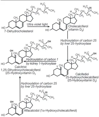
GENERAL METABOLISM OF VITAMIN D AND ITS ANALOGS (FIGS. 1-2)
Vitamins D2(ergocalciferol) and D3(cholecalciferol) are produced by the skin or absorbed from the gut. They are metabolized into their active form, calcitriol, by two successive steps: 25-hydroxylation in the liver to 25(OH)D, followed by 1α-hydroxylation in the renal proximal tubules to 1,25-(OH)2D, yielding the biologi-cally active form of vitamin D, calcitriol (1). Some oth-er cells exhibit 1α-hydroxylase activity, including os-teoblasts, placental cells, keratinocytes, macrophages and some tumor cells. The role of the extrarenal pro-duction of 1,25(OH)2D is still debated but, in normal conditions, it does not significantly contribute to the cir-culating levels of the hormone (2, 3). 24-hydroxylation, producing 24,25-dihydroxyvitamin D [24,25(OH)2D] or 1,24,25-trihydroxyvitamin D, occurs in a wide range of normal tissues and is believed to be important both to catabolise vitamin D metabolites and to regulate the ac-tive forms of vitamin D (4). 24,25(OH)2D is not in fact inactive. Recent animal studies have shown that this
metabolite can stimulate chondrocyte maturation (5), in-crease bone mineral density in vitamin D-replete rats, rabbits and dogs (6), and play a beneficial role in frac-ture repair in chicks (7). 24,25(OH)2D has also been shown to be a potent inhibitor of PTH secretion in hu-mans (8). The interactions among calcitriol, alfacalcidol and 24,25(OH)2D are still under study. Recent findings
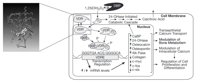
in animal models suggest that both metabolites must be present for optimal changes in bone metabolism (Fig. 2). The major enzymes involved in vitamin D hydrox-ylation are mitochondrial mixed-function oxidases con-taining cytochrome P450 with ferredoxin and heme-binding domains (9). Until today, four cytochrome P450 molecular species (CYP27A1, CYP2C11, CYP2D25, CYP3A4) have been identified as vitamin D3 25-hy-droxylases (10, 11). Alfacalcidol (1-α(OH)D3) is a syn-thetic derivate of vitamin D. Important for its metabo-lization is the fact that hepatic 25-hydroxylation is un-regulated and exclusively substrate-dependent. Con-versely, the renal 1α-hydroxylase enzyme is stringent-ly regulated by: the PTH cAMP-mediated pathway, calcitonin in a different region of the proximal tubule, and by 1,25(OH)2D itself, through negative feedback regulation via its receptor (VDR) (2, 4). Thus, in situa-tions in which the general vitamin D pathways are disturbed, exogenous alfacalcidol may bypass these regulatory systems to produce bioavailable calcitriol. Cir-culating vitamin D metabolites measured in clinical practice are 25(OH)D and 1,25(OH)2D (calcitriol). As 25(OH)D synthesis is substrate-, i.e., vitamin D-de-pendent, serum levels of this metabolite are taken as a measure of vitamin D status. Besides its classical actions in calcium metabolism, it is now suggested that the hor-monal form of vitamin D has many “uncommon” func-tions, which have only been revealed as a result of the identification and characterization of its receptor. The VDR complex was discovered in 1975 (12). Many tissues express VDR, including osteoblasts, intestinal, muscle, distal renal, liver, parathyroid, and T-cells and monocytes (13, 14). Calcitriol functions as a steroid hor-mone which binds to a cytosolic VDR, resulting in se-lective demasking of the genome of the nucleus (Fig. 3). CH3 CH3 CH3 CH 2 CH2 H CH2 CH3 7-Dehydrocholesterol HO CH H3C
Ultra violet light
Cholecalciferol (vitamin D3) H3C CH3 CH2 CH3CH 2 CH2 H CH2 CH3 HO CH C 25 H3C Hydroxylation of carbon 1 by kidney 1-hydroxylase CH3 CH2 CH3CH 2 CH2 H CH2 CH3 Calcitriol 1,25-Dihydroxycholecalciferol (25-Hydroxyvitamin D3 HO C OH OH C Hydroxylation of carbon 25 by liver 25-hydroxylase H3C CH3 CH2 CH3CH 2 CH2 H CH2 CH3 Calcifediol 25-Hydroxycholecalciferol (25-Hydroxyvitamin D3) HO C 1 OH C Hydroxylation of carbon 25 by liver 25-hydroxylase H3C CH3 CH2 CH3CH 2 CH2 H CH2 CH3 HO CH C 25 Alfacalcidol (1α-Hydroxycholecalciferol) OH C
Figure 1 -General vitamin D pathways.
Vitamin D3 25-OHaseliver 25 OH D3 1-OHase 1,25(OH)2D3
+ + 24-OHase Kidney, intestine 24-OHase Kidney, intestine – 24,25(OH)2D3 1-OHase Calcitonin PTH 1,24,25(OH)3D3 + + – – + + –
Figure 2 - Regulation of vitamin D metabolism.
Two subtypes of VDR have recently been discovered us-ing knock-out mice models. 1α,25(OH)2 D(3)-depen-dent regulation of DNA synthesis in chondrocytes re-quires the presence of the 1,25-nVDR, although other physiological responses to the vitamin D metabolite, such as proteoglycan sulfating, involve regulation via the 1,25-mVDR(5). Because of the wide variety of tissues in which VDR has been identified, the exploitation of vi-tamin D compounds in treating various diseases has been expanded.
VITAMIN D DEFICIENCY:
A HETEROGENEOUS CONDITION
“Vitamin D deficiency” collectively describes a number of pathological conditions which include primary vita-min D deficiency, calcitriol deficiency, and resistance to cal-citriol. Primary vitamin D deficiency is due to inadequate dietary intake or exposure to sunlight, and is not simply a biochemical abnormality. It is regularly associated with sec-ondary hyperparathyroidism, increased bone turnover, bone loss, osteoporosis, and increased risk of fracture (15). Vitamin D deficiency impairs the intestinal absorption of calcium. PTH maintains normal serum calcium levels for a time, despite decreased calcium absorption, by in-creasing bone resorption; however, vitamin D deficiency progressively causes resistance to the osteoclastic effects of PTH on bone and a decrease in calcium renal tubular reabsorption (16). PTH increases serum calcium but also stimulates phosphaturia, resulting in hypophosphatemia (16). These combined effects lead to a reduced bone mineralization. Unlike primary vitamin D deficiency,
pri-mary calcitriol deficiency is not due to the limitation of pcursors but to a defect in the synthesis of calcitriol, re-ducing intestinal Ca absorption and increasing PTH and bone resorption. The pathogenesis of primary calcitriol de-ficiency is related to impaired ability of the kidney to synthesize adequate amounts of calcitriol. It is common in patients with renal insufficiency or failure. Resistance to cal-citriol is related to aging-associated decline in the functions of various tissues and organs, leading to reduced calcitri-ol bicalcitri-ological action despite its normal serum levels (17). The potential cause of this resistance may be due to age-related or unknown defects in regulation in the number or a decrease in the affinity of VDR, which mediate ge-nomic actions of vitamin D (18, 19).
BIOLOGICAL EFFECTS OF ALFACALCIDOL IN POST-MENOPAUSAL OSTEOPOROSIS Alfacalcidol is a synthetic precursor of calcitriol, and is converted into 1,25(OH)2D3, predominantly in the liver, by 25-hydroxylation. 25-hydroxylation has been report-ed to be catalyzreport-ed by both mitochondrial CYP27A and a microsomal CYP2D25 vitamin D325-hydroxylase in the liver (20, 21). Northern blotting and reverse transcription-polymerase chain reaction experiments have revealed that porcine CYP2D25 mRNA (showing 77% identity with that of humans) is expressed at the highest level in the liver and in small amounts in other tissues, including muscle (20) and bone. As noted above, alfacalcidol by-passes endogenous regulation by renal 1-αhydroxylase and its pharmacokinetic profile is therefore very different from that of calcitriol: after oral ingestion of calcitriol, peak
VDR VDR RXR P P RXR VDR VDR D3 1,25(OH)2D3 DBP 24-OHase Initiated
Catabolic Cascade Calcitroic Acid
Cell Membrane Transepithelial Calcium Transport Modulation of Bone Metabolism Modulation of Bone Metabolism Modulation of Intracellular Calcium Regulation of Cell Proliferation and Differentiation Nucleus CaBP 24-OHase
GGGTGA ACG GGGGCA VDRE Transcription Regulation mRNA levels Osteocalcin Osteopontin Alk Pase Collagen c-myc c-sis c-fos D3 D3
Figure 3 - Vitamin D Receptor: structure and function.
DBP: vitamin D binding protein; VDRE: vitamin D responsive elements (specific DNA sequences); CaBP: calcium-binding protein; 24-OHase: 24-OH-hydroxylase; Alk Pase: Alkaline phosphatase; c-myc, c-fos, c-sis: protooncogen regulating cell proliferation and differentiation; RXR: Retinoid X Receptor
serum 1,25(OH)2D3is reached within 2 hours, whereas oral ingestion of alfacalcidol causes a slow rise in serum calcitriol with peak values after 8-18 hours. Calcitriol, up-on absorptiup-on, acts immediately and directly up-on the VDR in the intestinal mucosal cells to promote Ca absorp-tion, leading to a rapid increase in serum calcium. In contrast, alfacalcidol has very limited intestinal action, since 25-hydroxylase required for its metabolization into calcitriol acts predominantly in the liver. Compared with calcitriol, alfacalcidol allows for more progressive and longer pro-duction of calcitriol together with a lower risk of hyper-calcemia. This allows the lack of calcium absorption due to VDR deficiency to be counterbalanced.
It has been suggested (22) that pharmacological or suprapharmacological doses of 1,25(OH)2D(3) stimulate bone resorption by inducing RANKL, the ligand from pre-osteoblastic cells binding to RANK on pre-osteoclastic cells to promote the differentiation of osteoclasts (23). Conversely, a certain range of physiological doses of vi-tamin D inhibit PTH-induced bone resorption, the latter mechanism appearing to be mediated, at least partly, by suppression of PTH/PTHrP receptor-mediated signaling. This may highlight the central role played by 1,25(OH)2D3 in bone formation and resorption cou-pling. Indeed, the most important endocrine regulator of PTH is calcitriol (24), which regulates PTH through its re-ceptor by suppressing both the expression of the pre-proparathyroid gene and parathyroid cell proliferation (25). Alfacalcidol, by enhancing D-analog levels, indirectly suppresses secondary hyperparathyroidism, which is common in osteoporotic and elderly patients. The mech-anism includes inhibition of the proliferation of parathy-roid cells by reducing their apoptosis (26, 27) as well as PTH synthesis and release (28), and of the effects of PTH on bone (29, 30). Reduction of alkaline phos-phatase activity has also been shown (31). Vitamin D metabolites calcitriol and 24,25(OH)2D modulate the re-sponse of bone and cartilage cells to 17 beta-estradiol and dihydrotestosterone in both cell cultures and in vivo rat models (32). They both reduce, by one order of magnitude, the amount of sex steroids needed to stim-ulate cultured osteoblast-like cells or rat embryo epi-physeal cartilage cells, and synergistically increase the maximal response of these cells (32). Notwithstanding this, it has been shown that interactions among D-analogs, VDR and oestrogen receptor (ER) are largely de-pendent on gender groups, suggesting complex ER-VDR-sex, ER-age-sex and VDR-age-sex interactions may exist (33). The current data do not support the strong role of 24,25(OH)2D alone in the regulation of os-teoblast action and mineralization (34). Conversely, al-facalcidol induces an increase in calcitonin secretion (35) and normalization of uncoupled bone turnover through an increase in transforming growth factor beta (TGF-beta), which stimulates osteoblastic maturation,
and osteoprotegerin, which inhibits osteoclastic matu-ration by inhibiting the RANKL-RANK system (36). Calcitriol, by inhibiting bone-resorbing cytokines, specif-ically TNF-alpha or osteoblastic apoptosis, induces a modification in osteoclastic apoptosis, and an impact on the remodeling process has been observed (37). Cor-rection of the helper/suppressor ratio in patients with high bone loss due to an increase in CD8 also appears to be involved (38).
CLINICAL EFFECTS OF ALFACALCIDOL IN POST-MENOPAUSAL OSTEOPOROSIS Several studies have investigated the clinical effects of D-analogs (mainly calcitriol, alfacalcidol and 24,25(OH)2D) on bone mineral density, fractures and bone metabolism markers in PMOP. 24,25(OH)2D is still a poorly studied compound, which has not shown any beneficial effects on BMD and calcium metabolism in clinical studies (34).
We previously performed two meta-analyses review-ing the clinical effects of D-analogs calcitriol and alfa-calcidol (39), and compared their efficacies against that of native vitamin D (40). Our first systematic review in-cluded all randomized controled trials on alfacalcidol or calcitriol versus calcium or placebo. Eight studies of al-facalcidol (41-48) and a similar number of studies of calcitriol (49-56) specifically focused on their respec-tive clinical efficacies on bone loss and/or fracture. Among trials of alfacalcidol, four (41-44) investigated post-menopausal osteoporosis in women aged 60 and more. Meta-analysis of these studies revealed the highly significant effect of alfacalcidol on global and particularly on spinal BMD, at a median duration of 18 months. Re-garding non-spinal BMD, the data were too sparse for a proper meta-analysis. Regarding fracture prevention, we found a highly significant reduction (-47%) in the rel-ative risk of lumbar spine fractures in the alfacalcidol arms, compared with placebo or calcium alone, at a median follow-up of 12 months. We were not able to find relevant data on hip fracture prevention by alphacalcidol in PMOP. However, we did find a trend toward in-creased efficacy of alfacalcidol, compared with calcitriol, in preventing decrease with BMD, and more specifical-ly spinal BMD. In addition, studies on alfacalcidol, pooled together, provided remarkably homogenous results (pheterogeneity=0.66); studies on calcitriol did not (p hetero-geneity=0.01). These results suggested not only that alfa-calcidol and calcitriol have similar efficacies, but also that alfacalcidol may exert its BMD-preserving capabili-ties in a wider range of clinical patterns.
Our second quantitative review (40) assessed the rel-ative efficacies of D-analogs alfacalcidol and calcitriol against their parent compound, vitamin D. Regarding BMD, D-analogs exerted a significantly higher efficacy on BMD at any site compared with native vitamin D, at a me-dian duration of 24 months. When restricted to the
lum-bar spine, this intertreatment difference remained signif-icant whereas there were no signifsignif-icant differences re-garding their efficacies on other measurement sites, in-cluding hip. When comparing the adjusted global relative risks for spinal and non-spinal fracture, alfacalcidol and cal-citriol appeared to be significantly more effective ap-proaches compared with vitamin D. Analysis of the dif-ference between spinal and non-spinal fracture rates confirmed the benefits of D-analogs, with significantly low-er spinal and non-spinal fracture rates for D-analogs, on the basis of 30 to 36 months follow-up. Despite the lack of head-to-head trials in this field and the need for more careful follow-up of calcemia, alfacalcidol may be considered as an interesting alternative to native vita-min D in preventing bone loss and fractures in PMOP.
ALFACALCIDOL AND MUSCULAR FUNCTION
Muscle function, together with bone mineral density, is an important determinant of fracture risk, especially in the elderly (57). It was assumed for a century that vitamin D deficiency was linked to disturbed muscle metabolism (58). Vitamin D deficiency can impair intracellular Ca metabolism in muscle cells. The Ca-depleted content of mitochondria isolated from vitamin D-depleted chicks has been shown to be low (59), and Ca uptake into the sarcoplasmic reticulum is reduced during vitamin D defi-ciency (60, 61). Animal studies have shown that the actinomyosin content of myofibrils is reduced during ex-perimental rickets (62).
Several studies in humans have demonstrated the re-lationship between D-hormone analogs and muscle func-tion (63-65). Patients with osteomalacia suffer from muscle weakness and have low serum levels of muscle en-zymes (66). A recent study on chick embryonic muscle cells provided direct evidence for the participation of the VDR in non-genomic 1,25(OH)2D3signal transduc-tion. Activation of tyrosine phosphorylation cascades through this mechanism may contribute to hormone regulation of muscle growth (67). The results of a prospective, population-based study showed that lower 25-OHD and higher PTH levels increase the risk of sar-copenia in older men and women (68). A recent study has shown that congestive heart failure is associated with low vitamin D status (69).
Several trials have provided evidence for the involve-ment of D-analogs in preserving muscle function. Sup-plementation with 357 or 1250 µg vitamin D or 50 µg 25(OH)D for 1 or 2 months normalized muscle strength in patients with myopathy (66, 70). Leg extension pow-er was positively correlated with spow-erum 25(OH)D levels in elderly males and with serum 1,25(OH)2D3in the whole group of males and females (64). Grady et al. (71) and Lips et al. (72) did not show statistical improvement in muscle function using calcitriol or vitamin D. Glerup et al.
(73), Pfeifer et al. (74), and Bischof et al. (75) showed a significant decrease in body sway and number of falls, cor-responding to an improvement in muscle function using native vitamin D.
More recently, research has focused on alfacalcidol. Dukas et al. (76) demonstrated that, in calcium-replete pa-tients, alfacalcidol treatment significantly and safely reduced the number of fallers in an elderly community-dwelling pop-ulation (OR 0.45, 95% CI 0.21-0.97, p=0.042). Sato et al. (44) reported a highly significant differential efficacy of alfacalcidol on BMD depending on body side, in hemiplegic patients. BMD on the intact side increased by 3.5%, but decreased by 2.4% on the hemiplegic side over 6 months. Some local factors, such as paralysis and immobilization, may diminish the effect of alfacalcidol on bone formation on the hemiplegic side. This is an argument favoring the involvement of muscle 25-hydroxylase, which converts al-facalcidol into active calcitriol on a local basis. Janssen et al. (77) assessed the effects of alfacalcidol in vitamin D-de-ficient elderly people. Muscle strength improved, as well as walking distance and functional ability, which resulted in a reduction in the number of falls and fractures. Additional research is needed to clarify further to what extent alfa-calcidol supplementation can preserve muscle strength and prevent falls and fractures in elderly people.
IMMUNOLOGICAL ROLE OF ALFACALCIDOL (FIG. 4)
In recent years, there has been an effort to understand the possible non-calcemic roles played by vitamin D, in-cluding its role in the immune system and, in particular, on T-cell-mediated immunity. Vitamin D receptor is in fact found in significant concentrations in T-lymphocytes and macrophage populations (78, 79), but its highest con-centration is found in immature immune cells of the thy-mus and mature CD-8 T lymphocytes (80). Calcitriol has recently aroused great interest as an immune modu-lator with immunosuppressive activity, because of its
1,25(OH)2D3 IL-4 Th2 T Cell Th1 IL-12 IFN-γ Macrophage Monocyte
Figure 4 -Interactions between calcitriol and the immune system. Pietschmann et al. Bedeutung von Vitamin D im Immunsystem. Journal Für Mineralstoffwechsel 2003; 10: 13-15. Reproduced by kind permission of Krause & Pachernegg GMBH.
ability to shift T-cell responses from Th1 to Th2. The hor-mone inhibits the production of lymphokines (IL-2, IFN-gamma) and monocyte-derived cytokine (IL-12), leading to inhibition of helper T-cell subset type 1 (Th1) (81, 82). The significant role of vitamin D compounds as selective immunosuppressants is illustrated by their ability to pre-vent or even suppress animal models of autoimmune disease. Several studies on animal models have shown that 1,25-dihydroxyvitamin has a significant impact on the de-velopment of encephalomyelitis (83), rheumatoid arthri-tis (84), systemic lupus erythematosus (85), type I diabetes (86), and inflammatory bowel disease (87). Possible mechanisms of suppression of these autoimmune disor-ders by calcitriol have been presented. Notably, calcitriol stimulates transforming growth factor TGFß-1 (88) and terleukin production which, in turn, may suppress in-flammatory T-cell activity. In support of this, calcitriol was unable to suppress a murine model of human multiple sclerosis in IL-4-deficient mice (80). It was shown to sup-press proliferation of promyelocytes and promote their dif-ferentiation into monocytes (89). Peripheral cytes contain variable amounts of VDR. CD-8 lympho-cytes have the highest concentrations, whereas CD-4 and macrophages have lower ones (90).
The intrinsic effects of alfacalcidol, i.e., not those from its metabolite calcitriol, on various immunological pa-rameters including lymphocyte subsets are still not clear-ly demonstrated. However, interestingclear-ly, Yamauchi et al. (91) showed that, in patients with rheumatoid arthritis, the CD-4/CD-8 ratio remains stable in patients whose ini-tial value was normal, whereas it decreases after alfacalcidol treatment in patients whose initial values were abnor-mally high. It has been demonstrated that the delayed hy-persensitivity response to dinitrobenzene is impaired in vi-tamin D deficient mice (92), which tends to confirm the modulator control of 1,25-(OH)2D3 in T cell-mediated immunity. The T-cell immune response depending on D hormone levels displays a characteristic inverted “U” curve (93). While this field requires more research in-vestment, this particular activity of D-hormone analogs is the rationale for studies and trials on alfacalcidol and cal-citriol for treating autoimmune disorders, including RA.
ALFACALCIDOL IN RHEUMATOID ARTHRITIS
Patients with RA are at high risk of developing both generalized and periarticular osteoporosis (94) and are thus at even higher risk of fractures (95). Local and sys-temic osteoporosis are linked to increased production of inflammatory cytokines (TNF alpha, IL-1 beta, IL-6), re-sulting in increased formation and activation of osteo-clasts (96-100). TNF-alpha may also interfere with bone formation by promoting apoptosis of osteoblasts (101). Bone loss appears very early and is correlated di-rectly with disease activity (102). Later in the process, it
is associated with the negative effects of limited mobil-ity, which may be related to decreased muscle function in the elderly. Goertz et al. (103) and Lee et al. (104) showed that VDR polymorphisms do not play a major role in RA predisposition, but Gough et al. (105) did ob-tain inverse results in female patients with early RA. In parallel, predisposition towards osteoporosis has been shown in certain VDR genotypes (106). High disease ac-tivity in patients with RA has been associated with al-terations in vitamin D metabolism and increased bone re-sorption (84). The decrease in 1,25(OH)2D3 levels in these patients may lead to a negative calcium balance and inhibition of bone formation. Furthermore, low levels of 1,25(OH)2D3may raise levels of activated T-cells and proliferation of lymphokine-activated killer cells, thus accelerating the arthritic process (107).
Whether glucocorticoids work positively or nega-tively on generalized/periarticular osteoporosis in RA is still controversial (108-110). The pathogenesis of cor-ticosteroid-induced osteoporosis is complex. As a patho-genetic co-factor, corticosteroids reduce intestinal calcium absorption and increase renal calcium excretion, re-sulting in compensatory increased PTH release and in-creased sensitivity of bone to PTH. In addition, corti-costeroids inhibit osteoblastic function (111) as well as the favorable effects of growth factors and sex hor-mones on bone (112, 113). Thus, bone loss in RA is centered around primary inhibition of osteoblastic ac-tivity, compounded by the effects of secondary hyper-parathyroidism (114). It has recently been suggested that the expression of D-hormone receptors (VDR) may be decreased by corticosteroids, and that they probably reduce the number of functional VDR (115, 116). This may be an explanation for the efficacy of D-analogs in treating PMOP. Corticosteroids inhibit IL-12 production in human monocytes and enhance their capacity to in-duce IL-4 synthesis in CD4+ lymphocytes (117).
D-analogs have been shown to inhibit cytokines IL-1, IL-6, TNF-alpha and particularly IL-12 (118). At the cellular level, D-hormone may directly or indirectly reduce the expression of Th1 helper cells by inhibition of IL-12 from monocytes (119). Therapy with alfacalcidol or cal-citriol results in increased production of Th2 helper cells, which produce bone-protective cytokines like IL-4 and IL-10 (120). D-analogs have been shown to have a protective effect on osteoblasts against TNF-alpha-induced cell death (101).
Five intervention trials aimed at quantifying the effect of vitamin D and its metabolites on the clinical expres-sion of RA have been published. Andjelkovic et al. (121) (alfacalcidol 2 µg/day/3 months), Brohult et al. (122) (vitamin D 2500 µg/day/1-2 years) and Dottori et al. (123) (25(OH)D 50 µg/day/1 month) have shown re-ductions in disease activity or pain symptoms, whereas Yamauchi et al. (124) (alfacalcidol 2 µg/day/4 months)
and Hein et al. (125) (alfacalcidol 1 µg/day/2 months) have not. With respect to these contradictory findings, the exact role of alfacalcidol in the management of RA remains unclear.
Various clinical studies have investigated the efficacy of alfacalcidol in corticosteroid-induced bone loss, re-gardless of the underlying disease. The main results of its use in RA have been to preserve bone mass, not to in-crease it (126). Gukasian et al. (127) reported an anal-ysis of the anti-osteoporotic efficacy of alfacalcidol in 50 patients with RA. 30 RA patients received alfacalcidol (0.75-1.0 µg/day) for 12 months and 20 control RA pa-tients received a placebo. Alfacalcidol stabilized bone mineral density at the femoral neck and lumbar spine. A significant BMD increase was observed in those areas of the proximal femur where cortical bone tissue prevails. In a double-blind, placebo-controlled comparative trial of 16 weeks by Yamauchi et al. (91), 1.0-2.0 µg/day al-facalcidol revealed 10% more patients with improvement compared with the placebo group, but the difference be-tween them did not reach significance. The OKT-4/OKT-8 ratio was found not to change in patients whose initial value was normal, whereas it decreased af-ter alfacalcidol treatment in patients whose initial values were higher. Bone mineral density was conserved in the alfacalcidol group. Among all trials, taking efficacy on bone loss into consideration, a dose of 1.0 µg/day was judged to be suitable for safe, long-term treatment with regard to the limited risk of hypercalcemia.
REFERENCES
1. Stokstad E. Nutrition. The vitamin D deficit. Science 2003; 302: 1886-8.
2. Takeyama K, Kitanaka S, Sato T, Kobori M, Yanagisawa J, Kato S. 25-Hydroxyvitamin D3 1alpha-hydroxylase and vita-min D synthesis. Science 1997; 277: 1827-30.
3. Fu GK, Portale AA, Miller WL. Complete structure of the human gene for the vitamin D 1 alpha-hydroxylase, P450c1alpha. DNA Cell Biol 1997; 16: 1499-507.
4. St-Arnaud R, Arabian A, Travers R, et al. Deficient mineralization of intramembranous bone in vitamin D-24-hydroxylase-ablated mice is due to elevated 1,25-dihydroxyvitamin D and not to the absence of 24,25-dihydroxyvitamin D. Endocrinology 2000; 141: 2658-66.
5. Boyan BD, Sylvia VL, McKinney N, Schwartz Z. Membrane ac-tions of vitamin D metabolites 1alpha,25(OH)2D3 and 24R,25(OH)2D3 are retained in growth plate cartilage cells from vitamin D receptor knockout mice. J Cell Biochem 2003; 90: 1207-23.
6. Tanaka H. Vitamin D metabolites and bone. In: Feldman D, Glorieux FH, Pike JW, eds. Vitamin D. Academic Press, San Diego 1997: 305-12.
7. Seo EG, Norman AW. Three-fold induction of renal 25-hydrox-yvitamin D3-24-hydroxylase activity and increased serum 24,25-dihydroxyvitamin D3 levels are correlated with the healing process after chick tibial fracture. J Bone Miner Res 1997; 12: 598-606. 8. Carpenter TO, Keller M, Schwartz D, et al. 24,25 Dihydroxyvi-tamin D supplementation corrects hyperparathyroidism and
im-proves skeletal abnormalities in X-linked hypophosphatemic rickets- a clinical research center study. J Clin Endocrinol Metab 1996; 81: 2381-8.
9. Jones G, Ramshaw H, Zhang A, et al. Expression and activity of vitamin D-metabolizing cytochrome P450s (CYP1alpha and CYP24) in human nonsmall cell lung carcinomas. Endocrinology 1999; 140: 3303-10.
10. Yamasaki T, Izumi S, Ide H, Ohyama Y. Identification of a nov-el rat microsomal vitamin D3 25-hydroxylase. J Biol Chem 2004; 279: 22848-56.
11. Gupta RP, Hollis BW, Patel SB, Patrick KS, Bell NH. CYP3A4 is a Human Microsomal Vitamin D 25-Hydroxylase. J Bone Miner Res 2004; 19: 680-8.
12. Kream BE, Reynolds RD, Knutson JC, Eisman JA, DeLuca HF. Intestinal cytosol binders of 1,25-dihydroxyvitamin D and 25-hy-droxyvitamin D. Arch Biochem Biophys 1976; 176: 779-87. 13. DeLuca HF. New concepts of vitamin D functions. Ann NY
Acad Sci USA 1992; 669: 59-68.
14. Zittermann A. Vitamin D in preventive medicine: are we ignoring the evidence? Br J Nutr 2003; 89: 552-72.
15. Ooms ME, Lips P, Roos JC, et al. Vitamin D status and sex hor-mone binding globulin: determinants of bone turnover and bone mineral density in elderly women. J Bone Miner Res 1995; 10: 1177-84.
16. Beckerman P, Silver J. Vitamin D and the parathyroid. Am J Med Sci 1999; 317: 363-9.
17. Lau KH, Baylink DJ. Vitamin D therapy of osteoporosis: plain vi-tamin D therapy versus active vivi-tamin D analog (D-hormone) ther-apy. Calcif Tissue Int 1999; 65: 295-306.
18. Christakos S, Raval-Pandya M, Wernyj RP, Yang W. Genomic mechanisms involved in the pleiotropic actions of 1,25-dihy-droxyvitamin D3. Biochem J 1996; 316 (Pt 2): 361-71. 19. Kinyamu HK, Gallagher JC, Prahl JM, DeLuca HF, Petranick
KM, Lanspa SJ. Association between intestinal vitamin D re-ceptor, calcium absorption, and serum 1,25 dihydroxyvitamin D in normal young and elderly women. J Bone Miner Res 1997; 12: 922-8.
20. Hosseinpour F, Wikvall K. Porcine microsomal vitamin D(3) 25-hydroxylase (CYP2D25). Catalytic properties, tissue distribu-tion, and comparison with human CYP2D6. J Biol Chem 2000; 275: 34650-5.
21. Hosseinpour F, Ibranovic I, Tang W, Wikvall K. 25-Hydroxylation of vitamin D3 in primary cultures of pig hepatocytes: evidence for a role of both CYP2D25 and CYP27A1. Biochem Biophys Res Commun 2003; 303: 877-83.
22. Ueno Y, Shinki T, Nagai Y, Murayama H, Fujii K, Suda T. In vi-vo administration of 1,25-dihydroxyvitamin D3 suppresses the ex-pression of RANKL mRNA in bone of thyroparathyroidec-tomized rats constantly infused with PTH. J Cell Biochem 2003; 90: 267-77.
23. Kwan Tat S, Padrines M, Theoleyre S, Heymann D, Fortun Y. IL-6, RANKL, TNF-alpha/IL-1: interrelations in bone resorption pathophysiology. Cytokine Growth Factor Rev 2004; 15: 49-60. 24. Alvarez-Hernandez D, Naves M, Santamaria I, Menarguez J, Torregrosa V, Cannata J. Response of parathyroid glands to cal-citriol in culture: is this response mediated by the genetic poly-morphisms in vitamin D receptor? Kidney Int Suppl 2003; 85: S19-22.
25. Slatopolsky E, Brown AJ. Vitamin D and its analogs in chronic re-nal failure. Osteoporos Int 1997; 7 (Suppl 3): S202-8. 26. Ledger GA, Burritt MF, Kao PC, O’Fallon WM, Riggs BL, Khosla
women that are reversible by short term therapy with 1,25-dihy-droxyvitamin D3. J Clin Endocrinol Metab 1994; 79: 211-6. 27. Parfitt AM. The hyperparathyroidism of chronic renal failure: a
dis-order of growth. Kidney Int 1997; 52: 3-9.
28. Gallacher SJ, Cowan RA, Fraser WD, Logue FC, Jenkins A, Boyle IT. Acute effects of intravenous 1 alpha-hydroxychole-calciferol on parathyroid hormone, osteocalcin and calcitriol in man. Eur J Endocrinol 1994; 130: 141-5.
29. Shiraishi A, Higashi S, Okawa H. The effect of 1-a(OH)D3 on bone metabolism in parathyroidectomized rats with continuous PTH perfusion. Osteoporos Int 1997; 7: 35.
30. Suda T, Takahashi N, Martin TJ. Modulation of osteoclast dif-ferentiation. Endocr Rev 1992; 13: 66-80.
31. Lind L, Wengle B, Lithell H, Ljunghall S. Reduction in serum al-kaline phosphatase levels by treatment with active vitamin D (al-phacalcidol) in primary and secondary hyperparathyroidism and in euparathyroid individuals. Scand J Urol Nephrol 1991; 25: 233-6. 32. Somjen D, Weisman Y, Kaye AM. Pretreatment with 1,25(OH)2 vitamin D or 24,25(OH)2 vitamin D increases synergistically re-sponsiveness to sex steroids in skeletal-derived cells. J Steroid Biochem Mol Biol 1995; 55: 211-7.
33. Long J, Liu P, Zhang Y, et al. Interaction effects between es-trogen receptor alpha gene, vitamin D receptor gene, age, and sex on bone mineral density in Chinese. J Hum Genet 2003; 48: 514-9.
34. van Leeuwen JP, van den Bemd GJ, van Driel M, Buurman CJ, Pols HA. 24,25-Dihydroxyvitamin D(3) and bone metabolism. Steroids 2001; 66: 375-80.
35. Chen JT, Shiraki M, Hasumi K, et al. 1-alpha-Hydroxyvitamin D3 treatment decreases bone turnover and modulates calcium-reg-ulating hormones in early postmenopausal women. Bone 1997; 20: 557-62.
36. Hofbauer LC, Dunstan CR, Spelsberg TC, Riggs BL, Khosla S. Osteoprotegerin production by human osteoblast lineage cells is stimulated by vitamin D, bone morphogenetic protein-2, and cytokines. Biochem Biophys Res Commun 1998; 250: 776-81. 37. Cantorna MT, Woodward WD, Hayes CE, DeLuca HF. 1,25-di-hydroxyvitamin D3 is a positive regulator for the two anti-en-cephalitogenic cytokines TGF-beta 1 and IL-4. J Immunol 1998; 160: 5314-9.
38. Zofkova I, Kancheva RL. The effect of 1,25(OH)2 vitamin D3 on CD4+/CD8+ subsets of T lymphocytes in postmenopausal women. Life Sci 1997; 61: 147-52.
39. Richy F, Ethgen O, Bruyere O, Reginster JY. Efficacy of al-phacalcidol and calcitriol in primary and corticosteroid-induced os-teoporosis: a meta-analysis of their effects on bone mineral den-sity and fracture rate. Osteoporos Int 2004; 15: 301-10. 40. Richy F, Schacht E, Bruyere O, Ethgen O, Gourlay M, Reginster
JY. Vitamin D analogs versus plain vitamin D in preventing bone loss and osteoporosis-related fractures: a comparative meta-analysis. Calcif Tissue Int 2005 (in press).
41. Hayashi Y, Fujita T. Decrease of vertebral fracture in osteo-porotics by administration of 1-alpha-hydroxy-vitamin D3. JBMM 1992; 10: 184-8.
42. Menczel J, Foldes J, Steinberg R, et al. Alfacalcidol (alpha D3) and calcium in osteoporosis. Clin Orthop 1994; 300: 241-7. 43. Orimo H, Shiraki M, Hayashi Y, et al. Effects of 1
alpha-hy-droxyvitamin D3 on lumbar bone mineral density and vertebral fractures in patients with postmenopausal osteoporosis. Calcif Tis-sue Int 1994; 54: 370-6.
44. Shiraki M, Kushida K, Yamazaki K, Nagai T, Ioue T, Orimo H. Ef-fects of 2 years’ treatment of osteoporosis with 1a-hydroxy vitamin
D3 on bone mineral density and incidence of fracture: a placebo-controlled, double-blind prospective study. Endocr J 1996; 43: 211-20.
45. Reginster JY, Kuntz D, Verdickt W, et al. Prophylactic use of al-facalcidol in corticosteroid-induced osteoporosis. Osteoporos Int 1999; 9: 75-81.
46. Sato Y, Manabe S, Kuno H, Oizumi K. Amelioration of os-teopenia and hypovitaminosis D by 1a-hydroxyvitamin D3 in el-derly patients with Parkinson’s disease. J Neurosurg Psychiatry 1999; 66: 64-8.
47. Sato Y, Maruoka H, Oizumi K. Amelioration of hemiplegia-as-sociated osteopenia more than 4 years after stroke by 1-a-hy-droxyvitamin D3 and calcium supplementation. Stroke 1997; 28: 736-9.
48. Lakatos P, Nagy Z, Kiss L, Horvath C, Takacs I, Foldes J. Pre-vention of corticosteroid-induced osteoporosis by alphacalcidol. Z Rheumatol 2000; 59: 48-52.
49. Aloia JF. Role of calcitriol in the treatment of postmenopausal os-teoporosis. Metabolism 1990; 39 (4 Suppl 1): 35-8.
50. Ebeling PR, Wark JD, Yeung S, et al. Effects of calcitriol or cal-cium on bone mineral density, bone turnover, and fractures in men with primary osteoporosis: a two-year randomized, double blind, double placebo study. J Clin Endocrinol Metab 2001; 86: 4098-103.
51. Sambrook P, Henderson NK, Keogh A, et al. Effect of calcitriol on bone loss after cardiac or lung transplantation. J Bone Miner Res 2000; 15: 1818-24.
52. Gallagher JC, Riggs BL, Recker RR, Goldgar D. The effect of cal-citriol on patients with postmenopausal osteoporosis with special reference to fracture frequency. Proc Soc Exp Biol Med 1989; 191: 287-92.
53. Gallagher JC, Goldgar D. Treatment of postmenopausal osteo-porosis with high doses of synthetic calcitriol. A randomized controlled study. Ann Intern Med 1990; 113: 649-55. 54. Gallagher JC, Fowler SE, Detter JR, Sherman SS. Combination
treatment with estrogen and calcitriol in the prevention of age-re-lated bone loss. J Clin Endocrinol Metab 2001; 86: 3618-28. 55. Lambrinoudaki I, Chan DT, Lau CS, Wong RW, Yeung SS,
Kung AW. Effect of calcitriol on bone mineral density in pre-menopausal Chinese women taking chronic steroid therapy. A randomized, double blind, placebo controlled study. J Rheumatol 2000; 27: 1759-65.
56. Sambrook P. Alfacalcidol and calcitriol in the prevention of bone loss after organ transplantation. Calcif Tissue Int 1999; 65: 341-3.
57. Hedstrom M. Hip fracture patients, a group of frail elderly peo-ple with low bone mineral density, muscle mass and IGF-I levels. Acta Physiol Scand 1999; 167: 347-50.
58. Ritz E, Boland R, Kreusser W. Effects of vitamin D and parathy-roid hormone on muscle: potential role in uremic myopathy. Am J Clin Nutr 1980; 33: 1522-9.
59. Pleasure D, Wyszynski B, Sumner A, et al. Skeletal muscle calcium metabolism and contractile force in vitamin D-deficient chicks. J Clin Invest 1979; 64: 1157-67.
60. Curry OB, Basten JF, Francis MJ, Smith R. Calcium uptake by sarcoplasmic reticulum of muscle from vitamin D-deficient rabbits. Nature 1974; 249: 83-4.
61. Curry O, Francis MJ, Smith R. Proceedings: The effects of vita-min D deficiency on the isolated sarcoplasmic reticulum of mus-cle. Clin Sci Mol Med 1974; 46: 7P.
62. Stroder J. Hypothyroidism and rachitis. Arch Kinderheilkd [Archives of Pediatrics] 1965; 173: 105-9.
63. Bischoff HA, Borchers M, Gudat F, et al. In situ detection of 1,25-dihydroxyvitamin D3 receptor in human skeletal muscle tissue. Histochem J 2001; 33: 19-24.
64. Bischoff HA, Stahelin HB, Tyndall A, Theiler R. Relationship be-tween muscle strength and vitamin D metabolites: are there therapeutic possibilities in the elderly? Z Rheumatol 2000; 59 (Suppl 1): 39-41.
65. Bischoff HA, Stahelin HB, Urscheler N, et al. Muscle strength in the elderly: its relation to vitamin D metabolites. Arch Phys Med Rehabil 1999; 80: 54-8.
66. Rimaniol JM, Authier FJ, Chariot P. Muscle weakness in intensive care patients: initial manifestation of vitamin D deficiency. Intensive Care Med 1994; 20: 591-2.
67. Buitrago C, Vazquez G, De Boland AR, Boland R. The vitamin D receptor mediates rapid changes in muscle protein tyrosine phosphorylation induced by 1,25(OH)(2)D(3). Biochem Biophys Res Commun 2001; 289: 1150-6.
68. Visser M, Deeg DJ, Lips P. Low vitamin D and high parathyroid hormone levels as determinants of loss of muscle strength and muscle mass (sarcopenia): the Longitudinal Aging Study Ams-terdam. J Clin Endocrinol Metab 2003; 88: 5766-72. 69. Zittermann A, Schleithoff SS, Tenderich G, Berthold HK, Korfer
R, Stehle P. Low vitamin D status: a contributing factor in the pathogenesis of congestive heart failure? J Am Coll Cardiol 2003; 41: 105-12.
70. Ziambaras K, Dagogo-Jack S. Reversible muscle weakness in pa-tients with vitamin D deficiency. West J Med 1997; 167: 435-9. 71. Grady D, Halloran B, Cummings S, et al. 1,25-Dihydroxyvitamin D3 and muscle strength in the elderly: a randomized controlled tri-al. J Clin Endocrinol Metab 1991; 73: 1111-7.
72. Lips P, Graafmans WC, Ooms ME, Bezemer PD, Bouter LM. Vi-tamin D supplementation and fracture incidence in elderly persons. A randomized, placebo-controlled clinical trial. Ann Intern Med 1996; 124: 400-6.
73. Glerup H, Mikkelsen K, Poulsen L, et al. Hypovitaminosis D my-opathy without biochemical signs of osteomalacic bone involve-ment. Calcif Tissue Int 2000; 66: 419-24.
74. Pfeifer M, Begerow B, Minne HW, Abrams C, Nachtigall D, Hansen C. Effects of a short-term vitamin D and calcium sup-plementation on body sway and secondary hyperparathyroidism in elderly women. J Bone Miner Res 2000; 15: 1113-8. 75. Bischoff H, Stahelin H, Dick W, et al. Effects of vitamin D and
cal-cium supplementation on falls: a randomized controlled trial. J Bone Miner Res 2003; 18: 343-51.
76. Dukas L, Bischoff HA, Lindpaintner LS, et al. Alfacalcidol reduces the number of fallers in a community-dwelling elderly population with a minimum calcium intake of more than 500 mg daily. J Am Geriatr Soc 2004; 52: 230-6.
77. Janssen HC, Samson MM, Verhaar HJ. Vitamin D deficiency, muscle function, and falls in elderly people. Am J Clin Nutr 2002; 75: 611-5.
78. O’Kelly J, Hisatake J, Hisatake Y, Bishop J, Norman A, Koeffler HP. Normal myelopoiesis but abnormal T lymphocyte respons-es in vitamin D receptor knockout mice. J Clin Invrespons-est 2002; 109: 1091-9.
79. Towers TL, Freedman LP. Granulocyte-macrophage colony-stimulating factor gene transcription is directly repressed by the vi-tamin D3 receptor. Implications for allosteric influences on nuclear receptor structure and function by a DNA element. J Biol Chem 1998; 273: 10338-48.
80. DeLuca HF, Cantorna MT. Vitamin D: Its role and uses in im-munology. FASEB J 2001; 15: 2579-85.
81. Lemire JM. Immunomodulatory actions of 1,25-dihydroxyvitamin D3. J Steroid Biochem Mol Biol 1995; 53: 599-602. 82. Lemire JM, Archer DC, Beck L, Spiegelberg HL.
Immunosup-pressive actions of 1,25-dihydroxyvitamin D3: preferential inhi-bition of Th1 functions. J Nutr 1995; 125 (Suppl 6): 1704S-8S. 83. Garcion E, Sindji L, Nataf S, Brachet P, Darcy F, Montero-Menei CN. Treatment of experimental autoimmune en-cephalomyelitis in rat by 1,25-dihydroxyvitamin D3 leads to early effects within the central nervous system. Acta Neuropathol (Berl) 2003; 105: 438-48.
84. Oelzner P, Muller A, Deschner F, et al. Relationship between dis-ease activity and serum levels of vitamin D metabolites and PTH in rheumatoid arthritis. Calcif Tissue Int 1998; 62: 193-8. 85. Vaisberg MW, Kaneno R, Franco MF, Mendes NF. Influence of
cholecalciferol (vitamin D3) on the course of experimental systemic lupus erythematosus in F1 (NZBxW) mice. J Clin Lab Anal 2000; 14: 91-6.
86. Zella JB, DeLuca HF. Vitamin D and autoimmune diabetes. J Cell Biochem 2003; 88: 216-22.
87. Froicu M, Weaver V, Wynn TA, McDowell MA, Welsh JE, Can-torna MT. A crucial role for the vitamin D receptor in experi-mental inflammatory bowel diseases. Mol Endocrinol 2003; 17: 2386-92.
88. Demoor-Fossard M, Galera P, Santra M, Iozzo RV, Pujol JP, Re-dini F. A composite element binding the vitamin D receptor and the retinoic X receptor alpha mediates the transforming growth factor-beta inhibition of decorin gene expression in ar-ticular chondrocytes. J Biol Chem 2001; 276: 36983-92. 89. Tanaka H, Abe E, Miyaura C, et al. 1
alpha,25-Dihydroxyc-holecalciferol and a human myeloid leukaemia cell line (HL-60). Biochem J 1982; 204: 713-9.
90. Wiese RJ, Uhland-Smith A, Ross TK, Prahl JM, DeLuca HF. Up-regulation of the vitamin D receptor in response to 1,25-dihy-droxyvitamin D3 results from ligand-induced stabilization. J Biol Chem 1992; 267: 20082-6.
91. Yamauchi Y, Tsunematsu T, Konda S, Hoshino T, Itokawa Y, Hoshizaki H. A double blind trial of alfacalcidol on patients with rheumatoid arthritis (RA). Ryumachi [official journal of the Japan Rheumatism Association] 1989; 29: 11-24.
92. Yang S, Smith C, Prahl JM, Luo X, DeLuca HF. Vitamin D de-ficiency suppresses cell-mediated immunity in vivo. Arch Biochem Biophys 1993; 303: 98-106.
93. Yang S, Smith C, DeLuca HF. 1 alpha, 25-Dihydroxyvitamin D3 and 19-nor-1 alpha, 25-dihydroxyvitamin D2 suppress im-munoglobulin production and thymic lymphocyte proliferation in vivo. Biochem Biophys Acta 1993; 1158: 279-86.
94. Kameda H, Takeuchi T. Osteoporosis associated with rheumatoid arthritis. Nippon Rinsho [Japonese Journal of Clinical Medicine] 2003; 61: 292-8.
95. Haugeberg G, Orstavik RE, Kvien TK. Effects of rheumatoid arthritis on bone. Curr Opin Rheumatol 2003; 15: 469-75. 96. Brosch S, Redlich K, Pietschmann P. [Pathogenesis of
osteoporo-sis in rheumatoid arthritis]. Acta Med Austriaca 2003; 30: 1-5. 97. Hirayama T, Danks L, Sabokbar A, Athanasou NA. Osteoclast
formation and activity in the pathogenesis of osteoporosis in rheumatoid arthritis. Rheumatology (Oxf) 2002; 41: 1232-9. 98. Sugiyama T. Involvement of interleukin-6 and prostaglandin E2
in periarticular osteoporosis of postmenopausal women with rheumatoid arthritis. J Bone Miner Metab 2001; 19: 89-96. 99. Joe B, Griffiths MM, Remmers EF, Wilder RL. Animal models of
rheumatoid arthritis and related inflammation. Curr Rheumatol Rep 1999; 1: 139-48.
100. Yilmaz M, Kendirli SG, Altintas D, Bingol G, Antmen B. Cytokine levels in serum of patients with juvenile rheumatoid arthritis. Clin Rheumatol 2001; 20: 30-5.
101. Pascher E, Perniok A, Becker A, Feldkamp J. Effect of 1al-pha,25(OH)2-vitamin D3 on TNF alpha-mediated apoptosis of hu-man primary osteoblast-like cells in vitro. Horm Metab Res 1999;31:653-6.
102. Bottcher J, Malich A, Pfeil A, et al. Potential clinical rele-vance of digital radiogrammetry for quantification of periartic-ular bone demineralization in patients suffering from rheumatoid arthritis depending on severity and compared with DXA. Eur Ra-diol 2004; 14: 631-7.
103. Goertz B, Fassbender WJ, Williams JC, et al. Vitamin D receptor genotypes are not associated with rheumatoid arthritis or bio-chemical parameters of bone turnover in German RA patients. Clin Exp Rheumatol 2003; 21: 333-9.
104. Lee CK, Hong JS, Cho YS, Yoo B, Kim GS, Moon HB. Lack of re-lationship between vitamin D receptor polymorphism and bone ero-sion in rheumatoid arthritis. J Korean Med Sci 2001; 16: 188-92. 105. Gough A, Sambrook P, Devlin J, et al. Effect of vitamin D re-ceptor gene alleles on bone loss in early rheumatoid arthritis. J Rheumatol 1998; 25: 864-8.
106. Lisker R, Lopez MA, Jasqui S, et al. Association of vitamin D re-ceptor polymorphisms with osteoporosis in Mexican post-menopausal women. Hum Biol 2003; 75: 399-403.
107. Ravid A, Koren R, Maron L, Liberman UA. 1,25(OH)2D3 in-creases cytotoxicity and exocytosis in lymphokine-activated killer cells. Mol Cell Endocrinol 1993; 96: 133-9.
108. Rau R. Use of glucocorticoids in rheumatoid arthritis. Inhibition of disease progression versus risk of steroid osteoporosis. Z Rheuma-tol [German Journal of RheumaRheuma-tology] 2001; 60: 485-91. 109. Solomon DH, Katz JN, Jacobs JP, La Tourette AM, Coblyn J.
Management of glucocorticoid-induced osteoporosis in patients with rheumatoid arthritis: rates and predictors of care in an academic rheumatology practice. Arthritis Rheum 2002; 46: 3136-42. 110. Reginster JY, de Froidmont C, Lecart MP, Sarlet N, Defraigne
JO. Alphacalcidol in prevention of glucocorticoid-induced os-teoporosis. Calcif Tissue Int 1999; 65: 328-31.
111. Yudoh K, Matsuno H, Osada R, Nakazawa F, Katayama R, Kimura T. Decreased cellular activity and replicative capacity of osteoblastic cells isolated from the periarticular bone of rheuma-toid arthritis patients compared with osteoarthritis patients. Arthritis Rheum 2000; 43: 2178-88.
112. Ohlsson C, Bengtsson BA, Isaksson OG, Andreassen TT, Slootweg MC. Growth hormone and bone. Endocr Rev 1998; 19: 55-79.
113. Ohlsson C, Vidal O. Effects of growth hormone and insulin-like growth factors on human osteoblasts. Eur J Clin Invest 1998; 28: 184-6.
114.Toyoda T, Inokuchi S, Saito S, Horie Y, Tomita S. Bone
loss of the radius in rheumatoid arthritis. Comparison be-tween 34 patients and 40 controls. Acta Orthop Scand 1996; 67: 269-73.
115. Lee S, Szlachetka M, Christakos S. Effect of glucocorticoids and 1,25-dihydroxyvitamin D3 on the developmental expression of the rat intestinal vitamin D receptor gene. Endocrinology 1991; 129: 396-401.
116. Godschalk M, Levy JR, Downs RW, Jr. Glucocorticoids de-crease vitamin D receptor number and gene expression in human osteosarcoma cells. J Bone Miner Res 1992; 7: 21-7. 117. Blotta MH, DeKruyff RH, Umetsu DT. Corticosteroids inhibit
IL-12 production in human monocytes and enhance their capacity to induce IL-4 synthesis in CD4+ lymphocytes. J Immunol 1997; 158: 5589-95.
118. Schacht E. Osteoporosis in rheumatoid arthritis- significance of al-facalcidol in prevention and therapy. Z Rheumatol [German Journal of Rheumatology] 2000; 59 (Suppl 1): 10-20. 119. McKnight AJ, Zimmer GJ, Fogelman I, Wolf SF, Abbas AK.
Ef-fects of IL-12 on helper T cell-dependent immune responses in vi-vo. J Immunol 1994; 152: 2172-9.
120. Boonstra A, Barrat FJ, Crain C, Heath VL, Savelkoul HF, O’Garra A. 1alpha,25-Dihydroxyvitamin D3 has a direct effect on naive CD4(+) T cells to enhance the development of Th2 cells. J Immunol 2001; 167: 4974-80.
121.Andjelkovic Z, Vojinovic J, Pejnovic N, et al. Disease modify-ing and immunomodulatory effects of high dose 1 alpha (OH) D3 in rheumatoid arthritis patients. Clin Exp Rheumatol 1999; 17: 453-6.
122. Brohult J, Jonson B. Effects of large doses of calciferol on patients with rheumatoid arthritis. A double-blind clinical trial. Scand J Rheumatol 1973; 2: 173-6.
123. Dottori L, D’Ottavio D, Brundisini B. Calcifediol and calcitonin in the therapy of rheumatoid arthritis. A short-term controlled study. Minerva Med 1982; 73: 3033-40.
124. Yamauchi Y, Tsunematsu T, Honda S, Hoshino T, Itokawa Y. A double blind trial of alfacalcidol on patients with rheumatoid arthritis (RA). Ryumachi [Official Journal of the Japan Rheuma-tism Association] 1989; 29: 11-24.
125. Hein G, Oelzner P. Vitamin D metabolites in rheumatoid arthri-tis: findings—hypotheses—consequences. Z Rheumatol [Ger-man Journal of Rheumatology] 2000; 59 (Suppl 1): 28-32. 126.Buckley LM, Leib ES, Cartularo KS, Vacek PM, Cooper SM.
Calcium and vitamin D3 supplementation prevents bone loss in the spine secondary to low-dose corticosteroids in patients with rheumatoid arthritis. A randomized, double-blind, place-bo-controlled trial. Ann Intern Med 1996; 125: 961-8. 127. Gukasian DA, Nasonov EL, Balabanova RM, Smirnov AV,
Vlasova IS. Effects of alfacalcidol on mineral density of bone tis-sue in patients with rheumatoid arthritis. Klin Med [Clinical Medicine] 2001; 79: 47-50.