Université de Montréal
The role of ubiquitination and deubiquitination in the
regulation of BRCA1 function during genotoxic stress
Par Helen Pak
Université de Montréal Faculté des arts et des sciences
Mémoire présenté à la faculté des arts et des sciences en vue de l’obtention du grade de maîtrise en biochimie
Option génétique et moléculaire
Avril, 2012
Université de Montréal
Faculté des études supérieures et postdoctorales
Ce mémoire intitulé:
The role of ubiquitination and deubiquitination in the regulation of BRCA1
function during genotoxic stress
Présentée par: Helen Pak
Évalué par un jury composé des personnes suivantes:
Sylvie Mader, présidente-rapporteur El Bachir Affar, directeur de recherche
iii
RÉSUMÉ
BRCA1 est un suppresseur de tumeur majeur jouant un rôle dans la transcription, la réparation de l’ADN et le maintien de la stabilité génomique. En effet, des mutations dans le gène BRCA1 augmentent considerablement le risque de cancers du sein et de l’ovaire. BRCA1 a été en majorité caractérisé pour son rôle dans la réparation de l’ADN par la voie de recombinaison homologue (HR) en présence de bris double brins, par example, induits par l’irradiation gamma (IR). Cependant, la fonction de BRCA1 dans d’autres voies de réparation de l’ADN, comme la réparation par excision de nucléotides (NER) ou par excision de base (BER), demeurent toutefois obscures. Il est donc important de
comprendre la régulation de BRCA1 en présence d’agents génotoxiques comme le méthyle méthanesulfonate (MMS) ou l’UV, qui promouvoient le BER et le NER respectivement. Nos observations suggèrent que BRCA1 est dégradée par le protéasome après traitement avec le MMS ou les UV, et non avec l’IR. Par ailleurs, cette dégradation semble
compromettre le recrutement de Rad51, suggérant que la voie de HR est inhibée. Nos résultats suggèrent que la HR est inhibée afin d’éviter l’activation simultanée de multiples voies de réparation. Nous avons aussi observé que la dégradation BRCA1 est réversible et que la restauration des niveaux de BRCA1 coïncide avec le recrutement de Rad51 aux sites de dommages. Cela suggère que la HR est réactivée tardivement par les bris double brins générés suite à l’effondrement des fourches de réplication. Ayant observé que BRCA1 est hautement régulé par l’ubiquitination et est ciblé par le protéasome pour dégradation, nous avons émis une hypothèse que BRCA1 est régulé par des déubiquitinases. Cela amène à caractériser plus en profondeur par un criblage en déplétant les déubiquitinases
individuellement par RNAi et en observant leur effet sur le recrutement de BRCA1 et des protéines reliées à cette voie. Un criblage préliminaire nous a permi d’identifié candidats potentiels tel que BAP1, CXORF53, DUB3, OTUB1 et USP36.
Mots-clés : BRCA1, Dommage à l’ADN, Réparation de l’ADN, Recombinaison
Homologue, Ubiquitination, Ubiquitin ligase, Déubiquitinase, Dégradation protéasomale, Méthyle méthanesulfonate, Irradiation gamma.
iv
ABSTRACT
BRCA1 is a tumour suppressor involved in transcription, DNA repair and
maintenance of genomic stability. Indeed, BRCA1 mutation carriers have an exceptionally higher risk of breast and ovarian cancers. BRCA1 is mainly known for its role in
homologous recombination repair (HR) by recruiting HR proteins to chromatin upon double strand break (DSBs) formation, e.g., following treatment with ionizing irradiation (IR). However, the function of BRCA1 in other DNA repair pathways such as nucleotide excision repair (NER) or base excision repair (BER) is still obscure. It is thus of
fundamental and clinical importance to investigate BRCA1 function following exposure to diverse genotoxic agents. Using human cultured cell, we observed that BRCA1 is
downregulated by the proteasome upon treatment with MMS or UV, but not with IR. Moreover, this downregulation prevents Rad51 recruitment to chromatin following exposure to MMS. Given that DNA damage induced by UV and MMS trigger NER and BER pathways respectively, this implies that HR could be inhibited in order to prevent competition between independent DNA repair pathways. We also found that BRCA1 downregulation is reversible and the recovery of BRCA1 levels correlates with the reappearance of BRCA1 and Rad51 on chromatin. This implies that the HR has been reactivated at the late stage of DNA damage for the repair of double strand breaks generated by replication fork collapse. Since BRCA1 stability is highly regulated by ubiquitination and is downregulated following MMS treatment, one would expect that a deubiquitinase is responsible for relieving this downregulation to promote the reactivation of the HR pathway. To characterize this aspect further, we conducted DUB RNAi screens in which a particular DUB is depleted and the localization of BRCA1 and other related proteins were observed. According to a preliminary screen, a few DUBs (BAP1, CXORF53, DUB3, OTUB1, and USP36) were identified as potential regulators of the stability and localization of BRCA1 and proteins involved in homologous recombination. Keywords: BRCA1, DNA damage, DNA repair, Homologous recombination,
Ubiquitination, Ubiquitin ligase, Deubiquitinase, Proteasomal degradation, Methyl methanesulfonate, Ionizing radiation
v
LIST OF ABBREVIATIONS
ATM: Ataxia Telangiectasia Mutated
ATR: Ataxia Telangiectasia and Rad3 Related BER: Base Excision Repair
BRCA1: Brest Cancer Type1 Susceptibility BRCA2: Brest Cancer Type 2 Susceptibility BRCC: BRCA1 BRCA2 Containing Complex CAK: CDK Activating Kinase
CDK: Cyclin Dependent Kinase
CDKI: Cyclin Dependent Kinase Inhibitor CSA: Cockayne Syndrome type A
CSB: Cockayne Syndrome type B DDB1: DNA Damage-Binding Protein 1 DDB2: DNA Damage-Binding Protein 2 DNA: Deoxyribonucleic Acid
DSB: Double Strand Break DUB: Deubiquitinase
GG-NER: Global Genomic NER
HECT: Homologous to the E6-AP Carboxyl Terminus HR: Homologous Recombination
IR: Ionizing Irradiation IRIF: IR Induced Foci JAMM: JAB1/MPN/Mov34
MDC1: Mediator of DNA damage Checkpoint protein 1 MJD: Machado-Joshephin Domain
MMR: Mismatch Repair
MMS: Methyl Methanesulfonate NER: Nucleotide Excision Repair NHEJ: Non-Homologous End Joining
vi PCNA: Proliferating Cell Nuclear Antigen OTU: Ovarian Tumour Superfamily RING: Really Interesting New Gene RFC: Replication Factor C
RNA: Ribonucleic Acid RNAi: RNA interference RPA: Replication Protein A shRNA: Short hairpin RNA siRNA: Small Interfering RNA
TC-NER: Transcription Coupled-NER TLS: Translesion Synthesis
UCH: Ubiquitin C-terminal Hydrolase USP: Ubiquitin-Specific Processing Protease UV: Ultraviolet
vii
TABLE OF CONTENTS
RÉSUMÉ ... iii
ABSTRACT ... iv
LIST OF ABBREVIATIONS ... v
TABLE OF CONTENTS ... vii
LIST OF TABLES ... x LIST OF FIGURES ... xi ACKNOWLEDGEMENTS ... xiii A) INTRODUCTION ... 2 1 Ubiquitination ... 2 1.1 Ubiquitination process ... 2
1.2 Different types of E3s ... 2
1.2.1 The RING finger E3s ... 3
1.2.2 The HECT domain E3s ... 4
1.3 Ubiquitination and Signaling ... 5
2 Deubiquitination ... 7
2.1 Different Classes of DUBs... 7
2.1.1 Papain-like Cysteine Proteases ... 8
2.1.1.1 Ubiquitin-specific processing protease (USP)... 8
2.1.1.2 Ubiquitin C-terminal Hydrolase (UCH)... 9
2.1.1.3 Ovarian tumour (OTU) superfamily ... 9
2.1.1.4 Machado-Josephin domain (MJD)... 9
2.1.2 Metalloproteases JAB1/MPN/Mov34 (JAMM) ... 9
3 DNA damage and Repair... 10
3.1 DNA repair Pathways ... 11
3.1.1 Mismatch Repair (MMR) ... 12
3.1.2 Base Excision Repair (BER) ... 15
3.1.3 Nucleotide Excision Repair (NER) ... 17
viii
3.1.5 Homologous Recombination (HR) ... 22
3.1.6 Translesion Synthesis (TLS) ... 24
4 The cell cycle regulation and apoptosis ... 27
4.1 The cell cycle regulation ... 27
4.2 Cell cycle checkpoints ... 29
4.3 Apoptosis ... 31
5 BRCA1 ... 33
5.1 The Domains of BRCA1 ... 34
5.2 The mechanism of action of BRCA1 upon exposure to double strand breaks ... 37
5.3 The regulation of DNA double strand break repair pathways: choosing between non-homologous end-joining and homologous recombination ... 42
5.4 The role of BRCA1 in maintaining genomic integrity ... 44
5.5 The role of BRCA1 in transcription regulation... 46
5.6 The role of BRCA1 in cell cycle checkpoints... 49
6 Rationale and Hypothesis ... 51
B) MATERIAL AND METHODS ... 53
1) Chemicals and plasmids ... 53
2) Cell Culture ... 53 3) Immunoblot ... 54 4) Immunofluorescence ... 54 5) Immunoprecipitation ... 54 6) Chromatin Isolation ... 55 7) Cell Synchronization ... 55 8) siDUB Screen ... 55 C) RESULTS ... 57
I. BRCA1 is downregulated upon genotoxic stresses not directly inducing DNA double strand breaks... 57
II. BRCA1 downregulation in not caused by transcriptional silencing ... 60
III. Downregulation of BRCA1 is reversible and is not a consequence of apoptosis 61 IV. BRCA1 downregulation is independent of the cell cycle ... 63
ix
VI. The BRCT domain of BRCA1 is required for its downregulation ... 67
VII. BRCA1 downregulation is an early step to homologous recombination inhibition 69 VIII. BRCA1 regulation in presence of various genotoxic stress inducing agents .... 72
IX. Identification of the DUBs involved in BRCA1 regulation by a screen using a siRNA library ... 74
D) DISCUSSION ... 82
i. BRCA1 function is regulated by ubiquitination in a stress-dependent manner ... 82
ii. Potential DUBs regulating BRCA1 function ... 85
E) CONCLUSION ... 95
F) REFERENCES ... 98 G) ANNEX ... I
x
LIST OF TABLES
Table 1: Summary of the cell cycle checkpoints and the proteins involved. ... 30 Table 2: Description of the BRCA1 complexes. ... 36 Table 3: Potential hits of siDUB screen in untreated U2OS cells for spontaneous BRCA1
foci assembly ... 77 Table 4: Potential hits of siDUB screen in MMS-treated U2OS cells for BRCA1 and
γH2A.X foci assembly ... 78 Table 5: Potential hits for siDUB screen in IR-treated U2OS cells for BRCA1, γH2A.X,
Rad51 and 53BP1 foci assembly. ... 80 Table 6: List of antibodies used with their respective dilutions. ... I Table 7: siDUB screen in untreated U2OS cells for BRCA1 foci assembly ... III Table 8: siDUB screen in MMS-treated U2OS cells for BRCA1 foci assembly ... IV Table 9: siDUB screen in MMS-treated U2OS cells for γH2A.X foci assembly ... V Table 10: siDUB screen in IR-treated U2OS cells for BRCA1 foci assembly ... VI Table 11: siDUB screen IR-treated U2OS Cells for Rad51 foci assembly... VII Table 12: siDUB screen in IR-treated U2OS cells for γH2A.X foci assembly... VIII Table 13: siDUB Screen in IR-treated U2OS cells for 53BP1 foci assembly ... IX
xi
LIST OF FIGURES
Figure 1 : Description of the ubiquitination process and cellular outcomes of different
ubiquitination modifications. ... 6
Figure 2: Crystal structures of the catalytic sites of different classes of DUBs. ... 7
Figure 3 : Overview of the mismatch repair (MMR) pathway... 14
Figure 4: Overview of the base excision repair (BER) pathway. ... 16
Figure 5: Overview of the nucleotide excision repair (NER) pathway ... 19
Figure 6: Overview of the non-homologous end joining (NHEJ) repair pathway. ... 21
Figure 7: Overview of the homologous recombination (HR) repair pathway. ... 23
Figure 8: Overview of the translesion synthesis (TLS) mechanism ... 26
Figure 9: Cyclin levels and complexes during the cell cycle. ... 28
Figure 10: The domains of BRCA1 and its sites of phosphorylation in response of stress. 35 Figure 11: Overview of BRCA1 recruitment to the sites of DNA damage upon double strand breaks. ... 40
Figure 12: The role of BRCA1 in homologous recombination ... 41
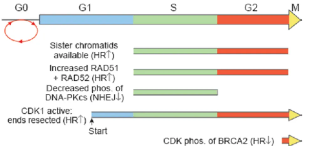
Figure 13: The role of the cell cycle in determining the choice of DNA double strand break repair between homologous recombination and non-homologous-end-joining. ... 43
Figure 14: The role of BRCA1 in marking post-duplicated centrosomes. ... 45
Figure 15: BRCA1 as a transcription co-factor. ... 48
Figure 16: BRCA1 and cell cycle checkpoints... 50
Figure 17: BRCA1 is downregulated with MMS and UVC treatment but not IR ... 59
Figure 18: BRCA1 downregulation is not caused by transcription inhibition. ... 60
Figure 19: BRCA1 downregulation is reversible ... 62
Figure 20: DNA damage-induced BRCA1 downregulation during cell cycle progression. ... 64
Figure 21: BRCA1 downregulation is mediated by proteasomal degradation ... 66
Figure 22: The BRCT domain of BRCA1 is crucial for BRCA1 downregulation ... 68
Figure 23: BRCA1 downregulation inhibits the recruitment of homologous repair proteins to the sites of DNA damage. ... 71
xii
Figure 24: Model describing BRCA1 regulation in the presence of diverse genotoxic stress-inducing agents... 73 Figure 25: Schema describing the siDUB screen ... 76 Figure 26: BRCA1 downregulation is mediated by the proteasome. ... II Figure 27: BRCA1 foci assembly in MMS-treated U2OS cells following DUB3 depletion.
... X Figure 28: Immunofluorescence for BRCA1 foci assembly in IR-treated U2OS cells ... XI
xiii
ACKNOWLEDGEMENTS
I would like to express gratitude to my supervisor, Dr. El Bachir Affar, for having me in his laboratory and for his constant presence during my learning experience. He has provided me guidance not only scientifically, but about life in general as well. I would also like to show gratitude to my lab members Ian Hammond-Martel, Helen Yu, Nazar Mashtalir, Salima Daou and Diana Adjaoud for their advices and support with the DUB screen. I especially want to thank Ian Hammond-Martel for his supervision and for introducing me to various techniques. It was all thanks to my supervisor and lab members that I had such a positive learning environment and it was a pleasure working with them. I also want to thank Raphael Rouget, Dr. Andrew A. Horwitz, Dr. Jeffrey Parvin and Dr. Elliot Drobetsky for their expertise in the DNA damage field and Dr. Natsuko Chiba for kindly providing the GFP-BRCA1 deletion constructs. Last but not least, I would like to show appreciation to the members of the jury, Dr. Sylvie Mader and Dr. Hugo Wurtele, for taking their time to correct this work.
2
A)
INTRODUCTION
1 Ubiquitination
Ubiquitination is a post-translational modification consisting of the covalent attachment of a small protein of 76 amino acids, ubiquitin, to a lysine residue of a target protein 1. Ubiquitination is known to regulate various fundamental cellular processes such as the cell cycle, DNA repair and chromatin structure. In fact, deregulation of the
ubiquitin pathway is greatly linked to various diseases including cancer 2,3.
1.1 Ubiquitination process
Three enzymes are required for the ubiquitination process. The ubiquitin activating enzyme, E1, activates the ubiquitin in an ATP dependent manner by
adenylation, which allows the interaction of ubiquitin with E1 through a thiolester bond between the active cysteine residue of E1 and the C-terminus of ubiquitin 1,4-6. The E1 then transfers the ubiquitin to the ubiquitin-conjugating enzyme E2 through a thioester bond formation with the active cysteine of E2 1,4-6. The ubiquitin ligase E3 catalyses the ubiquitination of the substrate as it promotes the formation of an isopeptide bond between the C-terminus of ubiquitin and a lysine residue of the target protein (Figure 1A) 1,4-6.
1.2 Different types of E3s
While there are only a few E1 and E2 enzymes, there are a myriad of E3 ligases given that they are responsible for specifically recognizing and ubiquitinating target proteins (Figure 1A). There are two major classes of E3 ubiquitin ligases mediating the transfer of ubiquitin to the substrate: the RING finger and the HECT domain.
3
1.2.1 The RING finger E3s
The RING (Really Interesting New Gene) finger enzymes are the most abundant class of ubiquitin ligases consisting of a domain of 40-60 amino acids with eight conserved cysteines and histidines residues required for coordinating two zinc ions 7,8. The
consensus sequence of the RING finger is: Cys-X(2)-Cys-X(9-39)-Cys-X(l-3)-His-X(2-3)-Cys-X(2)-Cys-X(4-48)-Cys-X(2)-Cys, where X represents any amino acids 7-9. The underlined residues interact with the first zinc ion, whereas the residues in italic interact with the second zinc ion; forming a finger-like structure required for the interaction with ubiquitin conjugating enzymes E2 7-9. The RING ubiquitin ligases do not directly interact with ubiquitin, but interact with E2 and promote ubiquitin transfer from the E2 to the substrate (Figure 1 A) 5. For example, SCF (skip1, cullin1, F-box) is a multi-component E3 ligase containing a RING finger protein RBX1 and a scaffold protein Cullin 1
connecting the RING finger with the adaptor protein SKP1, which interacts with different F-box proteins (i.e. SKP2, FBW7, β-TCR) to confer specificity. Indeed, each of the F-box protein recognizes a specific target to trigger its ubiquitination 6,10. Another ligase APC (anaphase promoting complex) has some similarities with the SCF as it contains a RING finger protein APC11 and a scaffold protein APC2 connecting the RING finger with several adaptor proteins interacting with the F-box proteins (e.g., cdc20, cdh1) 6,10. Both SCF and APC are important ligases required for cell cycle regulation.
4
1.2.2 The HECT domain E3s
The HECT (Homologous to the E6-AP Carboxyl Terminus) is a domain of approximately 350 amino acids and is termed after the ligase E6-AP due to structural similarity 11,12. The E6-AP ligase was first observed to interact with the E6 protein of human papillomavirus (HPV) and trigger the degradation of the tumour suppressor p53, suggesting a link between viral infection and cancer 11,13-16. The HECT domain consists of an N-terminal region required for interaction with E2 enzymes and a C-terminal region including a conserved catalytic cysteine residue 12. The N-terminal and the C-terminal regions of the HECT domain are linked by a flexible hinge that allows conformation change upon E2 interaction, bringing the catalytic cysteines of the E2 and the HECT domain closer to facilitate ubiquitin transfer12,17,18. The ubiquitination process by the HECT ligases starts with the interaction with ubiquitin-conjugated E2, allowing the transfer of ubiquitin to the E3 ligase through a thioester bond between ubiquitin and the catalytic cysteine of the HECT ligase 12. The HECT ligase then transfers the ubiquitin to the substrate 5,12. An example of a HECT class E3 ligase is HERC2, which was observed to ubiquitinate and degrade BRCA1 through the proteasome 19.
5
1.3 Ubiquitination and Signaling
The signaling outcome of ubiquitination depends on the type of ubiquitination. The ubiquitin can be attached individually (monoubiquitination) or as a chain
(polyubiquitination) on the target protein. Indeed, the ubiquitin possesses 7 lysine residues (K6, K11, K27, K29, K33, K48, and K63) capable of attachment with other ubiquitin molecules, allowing the formation of various ubiquitin chains 5,20. Depending on the type of chains, ubiquitination can trigger different cellular outcomes and signaling events. For example, monoubiquitination can signal endocytosis, virus budding, gene expression, DNA repair, or nuclear export5. In contrast, lysine 48 polyubiquitin chains signal
proteasomal degradation. Ubiquitination through lysine 11, 29 or 63 is less understood, but recent studies suggest that these polyubiquitin chains are involved in several cellular processes such as DNA repair, endocytosis, NF-kB activation, or ribosome function (Figure 1 B) 5.
6
Figure 1 : Description of the ubiquitination process and cellular outcomes of different ubiquitination modifications.
A) Ubiquitination is a sequential process consisting of the transfer of ubiquitin to an E1, E2 and E3 before attachment to the target protein.
B) Ubiquitin possesses 7 different lysine residues (K6, K11, K27, K29, K33, K48, and K63) that could potentially be attachment sites for other ubiquitin molecules, allowing the formation of diverse ubiquitin chains. Ubiquitination plays important roles in cell
function and the outcome is dependent on the nature of the ubiquitin chain. There is also evidence for the formation of mixed ubiquitin chains for which the cellular outcome is not well studied. Reference: Woelk et al. (2007) Cell Division 2:11. 5
7
2 Deubiquitination
Deubiquitination is the reverse process of ubiquitination4. The balance between the activity of the E3 ligases and the deubiquitinases often determines the outcome of cellular processes4. Deubiquitinases (DUB) are enzymes that remove ubiquitin from protein substrates through proteolytic activity4. These proteases are quite novel and some of their functions are still not well understood. Many DUBs are modified post-translationally through phosphorylation by ATM or ATR upon DNA damage and are regulators of cell cycle checkpoints4, suggesting roles in tumour development/suppression. DUBs can reverse protein degradation and thus ensure protein stabilization and function4. DUBs can also participate in signaling events in a proteasome-independent manner, e.g., by reversing monoubiquitination and K63 chains4. Finally, DUBs are also important for recycling free ubiquitin moieties to replenish the cellular pool of this critical signaling molecule4.
2.1 Different Classes of DUBs
The DUBs can be divided into two major classes: the cysteine proteases (USP, UCH, OTU, and MJD) and metalloproteases (JAMM). Each class of DUB has a specific secondary structures in which their conformation change following interaction with ubiquitin (Figure 2) 21,22.
Figure 2: Crystal structures of the catalytic sites of different classes of DUBs.
The catalytic structure of the proteases is in yellow and ubiquitin is in blue. The catalytic centers are shown as spheres (carbon, gray; nitrogen, blue; oxygen, red; sulfur, orange; zinc, purple) Reference: Sebastian M.B. Nijman et al. (2005) Cell 123: 773-786. 22
8
2.1.1 Papain-like Cysteine Proteases
The cysteine proteases’ catalytic activity involves 2 or 3 critical amino acids forming the catalytic diad or triad: i) a cysteine residue containing a reactive thiol group, ii) a histidine residue that lowers the pka of the catalytic cysteine by deprotonation, and iii) an asparagine or aspartic acid residue which polarizes the histidine, although the latter is not absolutely required as observed for the OTU class (Figure 2)21,22. The general mechanism consists of polarization and alignment of the histidine by an asparagine or aspartic acid, the polarized histidine then deprotonates the cysteine to lower the pka of the acidic thiol group to generate a nucleophile group for a nucleophilic attack between the substrate and the ubiquitin 21,22. This frees the substrate and promotes the formation of an acyl-intermediate between the ubiquitin and the DUB which is stabilized by the oxy-anion hole, which is an environment near the catalytic triad responsible for stabilizing the
negatively charged acyl-intermediate though a hydrogen bond donation by either a glutamine, glutamate or asparagine residue 21,22. Finally, a water molecule cleaves the acyl-intermediate by hydrolysis to free the DUB and Ubiquitin 21,22.
2.1.1.1 Ubiquitin-specific processing protease (USP)
USP is the largest class of DUBs and a typical USP DUB has three subdomains consisting of a finger, a palm, and a thumb 4,21-23. The catalytic center consists of the palm and thumb and the finger allows the interaction with ubiquitin 4,21-23. Studies have shown that ubiquitin binding is crucial for activating the catalytic triad. For example, ubiquitin binding on USP7 is required for bringing the catalytic cysteine closer to the histidine 4,21-23. Although the cysteine and histidine residues of the catalytic triad of USP14 are well
aligned, the catalytic center is blocked by the ubiquitin binding surface and is only freed upon interaction with ubiquitin 22,24 .
9
2.1.1.2 Ubiquitin C-terminal Hydrolase (UCH)
The UCH class has a large core catalytic domain of approximately 230aa and is responsible for cleaving small ubiquitin adducts 4,22. Structural studies on UCHL3 show that the catalytic center is blocked by a large loop and that only small ubiquitin chains can reach it 22,25,26. However, it is probable that UCH can deubiquitinate proteins from the end, where the chain can reach the catalytic center 22,27.
2.1.1.3 Ovarian tumour (OTU) superfamily
The OTU superfamily is named according to homology with the ovarian tumour gene4. Its core catalytic domain consists of five β-strands found between helical
domains4. Structural studies on yeast OTU1 bound to ubiquitin show that the surface for ubiquitin interaction is disordered in OTUB1 and OTUB2 when not bound to ubiquitin
22,28-31
.
2.1.1.4 Machado-Josephin domain (MJD)
The MJD class seems to have a similar structural domain as UCH. Studies on ATXN3 show that another ubiquitin binding site is present in a helical arm further away from the catalytic center. This suggests that 2 ubiquitins might be required to maintain it in an active conformation 22,32-34.
2.1.2 Metalloproteases JAB1/MPN/Mov34 (JAMM)
The JAMM DUBs are metalloproteases containing a zinc ion in the catalytic site which is stabilized by an aspartate and 2 histidine residues 21,35. The mechanism of JAMM requires a zinc ion to polarize and activate the water molecule21,35. The water molecule bound to zinc can interact with the substrate through non-covalent interaction and hydrolyze the isopeptide bond between the substrate and ubiquitin 21,35. The DUB is then released after a series of proton transfer 21,35. This class appears to bind specifically to K63-linked ubiquitin 4,22. An example of a metalloprotease would be BRCC36, which is known to be in the BRCA1-abraxas complex and regulates DNA damage signaling 22.
10
3 DNA damage and Repair
Every hour, each cell undergoes about 800 DNA lesions induced by endogenous DNA damaging agents (e.g., by-products of cellular metabolism and replication errors) 36. DNA damage is the foremost cause of tumourigenesis as it induces genomic instability (chromosomal aberrations or translocation) and mutations that impair the function of tumour suppressor genes or activate oncogenes. For instance, mutations in crucial tumour suppressor genes involved in cell cycle checkpoints and DNA repair such as BRCA1 highly promotes tumour formation37-41; mutations in the transcription factor c-Myc, which regulates genes involved in cell proliferation, can result in its constitutive activation42-44. A hyper-activated c-Myc would thus promote cells to proliferate continuously and eventually transformed into cancer cells42-44. DNA damage often impairs the normal structure of the DNA double helix and can provoke stalling of DNA and RNA
polymerases, blocking replication and transcription45. The replication fork collapse at the site of DNA damage results in the production of double strand breaks, which are
considered to be the most genotoxic of all DNA damages as they may result in loss of genomic information and chromosomal rearrangements 46,47. Exogenous damage can be caused by exposure to UV rays, chemicals, smoke or ionizing radiations. Endogenous damage can be an outcome of a metabolic process such as the generation of reactive oxygen species by the mitochondria during electron transport 48,49, chromosomal
rearrangement, or errors during DNA synthesis. Depending on the nature of the DNA damage, specific pathways are activated to repair DNA and to restore genomic stability.
11
3.1 DNA repair Pathways
DNA damage occurrence is extremely common. DNA repair plays a major role in maintaining the cells free of damage, thus preventing carcinogenesis. DNA repair proteins are crucial for detecting DNA damage and activating specific repair mechanisms. There are several DNA repair pathways that are activated in a tightly regulated manner
depending on the nature of the DNA damage in order to ensure a prompt and accurate repair of the lesion. For example, ionizing radiations induce DNA double strand breaks that are repaired by non-homologous end joining or homologous recombination 50. Reactive oxygen species induce single strand breaks, through oxidization of DNA bases, which are repaired by base excision repair 51,52. The chemical methyl methanesulfonate (MMS) induces DNA alkylation by methylating guanines or adenines at position N7 and N3 respectively and these DNA lesions are also repaired by base excision repair 53,54. DNA base mismatch caused by replication errors are repaired by DNA mismatch repair 55. UV rays or benzo[a]pyrene induce bulky DNA adducts distorting the DNA helix and are repaired by nucleotide excision repair. UV rays are well known to induce cross-linking of adjacent pyrimidines through their cyclobutane rings between position 5 and 6 referred to as cyclobutane pyrimidine dimers (CPD) 56,57. Benzo [a] pyrene is an environmental carcinogen consisting of a polycyclic aromatic hydrocarbon issued from combustion 58. Benzo [a] pyrene can intercalate into DNA through a nucleophilic attack at position 2 of a guanine, generating N2-dG lesions 58,59. UV and benzo [a] pyrene are highly genotoxic carcinogens present in everyday life as we are constantly exposed to the sun and smoke.
12
3.1.1 Mismatch Repair (MMR)
The DNA polymerases involved in DNA replication pol δ (lagging strand) and ε (leading strand) are not error proof 60-63. DNA polymerases can make errors approximately every 104-105 nucleotides by incorporating the wrong bases, by omitting or inserting extra bases; resulting in 100,000 to 1,000,000 mistakes during every replication 60,63,64.
Fortunately, the 5’ to 3’ exonuclease proofreading activity of these DNA polymerases and the DNA mismatch repair mechanism work together in order to significantly minimize the errors to approximately every 10-10 nucleotides per replication60,65. The DNA mismatch repair is the main mechanism involved in repairing mistakes of the polymerases55. MMR consists of recognizing the misincorporated base and excising it. The DNA is then re-synthesized using the parental strand as the template and then ligated (
Figure 3 A) 66. The error is recognized by MutS upon detection of the instability resulting from the kinked DNA structure near the site of mismatch 66,67. MutS is divided into 2 forms: MutSα (consisting of MSH2 and MSH6) and MutSβ (consisting of MSH2 and MSH3) 60,66. MutSα is involved in recognizing short mismatch loops of 1-2
nucleotides whereas as MutSβ is involved in recognizing longer mismatch loops up to 10 nucleotides 60,66. MutL (consisting of MLH1 and PMS2) is then recruited, forming a sliding clamp with MutS 60,66. It was shown that MLH1 or MSH2 are important
components of the MMR pathway as their deficiency leads to lethality upon the inhibition of polymerases involved in base excision repair such as polG and polB 66,68. The MutS possesses ATPase activity that is enhanced upon the recognition of a mismatched loop and exchanges ATP to ADP in order to trigger the release and sliding of the MutS/MutL clamp along the DNA in the opposite direction of DNA synthesis 66,69. The MutS/MutL clamp will eventually encounter an unreplicated single strand DNA gap along with replicative sliding clamp and replication factor C (RFC) 66,70. The MutS/MutL clamp displaces RFC while recruiting the exonuclease EXO1 and guides the latter in order to excise the DNA lesion 66,71. RPA is recruited to protect the single stranded DNA and once EXO1 has excised DNA beyond the lesion, EXO1 activity is inhibited by MutL 66,71. The replicative polymerases and PCNA then resume DNA replication to synthesize new strand and ends by ligating the gaps together (
13
Figure 3B) 66,72. It is still unclear how the MutS/MutL clamp distinguishes the daughter strand from the parental strand. The distinction between the parent and the daughter strand is a very crucial step, as the repair needs to be triggered on the newly synthesized daughter strand holding the damaged base and not on the undamaged parent strand. It has been well established that in Escherichia Coli, DNA methylation plays a major role in differentiating the daughther strand from the parent strand73,74. DNA is known to be hemimethylated after replication as the parent strand is methylated while the newly synthesized strand is temporarily unmethylated. The distinction between the parent and daughter strand is made by the endonuclease MutH which is activated and recruited by the ATPase activity of MutS 73,74. MutH recognizes hemimethylated dGATC sequences and incises the unmethylated strand close to the mismatch in order to initiate MMR72-74. However, there is no known human homolog of MutH up to date and thus it is still not well known whether the strand discrimination mechanism is conserved from bacteria to humans. However, it was suggested that MutS/MutL clamp could possibly sense the gaps in between the Okazaki fragments or the single strand gap when it encounters PCNA
66,71,72
. In fact, PCNA was suggested to interact with MutSα and MutSβ in order to assist their recruitment to the nascent daughter stand 75-79.
14
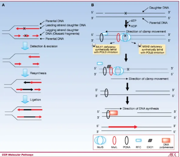
Figure 3 : Overview of the mismatch repair (MMR) pathway
A) Overview of the MMR pathway showing the detection and excision of the DNA lesion followed by resynthesis and ligation.
B) Overview of the MMR pathway including the proteins involved. The MutS recognizes the DNA region and promotes the formation of the MutS/MutL clamp. Upon ATP-ADP exchange by the ATPase activity of MutS, the MutS/MutL clamp slides along the DNA and eventually meets PCNA. The exonuclease EXO1 is then recruited and excises the DNA lesion. The MMR pathway is completed by DNA resynthesis and ligation. Reference: Martin S.A, Lord C.J, Ashworth A (2010) Clin Cancer Res. 16(21):5107-5113.66
15
3.1.2 Base Excision Repair (BER)
BER is activated throughout the cell cycle upon DNA alkylation, methylation, or oxidation that does not create distortion of the DNA helix backbone. The BER pathway is divided into two sub pathways: the short-patch (SP) pathway, also known as the single nucleotide (SN) pathway and the long-patch pathway (LP) 80. The short-patch pathway consists of replacing a single nucleotide whereas the long-patch pathway involves the displacement and the replacement of more than 1 nucleotide 80. The choice between these two pathways remains not so well understood although it was suggested to be dependent on the nature of the damage or the cell cycle 81. For example, synthetic abasic sites generated by tetrahydrofuran or methoxyamine are repaired by the long patch-pathway given that these reduced AP sites are resistant to the dRP lyase activity of DNA
polymerase β 81. In addition, the DNA polymerases δ and ε involved in the long patch repair are known to be the polymerases implicated in DNA synthesis and thus, the long-patch repair might be favoured during the S phase of the cell cycle 81. Upon DNA lesion, the damaged base is recognized and cleaved by a DNA glycosylase, which hydrolyzes the glycosidic bond between the base and the sugar phosphate backbone 80. The removal of the damaged base generates an abasic apurinic or apyrimidinic site known as an AP site
80,81
. The strand with the AP site is recognized and cleaved by AP endonucleases such as APE1 in order to generate a single stranded DNA with a 5’ deoxyribosephosphate (dRP)
80-82
. DNA polymerases are then recruited in order to fill in the gaps. In the short-patch pathway, the main polymerase involved is the DNA polymerases β 80-82. However, DNA polymerase λ can substitute pol β in the absence of the latter 80-82. In the long-patch pathway, the gap filling is mediated by the DNA polymerases δ or ε, along with their accessory proteins proliferating cell nuclear antigen (PCNA) and the replication promoting factor C (RFC) to coordinate DNA synthesis and increase the processivity of the DNA polymerases80,81. Of note, the bases are displaced as new DNA is synthesized, forming a flap cleaved by flap endonucleases such as FEN180,81. However, in the short patch
pathway, the dRP is cleaved by the dRP lyase activity of the DNA polymerase β or λ 80,81. Finally, the ends are ligated by the DNA ligase III in the short-patch pathway and the DNA ligase I in the long patch complex (Figure 4) 80,81.
16
Figure 4: Overview of the base excision repair (BER) pathway.
The left side shows the short-patch repair pathway whereas the right side shows the long-patch repair pathway. The damaged nucleotide is recognized and hydrolyzed by a DNA glycosylase, generating an abasic site. The abasic site is then incised by the AP
endonuclease APE1. In the short-patch repair, the DNA polymerases β or λ is recruited to fill in the gap and remove the abasic site. The DNA is then ligated by the DNA ligase III. In the long patch-repair, the DNA polymerases ε or δ, along with PCNA and RFC, are recruited to synthesize new nucleotides while the damaged strand is being displaced, generating a flap. The flap is then cleaved by the flap endonuclease FEN1 and the ends are ligated by DNA ligase I. Reference: Prasad R et al. (2011) Mol Biol (Mosk) 45(4): 586–600. 80
17
3.1.3 Nucleotide Excision Repair (NER)
NER is a repair mechanism activated in presence of DNA damage resulting in helix distortion. This type of damage is highly genotoxic as it blocks DNA replication and transcription 83. There are two different NER pathways: the global-genomic NER (GG-NER) repairs damage in the genome whereas the transcription-coupled NER (TC-(GG-NER) repairs damage of transcriptionally active genes 83-85. These two repair pathways differ in DNA damage recognition but share the same repair pathway called the core pathway. In the GG-NER, the damage is recognized by the XPE (CUL4-DDB-ROC1) complex and the XPC complex, which sense DNA lesions 83-86. The XPC complex has the role of
confirming the presence of damage by detecting DNA destabilization upon loss of
Watson-Crick base paring83. The XPC complex binds the undamaged strand of the DNA whereas the XPE complex interacts with and inserts its β-hairpin structure into the
damaged base in order to extrude and expose it on the surface83. The CUL4-DDB-ROC1 complex is an E3 ligase complex inhibited by COP9 83,86,87. In presence of damage, COP9 dissociates from the E3 ligase complex so it can be recruited to the lesion site and
monoubiquitinates histones 83,86,87. Histone monoubiquitination results in chromatin relaxation to allow exposure of the damaged site 83,86,87. The XPC and DDB2 are also ubiquitinated via K63 and K48 chains respectively 83,86-88. The XPE complex dissociates from DNA upon DDB2 degradation whereas XPC binding to DNA is enhanced, promoting NER activation 83,86-88. In the TC-NER pathway, upon encounter of DNA lesion, RNA pol II stalls and recruits CSB and the CUL4-CSA-ROC1 ubiquitin ligase complex 83,85. RNA pol II is then degraded by the E3 ubiquitin ligase Nedd4 89,90. This initiates the NER repair pathway. The ubiquitin ligase activity of the CUL4-CSA-ROC1 complex is still poorly understood. It was suggested that CSB is a target of the CUL4-CSA-ROC1 upon
dissociation of COP991. CSB ubiquitination and degradation was observed to occur during late TC-NER and was required for elimination of DNA damage signaling and allowing transcription to resume 91. Upon recognition of the damage and activation of NER, the transcription factor TFIIH is recruited along with the helicases XPB and XPD 83,85,91. TFIIH is recruited by XPC and CSB in GG-NER and TC-NER respectively 83,85,91-95. The helicases unwind the DNA at the damaged site. The DNA binding proteins XPA and
18
RPA bind to the damaged and undamaged strand respectively as a damage verification step and protecting DNA from degradation 83,85,91. The unwounded DNA facilitates the
recruitment and activity of the endonucleases XPF and XPG to excise the damaged DNA site 83,85,91. PCNA and DNA polymerase are then recruited to resynthesize the DNA and finally the ligase I is recruited in order to ligate the ends together (Figure 5) 83-85.
19
Figure 5: Overview of the nucleotide excision repair (NER) pathway
The DNA damage is first recognized by the CSA and CSB complex (during transcription) or the DDB1 and DDB2 complex (in the genome overall). The transcription factor TFIIH is then recruited along with the helicase XPB and XPD to unwind the damaged DNA to favour the recruitment of XPA and RPA, which bind and stabilize single stranded DNA. The endonucleases XPF and XPG are then recruited to excise the damaged DNA to allow the resynthesis by PCNA and DNA polymerase. The newly synthesized DNA is finally ligated by ligase I. Reference: Cleaver J.E (2009) Nature Reviews Genetics 10, 756-76885
20
3.1.4 Non-Homologous End joining (NHEJ)
NHEJ is a pathway activated upon double strand break functions throughout the cell cycle96. Although the NHEJ is error prone, it is the main pathway activated upon DNA double strand break96. The double strand break is recognized by the heterodimer Ku70/80 and recruits the DNA-dependent protein kinase subunit DNA-PKcs, a
serine/threonine kinase96. The Ku heterodimers and PKcs together form the DNA-PK complex, which is recruited at both ends of the DNA double strand break and is suggested to be important for bridging the DNA ends at a close proximity to allow proper repair 97-100. Upon interaction with DNA-PKcs, the Ku heterodimer translocates itself and positions DNA-PKcs at the extremities of the DNA100. The DNA-PKcs kinase activity is enhanced when two DNA-PKcs are in proximity100. Nucleases such as Artemis are recruited in order to process the ends by removing excess overhanging nucleotides that are not compatible for ligation 96-100. Artemis is a 5’ exonuclease; however, upon
phosphorylation by DNA-PKcs, Artemis can have exonuclease activity at both the 5’ and 3’ extremities 96,101. The gaps are then filled by DNA polymerases and DNA-PKcs autophosphorylates itself and is released allowing ligation of the DNA ends by the XRCC4/DNA ligase 4 complex100 (Figure 6).
21
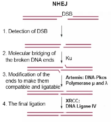
Figure 6: Overview of the non-homologous end joining (NHEJ) repair pathway. The NHEJ repair pathway specifically repairs double strand breaks. The MRN complex recognizes the double stranded break. The Ku heterodimers and DNA-PK is then recruited to bridge the broken DNA ends. The nuclease Artemis is then recruited to generate ends that are compatible for ligation and after DNA polymerases fill the gaps at the extremities, the XRCC/DNA ligase 4 complex ligates the ends together. Reference: Gu et al. (2008) PathoGenetics 1(1): 4. 102
22
3.1.5 Homologous Recombination (HR)
HR is another repair pathway activated upon DNA double strand break. This pathway requires the information of homologous sister chromatids. Since it uses the sister
chromatid as a template for repair, this pathway is error-free and is favoured during the S phase and the G2 phase of the cell cycle. Upon double strand break, the MRN complex is recruited and the 3’ to 5’ exonuclease MRE11 promotes DNA resection 103,104. DNA resection generates 3’ overhangs that are proficient for invasion and DNA synthesis as polymerases synthesize DNA in 5’ to 3’ direction103,104. The Replication Protein A (RPA) is then recruited to prevent degradation of the less stable single stranded DNA ends and formation of secondary structures104. Rad51 then displaces RPA, forming nucleoprotein filaments crucial for homology searching and invading the homologous sister
chromatids104. Another homologous recombination repair protein, Rad52, was reported to interact with Rad51 through its C-terminal domain to enhance Rad51 recombinase
activity105. The N-terminal domain of Rad52 contains a single strand DNA binding domain that allows annealing to the complementary strand 105. As the DNA invades the sister chromatid template, a D-loop is formed from the displacement resulting from DNA synthesis104. As the 3’ ends extends from each strand, the DNA strands will generate two crossing overs referred as Holliday junctions (HJs)104. The HJs will be cleaved by resolvases in order to recover the double strand structure of DNA. Depending on the cleavage direction (longitude or latitude), two possible products can be generated (Figure 7) 104.
23
Figure 7: Overview of the homologous recombination (HR) repair pathway.
Upon double strand breaks, the MRN complex (MRE11, RAD50, and NBS1) recognizes these lesions and generates overhangs by end resection to allow DNA synthesis. RPA is then recruited to protect the single strand DNA and is replaced by Rad51 to initiate strand inversion. The Holliday junctions generated during DNA synthesis are resolved in order to form a double strand DNA. Reference: Junran Zhang and Simon N. Powell (2005) Mol Cancer Res: 3 (10) 531-539. 104
24
3.1.6 Translesion Synthesis (TLS)
During replication, the DNA polymerase δ or ε encounters countless DNA lesions and results in replication stalling which impairs cell function 57,60-62,64. Although it is not directly a DNA repair mechanism, translesion synthesis is an approach to bypass DNA lesions and complete replication despite having damage57,60-62,64. Unlike DNA
polymerases, TLS polymerases are proficient in synthesizing DNA past DNA lesions (Figure 8) 57. Translesion synthesis is mediated by a different family of polymerases referred to as the Y family. In humans, the Y family polymerases include polη, polι, polκ, polζ, and REV1 57. It is still not well understood which TLS polymerase will be recruited to the site of lesion. It is speculated that the nature of damage mediates the choice of TLS polymerases recruited 57. It is also suggested that they might have redundant roles so that one can compensate for the loss of function of the other 57. Polη was reported to very accurately bypass cyclobutane pyrimidine dimers (CPD) generated by UV 57. Polι was found to have high fidelity replicating dA templates whereas it has low fidelity at
replicating dT templates 57. Polκ was shown to have a tendency to incorporate frameshifts whereas it could bypass N2-dG lesions induced by benzo [a] pyrene accurately 57-59. Polζ was suggested to form a complex with polι or polκ as an alternative to polη 57,106,107. REV1 was reported to incorporate dC opposite to dG although it is mostly observed to act as a scaffolding protein to recruit polη, polι, polκ to the DNA lesion 57. TLS is triggered upon the encounter of a DNA lesion by the replicative polymerase. The DNA replication polymerase stalls and signals PCNA monoubiquitination on lysine 164 by the E3 ligase Rad18 and the E2 conjugating enzyme Rad6 108-112. Proliferating cell nuclear antigen (PCNA) is a trimeric clamp that slides along the DNA and allows the anchoring of DNA polymerases, which recognize PCNA through their PCNA interacting motif referred as PIP boxes57,113. Upon monoubiquitination of PCNA, the recruitment of TLS polymerases is favoured as they possess an ubiquitin interacting motif and thus, promote a switch of position of DNA polymerases 57. The TLS then incorporates bases at the sites of DNA lesion and beyond before switching back to the DNA polymerases 57. It is still not clear how DNA synthesis is resumed but there are speculations that a deubiquitinase could be involved in antagonizing PCNA ubiquitination and/or the TLS polymerase is
25
polyubiquitinated and degraded by the proteasome 57. Of note, the TLS mechanism is error prone as the translesion synthesis polymerases are not as accurate as the replicative
polymerases and only promote the bypass of the lesion to allow the cell to progress in S phase 57. TLS polymerases might incorporate the correct base opposite of the DNA lesion or the wrong base which has to be repaired by the appropriate DNA repair pathway prior to the next replicative phase of the cell cycle in order to avoid genomic instability 57. It is interesting to note that, although still not well characterized, there is also an error free mechanism known as template switching regulated by the ubiquitin ligase complex RAD5-MMS2-UBC13 which triggers K63 polyubiquitination chains on lysine 164 of PCNA108. Template swithching is suggested to be dependent on Rad51; as the DNA polymerase encounters a DNA lesion and stalls, the newly synthesized strand temporatily invades and uses the sister chromatid on the opposing duplex as a template 114,115.
26
Figure 8: Overview of the translesion synthesis (TLS) mechanism
Upon encounter of a DNA lesion, the replicative polymerase stalls and triggers PCNA ubiquitination and promotes the switch between replicative polymerase and translesion synthesis polymerases. There are 5 translesion synthesis polymerases (polη, polι, polκ, polζ, and REV1) that could possibly be recruited depending on the type of DNA lesion. The translesion synthesis polymerase incorporates a base complementary to the DNA lesion and bypasses the DNA lesion before switching back with the replicative
27
4 The cell cycle regulation and apoptosis
The cell cycle is a process that exists in every living organism that allows cells to divide into identical cells. This important cycle consists of different phases in which each has a specific role and is regulated tightly in a unidirectional way in order to prevent chromosomal aberrations, which lead to tumourigenesis. Cell proliferation is regulated by different mechanisms that arrest cell cycle progression at different stages. These cell cycle arrest mechanisms, called checkpoints, play critical roles in preventing accumulation of mutations and genomic instability. In the presence of chromosomal abnormalities or genotoxic stress, the checkpoints are activated in order to arrest the cell cycle, providing time for DNA repair. Deregulation of checkpoint mechanisms by activation of specific oncogenes or inactivation of certain tumour suppressor genes cause genomic instability, which lead to cancer development.
4.1 The cell cycle regulation
The cell cycle is regulated by different cyclin dependent kinases (CDK) and cyclins. CDK and cyclins assemble as heterodimers and are activated by phosphorylation through CDK activating kinases (CAK) 116. The CDK/Cyclin dimer phosphorylates and regulates diverse proteins or transcriptional factors required for cell cycle progression. Each CDK/cyclin dimer has its own targets and thus, confers specificity in the cell cycle. Also, the cyclins are synthesized in a sequential manner when necessary and are
ubiquitinated and degraded by the proteasome once they have served their purpose to concede their place to other cyclins so that the cell cycle can go on (Figure 9) 116. The specificity of cyclin degradation is dependent on the E3 ligases, which confer specificity though their F-Box domains recognizing specific targets 6. The two main E3 ligases regulating the cell cycle are the RING type ligases SCF and APC/C whose F-BOX proteins are expressed in a cell cycle dependent manner 6(Table 1).
28
Figure 9: Cyclin levels and complexes during the cell cycle.
Overview of the cyclin levels and the cyclin/cdk heterocomplexes responsible for
regulating cell cycle phase transition. Reference: Verschuren E.W. et al. (2004) Journal of General Virology 85, 1347–1361. 116
The cell cycle can be regulated negatively by the CDK inhibitors or the kinases Wee1/Myt1. CDK inhibitors (CDKI) play an important role in cell cycle checkpoint by interacting with CDKs and inhibiting their activity 117,118. The CDKI include two families: the INK4 family and the cip/kip family. The INK4 family consists of p15, p16, p18, and p19 mainly known to mediate G1/S checkpoint 117,118. The cip/kip family consists of p21, p27, and p57 that mediate G1/S checkpoint or G2/M checkpoint 119-121. The Myt1 and Wee1 kinases are inhibitors of the cell cycle as they inhibit CDK activities by
phosphorylation of CDK1 on threonine 14 and tyrosine 15 respectively 122-124. This inhibition is reversed by the CDC25 family phosphatases (CDC25A, CDC25B, and CDC25C) and thus, plays an important role in promoting cell cycle progression 125,126. These phosphatases are expressed in a cell cycle dependent manner as CDC25A is mainly expressed from G1 to S; whereas CDC25B is mainly expressed from S to mitosis 125,126. CDC25C is constitutively expressed throughout the cell cycle 125,126.
29
4.2 Cell cycle checkpoints
Upon DNA damage, the cell cycle checkpoints are rapidly activated. The G1/S checkpoint is mediated by DNA damage activation of the ATM/ATR kinases which subsequently induce the phosphorylation of CHK2/CHK1 kinases 127. CHK1 and CHK2 were both observed to phosphorylate CDC25A on serine 126, resulting in CDC25A inhibition 128. Inhibition of CDC25A inhibits the activity of CDK2 and cell cycle
progression128. In addition, CHK1/CHK2 phosphorylation activates the tumour suppressor protein p53, which is well known to promote cell cycle arrest as p53 acts as a transcription factor promoting transactivation of the CDK inhibitor p21, known to inhibit the activity of the CDK2 129-131. ATM/ATR can as well phosphorylate and inhibit Mdm2, which is the E3 ligase for p53, preventing the degradation of p53 and thus promoting cell cycle checkpoint 132-134.
In the G2/M checkpoint, phosphorylation of CHK1/CHK2 inhibits CDC25B, resulting in CDK1-cyclin B inhibition128. Moreover, p21 can also inhibit G2/M transition by inhibiting CDK1 135. During the G2/M checkpoint, p53 can induce 14-3-3 and
GADD45, inhibiting CDK1 as GADD45 and 14-3-3 sequesters CDK1 and CDC25A respectively in the cytoplasm 41,136,137.
The mitotic checkpoint, also known as the spindle assembly checkpoint (SAC) is important for maintaining genomic integrity. When chromosomes are attached incorrectly on the kinetochores, this checkpoint is triggered to inhibit metaphase to anaphase transition
138
. This checkpoint is regulated by kinetochores. Kinetochores are chromosomal sites containing proteins that serve as attachment sites for the microtubules (the inner kinetochore is attached to the chromosome through the centromeres and the outer kinetochore is attached to the microtubules) 139. Kinetochores function as a sensor between the chromosomes and the microtubules in order to activate the SAC when
chromosomes are not attached properly to the microtubules 138-143. When the chromosome and the microtubules are attached properly, securin, an inhibitor of separase, is degraded by APC/Ccdc20 6,144. Separase, the enzyme responsible for degrading the cohesins and kinesins maintaining the chromosome together is then released and activated144. APC/Ccdc20 can also activate separase by degrading cyclin B as the latter is required for
30
maintaining separase inactivated by phosphorylation 6,144. The active separase degrades the kinesins and cohesins to facilitate chromosomal segregation thus promoting
anaphase144. In contrast, if the chromosomes are attached incorrectly, a tension is sensed by the kinetochores and the spindle checkpoint is activated by the formation of the mitotic checkpoint complex, Mad1, Mad2, Bub1, BubR1, Bubr3, which inhibits APC/Ccdc20 to prevent anaphase entry as the latter is the E3 ligase responsible for degrading cyclin B to promote anaphase entry 6,138. However, if the SAC fails (mitotic slippage), it might promote mitotic catastrophe, in which cell death is induced. Of note, it is possible for the cells to adapt and bypass cell death, leading to aneuploid cells that contain abnormal chromosome numbers, destabilizing genomic integrity and enhancing tumourigenesis.
Table 1: Summary of the cell cycle checkpoints and the proteins involved.
Summary table describing the phases and the checkpoints of the cell cycle along with the cyclin/CDK heterocomplexes and ubiquitin ligases regulating it 6,7.
31
4.3 Apoptosis
If the DNA damage is too massive to be repaired by DNA repair pathways, apoptosis will be triggered in order to prevent any further proliferation of the cell harbouring damaged DNA. Apoptosis is an irreversible process where cells are programmed to die when the damages are too important to be solved by the cell cycle checkpoints and DNA repair mechanisms. Apoptosis is initiated by particular proteases called caspases, which are found to be inactive under normal conditions. There are two types of caspases: initiator caspases such as Caspase-8 or Caspase-9 and effector caspases such as Caspase-3 145. The initiator caspases are first activated, triggering the cleavage and activation of the effector caspases 145. Apoptosis is negatively regulated by IAPs (inhibitor of apoptosis proteins) as they interact with caspases to constrain their active sites
146-148
. There are two pathways for apoptosis, the intrinsic pathway and the extrinsic pathway, which both ultimately lead to the cleavage and activation of Caspase-3 145-147. Caspase-3 then cleaves proteins and DNA in the cell, eventually resulting in cell death 145. The intrinsic pathway is mediated by the mitochondria. In presence of stress, the
mitochondria have increased membrane permeability, promoting the release of cytochrome c and SMAC (small mitochondria-mediated activator of caspases) 145,148. SMAC interact with IAPs in order to abolish the interaction of the latter with caspases 148. The
cytochrome c released by the mitochondria interacts with the apoptotic protease activating factor 1 (APAF-1) 149. APAF-1 promotes cleavage and activation of the caspase 9, resulting in Caspase-3 activation 149. The extrinsic pathway is mediated by membrane receptors such as Fas 150,151. Upon binding with its ligand FasL, the death-inducing signaling complex (DISC) is formed, allowing the interaction with and the activation of the initiator Caspase-8 150,151. Different markers of apoptosis include Caspase-3 and Poly (ADP-ribose) polymerase-1 (PARP-1) cleavage. PARP-1 is a protein required for ADP ribose synthesis for gene transcription and DNA repair152,153. In the presence of low DNA damage, PARP-1 activity is enhanced and is recruited to single strand and double strand DNA breaks in order to promote the formation of poly (ADP-ribose) chains 152,153. The recruitment of PARP-1 to the site of DNA damage promotes chromatin relaxation and
32
DNA repair 154,155. During apoptosis, PARP-1 is cleaved and inactivated by Caspase-3 and this cleavage is widely used as a marker for apoptosis 156,157.
33
5 BRCA1
Breast cancer susceptibility protein type 1 (BRCA1) is a major tumour suppressor. BRCA1 mutation carriers have a higher risk of developing breast cancer (65%) and/or ovarian cancer (39%) by the age of 70 andheterozygous BRCA1 mutation is sufficient for increasing the risk of developing cancer 37,38. Until now, there are approximately 1600 BRCA1 mutations recorded158. However, it is difficult to determine if all mutations are associated with an increased risk of cancer due to the lack of biochemical and functional studies on these mutations158. Nevertheless, there is no doubt that BRCA1 is an important tumour suppressor crucial for cell function and maintenance of genomic stability.
Consistent with this notion, BRCA1 null mice were reported to be embryonic lethal, likely due to the activation of several checkpoint responses that arrest cell proliferation as a consequence of DNA repair defect 159. Indeed, a few major roles of BRCA1 include: DNA damage signaling and repair, checkpoint activation, and transcription regulation.
34
5.1 The Domains of BRCA1
The N-terminal domain of BRCA1 possesses a RING domain and interacts with BRCA1 Associated RING Domain 1 (BARD1) to form a core complex, conferring
ubiquitin ligase activity. BARD1 is believed to be crucial for stabilizing the RING domain of BRCA1 for a proper conformation conferring E3 ligase activity (Figure 10) 160.
Although the role of the ubiquitin ligase activity of BRCA1 is still not well understood, it was proposed that it could have a role in amplifying ubiquitination of γH2AX at the site of DNA damage in order to facilitate the recruitment of additional BRCA1 and homologous recombination proteins161. Some studies proposed that BRCA1/BARD1 targets RNA polymerase II and has a role in regulating transcription 162-164. BRCA1/BARD1 ligase complex was also found to auto-ubiquitinate itself in a non-degradative manner in order to signal a yet to be known cellular function 165. In the middle region, BRCA1 contains a coiled coil domain, which is a hydrophobic region that promotes protein-protein
interaction; usually with the coiled coil region of another protein 166-169. The coiled coil middle region of BRCA1 was observed to interact with the N-terminal coiled-coil region of PALB2 whereas the C-terminal region of PALB2 interacts with the N-terminal region of BRCA2 166-170. Both PALB2 and BRCA2 are involved in the repair of double strand DNA breaks166-170. At the C terminus, BRCA1 contains 2 phospho-peptide binding domains that are typical of DNA repair proteins, called BRCT 104,171,172 (Figure 10). These BRCT domains recognize a consensus phosphoS-X-X-F motif and are of high importance as they allow the BRCA1/BARD1 core complex to interact with a multitude of proteins in order to form complexes involved in DNA damage repair171,172. As a matter of fact, mutations in the BRCT domains of BRCA1 predispose to cancer development 158,173. The BRCA1/BARD1 core complex interacts with phosphorylated Abraxas, BRIP1 and CtIP through the BRCT domains of BRCA1 to form the complexes A, B and C respectively
104,174
. The complex A is involved in triggering G2-M cell cycle checkpoint and recruiting BRCA1 to DNA damage sites 104,174. The complex B is involved in the S phase checkpoint of the cell cycle 104,174. The complex C is involved in DNA resection in order to initiate homologous recombination 104,174. Finally, the BRCC complex allows the recruitment of Rad51 through the intermediates PALB2 and BRCA2 in order to promote homologous
35
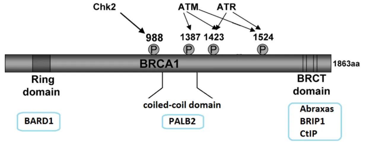
recombination 104,174 (Table 2). BRCA1 has been reported to be phosphorylated on serine 988 by Chk2 and on serine 1387, 1432 and 1524 by ATM or ATR 104,166 (Figure 10). Phosphorylation of BRCA1 on serine 988 by Chk2 was observed to be crucial for PALB2 interaction with BRCA1 and the recruitment of Rad51 but is not required for cell cycle checkpoint activation 166-169,175. In contrast, phosphorylation of BRCA1 on serine 1387, 1423 and 1524 were shown to be required for cell cycle checkpoint activation but not for homologous recombination repair 166,176,177 .
Figure 10: The domains of BRCA1 and its sites of phosphorylation in response of stress. Schema describing the different domains of BRCA1 along with its interaction sites with BARD1, PALB2, Abraxas, BRIP, and CtIP. The phosphorylation sites by Chk2, ATM, and ATR are also shown. Modified from: Zhang J, Powell S.N (2005) Mol Cancer Res. 3:531-39. 104
36
BRCA1 complex Function Components
Core complex Promotes E3 ligase activity BRCA1 and BARD1 (constitutive heterodimer) BRCA1 A Control of G2-M checkpoint
and BRCA1 accumulation at damage-induced foci
BRCA1, BARD1, Abraxas, RAP80 and BRCC36
BRCA1 B DNA replication and S phase progression
BRCA1, BARD1, BRIP and TOPBP1
BRCA1 C DNA resection and G2-M checkpoint
BRCA1, BARD1, CtIP and MRN complex (MRE11, Rad50, NBS1) BRCC (BRCA1/2 containing complex) Homologous recombination-mediated DNA repair
BRCA1, BARD1, BRCA2, PALB2 and Rad51
Table 2: Description of the BRCA1 complexes.
Description of the components and functions of the BRCA1/BARD1 core complex and the complexes A, B, C and BRCC. Modified from: Huen M.S.Y, Sy S.M.H, Chen J. (2010) Nature Review 11(2):138-148. 174
37
5.2 The mechanism of action of BRCA1 upon exposure to
double strand breaks
Although extensive studies have been conducted on BRCA1, the mechanism of action and the regulation of BRCA1 in presence of stress are still not well established. The following figure (Figure 11) shows a schema of how the BRCA1 complexes are being recruited and how they can recruit homologous recombination proteins. Upon IR, a
multitude of DNA damage proteins are recruited to the chromatin, forming IR induced foci (IRIF). MRE11, RAD50, and NBS1 (known as the MRN complex) are recruited to the damaged sites along with the BRCA1 complex C (BRCA1 and CtIP) 178-180. The MRN complex was shown to be recruited rapidly upon DNA breaks and thus, acts as an
important sensor of DNA damage 178-180. In addition, the MRN complex and CtIP possess exonuclease activity and work together for DNA resection in order to create 3’ single stranded DNA overhangs for strand invasion 39,40,174,178,181. A single strand DNA binding protein, replication protein A (RPA), is then recruited in order to prevent the formation of secondary structures and degradation of the single strand DNA ends40. The MRN complex also promotes the recruitment and the activation of the kinase Ataxia
Telangiectasia Mutated (ATM) by promoting its autophosphorylation on serine 1981 182. ATM is well known for its involvement in signaling DNA damage as it phosphorylates H2A.X on serine 139, an H2A histone variant; generating γH2AX foci at damaged sites on the chromatin 39,183. The formation of γH2A.X foci is one the of first steps in DNA
damage signaling as phosphorylation of H2A.X on serine 139 occurs as early as one minute after DNA double strand breaks induction and thus, γH2AX acts as a marker for DNA damage 184. Phosphorylation of H2A.X is crucial for the recruitment and
maintenance of DNA repair proteins at the sites of DNA damage as it provides binding sites for DNA repair proteins with BRCT domains 185,186. Subsequently, the DNA damage mediator MDC1 is recruited through its BRCT domain which recognizes phosphorylated H2A.X and stabilizes the MRN complex, creating a positive feed-back loop to amplify the recruitment of γH2A.X and DNA repair proteins 39,187. The MDC1 and γH2AX interaction also prevents the protein phosphatase 2A (PP2A) from dephosphorylating γH2AX and
38
thus, prevents γH2AX elimination from the site of DNA damage 187,188. MDC1 is phosphorylated by ATM on threonine 98, which promotes the recruitment of the ubiquitin conjugating enzyme UBC13 and ubiquitin ligase RNF8 189. The RNF8 ligase includes a forkhead associated (FHA) domain that recognizes phosphorylated threonines on proteins
190,191
. RNF8 monoubiquitinates H2A or H2A.X on lysine 119and monoubiquitination of H2A or H2A.X is recognized by another E3 ligase RNF168 through its
ubiquitin-interacting motif 179,191-195 . RNF168 then triggers K63 polyubiquitination chain formation on lysine 119 of H2A and H2A.X, which is crucial for the accumulation of BRCA1 complex A (BRCA1/BARD1/Abraxas/RAP80) at the sites of DNA damage via RAP80, which recognizes ubiquitinated H2A or H2A.X through its ubiquitin interacting motif (UIM) 179,191-196. It is important to note that lysine 63 polyubiquitin chains are not involved in proteasomal degradation, but involved in DNA damage and other signaling events. It is also interesting to note that H2A.X can also be phosphorylated on tyrosine 142 by the tyrosine kinase WSTF to trigger apoptosis 197,198. MDC1 only interacts with H2A.X unphosphorylated on tyrosine 142, suggesting that phosphorylation of H2A.X on tyrosine 142 determines the outcome between cell survival by DNA repair or cell death by apoptosis 197,198. On the other hand, it has been reported that the histone acetyl transferase TIP60 and the E2 conjugating enzyme UBC13 complex can interact with γH2A.X in presence of IR and acetylate histone γH2A.X on lysine 5 and this steps seems to promote histone ubiquitination through UBC1339,199. It was also shown that acetylation and
ubiquitination induces the eviction of γH2A.X and promotes chromatin reorganization near the site of DSB 39,199. Although it is still not too well understood, ubiquitination or
acetylation of γH2A.X could trigger chromatin modification in order to exposed dimethylated histone H4 to allow the recognition by the DNA damage protein
53BP139,191,200,201. 53BP1, p53 binding protein 1, also interacts with p53 and plays an important role in cell cycle checkpoint 202. 53BP1 interacts with the MRN complex and was shown to further promote ATM activity to phosphorylate CHK2 for checkpoint signaling 202-205. Although 53BP1 recruitment to H4K20me2 was shown to be dependent on H2A.X ubiquitination triggered by MDC1, RNF8 and RNF168, little is known on the exact mechanism of action involving 53BP1 191,200,201 (Figure 11). Finally, following
39
BRCA1 recruitment, Rad51 is recruited through the scaffolding proteins PALB2 and BRCA2 40,166. Rad51 displaces RPA and initiates strand invasion of the sister chromatid for homologous recombination repair 40,166 (Figure 12).
40
Figure 11: Overview of BRCA1 recruitment to the sites of DNA damage upon double strand breaks.
Upon double strand break, the MRN complex is recruited along with ATM.
Phosphorylation of H2A.X by ATM signals the recruitment of the ubiquitin ligases RNF8 and RNF168. Ubiquitination of γH2A.X by RNF8 and RNF168, serve as an interaction site for the BRCA1 complex A consisting of BRCA1, RAP80, Abraxas and BRCC36. BRCA1 recruitment triggers cell cycle checkpoints and DNA damage repair. Reference: Haico van Attikum and Susan M. Gasser (2009) Trends in Cell Biology 19(5): 207-217 39
41
Figure 12: The role of BRCA1 in homologous recombination
BRCA1 has a role is promoting homologous recombination upon double strand break. The BRCA1 complex C consisting of BRCA1, BARD1, CtIP, and the MRN complex promotes ends resection to allow strand invasion. In addition, BRCA1 can recruit RAD 51 through the BRCC complex to promote strand invasion. Reference: P.J.O’Donovan and D.M.Livingston (2010) Carcinogenesis 31(6): 961–967. 40