Echocardiographic reference ranges for normal
left ventricular layer-specific strain: results from
the EACVI NORRE study
Toshimitsu Tsugu
1,2, Adriana Postolache
1, Raluca Dulgheru
1, Tadafumi Sugimoto
1,3,
Julien Tridetti
1, Mai-Linh Nguyen Trung
1, Caroline Piette
1, Marie Moonen
1,
Roberta Manganaro
1,4, Federica Ilardi
1,5, Alexandra Maria Chitroceanu
1,6,
Simona Sperlongano
1,7, Yun Yun Go
1,8, George Kacharava
9,
George D. Athanassopoulos
10, Daniele Barone
11, Monica Baroni
12,
Nuno Cardim
13, Andreas Hagendorff
14, Krasimira Hristova
15, Teresa Lopez
16,
Gonzalo de la Morena
17, Bogdan A. Popescu
18, Martin Penicka
19,
Tolga Ozyigit
20, Jose David Rodrigo Carbonero
21, Nico van de Veire
22,
Ralph Stephan Von Bardeleben
23, Dragos Vinereanu
6, Jose Luis Zamorano
24,
Monica Rosca
18, Andreea Calin
18, Julien Magne
25,26, Bernard Cosyns
27,
Elena Galli
28, Erwan Donal
28, Ciro Santoro
5, Maurizio Galderisi
5,
Luigi P. Badano
29,30, Roberto M. Lang
31, and Patrizio Lancellotti
1,32,33*
1Department of Cardiology, GIGA Cardiovascular Sciences, University of Lie`ge Hospital, Heart Valve Clinic, CHU Sart Tilman, CHU Sart Tilman, 4000 Lie`ge, Belgium;
2Department of Cardiology, School of Medicine, Keio University, Tokyo, Japan;3Clinical Laboratory, Mie University Hospital, Mie, Japan;4Department of Clinical and
Experimental Medicine, Cardiology Unit, University of Messina, Via Consolare Valeria 1, 98125 Messina, Italy;5Department of Advanced Biomedical Sciences, Federico II
University Hospital, Naples, Italy;6Cardiovascular Research Unit, University and Emergency Hospital, University of Medicine and Pharmacy Carol Davila, Bucharest, Romania;
7Unit of Cardiology, Department of Translational Medical Sciences, University of Campania “Luigi Vanvitelli”, Monaldi Hospital, Naples, Italy;8National Heart Research Institute
Singapore, National Heart Centre Singapore, Singapore;9Department of the Cardiology, Tbilisi Institute of Medicine (TIM), 16 Tsintsadze, 0160 Tbilisi, Georgia;10Department of
Noninvasive Diagnostics, Onassis Cardiac Surgery Center, Athens, Greece;11Laboratory of Cardiovascular Ecography, Department of Cardiology, S. Andrea Hospital, La Spezia,
Italy;12Laboratorio Di Ecocardiografia Adulti, Fondazione Toscana “G.Monasterio” - Ospedale Del Cuore, Massa, Italy;13Hospital da Luz, Echocardiography Laboratory, Lisbon,
Portugal;14Department of Cardiology, University of Leipzig, Leipzig, Germany;15Department of Noninvasive Functional Diagnostic and Imaging, University National Heart
Hospital, Sofia, Bulgaria;16Cardiology Department, La Paz Hospital, IdiPAz, Ciber, Madrid, Spain;17Unidad de Imagen Cardiaca, Servicio de Cardiologia, Hospital Clinico
Universitario Virgen de la Arrixaca, IMIB-Arrixaca, Murcia, Spain;18University of Medicine and Pharmacy “Carol Davila” - Euroecolab, Institute of Cardiovascular Diseases “Prof.
Dr. C. C. Iliescu”, Sos. Fundeni 258, 022328, Bucharest, Romania;19Cardiovascular Center Aalst, OLV-Clinic, Aalst, Belgium;20VKV Amerikan Hastanesi, Kardiyoloji Bo¨lu¨mu¨,
Istanbul, Turkey;21Laboratorio de Ecocardiografia Hospital de Cruces, Barakaldo, Spain;22Echocardiography Unit, AZ Maria Middelares Gent, Gent, Belgium;23Emergency
Medical Department Cardiology, Universita¨tsmedizin of the Johannes Gutenberg-University Mainz, Mainz, Germany;24Department of Cardiology, University Alcala, Hospital
Ramo´n y Cajal, Madrid, Spain;25CHU Limoges, Hoˆpital Dupuytren, Service Cardiologie, Limoges, F-87042 France;26INSERM U1094, Univ. Limoges, CHU Limoges, IRD, U1094,
GEIST, 2, rue Marcland, 87000 Limoges, France;27CHVZ (Centrum voor Hart en Vaatziekten) – Universitair ziekenhuis Brussel; and ICMI (In Vivo Cellular and Molecular
Imaging) laboratory, 101 Laarbeeklaan, 1090b Brussels, Belgium;28Service de Cardiologie, INSERM 1414, CHU Pontchaillou - and- LTSI, Universite´ de Rennes 1 - INSERM, UMR
1099, Rennes, France;29Department of Cardiological, Neural and Metabolic Sciences, Istituto Auxologico Italiano, IRCCS, San Luca Hospital, Milan, Italy;30Department of
Medicine and Surgery, University of Milano-Bicocca, Milano, Italy;31Department of Medicine, University of Chicago Medical Center, Chicago, IL, USA;32Gruppo Villa Maria Care
and Research, Maria Cecilia Hospital, Cotignola, Italy; and33Anthea Hospital, Bari, Italy
Received 28 February 2020; editorial decision 2 March 2020; accepted 3 March 2020
Aims To obtain the normal range for 2D echocardiographic (2DE) measurements of left ventricular (LV) layer-specific
strain from a large group of healthy volunteers of both genders over a wide range of ages.
... Methods
and results
A total of 287 (109 men, mean age: 46 ± 14 years) healthy subjects were enrolled at 22 collaborating institutions of the EACVI Normal Reference Ranges for Echocardiography (NORRE) study. Layer-specific strain was analysed from the apical two-, three-, and four-chamber views using 2DE software. The lowest values of layer-specific strain calculated as ±1.96 standard deviations from the mean were -15.0% in men and -15.6% in women for epicardial
* Corresponding author. Tel: þ32 (4) 366 7194; Fax: þ32 (4) 366 7195. E-mail: [email protected]
Published on behalf of the European Society of Cardiology. All rights reserved.VCThe Author(s) 2020. For permissions, please email: [email protected].
Downloaded from ht tps: //academic. oup. com/ ehjcimaging/ advance-art icle-abst ract /doi/ 10. 1093/ ehjci/ jeaa050/ 5817446 by Universit y of Liege user on 09 A pril 2020
..
..
..
..
..
..
..
..
..
..
..
..
..
..
..
..
..
..
..
..
..
..
..
..
..
..
..
..
..
..
..
..
..
..
..
..
..
..
..
..
..
..
..
..
..
..
..
..
..
..
..
..
..
..
..
..
..
..
..
..
..
..
..
..
..
.
strain, -16.8% and -17.7% for mid-myocardial strain, and -18.7% and -19.9% for endocardial strain, respectively. Basal-epicardial and mid-myocardial strain decreased with age in women (epicardial; P = 0.008, mid-myocardial; P = 0.003) and correlated with age (epicardial; r = -0.20, P = 0.007, mid-myocardial; r = -0.21, P = 0.006, endocardial; r = -0.23, P = 0.002), whereas apical-epicardial, mid-myocardial strain increased with the age in women (epicardial; P = 0.006, mid-myocardial; P = 0.03) and correlated with age (epicardial; r = 0.16, P = 0.04). End/Epi ratio at the apex was higher than at the middle and basal levels of LV in men (apex; 1.6 ± 0.2, middle; 1.2 ± 0.1, base 1.1 ± 0.1) and women (apex; 1.6 ± 0.1, middle; 1.1 ± 0.1, base 1.2 ± 0.1).
... Conclusion The NORRE study provides useful 2DE reference ranges for novel indices of layer-specific strain.
!!!!!!!!!!!!!!!!!!!!!!!!!!!!!!!!!!!!!!!!!!!!!!!!!!!!!!!!!!!!!!!!!!!!!!!!!!!!!!!!!!!!!!!!!!!!!!!!!!!!!!!!!!!!!!!!!!!!!!!!!!!!!!!!!!!!!!!!!!!!!!!!!!!!!!!!!!!!!!!!!!!!!!!!!!!!!!!!!!!!!!!!!!!!!!!!!!!!!!!!!!!!!!!!!!!!
Keywords adult echocardiography
•
2D echocardiography•
deformation imaging•
reference valuesIntroduction
Two-dimensional (2D) speckle tracking echocardiography (STE) ena-bles quantitative evaluation of cardiac mechanics through
image-based analysis of myocardial deformation.1Although left ventricular
(LV) ejection fraction is the most commonly used parameter to as-sess LV mechanics, 2D-STE can detect latent LV dysfunction prior to a decline in LV ejection fraction by assessing mid-myocardial
longitu-dinal strain.2Recently, technological advances in 2D-STE has enabled
the assessment of layer-specific strain, thus allowing the measure-ment of epicardial, mid-myocardial, and endocardial longitudinal strain. The LV myocardium is divided into three myocardial layers consisting of circumferential fibres in the mid-myocardial layer and
longitudinal fibres in the epicardial and endocardial layers.3In most
heart diseases except some, such as sarcoidosis or hypertrophic car-diomyopathy, myocardial injury occurs predominantly in the
endo-cardial fibres in the early stages of the disease.4Endocardial strain
may have the potential to be more sensitive to assess myocardial function compared to epicardial or mid-myocardial strain in different
cardiovascular diseases.5–9However, normal ranges for each type of
layer-specific strain remain, to date, poorly defined.10,11The aim of
this study was to establish the normal ranges of layer-specific strain from a large group of healthy volunteers of both genders over a wide range of ages.
The NORRE (Normal Reference Ranges for Echocardiography) study is the first European, large prospective, multicentre study per-formed in 22 laboratories accredited by the European Association of Cardiovascular Imaging (EACVI) and in one American laboratory, which has provided reference values for all 2D echocardiographic
(2DE) measurements of all cardiac chambers,12 Doppler
parame-ters,13aortic dimensions,143D echocardiographic measurements of
the LV volumes and strain,152DE measurements of LV strain,162D
and 3D measurements of left atrial function,17and myocardial
indi-ces.18This study aimed to (i) establish normal reference limits for
layer-specific strain in healthy adults and (ii) examine the influence of age and gender on these normal reference ranges.
Methods
Patient population
A total of 734 healthy European subjects constituted the final NORRE study population. The local ethics committees approved the study proto-col. After the exclusion of patients that had incompatible image formats
and/or poor image quality, the final study population consisted of 287 (39%) healthy subjects.
Echocardiographic examination
A comprehensive echocardiographic examination was performed using state-of-the-art echocardiographic ultrasound system (GE Vivid E9; Vingmed Ultrasound, Horten, Norway) following a recommended proto-col approved by EACVI.19,20 All echocardiographic images were recorded in a digital raw-data format (native DICOM format) and central-ized for further analysis, after anonymization, at EACVI Central Core la-boratory at the University of Lie`ge, Belgium.
2D LV layer-specific strain
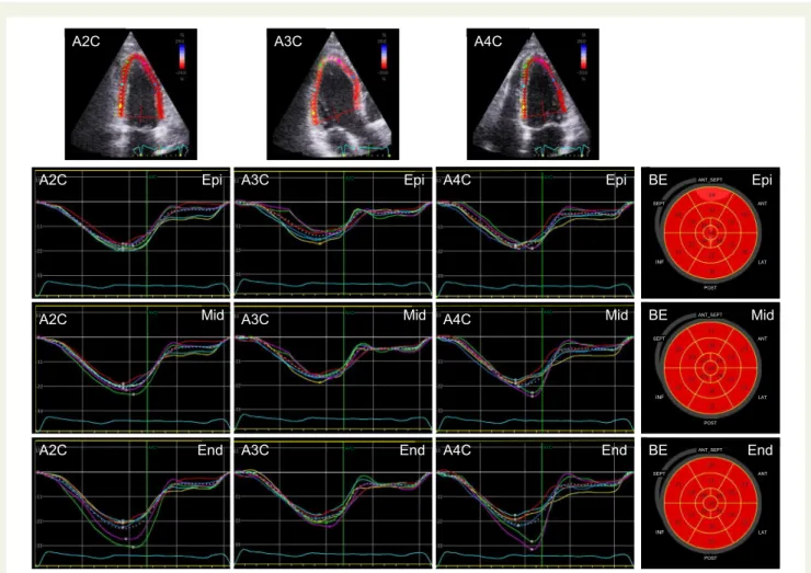
Quantification of layer-specific strain measurements were performed off-line with dedicated software (EchoPAC V.203, GE). For measuring layer-specific strain, attention was taken to cover the entire myocardial wall thickness with the region of interest (ROI) of each segment and to avoid to include the pericardium. Calculation of transmural variation of longitu-dinal strain across the entire myocardium was based on the assumption of linear distribution. Endocardial and epicardial strain were measured on the endocardial and epicardial ROI border, respectively, whereas the mid (centre line) of the ROI represented the average values of the transmural wall thickness. The layer-specific strain values were obtained by averaging the peak longitudinal strain of 17 segments (Figure1). The ratio of endo-cardial to epiendo-cardial was calculated using the End/Epi ratio for the assess-ment of the strain gradient.
Statistical analysis
Continuous variables were expressed as mean ± standard deviation (SD). The 95% confidence interval was calculated as ±1.96 SDs from the mean. Differences between groups were analysed for statistical significance with the unpaired t-test for normally distributed continuous variables. Comparison of continuous variables according to age groups was done with one-way analysis of variance test. When a significant difference was found, post hoc testing with Bonferroni comparisons to identify specific group differences was used. Correlation between continuous variables was performed using the Pearson correlation test. Multivariable linear re-gression analyses were performed to examine the independent corre-lates between layer-specific strain and baseline parameters. Intra-observer and inter-Intra-observer variability were assessed in 20 randomly selected subjects using Bland–Altman analysis. P < 0.05 was considered statistically significant. All statistical analyses were performed using JMP 11.0 statistical software (SAS Institute, Cary, NC, USA).
Downloaded from ht tps: //academic. oup. com/ ehjcimaging/ advance-art icle-abst ract /doi/ 10. 1093/ ehjci/ jeaa050/ 5817446 by Universit y of Liege user on 09 A pril 2020
..
..
..
..
..
..
..
..
..
..
..
..
..
..
..
..
..
..
..
..
..
..
..
..
..
..
..
..
..
..
..
..
Results
Demographic data
Table1summarizes the demographic data of the NORRE population
analysed in the present study. A total of 109 men (mean age 46 ± 14 years) and 178 women (mean age 45 ± 14 years) were included. Systolic blood pressure was higher in men (mean age 123 ± 10 mmHg) than in women (116 ± 15 mmHg). Strain values may be affected by LV afterload. However, it remains to be clarified whether the strain values correlate with the LV afterload, and few
studies have reported.21The mean frame rate was on the apical view
were 63 ± 10/s (men 63 ± 11/s, women 64 ± 9/s, P = 0.73). Layer-specific strain results from the entire study population are depicted in
Table2. All average layer-specific strains were significantly higher in
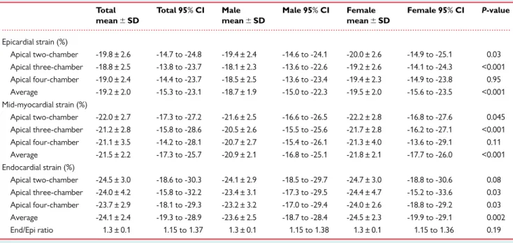
women than in men. The lowest values of layer-specific strains were -15.0% in men and -15.6% in women for epicardial strain, -16.8% and -17.7% for mid-myocardial strain, and -18.7% and -19.9% for endo-cardial strain, respectively. The highest values of layer-specific strain were -22.3% in men and -23.5% in women for epicardial strain, -25.1% and -26.0% for mid-myocardial strain, and -28.4% and -29.1% for endocardial strain, respectively.
Relationship between age, gender, and
layer-specific strain
Relationships between gender and age with layer-specific strain in all
apical views are shown in Table3and Figure2. No significant
correla-tions were observed between age and layer-specific strains for all ap-ical chamber views. In all age groups, layer-specific strain, including epicardial, mid-myocardial, and endocardial strain tended to be higher in women compared to men. In the age group between 20 and 40 years (epicardial, mid-myocardial, and endocardial strain) and in the age group >60 years, layer-specific epicardial and mid-myocardial strains were significantly higher in women than men.
Relationships between age and layer-specific strains in the apical,
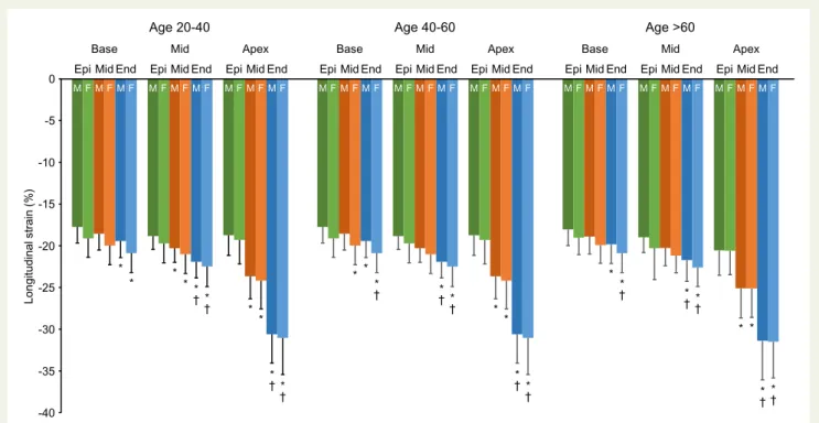
middle, and basal levels of the LV are shown in Table4and Figure3.
No significant age dependency was observed with respect to layer-specific strain in all segments in men. However, the basal-epicardial and mid-myocardial strain decreased with age in women (epicardial; P = 0.008, mid-myocardial; P = 0.003) and correlated with age (epi-cardial; r = -0.20, P = 0.007, mid-myo(epi-cardial; r = -0.21, P = 0.006, and endocardial; r = -0.23, P = 0.002). In contrast, theapical-epicardial and mid-myocardial strains increased with age in women (epicardial; P = 0.006 and mid-myocardial; P = 0.03) and correlated with age
Figure 1Layer-specific strain curves measurement by 2D speckle tracking echocardiography. A2C, apical two-chamber; A3C, apical three-cham-ber; A4C, apical four-chamthree-cham-ber; Epi, epicardial strain; Mid, mid-myocardial strain; End, endocardial strain; BE, bull’s eye of layer-specific strain.
Downloaded from ht tps: //academic. oup. com/ ehjcimaging/ advance-art icle-abst ract /doi/ 10. 1093/ ehjci/ jeaa050/ 5817446 by Universit y of Liege user on 09 A pril 2020
..
..
..
..
..
..
..
..
..
..
..
..
..
..
..
..
..
..
..
..
..
..
..
..
..
.
(epicardial; r = 0.16, P = 0.04). Although all strain values tended to in-crease from the epicardium to the endocardium, this tendency was stronger at the apical compared to the basal LV. Therefore, End/Epi ratio at the apex was higher than at the middle or the basal LV levels in men (apex; 1.6 ± 0.2, middle; 1.2 ± 0.1, base 1.1 ± 0.1) and women (apex; 1.6 ± 0.1, middle; 1.1 ± 0.1, base 1.2 ± 0.1), and this relationship
was preserved at all ages (Table4and Figure3).
Layer-specific strains determinants
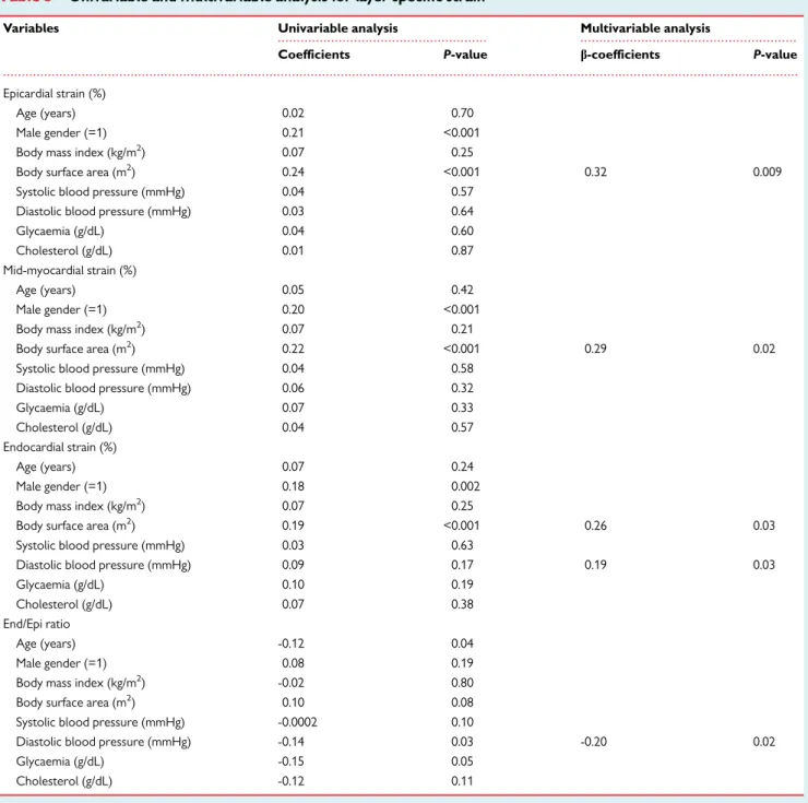
Multivariable analysis for layer-specific strain showed that epicardial, mid-myocardial, and endocardial strain increased with body surface area (epicardial; b-coefficient = 0.32, P = 0.009, mid-myocardial; b-co-efficient = 0.29, P = 0.02, endocardial; b-cob-co-efficient = 0.26, P = 0.03), whereas the End/Epi ratio was not related to body surface area. There was a significant increase in epicardial, mid-myocardial, and endocardial strain according to body surface area in univariable
analysis but no association was observed after adjustment for
con-founders (Table5).
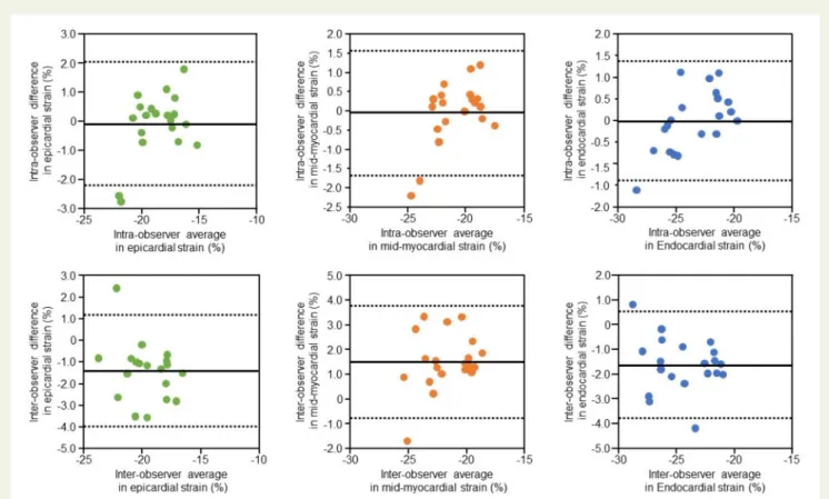
Repeatability and reproducibility
Intra-observer and inter-observer variability for layer-specific strain
are summarized in Table6. Intra-observer and inter-observer
analy-ses showed good repeatability and reproducibility in layer-specific
strain (Table6and Figure4).
Discussion
The present prospective, EACVI multicentre study provides contem-porary normal references values for 2DE measurements of layer-specific strain in a large cohort of healthy volunteers of both genders over a wide range of ages. Myocardial heterogeneity is characterized by higher deformation amplitude in the endocardial compared with
...
Table 1 Characteristics of the population
Parameters Total (n 5 287) Male (n 5 109) Female (n 5 178) P-value
Age (years) 46 ± 14 46 ± 14 45 ± 14 0.54
Height (cm) 170 ± 10 179 ± 8 165 ± 7 <0.001
Weight (kg) 69 ± 12 78 ± 10 63 ± 9 <0.001
Body surface area (m2) 1.8 ± 0.2 2.0 ± 0.1 1.7 ± 0.1 <0.001
Systolic blood pressure (mmHg) 119 ± 14 123 ± 10 116 ± 15 <0.001
Diastolic blood pressure (mmHg) 74 ± 9 75 ± 8 73 ± 9 0.02
Glucose (mg/dL) 91 ± 11 95 ± 9 89 ± 11 <0.001
Cholesterol (mg/dL) 182 ± 30 186 ± 26 180 ± 32 0.17
...
Table 2 2DE parameters of layer-specific strain
Total mean6 SD Total 95% CI Male mean6 SD Male 95% CI Female mean6 SD Female 95% CI P-value Epicardial strain (%) Apical two-chamber -19.8 ± 2.6 -14.7 to -24.8 -19.4 ± 2.4 -14.6 to -24.1 -20.0 ± 2.6 -14.9 to -25.1 0.03 Apical three-chamber -18.8 ± 2.5 -13.8 to -23.7 -18.1 ± 2.3 -13.6 to -22.6 -19.2 ± 2.6 -14.1 to -24.3 <0.001 Apical four-chamber -19.0 ± 2.4 -14.4 to -23.7 -18.5 ± 2.5 -13.6 to -23.4 -19.4 ± 2.3 -14.9 to -23.8 0.95 Average -19.2 ± 2.0 -15.3 to -23.1 -18.7 ± 1.9 -15.0 to -22.3 -19.5 ± 2.0 -15.6 to -23.5 <0.001 Mid-myocardial strain (%) Apical two-chamber -22.0 ± 2.7 -17.3 to -27.2 -21.6 ± 2.5 -16.6 to -26.5 -22.2 ± 2.8 -16.8 to -27.6 0.045 Apical three-chamber -21.2 ± 2.8 -15.8 to -28.6 -20.5 ± 2.6 -15.5 to -25.6 -21.7 ± 2.8 -16.2 to -27.1 <0.001 Apical four-chamber -21.1 ± 3.5 -14.2 to -28.1 -20.7 ± 2.7 -15.4 to -26.1 -21.3 ± 4.0 -13.6 to -29.1 0.11 Average -21.5 ± 2.2 -17.3 to -25.7 -20.9 ± 2.1 -16.8 to -25.1 -21.8 ± 2.1 -17.7 to -26.0 <0.001 Endocardial strain (%) Apical two-chamber -24.5 ± 3.0 -18.6 to -30.3 -24.1 ± 2.9 -18.5 to -29.7 -24.7 ± 3.0 -18.8 to -30.6 0.08 Apical three-chamber -24.0 ± 4.2 -15.8 to -32.2 -23.4 ± 3.1 -17.3 to -29.5 -24.4 ± 4.7 -15.2 to -33.6 0.03 Apical four-chamber -23.7 ± 2.9 -18.1 to -29.3 -23.2 ± 3.2 -17.0 to -29.4 -24.0 ± 2.6 -18.8 to -29.2 0.03 Average -24.1 ± 2.4 -19.3 to -28.9 -23.6 ± 2.5 -18.7 to -28.4 -24.5 ± 2.3 -19.9 to -29.1 0.002 End/Epi ratio 1.3 ± 0.1 1.15 to 1.37 1.3 ± 0.1 1.15 to 1.38 1.3 ± 0.1 1.15 to 1.36 0.19 CI, confidence interval; SD, standard deviation.
P-value differences between genders.
Downloaded from ht tps: //academic. oup. com/ ehjcimaging/ advance-art icle-abst ract /doi/ 10. 1093/ ehjci/ jeaa050/ 5817446 by Universit y of Liege user on 09 A pril 2020
... .... .... .... .... .... ... .... .... .. ... ... .... .... .... .... .... .... ... ... ... .... .... ... .... .... .... .... .... ... .... .... .... .... .... ... .... .... .... .... ... .... ... .... ... .... .. ... ... ... ... ... .... ... ... ... ... ... ... ... ... ... ... ... ... ... ... ... Ta ble 3 La y er -specific strain at th e apical tw o-chamber , apical thr ee-chamber , and apical four -chamber accor ding to g ender and a g e T otal (n 5 287) Ag e 20– 40 (n 5 115) Ag e 40–6 0 (n 5 122) A g e " 60 (n 5 50) P -v alue Male F ema le Male (n 5 110) , me an 6 SD F ema le (n 5 178) , mea n 6 SD Male (n 5 39), mea n 6 SD F emale (n 5 76), mea n 6 SD Male (n 5 50), mean 6 SD F em ale (n 5 72) , mean 6 SD Mal e (n 5 20) , m ean 6 SD F em ale (n 5 30) , m ean 6 SD Male F ema le RP -value RP -valu e Epicardi al lon gitudina lstrai n (%) Apical two-chamb er -19.4 ± 2.4 -20.0 ± 2.6 a -19.2 ± 2.5 -20.1 ± 2.6 -19.7 ± 2.5 -20.2 ± 2.6 -1 8.7 ± 2.0 -1 9.5 ± 2.6 0.2 3 0.59 -0.0002 1.00 0.08 0.28 Apical th ree-ch amber -18.1 ± 2.3 -19.2 ± 2.6 a -18.1 ± 2.1 -19.1 ± 2.8 a -18.5 ± 2.4 -19.4 ± 2.5 -1 7.3 ± 2.2 -1 8.9 ± 2.3 a 0.1 4 0.38 0.0 6 0.53 0.06 0.45 Apical four-cham ber -18.5 ± 2.5 -19.4 ± 2.3 a -17.7 ± 2.2 -19.0 ± 2.5 a -19.3 ± 2.6 -19.9 ± 2.0 -1 8.2 ± 2.0 -1 9.1 ± 2.1 0.0 1 0.81 -0.17 0.08 -0.04 0.60 Average -18.7 ± 1.9 -19.5 ± 2.0 a -18.3 ± 1.7 -19.4 ± 2.1 a -19.2 ± 2.1 -19.8 ± 1.9 -1 8.0 ± 1.5 -1 9.2 ± 1.8 a 0.0 3 0.73 -0.05 0.63 0.05 0.48 Mid-m yocardia llongitu dinal strai n (%) Apical two-chamb er -21.6 ± 2.5 -22.2 ± 2.8 a -21.6 ± 2.5 -22.4 ± 2.7 -21.9 ± 2.6 -22.3 ± 2.8 -2 0.7 ± 2.2 -2 1.6 ± 2.9 0.1 9 0.32 0.0 6 0.53 0.12 0.12 Apical th ree-ch amber -20.5 ± 2.6 -21.7 ± 2.8 -20.6 ± 2.3 -21.6 ± 2.8 -20.8 ± 2.7 -21.9 ± 2.9 a -1 9.8 ± 2.6 -2 1.3 ± 2.6 a 0.3 0 0.31 0.0 5 0.59 0.07 0.37 Apical four-cham ber -20.7 ± 2.7 -21.3 ± 4.0 -20.0 ± 2.4 -21.2 ± 2.7 a -21.4 ± 2.9 -21.5 ± 5.4 -2 0.4 ± 2.6 -2 1.3 ± 2.2 0.0 4 0.91 -0.14 0.14 0.02 0.78 Average -20.9 ± 2.1 -21.8 ± 2.1 a -20.7 ± 1.8 -21.8 ± 2.2 a -21.4 ± 2.3 -22.1 ± 2.1 -2 0.3 ± 1.9 -2 1.4 ± 2.0 a 0.1 1 0.57 -0.01 0.87 0.08 0.30 Endoca rdial longitu dinal strai n (%) Apical two-chamb er -24.1 ± 2.9 -24.7 ± 3.0 -24.4 ± 2.8 -25.0 ± 2.9 -24.3 ± 2.9 -24.6 ± 3.1 -2 2.9 ± 2.6 -2 4.0 ± 3.3 0.1 1 0.17 0.1 3 0.19 0.15 0.051 Apical th ree-ch amber -23.4 ± 3.1 -24.4 ± 4.7 -23.5 ± 2.8 -24.0 ± 6.1 -23.5 ± 3.3 -24.8 ± 3.4 a -2 2.7 ± 3.3 -2 4.3 ± 3.1 0.5 5 0.80 0.0 5 0.62 -0.00 02 1.00 Apical four-cham ber -23.2 ± 3.2 -24.0 ± 2.6 a -22.6 ± 2.7 -23.5 ± 2.8 -23.9 ± 3.3 -24.5 ± 2.4 -2 2.9 ± 3.3 -2 3.9 ± 2.5 0.1 4 0.75 -0.11 0.24 -0.05 0.51 Average -23.6 ± 2.5 -24.5 ± 2.3 a -23.5 ± 2.1 -24.4 ± 2.4 a -23.9 ± 2.8 -24.7 ± 2.3 -2 2.8 ± 2.4 -2 4.0 ± 2.3 0.2 5 0.45 0.0 2 0.83 0.09 0.21 End/Ep irati o 1.3 ± 0.1 1.3 ± 0.1 1.3 ± 0.1 1.3 ± 0.1 1.2 ± 0.1 1.2 ± 0.0 1.3 ± 0.1 1.3 ± 0.1 0.0 3 0.27 -0.15 0.12 -0.07 0.38 SD, standard deviation. aP < 0.05 vs. male. Downloaded from ht tps: //academic. oup. com/ ehjcimaging/ advance-art icle-abst ract /doi/ 10. 1093/ ehjci/ jeaa050/ 5817446 by Universit y of Liege user on 09 A pril 2020
..
..
..
..
..
..
..
.
the epicardial layer.22Layer-specific strain is a novel method that is
capable of assessing each layer of the myocardial function. Moreover, the absence of differences between vendors for layer-specific strain values makes this technique a useful tool for feasibility, accuracy, and
reproducibility.23
Our results are consistent with previous studies showing good concordance with the absolute values of layer-specific strain and that all layer-specific strains in women were consistently higher than in
men.10,24,25However, the relationship between layer-specific strain
and age dependency is inconsistent. As reported by Nagata et al. and
0 0 Lon git udinal st rain (%) 0 -20 -40 -35 -30 -25 -20 -15 -10 -5 0
Base Mid Apex
EpiMid End EpiMidEnd Epi MidEnd
M F MFM F M F MFM F
M FMFM F
Age 40-60
Base Mid Apex
EpiMid End EpiMidEnd Epi MidEnd
M F M F M F M F MFM F
M F M M F F
Age >60
Base Mid Apex
EpiMid End EpiMidEnd Epi MidEnd
M F FM F MFM FM F M MF MFM F Age 20-40 * † * * † * * * * * * † * † * † *††* * † * * * * * † * † * * * † * † * † * † * -25 -30 -35 -40 -15 -10 -5 0 -20
Figure 3Bar graphs showing average layer-specific strain at the apical, the mid-ventricular, and the basal levels of the left ventricle by 2D echocar-diography analysis according to gender and age categories. Epi, epicardial strain; Mid, mid-myocardial strain; End, endocardial strain. *P < 0.05 vs. epi-cardial strain.†P < 0.05 vs. mid-myocardial strain.
M F M F M F
Epi Mid End
Age 40-60
M F M F M F
Epi Mid End
Age >60 -30 -25 -20 -15 -10 -5 0 Epi F M F M F M Mid End Age 20-40 Longitudin al st ra in (%) * * * * *
Figure 2Bar graphs showing average layer-specific strain by 2D echocardiography analysis according to gender and age categories. End, endocar-dial strain; Epi, epicarendocar-dial strain; Mid, mid-myocarendocar-dial strain. *P-value differences between genders.
Downloaded from ht tps: //academic. oup. com/ ehjcimaging/ advance-art icle-abst ract /doi/ 10. 1093/ ehjci/ jeaa050/ 5817446 by Universit y of Liege user on 09 A pril 2020
... ... ... ... ... ... .... ... ... ... .. .... .... .... .... ... .... .... .... .... .... ... ... ... ... ... ... Ta ble 4 La y er -specific strain at the apical, mid-v entricular , and basal le v els of left v entricle accor ding to g ender and a ge T ot al (n 5 287) Ag e 20– 40 (n 5 115) Ag e 40–6 0 (n 5 122) Ag e " 60 (n 5 50) P -valu e Male F emale Mal e (n 5 109) , m ean 6 SD F em ale (n 5 178) , me an 6 SD Male (n 5 39), me an 6 SD F ema le (n 5 76), mea n 6 SD Male (n 5 50), mean 6 SD F em ale (n 5 72) , m ean 6 SD Male (n 5 20) , me an 6 SD F ema le (n 5 30), me an 6 SD Male F em ale RP -v alue RP -valu e Ba se Epi 17. 8 ± 2.0 -18.9 ± 2.2 a -17.7 ± 1.9 -19.1 ± 2.3 a -18.1 ± 1.9 -1 9.0 ± 2.0 a -17.3 ± 2.0 -17.8 ± 1.8 0.32 0.0 08 -0.02 0.8 3 -0.20 0.007 Mid 18. 6 ± 2.0 -19.7 ± 2.2 a -18.6 ± 2.0 -20.0 ± 2.3 a -18.9 ± 2.1 -1 9.9 ± 2.2 a -18.1 ± 2.0 -18.5 ± 1.8 0.30 0.0 03 -0.03 0.7 5 -0.21 0.006 End -1 9.5 ± 2.2 -20.7 ± 2.6 a -19.4 ± 2.0 -20.9 ± 2.3 a -19.8 ± 2.3 -2 0.9 ± 2.3 a -18.9 ± 2.1 -19.7 ± 3.7 0.27 0.2 3 -0.04 0.6 7 -0.23 0.002 End/Ep iratio 1.1 ± 0.1 1.2 ± 0.1 a 1.1 ± 0.1 1.1 ± 0.1 1.1 ± 0.1 1.1 ± 0.0 1.1 ± 0.0 1.2 ± 0.4 0.95 0.0 6 -0.05 0.6 1 0.1 3 0.09 M iddle Epi -18.8 ±1.8 -19.8 ± 3.0 a -18.8 ± 1.6 -19.7 ± 2.3 a -19.0 ± 1.8 -2 0.3 ± 3.8 a -18.1± 1.8 -18.9 ± 1.8 a 0.14 0.0 9 -0.10 0.3 1 -0.12 0.12 Mid -2 0.1 ± 2.0 -20.9 ± 2.1 a -20.3 ± 1.7 -21.0 ± 2.3 -20.3 ± 2.1 -2 1.2 ± 2.1 a -19.3 ± 2.1 -20.1 ± 1.8 0.12 0.0 6 -0.12 0.2 3 -0.16 0.04 End -2 1.7 ± 2.3 -22.4 ± 2.3 a -21.9 ± 1.9 -22.5 ± 2.4 -21.7 ± 2.5 -2 2.6 ± 2.3 a -21.0 ± 2.5 -21.5 ± 2.3 <0.001 0.0 8 -0.09 0.3 7 -0.14 0.07 End/Ep iratio 1.2 ± 0.1 1.1 ± 0.1 1.2 ± 0.1 1.1 ± 0.1 1.1 ± 0.1 1.1 ± 0.1 1.2 ± 0.1 1.1 ± 0.1 0.19 0.6 1 -0.02 0.8 5 -0.008 0.92 A pex Epi -1 9.6 ± 2.8 -20.1 ± 2.9 -18.7 ± 2.4 -19.3 ± 2.9 -20.5 ± 3.0 -2 0.6 ± 2.9 -19.1 ± 2.2 -20.9 ± 2.8 a 0.007 0.0 06 0.16 0.1 0 0.1 6 0.04 Mid -2 4.3 ± 3,2 -24.9 ± 3.5 -23.7 ± 2.8 -24.2 ± 3.4 -25.1 ± 3.6 -2 5.1 ± 3.5 -23.8± 2.6 -26.0 ± 3.6 a 0.06 0.0 3 0.12 0.2 2 0.1 3 0.08 End -3 0.9 ± 4.1 -31.6 ± 4.5 -30.6 ± 3.5 -31.0 ± 4.4 -31.4 ± 4.7 -3 1.5 ± 4.3 -30.1 ± 3.9 -33.0 ± 4.9 a 0.41 0.1 0 0.04 0.7 1 0.1 0 0.19 End/Ep iratio 1.6 ± 0.2 1.6 ± 0.1 1.6 ± 0.2 1.6 ± 0.1 1.5 ± 0.1 1.5 ± 0.1 1.6 ± 0.2 1.6 ± 0.1 0.01 0.0 02 -0.19 0.0 4 -0.05 0.61 SD, standard deviation. aP < 0.05 vs. male. Downloaded from ht tps: //academic. oup. com/ ehjcimaging/ advance-art icle-abst ract /doi/ 10. 1093/ ehjci/ jeaa050/ 5817446 by Universit y of Liege user on 09 A pril 2020
..
..
..
..
..
..
..
..
..
..
..
..
..
..
..
..
..
..
..
Shi et al.10,24no significant age dependency was observed concerning
all layer-specific strains. In contrast, as reported by Alcidi et al. all layer-specific strains were progressively reduced with increasing age. The relationship between layer-specific strain and age dependence was inconsistent and different from the previous NORRE study of
2D strain.16This difference may be due to the smaller number of
enrolled patients in this study than in previous NORRE study. Interestingly, the layer-specific strain gradient increased from the epi-cardial towards the endoepi-cardial layer. The mechanism underlying these findings remains unclear, but some considerations have been reported. The differences between epicardial and endocardial strain might be secondary to the ability of the endocardial fibres to stretch
more potently compared to the epicardial fibres during
end-dia-stole.26In addition, differences in coronary perfusion and metabolic
demands between the epicardial and endocardial layers may also
contribute to these differences.27,28In this context, the End/Epi ratio
at the apex was higher than that at the middle or basal LV levels in
both genders. (Table4and Figure3). The End/Epi ratio differs
depend-ing of the type of LV hypertrophic diseases, such as aortic stenosis29
or hypertrophic cardiomyopathy,30 and may have the potential to
diagnose, not only these disease but also other forms of hypertrophic diseases. The hypertrophied myocardium may remodel differently in response to a variety of aetiologies, resulting in different epicardial and endocardial strains. Moreover, the results of our multivariable
... ...
...
Table 5 Univariable and multivariable analysis for layer-specific strain
Variables Univariable analysis Multivariable analysis
Coefficients P-value b-coefficients P-value Epicardial strain (%)
Age (years) 0.02 0.70
Male gender (=1) 0.21 <0.001
Body mass index (kg/m2) 0.07 0.25
Body surface area (m2) 0.24 <0.001 0.32 0.009
Systolic blood pressure (mmHg) 0.04 0.57 Diastolic blood pressure (mmHg) 0.03 0.64
Glycaemia (g/dL) 0.04 0.60
Cholesterol (g/dL) 0.01 0.87
Mid-myocardial strain (%)
Age (years) 0.05 0.42
Male gender (=1) 0.20 <0.001
Body mass index (kg/m2) 0.07 0.21
Body surface area (m2) 0.22 <0.001 0.29 0.02
Systolic blood pressure (mmHg) 0.04 0.58 Diastolic blood pressure (mmHg) 0.06 0.32
Glycaemia (g/dL) 0.07 0.33
Cholesterol (g/dL) 0.04 0.57
Endocardial strain (%)
Age (years) 0.07 0.24
Male gender (=1) 0.18 0.002
Body mass index (kg/m2) 0.07 0.25
Body surface area (m2) 0.19 <0.001 0.26 0.03
Systolic blood pressure (mmHg) 0.03 0.63
Diastolic blood pressure (mmHg) 0.09 0.17 0.19 0.03
Glycaemia (g/dL) 0.10 0.19
Cholesterol (g/dL) 0.07 0.38
End/Epi ratio
Age (years) -0.12 0.04
Male gender (=1) 0.08 0.19
Body mass index (kg/m2) -0.02 0.80
Body surface area (m2) 0.10 0.08
Systolic blood pressure (mmHg) -0.0002 0.10
Diastolic blood pressure (mmHg) -0.14 0.03 -0.20 0.02
Glycaemia (g/dL) -0.15 0.05 Cholesterol (g/dL) -0.12 0.11 Downloaded from ht tps: //academic. oup. com/ ehjcimaging/ advance-art icle-abst ract /doi/ 10. 1093/ ehjci/ jeaa050/ 5817446 by Universit y of Liege user on 09 A pril 2020
..
..
..
..
..
..
..
..
..
..
..
..
..
..
..
..
..
analysis (Table5) suggest that the End/Epi ratio may have the
poten-tial to be a useful marker regardless of age, gender, or body surface area. Our data showed good reproducibility for the assessment of layer-specific strains, reinforcing the possibility of a promising applica-tion of this new advanced echocardiographic index in clinical practice.
Limitations
This study presents several limitations. First, only one-third of the patients included in the NORRE database could be analysed by the current available software. Second, since this study was
conducted only on GE equipment, data on other equipment, such as Philips, is not available. However, in our previous study, we reported that no differences were noted between GE and Philips
equipment with regard to longitudinal strain.16 Third, the number
of patients enrolled in this study was lower than in the previous
NORRE study of LV 2D strain.16 Therefore, the relationship
be-tween layer-specific strain and age dependency was inconsistent. The same tendency was observed for the basal and middle LV levels of all layer-specific strain. Fourth, whether the NORRE study results can be extrapolated to non-Caucasian European indi-viduals is still unknown.
Figure 4 Bland–Altman analysis for assessing intra-observer and inter-observer variability of layer-specific strain. Dotted lines represent bias and 95% limits of agreement for measurements performed in 20 patients.
...
Table 6 Repeatability and reproducibility of 2D echocardiographic data
Variables Mean6 SD Mean6 SD Bias P-value 95% LOA
Intra-observer
Average epicardial longitudinal strain (%) -19.4 ± 1.9 -18.6 ± 1.6 -0.46 0.005 -19.7 to -19.1 Average mid-myocardial longitudinal strain (%) -21.6 ± 2.1 -21.0 ± 2.0 -0.50 0.001 -22.1 to -21.5 Average endocardial longitudinal strain (%) -24.4 ± 2.4 -23.5 ± 2.2 -0.60 0.003 -24.9 to -24.1 Inter-observer
Average epicardial longitudinal strain (%) -19.4 ± 1.9 -18.3 ± 1.7 1.05 <0.001 -18.2 to -17.4 Average mid-myocardial longitudinal strain (%) -21.6 ± 2.1 -20.8 ± 1.9 0.82 0.001 -20.1 to -19.9 Average endocardial longitudinal strain (%) -24.4 ± 2.4 -23.5 ± 2.2 0.90 <0.001 -23.6 to -23.1 LOA, lower limits of agreement; SD, standard deviation.
Downloaded from ht tps: //academic. oup. com/ ehjcimaging/ advance-art icle-abst ract /doi/ 10. 1093/ ehjci/ jeaa050/ 5817446 by Universit y of Liege user on 09 A pril 2020
..
..
..
..
..
..
..
..
..
..
..
..
..
..
..
..
..
..
..
..
..
..
..
..
..
..
..
..
..
..
..
..
..
..
..
..
..
..
..
..
..
..
..
..
..
..
..
..
..
..
..
..
..
..
..
..
..
..
..
..
..
..
..
..
..
..
..
..
..
..
..
..
..
..
..
Conclusion
The NORRE study provides applicable 2DE reference ranges for layer-specific strain. Multivariable analysis did not show any significant association between layer-specific strain and age or gender.
Acknowledgements
The EACVI research committee thanks the Heart House for its support.
Funding
The NORRE study was supported by GE Healthcare and Philips Healthcare in the form of an unrestricted educational grant.
Conflict of interest:none declared.
References
1. Mor-Avi V, Lang RM, Badano LP, Belohlavek M, Cardim NM, Derumeaux G et al. Current and evolving echocardiographic techniques for the quantitative evalu-ation of cardiac mechanics: ASE/EAE consensus statement on methodology and indications endorsed by the Japanese Society of Echocardiography. J Am Soc Echocardiogr 2011;24:277–313.
2. Stanton T, Leano R, Marwick TH. Prediction of all-cause mortality from global longitudinal speckle strain: comparison with ejection fraction and wall motion scoring. Circ Cardiovasc Imaging 2009;2:356–64.
3. Ishizu T, Seo Y, Kameda Y, Kawamura R, Kimura T, Shimojo N et al. Left ven-tricular strain and transmural distribution of structural remodeling in hyperten-sive heart disease. Hypertension 2014;63:500–6.
4. Sengupta PP, Krishnamoorthy VK, Korinek J, Narula J, Vannan MA, Lester SJ et al. Left ventricular form and function revisited: applied translational science to car-diovascular ultrasound imaging. J Am Soc Echocardiogr 2007;20:539–51. 5. Shiino K, Yamada A, Scalia GM, Putrino A, Chamberlain R, Poon K et al. Early
changes of myocardial function after transcatheter aortic valve implantation using multilayer strain speckle tracking echocardiography. Am J Cardiol 2019;123: 956–60.
6. Huttin O, Girerd N, Coiro S, Bozec E, Selton-Suty C, Lamiral Z et al. Association between layer-specific longitudinal strain and risk factors of heart failure and dyspnea: a population-based study. J Am Soc Echocardiogr 2019;32: 854–65.
7. Zhang J, Zhu L, Jiang X, Hu Z. Layer-specific strain analysis of left ventricular myocardium after alcohol septal ablation for hypertrophic obstructive cardiomy-opathy. Medicine (Baltimore). 2018;97:e13083.
8. Zhang L, Wu WC, Ma H, Wang H. Usefulness of layer-specific strain for identify-ing complex CAD and predictidentify-ing the severity of coronary lesions in patients with non-ST-segment elevation acute coronary syndrome: compared with Syntax score. Int J Cardiol 2016;15;223:1045–52.
9. Sarvari SI, Haugaa KH, Zahid W, Bendz B, Aakhus S, Aaberge L et al. Layer-spe-cific quantification of myocardial deformation by strain echocardiography may re-veal significant CAD in patients with non-ST-segment elevation acute coronary syndrome. JACC Cardiovasc Imaging 2013;6:535–44.
10. Shi J, Pan C, Kong D, Cheng L, Shu X. Left ventricular longitudinal and circumfer-ential layer-specific myocardial strains and their determinants in healthy subjects. Echocardiography 2016;33:510–8.
11. Leitman M, Lysiansky M, Lysyansky P, Friedman Z, Tyomkin V, Fuchs T et al. Circumferential and longitudinal strain in 3 myocardial layers in normal subjects and in patients with regional left ventricular dysfunction. J Am Soc Echocardiogr 2010;23:64–70.
12. Kou S, Caballero L, Dulgheru R, Voilliot D, De Sousa C, Kacharava G et al. Echocardiographic reference ranges for normal cardiac chamber size: results from the NORRE study. Eur Heart J Cardiovasc Imaging 2014;15:680–90.
13. Caballero L, Kou S, Dulgheru R, Gonjilashvili N, Athanassopoulos GD, Barone D et al. Echocardiographic reference ranges for normal cardiac Doppler data: results from the NORRE Study. Eur Heart J Cardiovasc Imaging 2015;16:1031–41. 14. Saura D, Dulgheru R, Caballero L, Bernard A, Kou S, Gonjilashvili N et al.
Two-dimensional transthoracic echocardiographic normal reference ranges for prox-imal aorta dimensions: results from the EACVI NORRE study. Eur Heart J Cardiovasc Imaging 2017;18:167–79.
15. Bernard A, Addetia K, Dulgheru R, Caballero L, Sugimoto T, Akhaladze N et al. 3D echocardiographic reference ranges for normal left ventricular volumes and strain: results from the EACVI NORRE study. Eur Heart J Cardiovasc Imaging 2017;18:475–83.
16. Sugimoto T, Dulgheru R, Bernard A, Ilardi F, Contu L, Addetia K et al. Echocardiographic reference ranges for normal left ventricular 2D strain: results from the EACVI NORRE study. Eur Heart J Cardiovasc Imaging 2017;18:833–40. 17. Sugimoto T, Robinet S, Dulgheru R, Bernard A, Ilardi F, Contu L et al.; NORRE
Study. Echocardiographic reference ranges for normal left atrial function parame-ters: results from the EACVI NORRE study. Eur Heart J Cardiovasc Imaging 2018; 19:630–8.
18. Manganaro R, Marchetta S, Dulgheru R, Ilardi F, Sugimoto T, Robinet S et al. Echocardiographic reference ranges for normal non-invasive myocardial work in-dices: results from the EACVI NORRE study. Eur Heart J Cardiovasc Imaging 2019; 20:582–90.
19. Lancellotti P, Badano LP, Lang RM, Akhaladze N, Athanassopoulos GD, Barone D et al. Normal reference ranges for echocardiography: rationale, study design, and methodology (NORRE Study). Eur Heart J Cardiovasc Imaging 2013;14:303–8. 20. Cosyns B, Garbi M, Separovic J, Pasquet A, Lancellotti P; Education Committee of the European Association of Cardiovascular Imaging Association (EACVI). Update of the echocardiography core syllabus of the European Association of Cardiovascular Imaging (EACVI). Eur Heart J Cardiovasc Imaging 2013;14:837–9. 21. Burns AT, La Gerche A, D’hooge J, MacIsaac AI, Prior DL. Left ventricular strain
and strain rate: characterization of the effect of load in human subjects. Eur J Echocardiogr 2010;11:283–9.
22. Altiok E, Neizel M, Tiemann S, Krass V, Kuhr K, Becker M et al. Quantitative ana-lysis of endocardial and epicardial left ventricular myocardial deformation-comparison of strain-encoded cardiac magnetic resonance imaging with two-dimensional speckle-tracking echocardiography. J Am Soc Echocardiogr 2012;25: 1179–88.
23. Unlu S, Mirea O, Duchenne J, Pagourelias ED, Bezy S, Thomas JD et al. Comparison of feasibility, accuracy, and reproducibility of layer-specific global longitudinal strain measurements among five different vendors: a report from the EACVI-ASE strain standardization task force. J Am Soc Echocardiogr 2018;31: 374–80.e1.
24. Nagata Y, Wu VC, Otsuji Y, Takeuchi M. Normal range of myocardial layer-specific strain using two-dimensional speckle tracking echocardiography. PLoS One 2017;12:e0180584.
25. Alcidi GM, Esposito R, Evola V, Santoro C, Lembo M, Sorrentino R et al. Normal reference values of multilayer longitudinal strain according to age decades in a healthy population: a single-centre experience. Eur Heart J Cardiovasc Imaging 2018;19:1390–6.
26. Buchi M, Hess OM, Murakami T, Krayenbuehl HP. Left ventricular wall stress dis-tribution in chronic pressure and volume overload: effect of normal and depressed contractility on regional stress-velocity relations. Basic Res Cardiol 1990;85:367–83.
27. Kuwada Y, Takenaka K. [Transmural heterogeneity of the left ventricular wall: subendocardial layer and subepicardial layer]. J Cardiol 2000;35:205–18. 28. Path G, Robitaille PM, Merkle H, Tristani M, Zhang J, Garwood M et al.
Correlation between transmural high energy phosphate levels and myocardial blood flow in the presence of graded coronary stenosis. Circ Res 1990;67: 660–73.
29. Ozawa K, Funabashi N, Kobayashi Y. Left ventricular myocardial strain gradient using a novel multi-layer transthoracic echocardiography technique positively correlates with severity of aortic stenosis. Int J Cardiol 2016;221:218–26. 30. Ozawa K, Funabashi N, Takaoka H, Kamata T, Kanaeda A, Saito M et al.
Characteristic myocardial strain identified in hypertrophic cardiomyopathy sub-jects with preserved left ventricular ejection fraction using a novel multi-layer transthoracic echocardiography technique. Int J Cardiol 2015;184:237–43.
Downloaded from ht tps: //academic. oup. com/ ehjcimaging/ advance-art icle-abst ract /doi/ 10. 1093/ ehjci/ jeaa050/ 5817446 by Universit y of Liege user on 09 A pril 2020