HAL Id: dumas-02929561
https://dumas.ccsd.cnrs.fr/dumas-02929561
Submitted on 3 Sep 2020HAL is a multi-disciplinary open access archive for the deposit and dissemination of sci-entific research documents, whether they are pub-lished or not. The documents may come from teaching and research institutions in France or abroad, or from public or private research centers.
L’archive ouverte pluridisciplinaire HAL, est destinée au dépôt et à la diffusion de documents scientifiques de niveau recherche, publiés ou non, émanant des établissements d’enseignement et de recherche français ou étrangers, des laboratoires publics ou privés.
Percutaneous CT-guided biopsy of lytic bone lesions in
patients clinically suspected of lung cancer : diagnostic
performances for pathological diagnosis and molecular
testing
Stéphane Adam Asfari
To cite this version:
Stéphane Adam Asfari. Percutaneous CT-guided biopsy of lytic bone lesions in patients clinically suspected of lung cancer : diagnostic performances for pathological diagnosis and molecular testing. Human health and pathology. 2019. �dumas-02929561�
AVERTISSEMENT
Ce document est le fruit d'un long travail approuvé par le
jury de soutenance et mis à disposition de l'ensemble de la
communauté universitaire élargie.
Il n’a pas été réévalué depuis la date de soutenance.
Il est soumis à la propriété intellectuelle de l'auteur. Ceci
implique une obligation de citation et de référencement
lors de l’utilisation de ce document.
D’autre part, toute contrefaçon, plagiat, reproduction illicite
encourt une poursuite pénale.
Contact au SID de Grenoble :
bump-theses@univ-grenoble-alpes.fr
LIENS
LIENS
Code de la Propriété Intellectuelle. articles L 122. 4
Code de la Propriété Intellectuelle. articles L 335.2- L 335.10
UNIVERSITÉ GRENOBLE ALPES UFR DE MÉDECINE DE GRENOBLE
Année : 2019
PERCUTANEOUS CT-GUIDED BIOPSY OF LYTIC BONE LESIONS IN PATIENTS CLINICALLY SUSPECTED OF LUNG CANCER: DIAGNOSTIC PERFORMANCES
FOR PATHOLOGICAL DIAGNOSIS AND MOLECULAR TESTING
THÈSE
PRÉSENTÉE POUR L’OBTENTION DU TITRE DE DOCTEUR EN MÉDECINE
DIPLÔME D’ÉTAT
Stéphane Adam ASFARI
THÈSE SOUTENUE PUBLIQUEMENT À LA FACULTÉ DE MÉDECINE DE GRENOBLE
Le : 07/10/2019
DEVANT LE JURY COMPOSÉ DE
Président du jury :
M. Le Professeur Gilbert R. FERRETTI (directeur de thèse)
Membres :
Mme. Le Docteur Anne-Claire TOFFART Mme. Le Docteur Anne Mc LEER
M. Le Docteur Olivier STEPHANOV
L’UFR de Médecine de Grenoble n’entend donner aucune approbation ni improbation aux opinions émises dans les thèses ; ces opinions sont considérées comme propres à leurs auteurs.
5
TABLES DES MATIERES
Résumé en français ……… page 6 Abstract ……… page 8 Introduction ……… page 10 Materials and Methods
Subjects and study design ……… page 12 Data ……… page 12 Biopsy procedure ……… page 13 Histopathological and molecular biology analysis …. page 14 Complications ……… page 14 Statistical analysis……… page 15 Results
Patients……… page 16 Performance of histopathology and molecular analyses… page 16 Complications……… page 17 Therapeutic Impact……… page 17 Discussion………page 18 Conclusion……… page 21 Annexes……… page 22 Bibliographie……… page 27 Serment d’Hippocrate………page 30
6
Stéphane ASFARI
Biopsie percutanée scano-guidée de lésion osseuse lytique chez des patients soupçonnés de cancer du poumon: performances diagnostiques pour analyse histologique et moléculaire
RÉSUMÉ :
Introduction: Les os sont un lieu fréquent de métastase du cancer pulmonaire. Les cliniciens sont souvent réticents à la biopsie des métastases osseuses, car elle nécessite un processus de décalcification qui endommage les acides nucléiques, la rendant incompatible aux tests moléculaires. Nous avons réalisé cette étude pour évaluer les performances diagnostiques histologiques et moléculaires des biopsies osseuses percutanées guidées par tomodensitométrie (TDM) des lésions osseuses lytiques lors de l'évaluation initiale ou de la progression du cancer du poumon.
Méthodes: Cette étude rétrospective a porté sur les patients chez lesquels un cancer primitif du poumon et des biopsies guidées par scanner d'os lytique ont été réalisées entre janvier 2010 et juin 2017. Le critère de jugement principal était la performance diagnostique histologique. Les critères d'évaluation secondaires étaient la performance diagnostique moléculaire et l'incidence des complications.
Résultats: Cinquante patients ont été inclus. Le rendement pour l'analyse histologique était de 100%, permettant dans tous les cas d'établir un diagnostic de certitude. Le pourcentage de cellules tumorales (mentionné dans 31/50 cas) était supérieur au seuil de 20% dans 83,9% des cas. Le rendement de l'analyse moléculaire était de 94,6% (35 échantillons sur 37). Une mutation a été trouvée dans 60% des cas; le plus souvent KRAS (28,6%) et EGFR (14,3%). Le taux de complications était de 2% : un pneumothorax mineur non drainé.
Conclusion: les biopsies d'os lytique percutanées scanoguidées sont associées à un taux de complications très faible et des performances diagnostiques histologiques et moléculaires élevées.
MOTS CLÉS :
métastases osseuses, biopsie percutanée, biopsie sous scanner, histopathologie, biologie moléculaire, génotype tumoral
7
Percutaneous CT-guided biopsy of lytic bone lesions in patients
clinically suspected of lung cancer: diagnostic performances for
pathological diagnosis and molecular testing
Running title: Percutaneous CT-guided biopsy of lytic bone lesions
Stéphane ASFARI 1, Anne-Claire TOFFART 2, Anne Mc LEER 3, Emilie REYMOND 1, Adrien JANKOWSKI 1, Denis MORO-SIBILOT 2, Olivier STEPHANOV 4, Julien GHELFI 1, Sylvie LANTUEJOUL 5, Gilbert R. FERRETTI 6
1 Service de radiologie diagnostique et interventionnelle Nord, CS 10217, 38043 Grenoble
2 Unité d'oncologie thoracique, Service de pneumologie, CS 10217, 38043 Grenoble
3 pathologie moléculaire, Institut de Biologie et de Pathologie - Rdc Bas, CS 10217, 38043
Grenoble
4 anatomopathologie, Institut de Biologie et de Pathologie - Rdc Bas, CS 10217, 38043
Grenoble
5 Plateforme Anapath Recherche Synergie Lyon Cancer-Est, Département de Recherche
Translationnelle et d’Innovations Centre de Lutte Contre le Cancer UNICANCER Léon Bérard, 28 rue Laënnec, 69008 Lyon
6 INSERM U 1209 IAB, la Tronche, 38700 France
Corresponding author: G R Ferretti, Service de radiologie diagnostique et interventionnelle
8
Abstract
Introduction:
Bone is a common location for lung cancer metastasis. Clinicians are often reluctant to biopsy
bone metastases, as they are known to require a decalcification process that damages nucleic
acids, which makes it incompatible with molecular testing. We performed this study to assess
the diagnostic performance of histopathology and molecular testing of computed tomography
(CT)-guided percutaneous bone biopsies of lytic bone lesions during the initial assessment or
during the progression of lung cancer.
Methods:
This retrospective study included all patients suspected of having or known to have primary
lung cancer and CT-guided percutaneous bone biopsies of lytic bone from January 2010 to
June 2017. The main judgment criterion was the diagnostic performance of the pathological
analysis. Secondary endpoints were the diagnostic performance of molecular testing and
incidence of complications.
Results:
Fifty patients were included. The yield of CT-guided percutaneous bone biopsies for
pathological analysis was 100%, allowing for a diagnosis of certainty in all cases. The
percentage of tumor cells in samples (mentioned in 31/50 cases) was higher than the 20%
threshold in 83.9% of cases. The yield of molecular analysis was 94.6% (35 of 37 samples). A
mutation was found in 60% of cases; most frequently in KRAS (Kirsten rat sarcoma viral
oncogene homolog) (28.6%) and EGFR (epidermal growth factor receptor) (14.3%). The
complication rate was 2%, i.e. a minor undrained pneumothorax.
Conclusion: CT-guided percutaneous bone biopsies of lytic bone is associated with a very low
9
Key words: lung cancer; bone metastasis; core biopsy; computed tomography; CT-guided
10
Introduction
The management of advanced stage non-small cell lung cancer (NSCLC) has significantly
improved in the last decades with the introduction of personalized treatments showing better
efficacy than chemotherapy [1,2]. However, such treatments require precise histopathological
subtyping of the tumor, molecular testing and PD-L1 (Programmed death-ligand 1)
immunohistochemistry (IHC) to predict immune checkpoint inhibitor response. In cases of
tumor progression, a rebiopsy is often performed with specific molecular testing to detect
mechanisms of EGFR (epidermal growth factor receptor) or ALK (anaplastic lymphoma
kinase) inhibitor resistance. Finally, this multistep approach for the biological diagnosis of
lung cancer requires sufficient quantity and optimal quality of material [3]. Alternatively,
while the number of biological tests has increased, sample size has decreased with the
growing development of small biopsies under local anesthesia, such as those provided by
computed tomography (CT)-guided biopsies.
The contribution to the histopathological diagnosis and molecular testing of CT-guided
biopsies of NSCLC bone metastases (initial diagnosis or rebiopsy at progression) is, however,
poorly understood. Indeed, whereas bone is a frequent or exclusive site of lung cancer
metastasis, biopsies frequently require decalcification, with acidic reagents leading to
degradation of nucleic acids and DNA, incompatible with molecular testing [4].
Nonetheless, it has been shown that biopsies of lytic bone lesions perform better,
regarding diagnosis and theranostics, than biopsies of sclerotic bone lesions [5,6]. To our
knowledge, no studies have specifically examined the performances and complications of
CT-guided percutaneous bone biopsies in patients with suspected or known advanced lung cancer.
Complications of bone biopsies are on the order of 0.2 to 1.6% for percutaneous
biopsies, compared to 6 to 16% for open surgical biopsies [7], including hematomas,
11
Murphy [8] recorded severe neurological complications (paralysis, meningitis) in 0.08% of
cases and death in 0.02%. The theoretical risk of dissemination on the path of biopsy is low.
The purpose of our study was to determine the diagnostic performance and complication
rate of imaging-guided percutaneous needle core biopsy in patients with lytic bone metastases
12
Materials and Methods
Subjects and study design
This monocentric retrospective study was conducted at the University Hospital of
Grenoble Alpes, France. All patients known or suspected of having lung cancer who received
CT-guided percutaneous bone biopsies of a lytic bone between January 2010 and June 2017
were included. Lytic bone lesions were detected by regular contrast enhanced CT of the
thorax, abdomen and pelvis and defined as a bone defect filled with tissue density material,
with or without cortical rupture. Patients with lytic bone metastases of known
extra-pulmonary origin were excluded as well as those with entirely sclerotic bone lesions. The
main endpoint of this study was the diagnostic performance of the pathological analysis.
Secondary endpoints were the diagnostic performance of molecular testing and incidence of
complications.
According to the French regulations for retrospective non-interventional clinical research,
written information was delivered to the last known postal address to all selected patients,
with the possibility of sending back, within the specified time limits (1 month), their
opposition to the anonymous collection of their data. This study was registered at
www.ClinicalTrials.gov with the identifier NCT03386916.
Data
Data for each CT-guided percutaneous bone biopsies were collected from our
hospital's computerized patient record and the radiology information system (RIS-PACS).
The patients’ electronic medical records were reviewed to obtain clinical history.
Collected data included characteristics of the patient, biopsied lesion (location, size),
occurrence of a complication during and after the biopsy, histopathological and molecular
13 Biopsy procedure
Prior to each CT-guided percutaneous bone biopsy, the risks and benefits of
percutaneous biopsies were discussed and written informed consent was obtained from each
patient.
All CT-guided percutaneous bone biopsies were performed by a board-certified
radiologist (with 3 to 27 years of experience) or by a radiology resident under direct senior
radiologist supervision.
Coagulation screening was performed before all procedures (exclusion criteria were an
international normalized ratio > 1.5 or a platelet count <50,000/L). For all patients, the biopsy
was performed percutaneously under CT scan guidance.
All patients underwent a CT of the chest-abdomen-pelvis prior to biopsy to confirm
the site of biopsy and calculate the optimal trajectory of the needle. At the time of biopsy,
selected images were obtained in the area of interest with 1 mm-thick contiguous CT sections.
The procedure was performed with the patient in a prone, supine, or lateral position,
depending on the location of the lesion (Figure 1). The position allowed the easiest access
route to avoid neurovascular structures and viscera. All CT-guided percutaneous bone
biopsies were performed using a local anesthetic (20 mL of 1% lidocaine). We used a coaxial
biopsy system consisting of a 15- or 17-gauge (G) outer needle (Angiotech coaxial introducer
needle; Medical Device Technologies Inc., Gainesville, FL, USA) and a 16- or 18-G core
biopsy needle (Angiotech BioPinceTM full core biopsy instrument), respectively, which
allows for 13-mm or 23-mm or 33-mm long core tissue biopsies. The coaxial technique
allows for acquisition of multiple samples using only one transgression through the skin. The
number of samples depended on the will of the radiologist in charge of the patient according
to the visual quality and quantity of the material obtained. After the biopsy, the patients were
14
3 hours after the procedure in cases of thoracic biopsy. Biopsies were immediately fixed in
formalin and embedded in paraffin within 24 hours.
Histopathological and molecular biology analysis
As mentioned in our prescription form, pathologists received the information of a lytic
bone lesion to preclude decalcification.
Hematoxylin–eosin–saffron (HES) sections of formalin-fixed and paraffin-embedded
TTNB were analyzed histologically. The lengths of biopsies and the average percentage of
tumor cells present on the slides were recorded.
For nonsquamous NSCLC, samples were tested by pyrosequencing or NGS (next generation
sequencing) for EGFR, KRAS, BRAF (BRAF proto oncogene), and HER2 (human epidermal
growth factor receptor 2), and screened for ALK and ROS1 (ROS1 proto oncogene)
rearrangement by IHC and by FISH (Fluorescence in situ hybridization) for MET
amplification in cases of EGFR TKI (Tyrosine kinase inhibitor) resistance. Detection of MET
(hepatocyte growth factor receptor) exon 14 mutation was performed when requested by the
oncologist. PD-L1 IHC (using the 22C3 assay) was performed starting in May 2015 and only
for requested patients.
Complications
Immediate complications were collected on the radiologist's report. The clinical files
were reviewed for complications.
Complications were classified as minor or major according to the Society of
Interventional Radiology (SIR) Guidelines [9]. Minor complications consisted of pneumothorax without need for intervention, ground glass opacity around the target
15
consisted of pneumothorax requiring intervention, hemothorax, air embolism, needle tract
seeding, and death. Intervention was defined as treatment consequences (manual aspiration,
chest tube placement, or pain control) or hospital admission.
Statistical analysis
The diagnostic performance of CT-guided percutaneous bone biopsies of lytic bone
for histopathology was calculated by the number of samples that could be analyzed / total
number of bone biopsies performed x100. The diagnostic performance of molecular analysis
was calculated as the number of analyzable samples / total number of samples sent for
molecular testing x 100.
Tumor size is the sum of the larger tumor axis and the larger perpendicular axis divided by
two [10]. The percentage of tumor cells collected was classified according to the following
threshold values: less than 20%, between 20 and 50% and greater than 50%, acknowledging
that a minimum rate of 10 to 20% is commonly necessary for genetic testing.
Quantitative variables were expressed as median [interquartile (IQR) 25%-75%] and
qualitative variables as n (%).
16
Results
Patients
A total of 773 biopsies were collected in the thoracic imaging unit of our hospital during
the study period (Figure 2). Of these, 58 (8%) involved suspected bone metastases in patients
with known or suspected primary lung cancer. Eight patients were excluded because the
suspected bone lesions were not lytic. Finally, data from 50 patients with biopsies of lytic
bone were included.
The main characteristics of biopsies are reported in Table 1. The characteristics of the
biopsied lesions in terms of morphology and location are reported in Table 2. The average
size of the biopsied lesions was 33 mm (IQR 25%-75%, 24-43 mm), located 27 mm deep
(IQR 25%-75%, 20-36 mm). Bone lesions affecting the thoracic region (41 biopsies / 50,
82%) were at a median distance of 0 mm from the pleura (IQR 25%-75%, 0-6). Costal
location predominated (28 cases (56%)), followed by sternum and iliac bone locations. A
median of 3 biopsy samples were taken (1 to 5 samples, IQR 25%-75%, 2-3) with a median
sampling length of 13 mm [IQR 25%-75%, 13-23]. A majority of biopsies were performed
with a 16G needle (n=33, 66%).
Performance of histopathology and molecular analyses
The biopsy was informative in 100% of cases. Lung adenocarcinoma accounted for
the majority of the cases (31 patients (62%)). Table 3 summarizes the main results.
For 31 (62%) patients, the percentage of tumor cells was mentioned in the pathological
report. Among them, 26/31 (84%) patients had a tissue sample with more than 20% tumor
cells and higher than 50% in 58.1%, allowing for reliable molecular analysis.
Molecular testing was requested for 37 of the 50 patients (74%), consistent with the
current recommendations according to histological subtype of the tumor (Table 3). In 35/37
17
insufficient tissue material. In 21 patients, mutations were identified, mainly KRAS mutations
(n=10; 29%) and EGFR mutations (n=6; 17%). In one patient, an adenocarcinoma was
diagnosed on a biopsy performed during fibroscopy, but due to insufficient material for
molecular testing, a biopsy of the metastatic bone was subsequently performed and showed an
EGFR mutation.
Complications
Only one (2%) complication was detected. The complication was a pneumothorax of
low abundance (13 mm thick) complicating a biopsy of a rib and requiring no drainage or
specific management and therefore was minor according to the SIR classification.
Therapeutic Impact
Following the bone biopsy analyses, 8 patients received a specific targeted therapy, 28
received chemotherapy and 3 received immunotherapy. No follow-up information was found
for 1 patient.
In 8 patients, an EGFR mutation or ALK translocation was identified. Six received a
new targeted therapy. For the other 2 patients, no new targeted therapy could be proposed,
18
Discussion
Our retrospective study showed a 100% yield for histopathological diagnosis. Forty-five
biopsies (90%) showed metastases from a primary lung cancer, of which adenocarcinoma was
the most common subtype (62%). The diagnostic yield for molecular biology was 94.6%
(35/37). A mutation was found in 60% of cases, and the most frequent were in KRAS (28.6%)
and EGFR exons 19-21 (14.3%), in accordance with the literature [11]. The percentage of
tumor cells in samples (mentioned in 31/50 cases) was higher than the usually recommended
20% threshold [12] in 83.9% of cases. The complication rate was very low (2%), in the form
of a minor complication with spontaneous resolution of the pneumothorax type.
One of the criteria for inclusion in our study was the osteolytic nature of bone lesions on CT
images, i.e., the calcified bone structure was replaced by soft attenuation tissue, with or
without rupture of the cortical bone. This presentation at CT allowed for the following:
1/ Using a simple coaxial technique protocol of biopsy without specific tools that are required
to biopsy suspected osteoblastic metastasis on the bone.
2/ Using a selected needle-length of the sample (13, 23, 33 mm) adapted to the size of the
target lesion. As bone biopsies are at lower risk for complications than intrathoracic biopsies
[8-10], we used larger diameter needles (16G) to optimize the quantity and quality of tissue
samples. In our study, 66% of the samples (33 biopsies) were obtained with 16G needles and
34% (17 biopsies) with 18G needles.
3/ Specifying to the pathologist that the sample should not be decalcified before analysis. The
absence of decalcification and subsequent absence of degradation of nuleic acids and antigens
explains the excellent yield of histopathological and molecular analyses [3].
Indeed, the histopathological yield of bone biopsies in the literature varies according to the
series, depending on the osteoblastic or osteolytic nature of the targeted lesion. The yield of
19
(bone tumors) and 85% in the study by Fraser-Hill et al. [14]. For osteoblastic lesions, the
yield varies between 69 and 82% [14, 15]. The study by Vieillard et al. [6] evaluated the yield
of percutaneous bone biopsies for vertebral and peripheral bone tumors. The efficacy was
higher for lytic lesions (87%; 64/73) than for osteoblastic lesions (66%; 2/3) but with no
significant difference between these subgroups due to small figures. The lower diagnostic
yield of osteoblastic lesions is due to the difficulty in identifying the tiny spinal cord areas
within bone fragments containing a few groups of neoplastic cells [6].
When the amount of calcified bone tissue is small, it is not necessary to perform a
decalcification step, which deteriorates membrane antigens and nucleic acids [16]. The study
by VanderLaan et al. [5] evaluated the success rate of EGFR/KRAS mutations and ALK
rearrangements by comparing surgical specimens of all types (pulmonary / lymph node / bone
/ pleural and cerebral) vs. percutaneous biopsy vs. bronchoalveolar lavage in non-small cell
lung cancers. Failure rates for EGFR molecular testing were higher in percutaneous biopsy if
the biopsy sample was bone: 40% (4/10) compared to 0% (0/13) (p=0.024) in surgical
resection. Bone samples were of all types, lytic and condensing, with decalcification of tumor
tissue before sample preparation correlating with a higher failure rate for genotypic research:
60% (3/5) vs. 5.5% (1/18) (p=0.021) for EGFR with similar rates for ALK (44% vs. 0%) and
KRAS (22% vs. 0%).
Due to the infrequent availability of PET-CTs in our PACS system at the time of bone biopsy,
it was not possible to assess the correlation between the lytic nature of a bone lesion at CT
chosen as a target and the hypermetabolic activity at PET-CT. This correlation was performed
by Wei Guo et al. [17] in 51 patients with a strong suspicion of metastatic lung cancer and
bone lesions. Biopsies confirmed the metastatic nature of bone lesions in 96.1% of cases
(49/51 patients) after the first biopsy while a second sampling was needed in two patients.
20
performed in 23 cases, 7 of which were positive (31.4%). ALK rearrangements tested positive
in 31.6% of cases (6/19 patients). The results of the study by Wei Guo et al. [17] are
comparable to those of the present study regarding the pathological efficiency of biopsies. We
therefore question the interest of PET-CT to target the bone lesion in a lung cancer context,
regarding its cost and low availability [18].
In the context of recent diagnosis or progression of lung cancer with secondary bone
metastases, the question of the choice of the lesion to biopsy, either the primary lung tumor or
a secondary lytic bone lesion, arises. For many clinicians, a biopsy of a bone lesion is not
desirable because of the decalcification process that degrades the quality of the samples [16].
The study by Ferretti et al. performed in 2013 [19] included CT-guided lung biopsies.
Confirmation of lung cancer was obtained in 80 to 95% of cases, with successful molecular
biology tests in nearly 95% of cases for EGFR and KRAS and 100% for ALK. The cancer cell
count on the samples was approximately 42%. The results of the present study are similar and
demonstrate the importance of osteolytic bone sampling in the specific context of
bronchopulmonary cancers.
The benefit/risk ratio is an important issue for the choice of any invasive procedure. All of the published studies in the literature on percutaneous bone biopsies show extremely low
complication rates [6, 13, 18, 19-22], including Lorenzo Monfardini's retrospective study [23], which included 308 percutaneous bone metastases and reported complications in 3.5% (11/308), all considered minor. This result is consistent with our study, which found only one minor complication (2%), a limited pneumothorax. In contrast, CT-guided lung biopsies have a substantially higher complication rate as recently confirmed in the multicenter retrospective analysis by Fontaine-Delaruelle et al. [24], who showed complications in 34% of cases (pneumothorax and hemoptysis), including 6% that were severe complications (drained pneumothorax, hemoptysis or hemothorax).
21
Conclusion
CT-guided biopsy of lytic bone lesions occurring in a context of diagnosis or progression of
lung cancer is a simple, safe and cost-effective procedure for pathological diagnosis and
molecular biology analysis. The excellent yield of lytic bone biopsies associated with a low
complication rate should encourage biopsies of lytic bone lesions if present and accessible
instead of lung targets. In addition, these biopsies can be performed with the same material as
22
ANNEXES
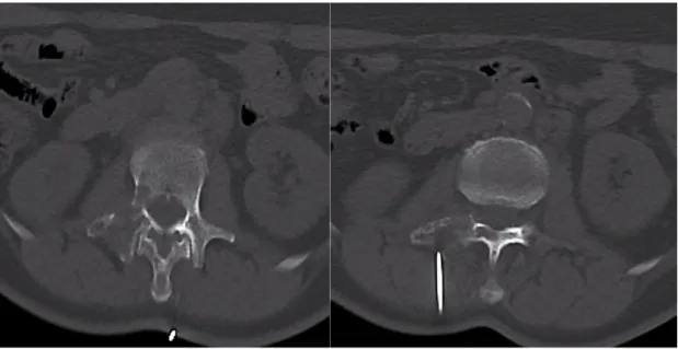
Figure 1: CT guided biopsy of focal osteolysis of right transverse process extended to pedicle.
24
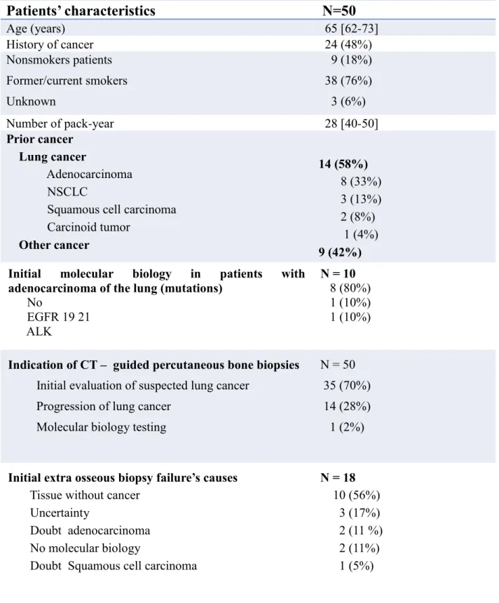
Patients’ characteristics
N=50
Age (years) 65 [62-73] History of cancer 24 (48%) Nonsmokers patients Former/current smokers Unknown 9 (18%) 38 (76%) 3 (6%) Number of pack-year 28 [40-50] Prior cancer Lung cancer Adenocarcinoma NSCLCSquamous cell carcinoma Carcinoid tumor Other cancer 14 (58%) 8 (33%) 3 (13%) 2 (8%) 1 (4%) 9 (42%) Initial molecular biology in patients with
adenocarcinoma of the lung (mutations)
No EGFR 19 21 ALK N = 10 8 (80%) 1 (10%) 1 (10%)
Indication of CT – guided percutaneous bone biopsies
Initial evaluation of suspected lung cancer Progression of lung cancer
Molecular biology testing
N = 50 35 (70%) 14 (28%) 1 (2%)
Initial extra osseous biopsy failure’s causes
Tissue without cancer Uncertainty
Doubt adenocarcinoma No molecular biology
Doubt Squamous cell carcinoma
N = 18 10 (56%) 3 (17%) 2 (11 %) 2 (11%) 1 (5%)
Table 1. Patients’ characteristics
*Qualitative variables were expressed as n (%) and quantitative variables as median (interquartile 25%-75%)
25
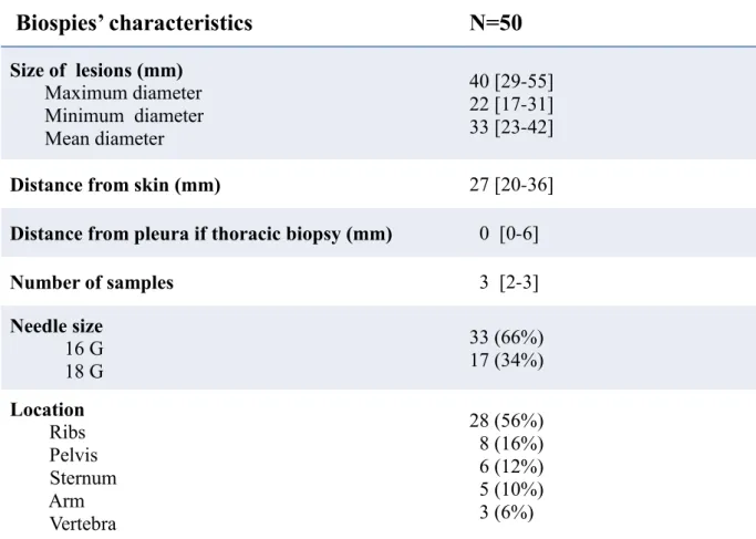
Biospies’ characteristics
N=50
Size of lesions (mm) Maximum diameter Minimum diameter Mean diameter 40 [29-55] 22 [17-31] 33 [23-42]Distance from skin (mm) 27 [20-36]
Distance from pleura if thoracic biopsy (mm) 0 [0-6]
Number of samples 3 [2-3] Needle size 16 G 18 G 33 (66%) 17 (34%) Location Ribs Pelvis Sternum Arm Vertebra 28 (56%) 8 (16%) 6 (12%) 5 (10%) 3 (6%)
Table 2. biopsies characteristics.
Qualitative variables were expressed as n (%) and quantitative variables as
median (IQR 25%-75%)
26
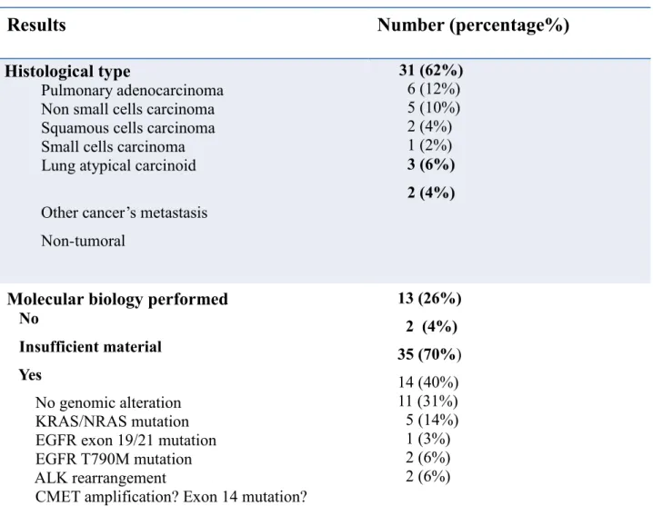
Results
Number (percentage%)
Histological type
Pulmonary adenocarcinoma
Non small cells carcinoma Squamous cells carcinoma Small cells carcinoma Lung atypical carcinoid
Other cancer’s metastasis
Non-tumoral 31 (62%) 6 (12%) 5 (10%) 2 (4%) 1 (2%) 3 (6%) 2 (4%)
Molecular biology performed
No
Insufficient material Yes
No genomic alteration KRAS/NRAS mutation EGFR exon 19/21 mutation EGFR T790M mutation ALK rearrangement
CMET amplification? Exon 14 mutation?
13 (26%) 2 (4%) 35 (70%) 14 (40%) 11 (31%) 5 (14%) 1 (3%) 2 (6%) 2 (6%)
27
REFERENCES BIBLIOGRAPHIQUES
1. Peters S, Camidge DR, Shaw AT, Gadgeel S, Ahn JS, Kim D-W, et al. Alectinib
versus Crizotinib in Untreated ALK-Positive Non-Small-Cell Lung Cancer. N Engl J Med. 31
2017;377(9):829‑38.
2. Soria J-C, Ohe Y, Vansteenkiste J, Reungwetwattana T, Chewaskulyong B, Lee KH,
et al. Osimertinib in Untreated EGFR-Mutated Advanced Non-Small-Cell Lung Cancer. N
Engl J Med. 11 2018;378(2):113‑25.
3. Bubendorf L, Lantuejoul S, de Langen AJ, Thunnissen E. Nonsmall cell lung
carcinoma: diagnostic difficulties in small biopsies and cytological specimens: Number 2 in
the Series « Pathology for the clinician » Edited by Peter Dorfmüller and Alberto Cavazza.
Eur Respir Rev Off J Eur Respir Soc. 30 juin 2017;26(144).
4. Singh VM, Salunga RC, Huang VJ, Tran Y, Erlander M, Plumlee P, et al. Analysis of
the effect of various decalcification agents on the quantity and quality of nucleic acid (DNA
and RNA) recovered from bone biopsies. Ann Diagn Pathol. août 2013;17(4):322‑6.
5. Vanderlaan PA, Yamaguchi N, Folch E, Boucher DH, Kent MS, Gangadharan SP, et
al. Success and failure rates of tumor genotyping techniques in routine pathological samples
with non-small-cell lung cancer. Lung Cancer Amst Neth. avr 2014;84(1):39‑44.
6. Vieillard M-H, Boutry N, Chastanet P, Duquesnoy B, Cotten A, Cortet B.
Contribution of percutaneous biopsy to the definite diagnosis in patients with suspected bone
tumor. Jt Bone Spine Rev Rhum. janv 2005;72(1):53‑60.
7. Mankin HJ, Mankin CJ, Simon MA. The hazards of the biopsy, revisited. Members of
the Musculoskeletal Tumor Society. J Bone Joint Surg Am. mai 1996;78(5):656‑63.
8. Murphy WA, Destouet JM, Gilula LA. Percutaneous skeletal biopsy 1981: a procedure
28
9. Sacks D, McClenny TE, Cardella JF, Lewis CA. Society of Interventional Radiology
clinical practice guidelines. J Vasc Interv Radiol JVIR. sept 2003;14(9 Pt 2):S199-202.
10. Bankier AA, MacMahon H, Goo JM, Rubin GD, Schaefer-Prokop CM, Naidich DP.
Recommendations for Measuring Pulmonary Nodules at CT: A Statement from the Fleischner
Society. Radiology. 2017;285(2):584‑600.
11. Barlesi F, Mazieres J, Merlio J-P, Debieuvre D, Mosser J, Lena H, et al. Routine
molecular profiling of patients with advanced non-small-cell lung cancer: results of a 1-year
nationwide programme of the French Cooperative Thoracic Intergroup (IFCT). Lancet Lond
Engl. 2 avr 2016;387(10026):1415-26.
12. Smits AJJ, Kummer JA, de Bruin PC, Bol M, van den Tweel JG, Seldenrijk KA, et al.
The estimation of tumor cell percentage for molecular testing by pathologists is not accurate.
Mod Pathol Off J U S Can Acad Pathol Inc. févr 2014;27(2):168-74.
13. Jelinek JS, Murphey MD, Welker JA, Henshaw RM, Kransdorf MJ, Shmookler BM,
et al. Diagnosis of primary bone tumors with image-guided percutaneous biopsy: experience
with 110 tumors. Radiology. juin 2002;223(3):731‑7.
14. Fraser-Hill MA, Renfrew DL. Percutaneous needle biopsy of musculoskeletal lesions.
1. Effective accuracy and diagnostic utility. AJR Am J Roentgenol. avr 1992;158(4):809‑12. 15. Leffler SG, Chew FS. CT-guided percutaneous biopsy of sclerotic bone lesions:
diagnostic yield and accuracy. AJR Am J Roentgenol. mai 1999;172(5):1389‑92. 16. Schrijver WAME, van der Groep P, Hoefnagel LD, Ter Hoeve ND, Peeters T,
Moelans CB, et al. Influence of decalcification procedures on immunohistochemistry and
molecular pathology in breast cancer. Mod Pathol Off J U S Can Acad Pathol Inc.
2016;29(12):1460‑70.
17. Guo W, Hao B, Chen H-J, Zhao L, Luo Z-M, Wu H, et al. PET/CT-guided
percutaneous biopsy of FDG-avid metastatic bone lesions in patients with advanced lung
29
18. Fournier L-S, Cuénod C-A, Clément O, Frija G. [Treatment response evaluation by
functional imaging]. Cancer Radiother J Soc Francaise Radiother Oncol. nov
2006;10(6‑7):484‑7.
19. Ferretti GR, Busser B, de Fraipont F, Reymond E, McLeer-Florin A, Mescam-Mancini
L, et al. Adequacy of CT-guided biopsies with histomolecular subtyping of pulmonary
adenocarcinomas: influence of ATS/ERS/IASLC guidelines. Lung Cancer Amst Neth. oct
2013;82(1):69‑75.
20. Logan PM, Connell DG, O’Connell JX, Munk PL, Janzen DL. Image-guided
percutaneous biopsy of musculoskeletal tumors: an algorithm for selection of specific biopsy
techniques. AJR Am J Roentgenol. janv 1996;166(1):137‑41.
21. Cox M, Pukenas B, Poplawski M, Bress A, Deely D, Flanders A. CT-guided Cervical
Bone Biopsy in 43 Patients: Diagnostic Yield and Safety at Two Large Tertiary Care
Hospitals. Acad Radiol. nov 2016;23(11):1372‑5.
22. Preteseille O, Barral FG, Court L, Russias B, Manet L, Tanji P, et al. [Value of
percutaneous core needle biopsy in the investigation of a suspected bone tumor]. J Radiol.
juin 2003;84(6):693‑7.
23. Monfardini L, Preda L, Aurilio G, Rizzo S, Bagnardi V, Renne G, et al. CT-guided
bone biopsy in cancer patients with suspected bone metastases: retrospective review of 308
procedures. Radiol Med (Torino). nov 2014;119(11):852‑60.
24. Fontaine-Delaruelle C, Souquet P-J, Gamondes D, Pradat E, de Leusse A, Ferretti GR,
et al. [Predictive factors of complications during CT-guided transthoracic biopsy]. Rev