Ann. N.Y. Acad. Sci. ISSN 0077-8923
A N N A L S O F T H E N E W Y O R K A C A D E M Y O F S C I E N C E S
Issue: Neuroimmunomodulation in Health and Disease
Presentation of neuroendocrine self in the thymus: a
necessity for integrated evolution of the immune
and neuroendocrine systems
Vincent Geenen
University of Liege, GIGA-Research Center of Immunoendocrinology, Sart Tilman, Belgium
Address for correspondence: Vincent Geenen, M.D., Ph.D., University of Liege, GIGA-Research Center of Immunoendocrinology, CHU-B34, B-4000 Liege-Sart Tilman, Belgium. vgeenen@ulg.ac.be
During evolution, from ancestor thymoids scattered in gill baskets of the lamprey, the first unique thymus appeared in jawed cartilaginous fishes around 450–500 millions years ago, concomitantly or shortly after the emergence of recombinase-dependent adaptive immunity. The major biological function of the thymus is to generate a diverse repertoire of T cell receptors that are self tolerant. The thymus achieves this role by using two complementary and intimately associated mechanisms: apoptotic deletion of T cell clones bearing a TCR with high affinity for self-antigens presented by MHC proteins on thymic epithelial cells (TECs) and dendritic cells (DCs); and generation of self-antigen–specific natural regulatory T (nTreg) cells. Moreover, the escape from thymic central self-tolerance
plays a primary role in the development of autoimmune diseases that are a significant burden for the quality of life and health-care cost. Our new knowledge in thymus physiology and physiopathology is currently translated into innovative therapeutic strategies against these devastating chronic diseases.
Keywords. thymus; antigen presentation; central self-tolerance; autoimmunity; AIRE; type 1 diabetes; IGF-2; Graves’ disease
The moving place of the thymus in the history of medicine
Claude Galen (129–199 or 217 AD), one of the “fa-thers” of medicine, and Hippocrates (ca. 460 BC– ca. 370 BC), first reported the observation of the thymus that he so named because of its close re-semblance with the leaf of the plant Thymus
vul-garis. Galen suspected the thymus to be the seat of
soul, humor, eagerness, and fortitude. Given Galen’s strong influence on Western medicine until the 18th century, this old misconception most probably ex-plains why the words thymie and troubles thymiques still resonate in French medical language mood and
mood disorders that are observed in
neuropsychi-atric diseases. Jacopo Berengario da Carpi (1460– 1530) then provided the first complete anatomical description of the thymus.
For a long period of time, the thymus was thought to be a vestigial organ that had become useless
and redundant during phylogeny and ontogeny af-ter puberty. In the early 1900s, J. August Ham-mar in Sweden highlighted the important neuroen-docrine regulation of the thymus, in particular, the relationship between thymic hyperplasia and acromegaly, Graves’ disease, and castration.1 The thymus was then considered a gland and an intrin-sic component of the endocrine system. However, despite the identification of several thymic “hor-mones,” the model of endocrine cell-to-cell sig-naling failed to characterize the complex molec-ular dialogue between thymic stromal cells and thymic T cells (thymocytes). In 1959 and 1961, af-ter Jacques F.A.P. Miller demonstrated the crucial role of the thymus in mouse leukemia and T cell development,2,3while the endocrine feature of the
thymus progressively vanished until the discovery of the intrathymic transcription of neuroendocrine genes.
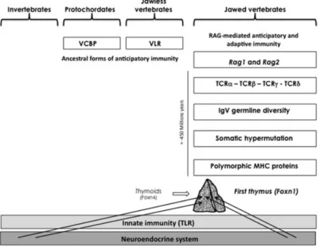
Figure 1. Integrated evolution of the immune and neuroendocrine systems. Essential components of the neuroendocrine system were established long ago and did not display important variation during evolution except for gene duplication and differential RNA splicing. The appearance of RAG-dependent adaptive immunity in jawed vertebrates was associated with a high risk of autotoxicity directed against the neuroendocrine system. Of note, from ancestor lamprey thymoids, the first unique thymus emerged concomitantly in jawed cartilaginous fishes, and the intrathymic presentation of neuroendocrine-related genes (arrows) may be viewed a posteriori as a very efficient and economical way to instruct the adaptive T cell system to tolerate neuroendocrine antigens as early as during intrathymic T cell development and differentiation. VCBP, variable-region-containing chitin-binding protein; VLR, variable lymphocyte receptor.
Emergence of the thymus in evolution
In all living species, the neuroendocrine and in-nate immune systems have evolved in parallel and still coexist today without any problem (Fig. 1). In-deed, Toll-like receptors (TLR), which are the most important mediators of innate immunity, do not have the ability to react against normal and undam-aged self. Some 450–500 million years ago, the emer-gence of transposon-like recombination-activating genes Rag1 and Rag2 in jawed fishes (sharks and rays) promoted the development of adaptive im-munity.4–6 The appearance of these elements in the genome of gnathostomes, and the subsequent development of the combinatorial immune sys-tem, has been sometimes described as to the “Big Bang” of immunology. Gene recombination in so-matic lymphoid cells is responsible for the ran-dom generation of the extreme diversity of immune receptors for antigens, B cell (BCR) and T cell re-ceptors (TCR). Because of its inherent
autotoxic-ity, the emergence of this new sophisticated type of immune response exerted an evolutive pressure so strong that, in accordance with Paul Ehrlich’s pre-diction of horror autotoxicus, novel structures and mechanisms appeared with the specific role of es-tablishing protection against potential autoimmune attacks to the host (immunological self-tolerance). Of note, the first unique thymus also appeared in the jawed cartilaginous fishes, but was preceded by thymus-like lymphoepithelial structures in the gill baskets of lamprey larvae, as recently demon-strated.7These structures named thymoids express the gene-encoding forkhead box N4 (Foxn4), the orthologue of Foxn1, the transcription factor re-sponsible for the differentiation of thymic epithe-lium in higher vertebrates. Thus, FOXN1 stands at a crucial place in the emergence of thymus epithe-lium that is an absolute requirement for the control of T cell differentiation and central self-tolerance induction.8
Neuroendocrine self in the thymus Geenen
Presentation of self in the thymus
Self-antigen presentation by major histocompatibil-ity complex (MHC) proteins on thymic stromal cells (epithelial cells (TECs) and dendritic cells (DCs), mainly) is the central mechanism determining the process of T cell differentiation, which includes three alternative and exclusive fates for developing thymo-cytes: negative selection of self-reactive T cells gen-erated during the random generation of TCR diver-sity, catalyzed by recombination-activating enzymes RAG1 and RAG2; selection of self-specific natural regulatory (nTreg) cells; and survival and positive se-lection of CD4+and CD8+effector and self-tolerant T cells. The first two events ensure the establishment of the thymus-dependent central arm of immuno-logical self-tolerance, while the avidity/affinity of the TCR–self-antigen–MHC interaction is the cen-tral determinant of T cell negative or positive selec-tion. One important unresolved question, however, is how the same MHC–self-antigen complexes are able to mediate negative selection of self-reactive T cells and yet generate self-specific nTreg cells (extensively discussed in Ref. 9).
Another question has long concerned the nature of self that is presented in the thymus to differen-tiating T cells, in particular during fetal life. Since its original formulation by Frank M. Burnet, self has been a seminal word, first coined in the im-munological language as a fecund metaphor with some equivocal correlations to the neurocognitive sciences and even philosophy. The precise identity of self was not elucidated before a series of stud-ies initiated in the late 1980s and 1990s.10–16 Our personal contribution to this field was to define the biochemical nature of the neuroendocrine self. First, thymic neuroendocrine self-antigens corre-spond to peptide sequences that are mostly con-served throughout evolution of their related family. Second, a hierarchy and an economic principle char-acterize their profile of expression in the thymus, as one dominant member per family is synthesized in TECs: that is, oxytocin (OT) for the neurohy-pophysial family, neurokinin A for tachykinins, neu-rotensin for neuromedins, corticostatin for somato-statins, and insulin-like growth factor 2 (IGF-2) for the insulin family. This hierarchy is very im-portant because the strength of immunological tol-erance to a protein/peptide is proportional to its intrathymic concentration.17 Third, the autoim-mune regulator gene/protein (AIRE) controls the
intrathymic transcription of most of the genes en-coding neuroendocrine self-antigens.18 Following AIRE-regulated gene transcription, thymic neu-roendocrine precursors are not processed according to the classic model of neuroendocrine secretion but as antigens for presentation by, or in association with, thymic MHC proteins.14 Fourth, according to the cryptocrine model of cell-to-cell signaling,19 thymic T cells express functional neuroendocrine cognate receptors.20For example, binding of thymic OT to the OT receptor expressed by thymic pre-T cells phosphorylates focal adhesion kinases,21and this might promote the establishment of immuno-logical synapses between TECs and T cells. Finally, for some neuroendocrine-related precursors, their transcription in TECs precedes their eutopic expres-sion in peripheral neuroendocrine glands/cells,20 and this is also very relevant with regard to the in-duction of self-tolerance to neuroendocrine princi-ples. Therefore, depending on their behavior as the source of self-antigens or cryptocrine ligands, re-spectively, the thymic repertoire of neuroendocrine-related precursors transposes at the molecular level the multiple roles of the thymus in T cell differenti-ation (Fig. 2).22,23
The organization of the thymic repertoire of neuroendocrine self-precursors is also significant from an evolutionary point of view. Because neuroendocrine hormones were implicated in the regulation of many physiological functions before the appearance of the anticipatory adaptive immune response, they had to be protected from the risk of autoimmunity inherent to this type of immune lottery. The hypothalamic peptide OT controls different steps of the reproductive process, starting from social affiliation and bonding to parturition and lactation.24 Consequently, self-tolerance toward OT is important for the preservation of animal and human species. Via its dominant expression in TECs, OT is more tolerated than its homologue vasopressin, which essentially controls water balance and vascular tone. Of note, rare cases of autoimmune diabetes insipidus have been reported,25–27 whereas autoimmunity toward hypothalamic OT-ergic neurons have never been described. With regard to the insulin family, no autoimmunity has been described against IGF-2, the dominant self-antigen of the insulin family during fetal life, whereas insulin is the primary autoantigen of type 1 diabetes
Figure 2. The role of thymic neuroendocrine precursors in T cell differentiation. A precursor X encoded by a neuroendocrine-related gene in a TEC is the source of two distinct types of signaling with thymocytes. First, it delivers a cryptocrine ligand X that is not secreted but targeted at the outer surface of TEC plasma membrane. Through direct membrane-to-membrane contact, this ligand binds with high affinity to a cognate neuroendocrine receptor expressed by thymocytes. For example, OT-mediated cryptocrine signaling activates phosphoinositide turnover with an increase of IP3in pre-T cells and phosphorylates focal adhesion-related kinases, which may promote the formation of synapses between TEC and thymocytes. Second, the same precursors may be processed for presentation of neuroendocrine self-epitopes by thymic MHC proteins. Deletion of T cell clones bearing a TCR specific for MHC—neuroendocrine self-antigen complexes, together with generation of self-antigen–specific nTreg, is responsible for the establishment of central self-tolerance toward neuroendocrine gene/protein families. How precisely the same MHC–self-antigen complexes are able to delete self-reactive T cells and select self-specific nTregcells remains a major unsolved question. FAK, focal adhesion kinase; IP3, inositol triphosphate.
(T1D).28However, through cross-tolerance, thymic neuroendocrine self-antigens promote self-tolerance to all the homologous members of their family as evidenced by the weaker tol-erance to insulin in Igf2−/– than in normal mice.29
The escape from central self-tolerance as a primary event in autoimmunity, and the concept ofnegative self-vaccination As already theorized by Burnet, the pathogenesis of autoimmune diseases may first depend on a failure of self-tolerance and the development of “forbid-den” self-reactive immune clones.30The progressive increase in immune complexity during evolution is associated with a higher incidence of self-tolerance failures, most of them occurring in the human species. There is more and more evidence that a thymus dysfunction in the establishment of central self-tolerance drives the development of the au-toimmune response toward many organs. Thymus
transplantation from nonobese diabetic (NOD) mice, an animal model of T1D, was shown to induce diabetes in normal recipients.31Igf2 transcription is deficient in the thymus of diabetes-prone BioBreed-ing (DPBB) rats, another animal model of T1D, such a defect might contribute to both the absence of tolerance toward cells and the usual lymphope-nia (including RT6+ Treg cells) observed in these animals.32 Mice with a thymus-restricted insulin defect develop strong proinsulin-specific T cell reactivity,33and thymus-specific deletion of insulin induces rapid development of an autoimmune di-abetes.34Nevertheless, despite the current evidence for a role of thymic insulin in the induction of cell tolerance (even at a very low level of transcription in medullary TECs), it is important to note that insulin per se failed to restore self-tolerance toward  cells in all animal or clinical trials to date.
Loss-of-function AIRE single mutations are re-sponsible for a very rare autosomal recessive disease named autoimmune polyendocrinopathy,
Neuroendocrine self in the thymus Geenen
candidiasis, and ectodermal dystrophy or autoim-mune polyglandular syndrome type 1. Depending on their genetic background, Aire−/– mice exhibit several signs of peripheral autoimmunity, which are associated with a significant decrease in the intrathymic transcription level of neuroendocrine genes, including those encoding OT, proinsulin 2, and IGF-2.18,35Of note, with regard to autoimmune thyroiditis, which is the most frequent autoim-mune disease, all major thyroid-related antigens (thyroperoxydase, thyroglobulin, and thyrotropin receptor (TSHR)) are also transcribed in TECs in normal conditions.15,36Thymic hyperplasia is com-monly observed in Graves’ disease,37and it was re-cently shown that homozygotes for an SNP allele predisposing to Graves’ disease have significantly lower intrathymic TSHR transcripts than carriers of the protective allele.38Another credit to a defective central tolerance driving the development of au-toimmunity was recently provided with the demon-stration of the central role played by a defect in in-trathymic␣-myosin expression in the pathogenesis of autoimmune myocarditis in mice and humans.39 Our current in-depth knowledge of thymus phys-iology and physiopathology has now been trans-lated into the design of innovative tolerogenic and regulatory strategies aimed at restoring central self-tolerance that is absent or defective in autoimmu-nity.40–42 The concept of negative self-vaccination has been proposed and is based both on the compe-tition between thymic self-antigens and peripheral target antigens for presentation by MHC proteins, as well as a tolerogenic response—including recruit-ment of Tregcells—induced by MHC presentation of thymic self-epitopes.43,44With this perspective, a re-search consortium in Wallonia is working on the de-velopment of a negative/tolerogenic self-vaccination with the thymic self-peptides related to T1D (Tole-diab project).
Thymus involution and immunosenescence
Although thymopoiesis is maintained until late in life,45–47 thymus involution remains the hallmark of immunosenescence that is characterized by a higher susceptibility to infections, as well as a de-crease in vaccine and antitumor immune responses. Thymic fat and fibrous involution is associated with a marked decrease in the generation of diverse T cells (in particular, naive CD4+T cells), an
expan-sion of memory CD8+ T cells, and a diminished influence of central self-tolerance. Involution of the thymus after hypophysectomy was early evidence for the control of the thymus function by a neu-roendocrine gland.48 Since then, numerous stud-ies have unambiguously demonstrated that the hy-pophysial growth hormone (GH) is able to reverse the age-dependent involution of the thymus.49–51 Intrathymic proliferation of T cell precursors and thymic output of naive T cells are significantly de-creased in adults with GH deficiency, and GH re-placement restores these two parameters.52 Today, restoration of thymus function appears more and more to be an important objective in the elderly, as well as in patients suffering with acquired im-munodeficiency syndrome or several hematological diseases.53,54 It can be anticipated that GH, IGF-1,
GH secretagogues (such as ghrelin), GH and ghrelin receptor agonists, as well as other thymus-specific growth factors will be used in the near future for re-generating thymopoiesis and, secondarily, immune functions, including response to vaccines in aged and other immunocompromised patients.
Conclusion
As evidenced in this short overview, a novel era is now beginning for a quantifiable clinical investiga-tion of thymus funcinvestiga-tion in the context of a series of immune-mediated and infectious diseases. Further-more, pharmacological manipulation of thymus-dependent thymopoietic and tolerogenic functions can now be exploited to provide the scientific com-munity with innovative strategies in the treatment of a large number of immune-mediated disorders.
Acknowledgments
These studies have been supported by the Fund of Scientific Research (F.R.S.-FNRS, Belgium), the Fund for Research in Industry and Agronomy (FRIA, Belgium), the Fund Leon Fredericq for biomedical research at the University Hospital of Liege, the Special Research Fund of the University of Liege, Wallonia (Tolediab, Senegene, ThymUP, and Raparray projects), the Belgian Association of Di-abetes, an Independent Research Grant (Pfizer Eu-rope), the European Commission (Eurothymaide FP6 Integrated Project, www.eurothymaide.org), the Juvenile Diabetes Research Foundation (JDRF, New York), and the European Federation for the Study of Diabetes (EFSD, D¨usseldorf).
Conflicts of interest
The author declares no conflicts of interest.
References
1. Hammar, J.A. 1921. The new views as to the morphology of the thymus gland and their bearing on the problem of the function of the thymus. Endocrinology 5: 543–573. 2. Miller, J.F.A.P. 1959. Role of the thymus in murine
leukaemia. Nature 183: 1069.
3. Miller, J.F.A.P. 1961. Immunological function of the thymus.
Lancet 2: 748–749.
4. Agrawal, A., Q.M. Eastman & D.G. Schatz. 1998. Transposi-tion mediated by RAG1 and RAG2 and its implicaTransposi-tions for the evolution of the immune system. Nature 394: 744–751. 5. Boehm, T. & C. Bleul. 2007. The evolutionary history of
lymphoid organs. Nat. Immunol. 8: 131–135.
6. Hirano, M., S. Das, P. Guo & M.D. Cooper. 2011. The evo-lution of adaptive immunity in vertebrates. Adv. Immunol.
109: 125–157.
7. Bajoghli, B., P. Guo, N. Aghaallaei, et al. 2011. A thymus candidate in lampreys. Nature 470: 90–95.
8. Boehm, T. 2011. Design principles of adaptive immune sys-tems. Nat. Rev. Immunol. 11: 307–317.
9. Klein, L., M. Hinterberger, G. Wirnsberger & B. Kyewski. 2009. Antigen presentation in the thymus for positive selec-tion and central tolerance inducselec-tion. Nat. Rev. Immunol. 9: 833–844.
10. Geenen, V., J.J. Legros, P. Franchimont, et al. 1986. The neuroendocrine thymus: coexistence of oxytocin and neu-rophysin in the human thymus. Science 232: 508–511. 11. Ericsson, A.E., V. Geenen, F. Robert, et al. 1990. Expression
of preprotachykinin-A and neuropeptide-Y in mRNA of the thymus. Mol. Endocrinol. 4: 1211–1218.
12. Geenen, V., I. Achour, F. Robert, et al. 1993. Evidence that insulin-like growth factor 2 (IGF-2) is the dominant thymic member of the insulin superfamily. Thymus 21: 115–127. 13. Jolicœur, C., D. Hanahan & K.M. Smith. 1994. T cell
toler-ance toward a transgenic beta-cell antigen and transcription of endogenous pancreatic genes in thymus. Proc. Natl. Acad.
Sci. U.S.A. 91: 6707–6711.
14. Vanneste, Y., A. Ntodou-Thome, E. Vandersmissen, et al. Identification of neurotensin-related peptides in human thymic epithelial cell membranes and relationship with ma-jor histocompatibility complex class I molecules. J.
Neuroim-munol. 76: 161–166.
15. Sospedra, M., X. Ferrer-Francesch, O. Dominguez, et al. 1998. Transcription of a broad range of self-antigens in the thymus suggests a role for central mechanisms in tolerance toward peripheral antigens. J. Immunol. 161: 5918–5929. 16. Derbinski, J., A. Schulte, B. Kyewski & L. Klein. 2001.
Promis-cuous gene expression in medullary thymic epithelial cells mirrors the peripheral self. Nat. Immunol. 2: 1032–1039. 17. Ashton-Rickardt, P., A. Bandeira, J.R. Delaney, et al. 1994.
Evidence for a differential avidity model of T cell selection in the thymus. Cell 74: 651–663.
18. Anderson, M.S., E.S. Venanzi, L. Klein, et al. 2002. Projection of an immunological self shadow in the thymus by the Aire protein. Science 298: 1395–1401.
19. Funder, J.W. 1990. Paracrine, cryptocrine, acrocrine. Mol.
Cell Endocrinol. 70: C21–C24.
20. Hansenne, I., G. Rasier, C. Pequeux, et al. 2005. Ontogenesis and functional aspects of oxytocin and vasopressin gene expression in the thymus network. J. Neuroimmunol. 158: 67–75.
21. Martens, H., O. Kecha, C. Charlet-Renard, et al. 1998. Neu-rohypophysial peptides stimulate the phosphorylation of pre-T cell focal adhesions kinases. Neuroendocrinology 67: 282–289.
22. Geenen, V., B. Goxe, H. Martens, et al. 1995. Cryptocrine signaling in the thymus network and T cell education to neuroendocrine self-antigens. J. Mol. Med. 73: 449–455. 23. Martens, H., B. Goxe & V. Geenen. 1996. The thymic
reper-toire of neuroendocrine-related self-antigens: Physiological implications in T cell life and death. Immunol. Today 17: 312–317.
24. Gimpl, G. & F. Fahrenholz. 2001. The oxytocin receptor system: structure, function, and regulation. Physiol. Rev. 81: 629–683.
25. Scherbaum, W.A. & G.F. Bottazzo. 1983. Autoantibodies to vasopressin cells in idiopathic diabetes insipidus: evidence for an autoimmune variant. Lancet 1: 897–901.
26. Imura, H., K. Nakao, A. Shimatsu, et al. Lymphocytic in-fundibuloneurohypophysitis as a cause of central diabetes insipidus. N. Engl. J. Med. 239: 683–689.
27. De Bellis, A., A. Bizzaro & A. Bellastella. 2004. Autoim-mune central diabetes insipidus. In Immunoendocrinology
in Health and Disease. V. Geenen & G.P. Chrousos, Eds.:
439–459. Marcel Dekker. New York.
28. Nakayama, M., N. Abiru, N. Moriyama, et al. 2005. Prime role for an insulin epitope in the development of type 1 diabetes in mice. Nature 435: 220–223.
29. Hansenne, I., C. Charlet-Renard, R. Greimers & V. Gee-nen. 2006. Dendritic cell differentiation and tolerance to insulin-related peptides in Igf2-deficient mice. J. Immunol.
176: 4651–4657.
30. Burnet, F.M. 1973. A reassessment of the forbidden clone hypothesis of autoimmune diseases. Aust. J. Exp. Biol. Med.
50: 1–9.
31. Georgiou, H.M. & T.E. Mandel. 1995. Induction of insulitis in athymic (nude) mice. The effect of NOD thymus and pancreas transplantation. Diabetes 44: 49–59.
32. Kecha-Kamoun, O., I. Achour, H. Martens, et al. 2001. Thymic expression of insulin-related genes in an animal-model of type 1 diabetes. Diab. Metab. Res. Rev. 17: 146–152. 33. Chentoufi, A. & C. Polychronakos. 2002. Insulin expression levels in the thymus modulate insulin-specific autoreactive T cell tolerance: the mechanism by which the IDDM2 locus may predispose to diabetes. Diabetes 41: 1383–1390. 34. Fan, Y., W.A. Rudert, H. Grupillo, et al. 2009.
Thymus-specific deletion of insulin induces autoimmune diabetes.
EMBO J. 28: 2812–2824.
35. Ramsey, C., O. Winqvist, M. Puhakka, et al. Aire-deficient mice develop multiple features of APECED phenotype and show altered immune response. Hum. Mol. Genet. 11: 397– 409.
36. Paschke, R. & V. Geenen. 1995. Messenger RNA expression for a TSH receptor variant in the thymus of a two-year old child. J. Mol. Med. 73: 577–580.
Neuroendocrine self in the thymus Geenen
37. Murakami, M., Y. Hosoi, T. Negishi, et al. 1996. Thymic hyperplasia in patients with Graves’ disease. Identification of thyrotropin receptors in human thymus. J. Clin. Invest.
98: 2228–2234.
38. Colobran, R., M. del Pilar Armengol, R. Faner, et al. 2011. Association of an SNP with intrathymic transcription of TSHR and Graves’ disease: a role for defective thymic toler-ance. Hum. Mol. Genet. 20: 3415–3423.
39. Lv, H., E. Havari, S. Pinto, et al. 2011. Impaired thymic tolerance to ␣-myosin directs autoimmunity to the heart in mice and humans. J. Clin. Invest. 21: 1561– 1573.
40. Geenen, V., M. Mottet, O. Dardenne, et al. 2010. Thymic antigens for the design of a negative/tolerogenic self-vaccination gainst type 1 diabetes. Curr. Opin. Pharmacol.
10: 461–472.
41. Chentoufi, A. & V. Geenen. 2011. Thymic self-antigen expression for the design of a negative/tolerogenic self-vaccination against type 1 diabetes. Clin. Dev. Immunol. doi:10.11555/2011/349368.
42. Daniel, C., B. Weigmann, R. Bronson & H. von Boehmer. 2011. Prevention of type 1 diabetes in mice by tolerogenic vaccination with a strong insulin mimetope. J. Exp. Med.
208: 1501–1510.
43. Geenen, V. 2006. Thymus-dependent T cell tolerance of neu-roendocrine functions. Principles, reflections, and impli-cations for tolerogenic/negative self-vaccination. Ann. N.Y.
Acad. Sci. 1088: 284–296.
44. Geenen, V., C. Louis, H. Martens & The Belgian Diabetes Registry. 2004. An insulin-like growth factor 2-derived self-antigen inducing a regulatory cytokine profile after presen-tation to peripheral blood mononuclear cells from DQ8+ type 1 diabetic adolescents: preliminary design of a
thymus-based tolerogenic self-vaccination. Ann. N.Y. Acad. Sci. 1037: 59–64.
45. Douek, D.C., R.D. McFarland, P.H. Keiser, et al. 1998. Changes in thymic function with age and during the treat-ment of HIV infection. Nature 396: 690–695.
46. Poulin, J.F., J.M. Viswanathan, J.M. Harris, et al. 1999. Direct evidence for thymic function in adult humans. J. Exp. Med.
190: 479–486.
47. Geenen, V., J.F. Poulin, M.L. Dion, et al. 2003. Quantifica-tion of T cell receptor rearrangement excision circles to esti-mate thymic function: an important new tool for endocrine-immune physiology. J. Endocrinol. 176: 305–311.
48. Smith, P. 1930. The effect of hypophysectomy upon the in-volution of the thymus in rat. Anat. Rec. 47: 119–143. 49. Kelley, K.W., D.A. Weigent & R. Kooijman. 2007. Protein
hormones and immunity. Brain Behav. Immun. 21: 384– 392.
50. Savino, W. & M. Dardenne. 2000. Neuroendocrine control of thymus physiology. Endocr. Rev. 21: 412–443.
51. Taub, D.D., W.J. Murphy & D.L. Longo. 2010. Rejuvena-tion of the aging thymus: growth hormone-mediated and ghrelin-mediated signaling pathways. Curr. Opin.
Pharma-col. 10: 408–424.
52. Morrhaye, G., H. Kermani, J.J. Legros, et al. 2009. Impact of growth hormone (GH) deficiency and GH replacement upon thymus function in adult patients. PLoS ONE 4: e5668. 53. Napolitano, L.A., D. Schmidt, M.B. Gotway, et al. 2008. Growth hormone enhances thymic function in HIV-1-infected patients. J. Clin. Invest. 118: 1085–1098.
54. Castermans, E., M. Hannon, J. Durieux, et al. 2011. Thymic recovery after allogeneic hematopoietic cell transplantation with non-myeloablative conditioning is limited to patients younger than 60 years of age. Haematologica 96: 298–306.