ACKNOWLEDGEMENT
I want to thank all the people whose assistance was a milestone in the completion of this project.
I would like to thank CONACyT for making this research possible.
The ITAV and the CNRS for offering me facilities during this project, and Manuel Rodriguez and Pierre Lutz for supervising this thesis.
I wish to express my deepest gratitude to Jean Emmanuel Sarry, for always having the time for this research, and to all the members of the 18th team, for all their support, help and patience.
Thanks to Clement Larrue for his help during this research, and to Carine Joffre for her help during the completion of this project
I finally would like to thank all the members of the UbiCARE team. And of course, the evaluators, for taking the time to evaluate this thesis.
SPECIAL THANKS
“Sweet are the uses of adversity which, like the toad, ugly and venomous, wears yet a precious jewel in his head.”
William Shakespeare Thanks to the Supreme Being that has allowed me to be 29 years on earth. My birth is a miracle, and my existence is proof of his mercy. He has given me the strength to continue, thank you for sending me with such great human beings: my parents. Thank you for covering me with your heavenly mantle.
A long time ago I read an African proverb that said: “If you want to go fast, go alone if you want to go far, go accompanied,” I have had the blessing of walking beside my parents: Amanda Reyes Vázquez and Juan López Velázquez. Thank you for fighting battles for and with me, for believing in my dreams (no matter how crazy they sound, no matter how scary they are), for being by my side in times of frustration, and pain, for never letting me give up, because you never cut my wings, on the contrary, they have driven me to fly higher and higher. Because of you I am who I am, because of you I will be, because you are my greatest motivation. Thanks, mom, thanks dad, here is the fruit of teamwork, I love you.
To the rest of my family: Williams, Vianey, Amandita, and Jonathan, for understanding my absence in important family events. Distance is very hard without you.
To my French family, thank you for all the support, and your kind words.
“Guenda nabani xhianga sicarú, ne gasti rú ni Ugaanda laa, Diuxhi biseenda laanu idxi layú, ne laa cuidxi laanu ra nuu” (Life is wonderful, and there is nothing to compare, God sent us to earth and He will call us to his side). To my uncle Wilfrido López Velázquez, “Tío Willy,” because although years ago the circumstances of life snatched you from my side on this earthly plane, I know that you are always with me. Your teachings are still present in me, I live to honor your memory, I always carry your heart with me. It never leaves me and, wherever I go, you are always with me. I know that this moment would have been so special for you, as it is for me. This achievement is also yours, I love you, uncle.
To my husband, Anthony Gallais, thank you for supporting me, for believing in me, for understanding me. Your help has been fundamental and, you have been with me even in the most challenging moments. This project was not easy, but you were always there, motivating me and helping me.
To Dr. Guadalupe Soto Rodriguez, for being crucial in my life to continue on the path of research, for not only being my guide in the degree but for the friendship you have given me these years. Despite the distance you have never left me alone. This dream is possible because of you. Thank you, a paragraph does not summarize all the profound grattitude I have!
To Dr. Marisela Ahumada Santiago, thank you for believing in my dream, thank you for your friendship even in the distance.
To my aunt, Lelia López Velasquez, thank you for believing in my dreams and supporting me.
Thanks to all the angels that God has sent me in my everyday life.
Life has taught me that I do not need many friends, but the right ones. Thank you, Miguel Madrid, for all your support, for being beside me in the most challenging times. You have shown me the meaning of loyalty. Thank you for being my friend.
Thank you, Maria Gonzalez Santamarta; in a short period, you have become a very great friend. I want to thank you not only for the help provided but for the good times we have lived. You are a great person, and I love having you by my side as a friend.
Thanks, Clémence Coutelle-Rebut for your kindness, patience and time for reading this thesis.
Thank you, Gabrielle Sueur, for your friendship and kindness, for being there since I have not friends here.
Thank you, Javier Morgado, you have been like a brother, you have supported me, you have never let me down even when I want. Thank you for being my friend.
To Latifa, thank you for your support during these years, thank you for listening to me. I want to give a special thanks to Claudie, Marie, Estelle, Mathilde, and Emeline for your help and support during these years.
Thank you to the doctoral school for all your support and help during this thesis.
I am pretty sure that I am forgetting too many people who have helped me during all these years but thank you for making this thesis possible.
TABLE OF CONTENTS
ABBREVIATIONS ... 7 FIGURES... 9 LIST OF TABLES ... 11 SUMMARY ... 12 RÉSUMÉ ... 13 PREAMBLE ... 15 I. INTRODUCTION ... 17 1. CARCINOGENESIS ... 172. From hematopoiesis to Leukemia ... 19
2.1 Hematopoietic regulation ... 19
2.2 Leukemia and leukemic development ... 21
2.3 Leukemia Classification... 22
3. Acute Myeloid Leukemia ... 23
3.1 Epidemiology ... 23
3.2 AML classification ... 24
3.2.1 The French-American-British (FAB) classification of AML ... 24
3.2.2 The World Health Organization (WHO) classification ... 25
3.3 Cytogenetic characteristics and their prognostic impact in AML ... 27
3.4 Molecular basis of Acute myeloid leukemia ... 28
3.5 AML in pathway signaling ... 29
3.6 Treatment in AML ... 31
4. Tyrosine Kinases ... 32
4.1 Tyrosine kinase classification ... 32
4.2 Mechanism of RTK activation under normal physiological conditions ... 33
5. The FLT3 receptor ... 34
5.1 Protein and gene structure of FLT3 ... 34
5.2 Signal transduction networks activated by FLT3-WT ... 35
5.3 FLT3 mutations ... 37
5.4 FLT3-ITD ... 37
5.5 FLT3-ITD signaling ... 39
6. Intracellular protein degradation ... 40
7. Autophagy... 42
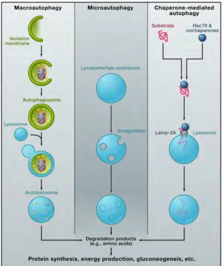
7.1 Types of autophagy ... 42
7.2 Autophagy induction ... 45
7.4 LC3/GABARAP and autophagy receptors ... 48
7.5 The role of Autophagy in cancer ... 53
7.6 Autophagy inhibitors ... 56
8. The proteasome ... 58
8.1 Function, structure and organization ... 58
8.2 The CP or 20S proteasome ... 59
8.3 The RP or 19S regulatory complex... 59
8.3.1 The lid and base subcomplex ... 60
9. Protein Ubiquitylation... 61
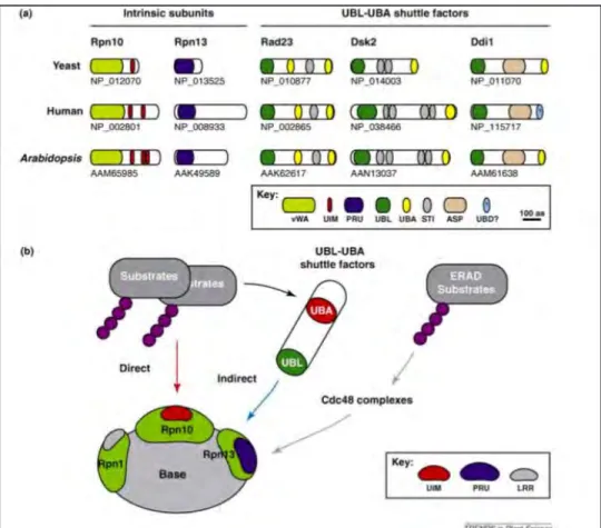
9.1 Major Ubiquitin receptors for proteasomal degradation ... 63
9.2 Purification of Ubiquitylated proteins: TANDEM UBIQUITIN BINDING ENTITIES (TUBE) ... 65
10. Ubiquitin-Proteasome System (UPS) ... 67
10. 1 UPS inhibitors ... 68
10.2 The role of the proteasome in acute myeloid leukemia (AML) ... 71
11. Crosstalk between Autophagy and Ubiquitin Proteasome System ... 74
11.1 Proteaphagy ... 76
II. SCIENTIFIC CONTEXT ... 79
Hypothesis ... 81
III. MATERIAL AND METHODS ... 82
IV. RESULTS ... 90
Setting up conditions for detection of selective autophagy events ... 90
Apoptosis in FLT3-ITD cells after 8h treatment with inhibitors ... 95
Proteaphagy in other AML cells ... 96
Proteaphagy in FLT3-ITD AML cells using an autophagy inhibitor acting on p62 ... 100
Proteaphagy is not constitutively active in FLT3-ITD cells ... 103
Evidence of proteaphagy and other selective autophagy events by Immunofluorescence in FLT3-ITD cells... 105
Impact of autophagy inhibition on proteasome activity ... 111
Identification of ubiquitylated proteins using TANDEM UBIQUITIN BINDING ENTITIES (TUBEs) ... 113
Ubiquitin-dependent regulation of p62 complexes regulating autophagy activated by Bz 115 Inhibition of UPS and ALS enhances apoptosis in FLT3-ITD AML cells ... 119
V. GENERAL DISCUSSION ... 122
VI. BIBLIOGRAPHY ... 134
ABBREVIATIONS
ALS: Autophagy Lysosome System AML: Acute Myeloid Leukemia Atg: Autophagy-related genes ATP: Adenosine triphosphate BafA: Bafilomycin A1
BB: Boiling buffer BM: Bone Marrow
BSA: Bovine serum albumin Bz: Bortezomib
CP: Core particle CQ: Chloroquine
CMML: Chronic myelomonocytic leukemia
CREB: Cyclic adenosine monophosphate response element-binding protein CSF: Colony stimulating factor
DUB: Deubiquitinating enzyme DNA: Deoxyribonucleic acid DTT: Dithiothreitol
ER: Endoplasmic reticulum
Erk: Extracellular signal-regulated kinase FAB: The French-American-British
FBS: Fetal bovine serum
FDA: Food and Drug Administration FL: FLT3 ligand
FLK2: Fetal Liver Tyrosine Kinase 2 FLT3: Fms-Like Tyrosine kinase 3
FLT3-ITD: Fms-Like Tyrosine kinase 3 with Internal Tandem Duplications FLT3L: Fms-Like Tyrosine Kinase 3 with Tyrosine Kinase Domain mutation FOX3 A: Forkhead box 03
GM-CSF: Granulocyte-macrophage colony-stimulating factor GRB2: Growth factor receptor-bound 2
HCQ: Hydroxychloroquine HSCs: Hematopoietic stem cells ITD: Internal Tandem Duplication IP: Immunoprecipitation
JAK: Janus Kinase JM: JuxtaMembrane
LSCS: Leukemic Stem cells
MAPK: Mitogen-Activated Protein kinase MCH: Major complex histocompatibility MDR: Multidrug resistance gene complex MDS: Myelodysplastic syndromes
MC: Mantel cell lymphoma MM: Multiple Myeloma
MP: Myeloproliferative neoplasms MS: Mass Spectrometry
NBR1: Neighbor of BRCA gene-one protein mTOR: Mammalian Target of Rapamycin
NF-ĸB: Nuclear factor-B OPTN: Optineurin
PAS: Pre-autophagosomal structure PE: Phosphatydiletanolamine
PGDFR: Derived growth factor platelet receptor PI: Proteasome inhibitors
PI3K: Phosphoinositide 3-kinase PK: Protein Kinase
RP: Regulatory particle
Rpn: Regulatory particle of non-ATPase subunits Rpt: Regulatory particle of triple-ATPase subunits RTKs: Receptor Tyrosine Kinase
SCFR: Stem cell factor receptor SQSTM1: Sequestosome 1
Stat 5: Signal transducer and activator of transcription 5 TBS: Tris-buffered saline
TUBE: Tandem Ubiquitin Binding Domain TK: Tyrosine Kinase
TKD: Tyrosine Kinase Domain
TUBE: Tandem ubiquitin binding entities Ub: Ubiquitin
UBA: Ubiquitin associated domain UBD Ubiquitin-Binding domain UBL: Ubiquitin-Like proteins
UPS: Ubiquitin-Proteasome System VEGF: Vascular Endothelial Grow Factor VPS34: Vacuolar Protein Sorting 34 VT: Verteporfin
WB: Western Blot
WHO: World Health Organization WT: Wild Type
FIGURES
Figure 1. The hallmarks of cancer………..…………..17
Figure 2: A general model of Hematopoiesis……….……20
Figure 3: Schematic representation of the FLT3 receptor……….……..35
Figure 4: Ligand independent FLT3 activation……….….35
Figure 5: Signaling pathways activated by FLT3-WT………...36
Figure 6: Constitutive activation of the FLT3 receptor by the ITD mutation………..38
Figure 7: The Two major protein degradation pathways……….….41
Figure 8: Different Types of Autophagy………..43
Figure 9: The process and regulation of selective autophagy……….44
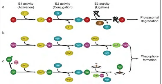
Figure 10: Assembly and elongation of autophagic membranes are accomplished via sequential action of UPS-like E1-E2-E3 cascades……….48
Figure 11: Potential mechanisms of selective macroautophagy………49
Figure 12: Selective autophagy receptors: ubiquitin-mediated cargo recognition…………50
Figure 13: Ubiquitin-dependent and Ubiquitin independent autophagy……….51
Figure 14: The autophagy receptor p62………..52
Figure 15: Autophagy has multiple roles during tumorigenesis………55
Figure 16: 26S proteasome structure………...58
Figure 17: Ubiquitin chains and its major functions………....62
Figure 18: Recognition of the ubiquitylated substrates for the 26S proteasome-mediated proteolysis……….64
Figure 19: The tandem disposition of associated domains preserves their ubiquitin-binding capacity………66
Figure 20: Functional representation of the mechanism of ubiquitin conjugation and presentation to the proteasome……….67
Figure 21: Cellular consequences of proteasome inhibition………69
Figure 22: The proteasome has several roles in AML………..73
Figure 23: The compensatory balance between the activities of autophagy and the UPS in order to maintain cellular homeostasis……….75
Figure 24: FLT3-ITD positive AML cells are targeted to apoptosis by Bz-mediated autophagy………..…80
Figure 25: Immunoprecipitations of p62 in the presencse or absence of TUBEs………….86
Figure 26: Setting up conditions for detection of proteaphagy in MOLM-14 cells using high serum concentration………92
Figure 27: Set up conditions for detection of proteaphagy in MOLM-14 cells using low serum concentration………94
Figure 28: Cell death evaluation after 8h of treatment with proteasome and autophagy inhibitors in MOLM-14 cells ……….95
Figure 29: Proteaphagy evaluation in other FTL3-ITD AML cell lines………97
Figure 30: Analysis of proteaphagy in FLT3 WT cells………...99
Figure 31: Verteporfin (VT) IC50 in FLT3-ITD AML cells ………100
Figure 32: Verteporfin blocked proteaphagy in AML cells………..102
Figure 33: Analysis of basal levels of proteaphagy in MOLM-14 cells……….104
Figure 34: Colocalization of β2 proteasome subunit and LC3B after Bz and VT treatment in FLT3-ITD AML cells………...107
Figure 35: Colocalization of α2 proteasome subunit and p62 after Bz treatment in FLT3-ITD AMLcells ……….108
Figure 36: Colocalization of Rpn1 and LC3B after Bz treatment in FLT3-ITD AMLcells………..109
Figure 37: Colocalization of FLT3 and LC3B after Bz and VT treatment in FLT3-ITD AML cells………..…109 Figure 38: Colocalization mytophagy indicator COXIV and p62 after Bz and autophagy inhibitors treatment in FLT3-ITD AML cells………...110 Figure 39: Proteasome activity after different doses of Bz in MOLM-14 cells…………...112 Figure 40: The role of protein ubiquitylation in proteaphagy and FLT3-ITD degradation under proteasome inhibition conditions………..114 Figure 41: Setting up conditions for immunoprecipitations of p62 and Beta 2 under proteasome inhibition conditions……….117 Figure 42: Ubiquitin role in proteaphagy and degradation of FLT3-ITD under proteasome
inhibition conditions………...118
Figure 43: Cell death evaluation after 24h treatments, FBS 10% in AML FLT3 wild type, and
FLT3-ITD positive ………..120
Figure 44: Proteasome and autophagy inhibitors cooperate to improve the apoptosis of
FLt3-ITD expressing cells..………...121
LIST OF TABLES
Table 1: Main indicators in 2012 of AML……….24
Table 2: FAB classification for AML……….25
Table 3: WHO classification for AML………...26
Table 4: The cytogenetic classification of AML………..27
Table 5: Autophagy inhibitors………57
Table 6: Mono/multiple monoubiquitylated Proteasomal substrates………..62
Table 7: Antibodies used during this study……….82
Table 8: Drugs used during this research..……….83
SUMMARY
Acute myeloid leukemia (AML) represents a heterogeneous group of malignant hematopathies characterized by a clonal proliferation of hematopoietic progenitors blocked in their differentiation (blasts), which accumulate in the bone marrow, blood, and other organs. The AML causes more than 3000 death every year in France. Different genetic mutations are found in the AML, among them the ones affecting the kinase receptor activity: KIT and FLT3-ITD. The internal tandem duplication in the FLT3 receptor (FLT3-ITD), represents 30% of cases of AML, and has a poor prognosis, compared to patients expressing the wild-type receptor. Previous studies have demonstrated that autophagy regulates cytotoxicity in FLT3-ITD AML cells after proteasome inhibition. In this study proteotoxic stress conditions generated by Bortezomib (Bz) resulted in the degradation of proteasome subunits in FLT3-ITD MOLM-14 cells but not in FLT3-WT OCI-AML3 cells, suggesting ITD mutation contributes to activate autophagy-mediated proteolysis of the proteasome, known as proteaphagy. In this study, using chemical inhibition of autophagy with Bafilomycin A (BafA) we blocked proteaphagy and accumulated proteasome core subunits into autophagosomes of Bz-treated MOLM-14 cells.
To investigate the role of protein ubiquitylation in proteaphagy, we used distinct TUBEs (Tandem Ubiquitin Binding Entities). While TUBE-HHR23 captures p62, proteasome subunits and ubiquitylated forms of FLT3, TUBE-p62 does not. Nevertheless, TUBE-p62 protected the unmodified form of FLT3-ITD from degradation driven by Bz. According to our results, the p62 inhibitor Verteporfin (VT) blocked proteaphagy and reduced the colocalization of p62/2 core subunit but did not affected the one of p62/Rpn1. VT also protected FLT3-ITD from Bz-induced degradation and colocalized within p62 in MOLM-14 cells. Both autophagy inhibitors enhanced Bz-induced apoptosis in MOLM-14 cells suggesting that these combinatorial treatments could be a therapeutic strategy to sensitize FLT3-ITD positive cells. This study allowed us to understand that the proteasome- and autophagy-mediated proteolysis are most likely in a dynamic equilibrium and play an important role when one or the other pathway is impaired.
RÉSUMÉ
La leucémie myéloïde aiguë (LAM) représente un groupe hétérogène d'hématopathies malignes caractérisées par une prolifération clonale de progéniteurs hématopoïétiques bloqués dans leur différenciation (blastes), qui s'accumulent dans la moelle osseuse, le sang et d'autres organes. Le LAM fait chaque année plus de 3000 morts en France. Différentes mutations génétiques se trouvent dans le LAM, parmi lesquelles celles affectant l'activité des récepteurs de kinase: KIT et FLT3-ITD. La duplication interne en tandem dans le récepteur FLT3 (FLT3-ITD), représente 30% des cas de LAM, et a un mauvais pronostic, par rapport aux patients exprimant le récepteur de type sauvage.
Les patientes atteintes de leucémie aiguë myéloïde présentant une duplication interne en tandem dans le récepteur FLT3 (FLT3-ITD), les patients atteints de FLT3-ITD représentent 30% des cas de leucémie aiguë myéloïde (LAM), et ont un mauvais pronostic de survie par rapport aux patients exprimant le récepteur de sauvage.
Des études antérieures ont démontré que l'autophagie régule la cytotoxicité induite par l’inhibition du protéasome dans les cellules de LAM FLT3-ITD. Dans cette étude, les conditions de stress protéotoxique générées par le bortézomib (Bz) ont entraîné la dégradation des sous-unités du protéasome dans les cellules FLT3-ITD MOLM-14 mais pas dans les cellules FLT3-WT OCI-AML3, ce qui suggère que la mutation ITD contribue à activer la protéolyse par autophagie du protéasome, connue sous le nom de proteaphagie. L'inhibition chimique de l'autophagie avec la bafilomycine A (BafA) a bloqué la protaphagie et favorisé l’accumulation des sous-unités centrales du protéasome accumulées dans les autophagosomes des cellules MOLM-14 traitées au Bz.
Pour étudier le rôle de l'ubiquitylation des protéines dans la protaphagie, nous avons utilisé des TUBE distincts (Tandem Ubiquitin Binding Entities). Alors que TUBE-HHR23 capture p62, les sous-unités de protéasome et les formes ubiquitylées de FLT3, TUBE-p62 ne le fait pas. Néanmoins, TUBE-p62 a protégé la forme non modifiée de FLT3-ITD de la dégradation provoquée par Bz. Selon nos résultats, l'inhibiteur de la p62 Verteporfin (VT) a bloqué la protaphagie et a réduit la colocalisation de la sous-unité centrale p62 avec α2 mais n'a pas affecté celle de p62 avec Rpn1. VT a également protégé FLT3-ITD de la dégradation induite par Bz et augmenté la colocalisation de p62 dans les cellules MOLM-14. Les deux inhibiteurs de l'autophagie ont augmenté l'apoptose induite par le Bz dans les cellules MOLM-14, suggérant que ces traitements combinatoires pourraient être une stratégie thérapeutique
pour cibler les cellules positives FLT3-ITD. Ainsi, la protéolyse médiée par le protéasome et l'autophagie est très probablement dans un équilibre dynamique et joue un rôle important lorsque l'une ou l'autre voie est altérée.
PREAMBLE
Protein homeostasis is necessary for the proper functioning and wellbeing of the cells. In the 19th century, Claude Bernard underlined the need to maintain a stable internal environment - milieu intérieur - which would allow biological processes to take place despite variations in the external environment (Gross, 1998). Bernard’s concept was further explored, developed, and popularized by Walter Cannon, who coined the term “homeostasis” to describe how critical physiological variables are maintained within a predefined range by feedback mechanisms (Cannon, 1929). Nearly two decades after Cannon, James Hardy proposed a model in which homeostatic mechanisms maintain physiological variables within an acceptable range by comparing the actual value of the variable to the desired value or ‘set point,’ (Hardy, 1953). Homeostasis is a unifying theme of modern physiology, and much has been elucidated about molecular mechanisms of homeostatic control.
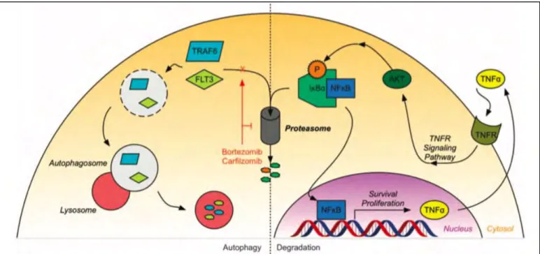
Several disorders occur when cell homeostasis is disrupted, including cancer. Blood cancers encompass an extensive collection of diseases affecting the equilibrium and correct functioning of this tissue. Acute myeloid leukemia (AML) is a malignant disease characterized by differentiation blockage and proliferation of clonal hematopoietic stem or progenitor cells (HSPCs), which rapidly lead to bone marrow (BM) failure and eventually to death if left untreated. Internal tandem duplication (ITD) of the Fms-like tyrosine kinase-3 receptor (FLT3) is found in 30% of acute myeloid leukemia (AML) and is associated with poor outcomes. Multiple approaches based on new pharmacological targets have been considered to tackle this disease, and new treatments are being developed. Among the most promising are autophagy and proteasome inhibitors, which directly affect protein homeostasis and contribute to optimizing the apoptotic response of AML cells.
Studies conducted by Larrue and his collaborators showed that AML cell lines and samples from patients bearing FLT3-ITD mutations are more sensitive to the proteasome inhibitor Bortezomib (Bz) than wild-type samples and that this sensitivity is strongly correlated with a higher FLT3-ITD allelic burden. Indeed, FLT3-ITD molecules were detectable within autophagosomes after Bz treatment indicating that autophagy induced by Bz was responsible for the early degradation of FLT3-ITD. This degradation preceded the inhibition of mitogen-activated protein kinase (MAPK), extracellular signal-regulated kinase (Erk), PI3K/Akt, and Stat5 pathways, as well as the subsequent activation of cell death. Based on this work, we aimed to explore the different molecular mechanisms involved in the crosstalk
between the ubiquitin-proteasome system and autophagy in AML, under conditions of proteasome inhibition. In concrete terms, we investigated the participation of selective autophagy events like proteaphagy or aggrephagy activated after Bz treatment. The evidence suggests that chemical inhibitors of autophagy accumulate proteasome subunits into autophagosomes of bortezomib-treated FLT3-ITD AML cells but not in FLT3-WT AML cells. Even if bafilomycin A (BafA) blocks the degradation of the 20S proteasome subunits, 19S subunits appear to be more vulnerable to bortezomib-induced degradation. In particular, the Rpn1 subunit forms high molecular weight complexes with the autophagy receptor p62 after treatment with verteporfin (VT), underlining its implication in proteaphagy.
To investigate the role of ubiquitin in this regulatory mechanism, we used a technology previously developed by our laboratory, based on ubiquitin-binding domains of the proteasome adaptor HHR23, or the autophagy adaptor p62. We could observe that ubiquitylated FLT3-ITD or proteasome subunits were captured after Bz+BafA treatment by Tandem Ubiquitin Binding Entities (TUBEs)-HHR23 but not by TUBEs-p62. Nevertheless, both TUBEs protect proteasome subunits, p62 or FLT3-ITD in different proportions. Altogether our evidence supports a role of FLT3-ITD in predisposing the activation of Bz-induced proteaphagy. This route can potentially be targeted using combinatorial approaches with autophagy inhibitors to improve previous results obtained with Bz.
I.
INTRODUCTION
1. CARCINOGENESIS
Over the years, cancer research has provided evidence that tumor development is a multi-step process that transforms a normal cell into a malignant derivative. This process was comprehensively schematized (Figure 1) by Hanahan and Weinberg. They constituted the well-established hallmarks of cancer (Hanahan, 2014), where they attempted to organize the dense complexities of cancer biology into six acquired capabilities of cancer cells. These are the major hallmarks:
1. Cancer cells adopt alternative ways to self-sustain proliferation. For example, cell proliferation depends on extracellular stimulus transmitted to the cell by interactions of transmembrane receptors and signaling molecules (growth factors, extracellular matrix components, and cell-cell adhesion molecules). Transmembrane receptors responsive to proliferative signals are deregulated in cancer cells and often contain intracellular tyrosine kinase activity promoting tumor progression.
Figure 1: The hallmarks of cancer. Taken from (Hanahan, 2014).
Cancers acquire ten damaging functional capabilities and facilitators (shown in white ring and indicated by symbols in coloured ring) that collectively manifest successful attacks on the aff ected individual. Each capability can be counteracted by various mechanism-targeted treatments (shown figuratively as explosion shapes) which, generally, do not act as curative magic bullets because of the countervailing development of advanced strategies of resistance. One battlespace plan involves multitargeting of all of these capabilities and facilitators (A). A major challenge, however, is limitation of collateral damage—toxicity to normal tissue and physiological functions. Realistically, tactical variations will involve more selective multitargeting (exemplified in B, C, and D), fine-tuned both by enabling of military intelligence of a patient’s tumour aff orded by increasingly accurate, high-resolution molecular diagnostics, and by the nature of the drugs and the tactical regimens in which they are launched, to optimise eff ectiveness while restricting toxicity
2. Simultaneously, to sustaining proliferation, cancer cells acquire the capacity to evade apoptosis. Tissue homeostasis is tightly regulated by the elimination of non-healthy cells originating from infectious and non-infectious insults, a potential cause of oncogenic lesions. In such cases, impairment of tumor suppressors activity enables altered cells to escape apoptosis by acting upon inhibitors of apoptosis.
3. Oncogene signaling can drive angiogenic regulators involved in perpetuating the sprouting of new vessels to give nutrition for the growing tumor.
4. Activation of invasion and metastasis enables the tumor to spread. The multistep process of invasion and metastasis has been schematized as a sequence of discrete steps, often termed the invasion-metastasis cascade (Fidler, 2003; Talmadge & Fidler, 2010). This depiction envisions a succession of cell-biologic changes, beginning with local invasion, then intravasation by cancer cells into nearby blood and lymphatic vessels and the transit of cancer cells through the lymphatic and hematogenous systems. These are followed by the escape of cancer cells from the lumina of such vessels into the parenchyma of distant tissues (extravasation), the formation of small nodules of cancer cells (micrometastases), and finally the growth of micrometastatic lesions into macroscopic tumors. This last step is called ‘‘colonization.’’
5. Evading growth suppressors: Cancer cells must also circumvent powerful programs that negatively regulate cell proliferation; many of these programs depend on the actions of tumor suppressor genes.
6. Enabling replicative immortality equipping the cancer cells to overcome the Hayflick limit. Tumor cells overcome the finite replicate potential at some point during multistep tumor progression, evolving to premalignant. Cell populations exhaust their endowment of allowed doublings and can only complete their tumorigenic agenda by breaching the mortality barrier and acquiring unlimited replicative potential (Hayflick, 1997).
More recently, Hanahan and Weinberg provided a substantial body of evidence (Hanahan & Weinberg, 2011) to pinpoint new capabilities as emerging hallmarks of cancer, being those related to the involvement of the immune system as a perpetrator of an inflammation condition before tumor development and during its establishment. Furthermore, cancer cells have a selective advantage enabled by specific mutant genotypes, categorized as evading immune destruction and reprogramming energy metabolism (Fouad & Aanei, 2017).
2. From hematopoiesis to Leukemia
2.1 Hematopoietic regulationHematopoiesis is the process by which all lineages of blood cells are generated in a hierarchical and stepwise manner from immature cells present in the bone marrow (BM) and subsequently released into circulating blood and peripheral organs for further maturation steps and or effector function (Orkin & Zon, 2008). The BM is the primary site of hematopoiesis and normal immature precursors of hematopoietic cells. The hematopoietic system is organized by a pyramidal hierarchy, in the apex of this hierarchy are hematopoietic stem cells (HSCs), which are the only self-renewing cells capable of lifelong production of all lineage of blood cells (Figure 2). Control of cell proliferation, growth, and survival is vital for the maintenance of homeostasis in every tissue. Hematopoiesis is controlled at different levels via the production of growth factors and extracellular cytokines that will control transcription and lead the fate of the cells.
Growth factors are required for the survival and proliferation of hematopoietic cells at all stages of development. Among the factors that affect multipotent cells, the best characterized are steel factor, Fms-like tyrosine kinase 3 (FLT3) ligand, granulocyte-macrophage colony-stimulating factor (GM-CSF), interleukin-2, interleukin-3, and interleukin-7. Each of these proteins supports the survival and proliferation of many distinct target cells, and except for interleukin-7 and steel factor, the elimination of any one of them does little harm because of the redundancy in the functions of these early-acting growth factors (Kaushansky, 2006). Growth factors and cytokines secreted by hematopoietic cells or by environmental cells can be recognized by different surface receptors that regulate different pathways, like PI3K, JAK/Stats and MAPKs, involved in proliferation and differentiation.
Figure 2: A general model of Hematopoiesis. Taken from (Kaushansky, 2006).
Blood-cell development progresses from a hematopoietic stem cell (HSC), which can undergo either self-renewal or differentiation into a multilineage committed progenitor cell: a common lymphoid progenitor (CLP) or a common myeloid progenitor (CMP). These cells then give rise to more-differentiated progenitors, comprising those committed to two lineages that include T cells and natural killer cells (TNKs), granulocytes and macrophages (GMs), and megakaryocytes and erythroid cells (MEPs). Ultimately, these cells give rise to unilineage committed progenitors for B cells (BCPs), NK cells (NKPs), T cells (TCPs), granulocytes (GPs), monocytes (MPs), erythrocytes (EPs), and megakaryocytes (MkPs). Cytokines and growth factors that support the survival, proliferation, or differentiation of each type of cell are shown in red. For simplicity, the three types of granulocyte progenitor cells are not shown; in reality, distinct progenitors of neutrophils, eosinophils, and basophils or mast cells exist and are supported by distinct transcription factors and cytokines (e.g., interleukin-5 in the case of eosinophils, stem-cell factor [SCF] in the case of basophils or mast cells, and G-CSF in the case of neutrophils). IL denotes interleukin, TPO thrombopoietin, M-CSF macrophage colony-stimulating factor, GM-CSF granulocyte-macrophage CSF, and EPO erythropoietin.
2.2 Leukemia and leukemic development
The etymological root of the term leukemia comes from the Greek leukos (λευκός), meaning "white,” and haima (αἷμα), meaning "blood." Leukemia is the consequence of stepwise genetic alterations that confer both proliferative and survival advantage, as well as self-renewal capacity to the malignant cells (S. W. Lane et al., 2009). In the case of deregulation of the HSCs processes of self-renewal and differentiation, individuals can develop acute myeloid leukemia, a disease characterized by an accumulation of immature blast cells that fail to differentiate into functional cells. Two types of abnormal events can lead to leukemia:
Firstly, a normal stem cell acquires several mutations, including gene mutation or abnormal expression of the genes/non-coding RNAs (Marcucci et al., 2011). Thus, epigenetic changes alter signaling pathways, affecting growth control, apoptosis and ability to differentiate.
Secondly, partially differentiated cells can restore gene expression patterns, allowing them to reacquire the unique self-renewal properties of stem cells while also interferes with their subsequent ability to differentiate (Testa & Pelosi, 2013).
Hematopoietic stem cells (HSCs) are unique, multipotent cells that generate via progenitor and precursor cells of all blood lineages (McCulloch & Till, 2005; Riether et al., 2015). Leukemic hematopoiesis retains process characteristics of normal hematopoiesis. Similar to normal hematopoiesis, leukemia is also hierarchically organized, and a subpopulation, the leukemic stem cells (LSCs), is responsible for disease initiation and maintenance. Indeed, the leukemic clone is organized hierarchically into three different compartments. One of these compartments is composed of immature phenotype leukemic stem cells, mostly quiescent but capable of self-renewal or commitment in the process of differentiation. A more mature compartment of leukemic progenitors (CFU-L) having lost self-renewing capacities, but high proliferation properties. Finally, a major compartment of leukemic cells blocked at a stage of granulo-monocyte differentiation (Bonnet & Dick, 1997; J. C. Y. Wang & Dick, 2005). Leukemogenesis is a process in which multiple events involving independent genetic alterations in proto-oncogene or suppressor genes, together with epigenetic or environmental factors, contribute to the development of the full malignant phenotype (Irons & Stillman, 1996). Genes involved in leukemogenesis are related to various cellular functions, including ligand-receptor interaction, signal transduction, intracellular localization, cell cycle control, and apoptosis. In detail, the oncogenic events that underlie the onset of
acute myeloid leukemia are often divided into two classes of mutations, following the two-hit model of leukemogenesis.
1. Class I mutations: Confer a proliferation and survival advantage to blast cells, typically as a result of aberrant activation of signaling pathways.
2. Class II mutations: Lead to an impaired differentiation via interference with transcription factors or co-activators.
The cooperation between these two main classes of mutations leads to the emergence of leukemic cells capable of proliferation but not differentiation (Dash & Gilliland, 2001).
2.3 Leukemia Classification
Leukemia is clinically and pathologically divided into different subcategories. It is, therefore, possible to differentiate acute from chronic forms (Daniel A. Arber et al., 2003)
Chronic leukemia (CML): Is characterized by an excessive buildup of relatively
mature but still abnormal white blood cells. This form typically takes months or years to progress, the cells are produced at a much higher average rate, resulting in many abnormal white blood cells.
Chronic lymphocytic leukemia (CLL): Is a malignancy of CD5+ B cells that is characterized by the accumulation of small, mature-appearing lymphocytes in the blood, marrow, and lymphoid tissues (Kipps et al., 2017).
Acute lymphoblastic leukemia (ALL): This is a malignant transformation and
proliferation of lymphoid progenitor cells in the BM, blood, and extramedullary sites (Terwilliger & Abdul-Hay, 2017).
Acute myeloid leukemia (AML): Is characterized by a rapid increase in the number
of immature blood cells. This overcrowding makes the BM unable to produce healthy blood cells. Immediate treatment is then required due to the rapid progression and accumulation of malignant cells, which then spill over into the bloodstream and spread to other organs.
3. Acute Myeloid Leukemia
Myeloid malignancies are clonal diseases of hematopoietic stem or progenitor cells (Murati et al., 2012). They result from genetic and epigenetic alterations that disrupt vital processes such as self-renewal, proliferation, and differentiation. Myeloid malignancies comprise chronic stages such as myeloproliferative neoplasms (MPN), myelodysplastic syndromes (MDS) and chronic myelomonocytic leukemia (CMML) and acute stages, i.e., AML.
AML is a clonal disorder affecting hematopoietic stem cells or myeloid progenitors, characterized by an accumulation of immature leukemic cells in the BM and peripheral blood, consequently leading to BM failure (Récher et al., 2005). AML is a heterogeneous condition at both the phenotypic and molecular levels, with a variety of distinct genetic alterations giving rise to the disease (S. J. Horton & Huntly, 2012). It is poorly represented in children, but it is the main acute leukemia in adults, among whom it mainly affects people over 60 years of age (the median age for diagnosis is around 70 years). Despite efforts made to improve treatment for several years, overall 5-year survival is only around 40-50% in young people treated with chemotherapy, and much less in the elderly (DiNardo & Cortes, 2016).
3.1 Epidemiology
AML is the most common form of acute leukemia in adults and constitutes approximately 80 percent of cases (Siegel et al., 2016). AMLs are rare diseases but disproportionally have a significant effect on cancer survival statistics (Jemal et al., 2006).
The American Cancer Society estimates that 31,500 individuals in the United States are annually diagnosed with a form of leukemia. Approximately 21,500 patients will die of this disease (Greenlee et al., 2001; Hernández et al., 1995).
The incidence in France is similar to that observed in Europe with standardized incidence rates on the European population of 3.9 in men and 3.35 in women. These rates are equivalent to the European average and those in central and southern Europe (Gatta et al., 2011). There are around 3,000 new cases of AML per year in France (Data from the National League Against Cancer), including around 200 listed in Occitanie Ouest (formerly Midi-Pyrénées region). AML accounts for about 1 percent of all incident cancers in France and just under 10 percent of all hematological malignancies. The standardized incidence rate of AML for the world population is 2.6 per 100,000 in men and 2.3 per 100,000 in women, i.e.,
a male / female ratio of 1.1. In France, in 2012, 2.791 new cases of AML were estimated, of which 49% are in men (Sant et al., 2010). (Table 1).
Leading indicators in 2012 of Acute Myeloid Leukemia
Sex Gross
rate Standardized rates Europe Standardized rates World Number of cases Incidence Men 4.5 3.5 2.6 1381 Women 4.3 3.0 2.3 1410
Table 1: Main indicators in 2012 of AML
At present, AML can be cured in approximately 40 percent of AML patients who are under 60 years of age. However, the prognosis remains poor in patients who are older than 60 (Saultz & Garzon, 2016). AML is therefore primarily a disease of later adulthood (Forman et al., 2003)
3.2 AML classification
The examination of the myelogram to confirm the diagnosis of AML provides information to characterize AML: line, immunophenotyping and morphological appearance of blasts, genetic and molecular abnormalities. The pooling of this information obtained from many patients over the years has made it possible to develop classifications of subtypes of this disease. These classifications are crucial for developing management recommendations adapted to each patient (Papaemmanuil et al., 2016).
Two staging systems are commonly used for AML. The first one proposed by The French-American-British (FAB) classification system is based on morphology to define specific immunotypes. The second one by The World Health Organization (WHO) classification reviews chromosome translocations and evidence of dysplasia (Brunning, 2003).
3.2.1 The French-American-British (FAB) classification of AML
In the 1970s, a group of French, American, and British leukemia experts divided AML into subtypes, from M0 to M7, based on the type of cell leukemia development and how mature the cells are. The FAB classification was mainly based on how the leukemia cells looked under the microscope after routine staining (Table 2).
FAB Subtype Name Adult AML patients (%) M0 Undifferentiated acute myeloblastic
leukemia 5%
M1 Acute myeloblastic leukemia with
minimal maturation 15%
M2 Acute myeloblastic leukemia with
maturation 25%
M3 Acute promyelocytic leukemia
(APL) 10%
M4 Acute myelomonocytic leukemia 20%
M4 eos Acute myelomonocytic leukemia
with eosinophilia 5%
M5 Acute monocytic leukemia 10%
M6 Acute erythroid leukemia 5%
M7 Acute megakaryoblastic leukemia 5%
Table 2: FAB Classification for AML (Bennett et al., 1976; Saultz & Garzon, 2016)
3.2.2 The World Health Organization (WHO) classification
The FAB classification can be useful, but it does not take into account many of the factors that are known to affect prognosis. To allow better stratification of patients, to evaluate prognosis, and to consider specialized treatment of AML, the WHO developed in 2001 a new classification of AML according to the number of genetic and cytogenetic abnormalities (D. A. Arber, 2001). Furthermore, because of the increasing recognition of the importance of genetic events in the diagnosis and treatment of AML, the proposed new WHO classification incorporates genetic aberrations and immunology as major defining features in addition to morphology (Table 3) (Hong & He, 2017).
WHO myeloid neoplasm and acute leukemia classification
Acute myeloid leukemia (AML) and related neoplasms AML with recurrent genetic abnormalities
AML with t(8;21)(q22;q22.1);RUNX1-RUNX1T1
AML with inv(16)(p13.1q22) or t(16;16)(p13.1;q22);CBFB-MYH11 APL with PML-RARA
AML with t(9;11)(p21.3;q23.3);MLLT3-KMT2A AML with t(6;9)(p23;q34.1);DEK-NUP214
AML with inv(3)(q21.3q26.2) or t(3;3)(q21.3;q26.2); GATA2, MECOM AML (megakaryoblastic) with t(1;22)(p13.3;q13.3);RBM15-MKL1 Provisional entity: AML with BCR-ABL1
AML with mutated NPM1
AML with biallelic mutations of CEBPA Provisional entity: AML with mutated RUNX1 AML with myelodysplasia-related changes Therapy-related myeloid neoplasms AML, NOS
AML with minimal differentiation AML without maturation
AML with maturation
Acute myelomonocytic leukemia Acute monoblastic/monocytic leukemia Pure erythroid leukemia
Acute megakaryoblastic leukemia Acute basophilic leukemia
Acute panmyelosis with myelofibrosis Myeloid sarcoma
Myeloid proliferations related to Down syndrome Transient abnormal myelopoiesis (TAM)
Myeloid leukemia associated with Down syndrome Blastic plasmacytoid dendritic cell neoplasm
Acute leukemias of ambiguous lineage Acute undifferentiated leukemia
Mixed phenotype acute leukemia (MPAL) with t(9;22)(q34.1;q11.2); BCR-ABL1 MPAL with t(v;11q23.3); KMT2A rearranged
MPAL, B/myeloid, NOS MPAL, T/myeloid, NOS
B-lymphoblastic leukemia/lymphoma
B-lymphoblastic leukemia/lymphoma, NOS
B-lymphoblastic leukemia/lymphoma with recurrent genetic abnormalities B-lymphoblastic leukemia/lymphoma with t(9;22)(q34.1;q11.2);BCR-ABL1 B-lymphoblastic leukemia/lymphoma with t(v;11q23.3);KMT2A rearranged B-lymphoblastic leukemia/lymphoma with t(12;21)(p13.2;q22.1); ETV6-RUNX1 B-lymphoblastic leukemia/lymphoma with hyperdiploidy
B-lymphoblastic leukemia/lymphoma with hypodiploidy
B-lymphoblastic leukemia/lymphoma with t(5;14)(q31.1;q32.3) IL3-IGH B-lymphoblastic leukemia/lymphoma with t(1;19)(q23;p13.3);TCF3-PBX1 Provisional entity: B-lymphoblastic leukemia/lymphoma, BCR-ABL1–like Provisional entity: B-lymphoblastic leukemia/lymphoma with iAMP21 T-lymphoblastic leukemia/lymphoma
Provisional entity: Early T-cell precursor lymphoblastic leukemia
Provisional entity: Natural killer (NK) cell lymphoblastic leukemia/lymphoma
3.3 Cytogenetic characteristics and their prognostic impact in AML
Cytogenetically speaking, AML is a very heterogeneous disease, with more than 160 recurrent structural chromosomal abnormalities. An important mutational analysis of 18 genes, performed by Patel and his collaborators in the Eastern Cooperative Oncology Group (ECOG) E1900 patient population, showed that a more extensive mutational analysis can better discriminate patients with AML into various prognostic groups (Patel et al., 2012). A cytogenetic evaluation of myeloid disorders is useful for diagnosis to identify a proliferation as clonal or not, especially when there is a diagnostic dilemma between a neoplastic or a reactive process to choose the therapy approach (Gupta et al., 2019). Chromosomal abnormalities allow AML to be classified into three subgroups with different prognoses (Table 4):
Cytogenetic abnormality Frequency Survival at five years Favorable T (15;17) (q22;q21) T (8;21) (q22;22)
Inv (16) (p13;q22)/t (16 ; 16) (p13 ; q22)
25% 70%
Intermediate Normal karyotypes and entities not classified as favorable or
derogatory 50% 40% Unfavorable Abn (3q) excluding T(3;5)(q2125;q3135) Inv (3) (q21q226)/ t (3;3) (q21; q26) Add (5q), of the (5q), -5, -7add(7q) of the (7q) T(6;11) (q27;q23) T(10;11)(p1113;q23) T(11q23) excluding t(9 ;11)(p2122 ;q23) et t(11 ;19)(q23 ;p13) T(9 ;22)(q34 ;q11) -17/abn(17p) Complexe (>3 abnormalities) 25% <15%
Table 4: The cytogenetic classification of AML (Grimwade et al., 2016)
3.4 Molecular basis of Acute myeloid leukemia
The molecular pathogenesis of AML has not yet been completely defined (Harada & Harada, 2015; Welch et al., 2012). Current opinions on the molecular basis of leukemia suggest that AMLs derive from at least two critical mutational events (Gilliland & Tallman, 2002).
Tyrosine kinase receptor (class I mutation):
Class I mutations confer a proliferation and survival advantage to blast cells, typically as a result of aberrant activation of signaling pathways.
Transcription factor (class II mutation)
Class II mutations lead to an impaired differentiation via interference with transcription factors or co-activators (Grafone et al., 2012).
The cooperation between these classes of mutations leads to the emergence of leukemic cells capable of proliferation but not differentiation (Gilliland & Tallman, 2002). During the last decade, several studies have shown that the presence or absence of specific gene mutations or changes in gene expression can further classify AML cases and have an effect on the patient's prognosis (Lindsley et al., 2015; Marcucci et al., 2011). Advances in genomic sequencing technologies have helped to identify mutations in AML. Whole genome sequencing analysis has revealed that mutations are common in signaling genes that encode for the tyrosine kinases, FLT3, JAK2, cKIT, for phosphatases, PTPN11, PTPRT, PTPN14, and for Ras GTPases, KRAS and NRAS, and represent 59% of all gene mutations (Cancer Genome Atlas Research Network et al., 2013; Papaemmanuil et al., 2016). Those mutations are often independently associated with poor outcomes (Hatzimichael et al., 2013). The commonality of these mutations, particularly of tyrosine kinases, make them attractive molecular targets. However, as it stands, targeting individual mutations using precision therapies has failed to deliver the anticipated increased survival. Here we will focus on the study of FLT3, in particular the ITD mutation.
Activating mutations in the FLT3 gene
Internal tandem duplication of the FLT3 gene is found in approximately 25- 45% of adult AML and related to adverse prognosis (Gale et al., 2008; Ishikawa et al., 2009; Parcells et al., 2006). Mutations within the FLT3 gene represent one of the most frequently identified genetic alterations that disturb intracellular signaling networks with a role in leukemia pathogenesis. These mutations are required for the genesis of leukemia as they confer survival and or
proliferative advantages and impair cell differentiation. Indeed, RAS, FLT3, and c-KIT mutations are exclusive and account for up to 50–60 % of AML cases (D. L. Stirewalt et al., 2001).
Missense point mutations in the tyrosine kinase domain (TKD)
The second most common type of FLT3 mutation is the missense point mutation in exon 20, within the activation loop of the tyrosine kinase domain (TKD), found in 5-10% of AML patients (Yamamoto et al., 2001). These mutations lead to the overexpression or constitutive activation of the tyrosine kinase receptor. Many studies indicate that patients with FLT3 mutations have a worse prognosis than patients without FLT3 alterations. Although most affected AML patients have only one type of FLT3 mutation, some patients have both FLT3-ITD and TKD mutations (Kang et al., 2005; Whitman et al., 2008). Cellular models of AML may differ in FLT3 mutation. For example, MOLM-14 is heterozygous for FLT3-ITD, MV4-11 homozygous for FLT3-ITD, and OCI-AML3 expresses the Wild Type FLT3 receptor.
3.5 AML in pathway signaling
In AML, aberrant signal transduction enhances the survival and proliferation of hematopoietic progenitor cells (Scholl et al., 2008). The activation of signal transduction in AML may occur through a variety of genetic alterations affecting different signaling molecules, such as the FLT3 and KIT receptor tyrosine kinases (RTKs) and members of the RAS family of guanine nucleotide-binding proteins (Scholl et al., 2008). These mutant signaling proteins are attractive therapeutic targets. However, developing targeted therapies for each genotypic variant and determining the relationships between different genotypes and critical functional dependencies of leukemic cells, remain significant challenges.
Several mutations lead to the constitutive activation of signaling pathways, which promote the expansion and survival of the leukemic clones. Most patients with AML show constitutive activation of the RAF/MEK/Erk and PI3K/Akt signaling cascades (A. M. Martelli et al., 2006; Platanias, 2003), known as the effector pathways of RTKs and RAS family members (Shaw & Cantley, 2006). Activating JAK2 mutations are less commonly seen, with one large study reporting an incidence of 3% in de novo AML (Sun et al., 2011). Despite this low incidence of JAK mutations, an intriguing report demonstrated that Stat3 activation is common in de novo AML (Steensma et al., 2006). Activating phosphorylation of Stat3 and Stat5a/b has been reported in various malignant cells, including several AML cell lines and a substantial
proportion (44-76%) of primary AML samples (Benekli et al., 2002; Gouilleux-Gruart et al., 1997; Spiekermann et al., 2002, 2002, 2003). The involvement of the JAK-Stat pathway was described in leukemia as a hematopoietic defect caused by an amino acid substitution in the Drosophila homolog of the JAK kinase (Luo et al., 1995). Several studies have revealed that other oncogenic events can drive enhanced Stat phosphorylation. For example, Gouilleux-Gruart and her collaborators, studying nuclear extracts derived from patients with AML, examined their content in Stat-DNA binding activity by using specific oligonucleotide probes and antibodies for Stat protein used in bandshift assay (Gouilleux-Gruart et al., 1996). They found that Stat proteins need to be phosphorylated before they translocate to the nucleus and bind to DNA. This statement implied that the Stat proteins detected in nuclear extracts from AML cells were constitutively phosphorylated. FLT3-ITD also leads to enhanced Stat5 signaling. Clinical trials of JAK inhibitor in AML have been initiated based on the fact that the JAK/Stat pathway is activated in many cases of AML and on data from pre-clinical studies, which have shown an anti-tumor effect of JAK-inhibitors. As a result, it has been postulated that aberrant Stat activation might play a central role in the apoptosis, resistance, growth factor independence, and deregulated cell proliferation that characterizes AML (Bar-Natan et al., 2012; Gouilleux-Gruart et al., 1996, 1997; Kirito et al., 2002; Minami et al., 1996).
3.6 Treatment of AML
Chemotherapy is the main treatment of AML. For more than ten years, the backbone of AML therapy has remained the same, with an initial remission, induction therapy followed by several months of consolidation (Döhner et al., 2010). There are significant issues in the treatment of AML, as the frequent resistance to available chemotherapeutic agents and the occurrence of relapses following chemotherapy due to resistant leukemic cells localized in the BM. The initial remission induction uses a combination of nucleoside analogs drugs (e.g., cytosine arabinoside) and anthracyclines antibiotics (e.g., idarubicin, daunorubicin), which interfere with DNA replication to induce apoptosis primarily in replicating cells. The consolidation therapy consists of cytosine arabinoside in multiple cycles (Siveen et al., 2017). Remission means that there is no evidence of leukemic cells in the blood and BM. The average blood cell production and normal blood counts are restored. Once a remission has been achieved, more chemotherapy is given to prevent relapse; this is called post-remission or consolidation therapy (Leukemia foundation). Treatments with modern chemotherapy regimens (cytarabine and daunorubicin) usually achieve high remission rates. However, a majority of patients relapse, resulting in only 40–45% overall 5-year survival in young patients and less than 10% in the elderly AML patients (Siveen et al., 2017). With the advent of FLT3 inhibitors, the treatment armamentarium for FLT3-mutated (FLT3+) AML is beginning to expand. Midostaurin, a multikinase inhibitor that targets FLT3, became the first targeted therapy approved by the FDA for FLT3+AML in 2017 (Garcia & Stone, 2017). Nowadays, new therapies have been developed, among which the use of autophagy inhibitors, playing a role in the pathogenesis, differentiation, relapse and drug resistance of AML (Evangelisti et al., 2015; S.-P. Zhang et al., 2013).
4. Tyrosine Kinases
Tyrosine kinases are a family of enzymes, which catalyze the phosphorylation of select tyrosine residues in target proteins using ATP (Paul & Mukhopadhyay, 2004). Receptor tyrosine kinases (RTKs) are essential components of signal transduction pathways that mediate cell-to-cell communication. These single-pass transmembrane receptors, which bind polypeptide ligands, play critical roles in processes such as cellular growth, differentiation, metabolism, and motility (Hubbard & Miller, 2007). Due to deregulated processes, tyrosine kinases are implicated in several steps of neoplastic development and progression. In normal conditions tyrosine kinases regulate proliferation and cellular survival, when deregulated like in AML, tyrosine kinases help to escape apoptosis, increase proliferation and cellular survival (Staudt et al., 2018).
4.1 Tyrosine kinase classification
The structural organization of the receptor tyrosine kinase exhibits a multidomain extracellular ligand for conveying ligand specificity, a single-pass transmembrane hydrophobic helix, a cytoplasmic portion containing a kinase domain domain, the regulatory sequence both on the N and C terminal end, and the regulator juxtamembrane. Based on their function and structure, tyrosine kinases are primarily classified into two families. As receptor tyrosine kinase (RTK) e.g., EGFR, PDGFR, FGFR, even FLT3, and the IR. And non-receptor tyrosine kinase (NRTK) e.g., SRC, ABL, FAK, and Janus kinase (JAK). RTKs include approximately 21 families; non-receptor tyrosine kinases (NRTKs) include approximately ten groups or families based on their structure, and all share the Src-Homology 2 domain or SH-2 (Sater, 2017).
Receptor tyrosine kinase (RTK):
RTK span the cell membrane with three domains: an extracellular receptor domain, a membranous anchoring domain, and an intracellular kinase domain. Ligand binding induces activation typically via structural changes in one or different neighbor receptors. The resulting alterations may lead to the proximity of membranous parts, and thus open inactive domains and turn them on to an active mode, again the “switch analogy.” The resulting process of approximating two receptor structures together is called dimerization (Lemmon & Schlessinger, 2010).
Nonreceptor tyrosine kinase (NRTK):
NRTK are cytoplasmic proteins, exhibiting considerable structural variability. The NRTK has a kinase domain and often possesses several additional signaling or protein-protein interacting domains such as SH2, SH3, and the PH domain (Jagade et al., 2010). The tyrosine kinase domain spans approximately 300 residues and consists of an N terminal lobe comprising of a five stranded β sheet and one α helix, while the C terminal domain is a large cytoplasmic domain that is mainly α helical. ATP binds in the cleft in between the two lobes, and the tyrosine containing a sequence of the protein substrate interacts with the residues of the C terminal lobe. NRTK activation involves heterologous protein-protein interaction to enable transphosphorylation (Heldin, 1995).
4.2 Mechanism of RTK activation under normal physiological conditions
RTKs are considered as protein platforms, or the starting point for many cellular signaling pathways by recruiting enzymatic effectors (PLCγ, PI3K, Src, etc.). Either directly onto their intra cytoplasmic domain, or indirectly through adapter proteins (Grb2, Shc, etc.), forming complexes capable of activating intracellular enzymes (For example Ras). RTKs usually rest in an inactive mode until a trigger activates them. Growth factor ligands bind to extracellular regions of RTKs, and the receptor is activated by ligand-induced receptor dimerization and or oligomerization (Schlessinger, 2000). For most RTKs, the resultant conformational changes enable trans-autophosphorylation of each TKD and release of the cis-autoinhibition following ligand-induced receptor dimerization (Lemmon & Schlessinger, 2010). This change allows the TKD to assume an active conformation. Autophosphorylation of RTKs recruits and activates a wide variety of downstream signaling proteins, which contain Src homology-2 (SH2) or phosphotyrosine binding (PTB) domains. These domains bind to specific phosphotyrosine residues within the receptor and engage downstream mediators that propagate critical cellular signaling pathways (Du & Lovly, 2018; Pawson et al., 2001).
5. The FLT3 receptor
5.1 Protein and gene structure of FLT3FLT3 is a receptor tyrosine kinase of 130-155 kDa and a member of the type III receptor tyrosine kinase (RTK) family (J. Zhou & Chng, 2018). Six members integrate the RTK family class III: the FLT3 receptor, C-kit, the receptor of Stem Cell Factor (SCFR) , the receptor of CSF (Colony-stimulating factor-1), PGDFR (Derived Growth Factor Platelet Receptor) alpha and beta, and the VEGF receptor (Vascular Endothelial Growth Factor 1 and 2). They are all transmembrane receptors which, like all RTK, have the function of activating cell signaling cascades after interaction with their ligand. They are characterized by the presence of five to seven immunoglobulin-like domains (De Kouchkovsky & Abdul-Hay, 2016; Terman et al., 1992).
Although most cell types express the FLT3 ligand (FL), the FLT3 tyrosine kinase is expressed in multipotent hematopoietic stem cells and progenitors, but also in blast cells of most patients with AML (Birg et al., 1992; Carow et al., 1996; Garcia & Stone, 2017, p. 3; Tse et al., 2000). The FLT3 receptor, also known as FLK2 (fetal liver tyrosine kinase 2), STK-1 (stem cell tyrosine kinase STK-1) or CDSTK-135, is encoded by the FLT3 gene located on chromosome 13q12 (Birg et al., 1992; Carow et al., 1995; Small, 2006). The FLT3 as a gene encodes a tyrosine kinase receptor, which plays a crucial role in controlling the survival, proliferation, and differentiation of hematopoietic cells. The FLT3 structure consists of four regions (Figure 3): i) an N-terminal extracellular region (541aa) consisting of five immunoglobulin-like domains, of which the three most distal from the plasma membrane are involved in ligand binding, while the proximal domains are involved in dimerization of the receptor; ii) a transmembrane portion (21aa); iii) a juxtamembrane (JM) domain, and iv) an intracellular C-terminal region (431aa) with a split kinase domain (S. Takahashi, 2011).
Figure 3: Schematic representation of the FLT3 receptor. Taken from (S. Takahashi, 2011)
General structure of FLT3 receptor. ECD: Extracellular domain; PM, Plasma membrane; CP: cytoplasm, TM:
Transmembrane domain; JM: Juxtamembrane domain.
5.2 Signal transduction networks activated by FLT3-WT
Like other class III members, and upon activation, the FLT3 tyrosine kinase promotes the activation of downstream pathways involving phosphatidylinositol-3 kinase (PI3K), Akt, mammalian target of rapamycin (mTOR), RAS, and extracellular signal-related kinase (Erk) (Figure 4).
Figure 4: Ligand independent FLT3 activation. Taken from (El Fakih et al., 2018).
Ligand independent activation of FLT3 activates PI3/AKT, RAS/RAF/MEK, and JAK/STAT5 pathways that activate cell proliferation and anti-apoptotic effects.
In normal physiology, after binding to its cognate ligand (FL), FLT3 dimerizes and undergoes a conformational change, which exposes the ATP-binding pocket in the activation loop. Although the activation of the receptor is ordinarily ligand-dependent, mutations also constitutively activate the receptor and the uncontrolled proliferation of cells. Ligand-activated FLT3 next undergoes autophosphorylation and transduces downstream signals, which promotes cell growth and prevents apoptosis through various intermediaries, including RAS/RAF/MEK/extracellular signal-regulated kinase (Erk), signal transducer and activator of transcription 5 (Stat5), and phosphoinositide (PI3)-kinase (Rosnet et al., 1996). FLT3 has a crucial role in many regulatory processes of hematopoietic cells, including phospholipid metabolism, transcription, proliferation, apoptosis, and by establishing a connection with the RAS pathway. Given its apparent pivotal role in normal hematopoiesis, it is perhaps not surprising to find that FLT3 appears to play an essential role in leukemia (Figure 5). FLT3-WT causes the activation of signal transduction networks mainly through phosphatidylinositol-3-kinase (PI3K) and the cascade of RAS, supporting the activation of Akt (protein kinase B, PKB) and signal transducer and activator transcription factor (Stat) (Scholl et al., 2008).
Figure 5: Signaling pathways activated by FLT3-WT. Taken from (Grafone et al., 2012).
This illustration encompasses the activation of signal transduction networks by FLT3-WT, mainly through PI3K and RAS that likewise activates transcription of apoptotic genes, cell survival, and proliferation.
Activated FLT3 is associated with the growth factor receptor-bound protein-2 (GRB2). The adaptor protein GRB2 also contains an SH3 domain capable of binding of other proteins, such as SOS (guanine nucleotide exchange factor), stimulating the dissociation of GDP and the subsequent binding of GTP to RAS, leading to the activation of RAS, which stimulates downstream effectors RAF, MAPK/Erk kinase (Belov & Mohammadi, 2012). These effectors activate CREB (cyclic adenosine monophosphate response element-binding protein), ELK, and Stat, leading to the transcription of genes involved in proliferation.
5.3 FLT3 mutations
As previously stated, FLT3 is highly expressed in most cases of AML. The FLT3 mutation results in constitutive activation of the receptor, ligand-independent dimerization, and auto-phosphorylation leading to uncontrolled proliferation and apoptosis (Gilliland & Griffin, 2002; Small, 2006; Derek L. Stirewalt et al., 2006; Yohe, 2015). Several mutations can affect the FLT3 receptor in AML. Those mutations can be divided into two families:
1. FLT3-TKDs (Tyrosine kinase domain): Mutations in the tyrosine kinase domain (TKD) of FLT3 are less frequent (7%) and currently have no clinically significant impact (Breitenbuecher et al., 2009; El Fakih et al., 2018).
2. FLT3-ITD (Internal Tandem Duplication): Approximately 30% of AML patients harbor FLT3 mutations, with the most frequently occurring as internal tandem duplications (ITDs) of sequences within the juxtamembrane domain (Nakao et al., 1996).
5.4 FLT3-ITD
Several FLT3 activating mutations are found in leukemia patients and human leukemia-derived cell lines. The FLT3-ITD mutation was the first to be identified (Nakao et al., 1996). The FLT3-ITD mutations were discovered through the use of a reverse transcriptase-polymerase chain reaction (RT-PCR) assay to screen a series of primary leukemia specimens for FLT3 expression by Nakao et al. They first reported the presence of internal tandem duplications (ITDs) in the juxtamembrane domain of FLT3 in AML and suggested that these mutations might play an essential role in the pathogenesis of AML. ITD its a duplication of 3 to 400 base pairs in a part of the FLT3 gene sequence coding for the JM domain (exon 14 or 15), not modifying the framework of reading (Nakao et al., 1996). This modification of the JM domain suppresses the receptor’s function of inhibiting the activation of the receptor in the absence of a ligand: the mutated receptor can dimerize and
autophosphorylate its TK domains constitutively (H. Kiyoi et al., 1998), and then activate the downstream signaling cell (Figure 6). In the case of a heterozygous mutation, the FLT3-ITD receptor can either homodimerize or form heterodimers with the wild form of the receptor. Indeed, the structural modification of the JM domain on FLT3-ITD makes it possible to lift the auto-inhibition of the wild-type receptor in the absence of FL (Hitoshi Kiyoi et al., 2002). The ITD mutations of the FLT3 receptor found in AML patients are associated with a poor prognosis (J. Fan et al., 2010; Gilliland & Griffin, 2002). The FLT3-ITD modulates cell proliferation, survival and differentiation through constitutive activation of canonical pathways, such as PI3K/Akt, Stat5 or MAP/Erk, and cooperates with other recurrent molecular abnormalities to induce leukemia in preclinical models (Kelly et al., 2002; H.-G. Kim et al., 2007; Ono et al., 2005; Schessl et al., 2005).
Figure 6: The Constitutive activation of the FLT3 receptor by the ITD mutation. Inspired and modified from
(Hitoshi Kiyoi et al., 2002) (Suer 2019)
This schema shows the dimerization of FLT3-WT by the ITD Mutation when it loses the self-inhibiting capacity of the JM domain.
Previous studies showed that FLT3-ITD facilitates the abnormal proliferation of hematopoietic cells and hinders the normal differentiation through consecutively activating: PI3K/ Akt/ Stat5 pathway and MAPK pathway (Nogami et al., 2015). A study realized by (Smith et al., 2012) validated FLT3-ITD as a therapeutic target in human AML. Their findings suggested that FLT3-ITD is capable of conferring a state of “oncogene addiction,” whereby cellular survival pathways associated with normal or precancerous cells can become hijacked, leading to a state of reliance upon crucial signaling molecules which can be