HAL Id: hal-02683276
https://hal.inrae.fr/hal-02683276
Submitted on 1 Jun 2020
HAL is a multi-disciplinary open access
archive for the deposit and dissemination of
sci-entific research documents, whether they are
pub-lished or not. The documents may come from
teaching and research institutions in France or
abroad, or from public or private research centers.
L’archive ouverte pluridisciplinaire HAL, est
destinée au dépôt et à la diffusion de documents
scientifiques de niveau recherche, publiés ou non,
émanant des établissements d’enseignement et de
recherche français ou étrangers, des laboratoires
publics ou privés.
Distributed under a Creative Commons Attribution - ShareAlike| 4.0 International
License
Distribution and sequence analysis of a family of type
III-dependent effectors correlate with the phylogeny of
Ralstonia solanacearum strains
M. Lavie, B. Seunes, Philippe Prior, C. Boucher
To cite this version:
M. Lavie, B. Seunes, Philippe Prior, C. Boucher. Distribution and sequence analysis of a
fam-ily of type III-dependent effectors correlate with the phylogeny of Ralstonia solanacearum strains.
Molecular Plant-Microbe Interactions, American Phytopathological Society, 2004, 17 (8), pp.931-940.
�10.1094/MPMI.2004.17.8.931�. �hal-02683276�
MPMI Vol. 17, No. 8, 2004, pp. 931–940. Publication no. M-2004-0519-02R. © 2004 The American Phytopathological Society
Distribution and Sequence Analysis of a Family
of Type III-Dependent Effectors Correlate
with the Phylogeny of Ralstonia solanacearum Strains
Muriel Lavie,1 Benjamin Seunes,1 Philippe Prior,2 and Christian Boucher1
1Laboratoire des Interactions Plantes Microorganismes INRA-CNRS, BP27, 31326 Castanet Tolosan Cedex, France; 2Station de Pathologie végétale Domaine Saint-Maurice BP 94 84143 Montfavet Cedex, France
Submitted 12 November 2003. Accepted 7 March 2004.
In Ralstonia solanacearum, we previously have reported on the characterization of popP1 and popP2 genes. These genes encode type III-dependent pathogenicity effectors related to the large family of AvrRxv/YopJ cysteine prote-ases that are shared among pathogens of plants and ani-mals. In this study, we identify a third gene, named
popP3, that is inactivated in the genome sequence of
strain GMI1000 by insertion of a copy of the insertion se-quence ISRso13. The three popP genes are localized on two large chromosomal pathogenicity islands, with popP1 and popP2 being present on the same island. Phylogenic analysis demonstrated that the PopP2 and PopP3 proteins are clearly distinct from other effectors of this family pre-viously characterized in plant and animal pathogens. Analysis of the distribution and allelic variations of the three genes in 30 strains representative of the biodiversity of R. solanacearum established that popP genes are dis-tributed widely among strains from two of the three phyla previously defined on the basis of the structure of the core genome. Sequencing of the popP genes from the different strains revealed limited allelic variations at the three loci but did not show evidence of recombination between the
popP genes. Limited allelic variation together with
occur-rence of insertion sequences within or in the close vicinity of popP genes and the presence of gene duplications in these pathogenicity islands suggest that genomic rear-rangements might be a major evolutionary driving force controlling evolution of the genes encoded in these re-gions. The implications of these observations in terms of bacterial evolution, gene acquisition, and horizontal gene transfers are discussed.
Sequencing and the subsequent comparison of complete bacterial genomes has dramatically changed our vision con-cerning the notion of bacterial species and of the processes that govern evolution of these organisms. A new light has been thrown on the major role played by horizontal gene transfers (Gogarten et al. 2002; Hacker et al. 2003; Jain et al. 2002; Ochman et al. 2000). For bacteria pathogenic to humans, com-parison of their genomes to closely related nonpathogenic strains revealed that these organisms share a large number of
common genes, referred to as the “core genome”, that includes genes encoding basic housekeeping functions. Apart from the core genome, which can be considered as the skeleton for each particular species, different strains from the same species often differ from each other by a large number of genes that are pre-sent in one strain but abpre-sent in another (Buchrieser et al. 2003; Perna et al. 2001). These genes often are organized in clusters and often have a biased base composition compared with the core genome. In addition, they often encode functions that im-prove the fitness of the bacteria under particular environmental conditions, including the ability to invade the tissues of a liv-ing organism in the case of pathogenic bacteria (Jain et al. 2003). Moreover, these frequently are associated with mobile genetic elements such as prophages and insertion sequences (IS), suggesting that they were acquired from various sources through horizontal gene transfers. These observations led Hacker and associates (2003) to reinforce the concept of a pathogenicity island and to stress the crucial role that these elements may play in bacterial evolution (Hacker and Kaper 2000).
Very limited data is available concerning the role such gene islands could play in the evolution of pathogenicity and host specificity toward plants. The most documented set of data comes from the pathogenicity island of Pseudomonas syringae and is centered on hrp genes that encode a type-III protein secretion system (Alfano et al. 2000; Charity et al. 2003; Hutcheson 1999).
Sequencing of the genomes of the plant-pathogenic bacte-ria Ralstonia solanacearum (Salanoubat et al. 2002),
Xantho-monas campestris, X. axonopodis (da Silva et al. 2002), and P. syringae (Buell et al. 2003) has established the presence of
genomic islands with a G+C biased composition in the ge-nome of each of these organisms. Candidate or established pathogenicity genes are present in several of these genomic islands. In R. solanacearum, these regions, named alternative codon usage regions (ACURs), encompass 7% of the genome and are distributed over the two large replicons that consti-tute the bacterial genome. In contrast to the situation re-ported for P. syringae (Alfano et al. 2000; Hutcheson, 1999),
R. solanacearum hrp gene cluster cannot be distinguished
from the core genome on the basis of DNA composition or on the basis of associated mobile genetic elements. A recent study has established that, in this bacterium, hrp genes have co-evolved with ribosomal genes (Gophna et al. 2003) and, therefore, are part of the core genome. On the other hand, a significant proportion of candidate effector genes identified in R. solanacearum are located within ACURs (Cunnac et al. in press; Salanoubat et al. 2002). This, together with the abil-Corresponding author: C. Boucher; Telephone: (33) 561 28 54 16; Fax:
(33) 561 28 50 61; E-mail: [email protected]
* The e-Xtra logo stands for “electronic extra” and indicates the HTML abstract available on-line contains two supplemental tables not included in the print edition.
ity of this bacterium to be naturally competent for DNA transformation (Bertolla et al. 1997), strongly suggests that several type-III effector genes might have been acquired rela-tively recently through horizontal gene transfers and that this could play a significant role in adaptation of this versatile plant pathogen to a wide range of potential hosts.
R. solanacearum is a complex species, grouping strains
with differences in virulence, host range, mode of transmis-sion, and metabolic properties. Based on these differences, several classification systems have been proposed. One is based on the host range of strains and originally distin-guished three “races” (Buddenhagen et al. 1962). It later was extended to five races to include strains pathogenic on ginger (Pegg and Moffett 1971) and mulberry (He et al. 1983). An independent classification into six “biovars” is based on the ability of strains to metabolize disaccharides and hexose-alcohols (Hayward et al. 1964, 1991, 1994). A new classifi-cation scheme, based on analysis of restriction fragment length polymorphisms (RFLP) at the hrp locus and addi-tional loci from the core genome (Cook et al. 1989; Cook and Sequeira 1994), revealed the existence of two divisions corresponding to two evolutionary phyla. Division I, also named “Asiaticum”, comprises strains mainly isolated from Asia and Australia, whereas strains from division II, also known as “Americanum”, mainly originate from South and Central America.
In addition, within each main division, this classification allows the identification of a subset of strains having a com-mon RFLP patterns at different loci which, therefore, are re-ferred to multilocus genotype or MLG groups. This grouping actually associates strains that share a common core genome structure (Cook and Sequeira 1994; M. Fegan and P. Prior,
personal communication). The genetic classification thus
de-fined correlates nicely with the biovar classification because strains from division Asiaticum match biovars 3, 4, and 5 and strains from division Americanum match biovars 1, 2, and N2. The diversification of R. solanacearum into two major phyla later was confirmed using additional molecular criteria addressing various elements of the core genome including i) polymerase chain reaction (PCR)-RFLP analysis of pglA polygalacturonase and hrp genes (Gillings et al. 1993; Poussier et al. 1999), ii) comparisons of rRNA sequences (Li et al. 1993; Poussier et al. 2002b; Seal et al. 1993; Taghavi et al. 1996), iii) amplified fragment length polymorphism (AFLP) analysis on genomic DNA (Poussier et al. 2000b), and iv) comparison of a partial nucleotide sequence of the
hrpB and eglA endoglucanase genes (Poussier et al. 2000a).
Some of these studies allowed identification of a third phy-lum corresponding to strains originating from Africa; how-ever, the position of this phylum relative to the two other phyla is still unclear (Poussier et al. 1999, 2000a, 2000b).
Although race 3 classifies as biovar 2, no other correlation can be drawn between host range (races) and the other classifi-cations based on analysis of genes from the core genome. This, together with the fact that a large proportion of effector genes are present in ACURs, suggests that, in R.
solanacea-rum, host range might be a biological trait under fast
evolu-tionary adaptation depending on high-frequency horizontal gene acquisitions and deletions. In order to test this hypothe-sis, we investigatedthe distribution and allelic variations of a family of three effector proteins among a collection of R.
so-lanacearum strains representative of the biodiversity of this
species. The three genes chosen for this study encode mem-bers of a family of potential cysteine proteases referred to as AvrRxv/YopJ effectors. Members of this family are widely conserved among a type-III secretion system harboring gram-negative pathogens of plants and animals (Orth 2002).
RESULTS
R. solanacearum encodes at least three evolutionarily distant
genes for AvrRxv/YopJ-related effectors.
Three structural genes for members of the AvrRxv/YopJ protein family are present in R. solanacearum strain GMI1000. Two of them have been identified previously as popP1 and
popP2 and encode avirulence proteins recognized by petunia
(Lavie et al. 2002) and Arabidopsis thaliana (Deslandes et al. 2003), respectively. In addition, it was shown that the PopP1 protein modulates aggressiveness of the bacterium toward tomato (Lavie et al. 2002). The third gene, named popP3, is inactivated in strain GMI1000 by a copy of the insertion se-quence ISRso13, and initially was described as RS02458 and RS02460 in the original annotation of the genome sequence. Analysis of the seven ISRso13 sequences that are present in the genome of strain GMI1000 shows that insertion of this IS generates an 11-bp duplication at the insertion site. Therefore, it was possible to reconstitute the DNA sequence of the native
popP3 gene before insertion of the IS occurred. The
corre-sponding functional gene codes for a protein of 386 amino acid (aa) residues, whereas popP1 and popP2 encode 368 and 483 aa proteins, respectively. For convenience, these three genes will be further referred to collectively as “popP genes” and the corresponding proteins as “PopP proteins”. When the three proteins are aligned with the pairwise alignment program of the Bioedit software package using the PAM250 similarity matrix, global identity of 17% was found between PopP2 and PopP3, 18% between PopP1 and PopP2, and 20% between PopP1 and PopP3. PopP1, PopP2, and PopP3 share 42% global similarity. The highest homology was found in the cen-tral region of the proteins which contain the catalytic site of C55 cysteine proteases that is conserved among all members of the AvrRxv/YopJ effector family. In contrast, the three pro-teins are highly divergent at their N-terminal ends. PopP2 har-bors a long N-terminal extension that includes a nuclear local-ization signal at position 33-53 that promotes addressing of the protein into plant cell nucleus after translocation into the plant cell (Deslandes et al. 2003). On the other hand, PopP3 harbors an N-terminal potential myristoylation site as defined by Johnson and associates (1994) located at position 2-7. Neither of these two signals are predicted in PopP1.
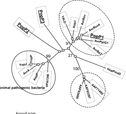
When a multiple alignment of the PopP proteins with other members of the AvrRxv/YopJ family of effectors was gener-ated, the resulting neighbor-joining tree clearly established that PopP1 clusters with one of the two groups of the other members of the family originating from plant-pathogenic and symbiotic bacteria. PopP2 and Pop3 are clearly distinct from these two groups and from the cluster of proteins identified in animal pathogens as well as from the HopPmaD protein from
P. syringae pv. maculicola (Fig. 1).
PopP genes are encoded
within two distinct pathogenicity islands.
With a respective G+C base composition of 55, 60, and 62% that is markedly different from the average value of 67% char-acteristic for the entire genome of strain GMI1000, popP1,
popP2, and popP3 are located within ACURs. In addition, popP1 and popP2 are part of a 79-kb chromosomal
patho-genicity island extending between the RSc0821 gene and the tmRNA gene ssrA. In addition to harboring several ACURs, this region is characterized by the presence of several genes originating from phages, two insertion sequences (ISRso9 and ISRso14), and one partial copy of ISRso1. It also carries sev-eral potential pathogenicity genes, such as the genes for the hemaglutinin-related protein RSc0887, and the RSc0824 and RSc0895 proteins that share a high homology in their
N-ter-minus with the N-terN-ter-minus of PthG, a protein controlling both aggressiveness and host specificity in the plant pathogen
Erwinia herbicola pv. gypsophylae (Ezra et al. 2000). This
pathogenicity island also harbors several stretches of DNA that are duplicated elsewhere in the genome.
Similarly, popP3 is part of an 83-kb genomic island located near the origin of replication of the chromosome. This island is bordered on one side by the cysC1 gene that codes for an adenylylsulfate kinase and by an Ala-tRNA (RS02400) on the other side. This region is characterized by the presence of sev-eral long ACURs and two copies of both ISRso1 and ISRso13. Furthermore, several phage genes and numerous structural genes for proteins with unknown functions are present in this island. This region also is particularly rich in DNA stretches that are duplicated elsewhere in the genome. In particular, two duplications are shared between the popP1–popP2 and the
popP3 pathogenicity islands, with rs16 occurring in direct
ori-entation and rs22 in reverse oriori-entation.
popP genes are present
in most Asiaticum and African isolates but are generally absent from Americanum isolates.
As a first step toward the analysis of the potential contribu-tion of horizontal gene transfer to strain variability in R.
solana-cearum, we used a collection of 30 strains representative of the
different races, biovars, and genetic groups previously described to monitor the distribution of popP genes in this species. The positioning of each of these strains according to the Ameri-canum, Asiaticum, or African divisions is shown in Table 1 and was confirmed using multiplex PCR as defined by M. Fegan and P. Prior (personal communication). In the case of race 2 strains originally described as pathogenic on Musa spp., further genomic characterization was performed to differentiate them according to the MLG classification (Cook and Sequeira 1994; M. Fegan and P. Prior, personal communication).
The distribution of popP genes among this strain collection was first analyzed by Southern hybridization of total genomic DNA with various probes corresponding to PCR amplicons of
popP1 and popP2 genes obtained from strain GMI1000 and to
the popP3 gene amplified from strain GMI8084. The nucleo-tide sequences of these three probes are highly divergent so that no cross-hybridization occurred under the experimental conditions used (data not shown). Clear hybridization signals were detected with at least one of the probes in all but one (Rd15) of the strains from the Asiaticum and African divisions (Table 1). The majority of these strains gave a positive signal with two of the three probes. Three other strains only hybrid-ized with popP1 and two strains (GMI8160 and GMI1000) gave a positive signal for the three genes. Another strain (JT685) gave a positive signal only with popP1. All three genes appear to be almost equally represented among the Asiaticum strains tested. Positive hybridization signals with the popP1 probe also were obtained with the three African strains included in this study. These strains did not hybridize with the popP2 probe and only one of them hybridized with the popP3 probe.
Conversely, with the exception of strains from MLG25, all of the Americanum strains tested were consistently negative when probed with the three popP genes.
A gene family with limited allelic variability at each locus.
To confirm the positive signals observed in Southern hy-bridization and to establish the nucleotide sequences at each
popP locus, PCR experiments were conducted to amplify the
genes, or parts thereof, from strains that were positive in Southern hybridizations. This allowed us to determine the se-quence for the full-length PopP1 and PopP2 coding genes for strains from the Asiaticum and the African divisions. For
popP3 genes, sequences covering 1,137 bp of the 3′ end of
the 1,161-bp popP3 gene were obtained. In the case of strains
Fig. 1. Phylogenic tree of members showing the relationship between members of AvrRxv/YopJ family of type III-dependent pathogenicity effectors.
Acces-sion numbers for the proteins are: Yersinia pseudotuberculosis YopJ, P31498; Y. enterocolitica YopP, NP-052382; Salmonella enterica AvrA, AAB83970;
Xanthomonas axonopodis (campestris) AvrRxv, Q08678; AvrBsT, AAD39255; XopJ, AAK72486; AvrXv4, AAG39033; Pseudomonas syringae AvrPpiG1,
CAC16700; HopPmaD, AAL84243; AAF71492, AAF71492; Erwinia amylovora AAF63400, AAF63400; AAF63397, AAF63397; Aeromonas salmonicida sp. salmonicida AopP, NP_710166; Vibrio parahaemolyticus VPA1346, NP_800856; Rhizobium sp. Y4LO, NP-443964. Relevant bootstrap values are shown on the graph.
from the subgroup MLG25, only partial popP1 gene frag-ments of 771 bp covering the 3′ end of the 1,107-bp PopP1 coding sequence could be amplified and sequenced. The results from PCR amplifications confirmed the presence of
popP genes in all the strains that gave a positive
hybridiza-tion signal in Southern hybridizahybridiza-tion, with the excephybridiza-tion of
popP2 amplicons from the MLG25 strains (discussed below).
All these sequences are available in the GenBank database under accession numbers AJ628876 to AJ628895.
In several cases, the sequences thus generated revealed the presence of an insertion sequence within or flanking the corre-sponding gene (Table 1). Where this occurred, the sequence of the native gene was established by removal of the IS sequence and of its flanking base pair duplication generated by the inser-tion process. The sequences obtained for each locus were aligned using Clustal X software and the corresponding neigh-bor-joining trees were constructed.
The neighbor-joining tree corresponding to the alignment of 771 bp covering the last codons of the popP1 loci is shown in Figure 2A. It establishes that, with a bootstrap value of over 70, the popP1 genes can be classified in three major allelic groups. Group 1A includes three Asiaticum
iso-lates, group 1B includes both African and Asiaticum isoiso-lates, and group 1C is characteristic of the MLG25 subgroup of Americanum isolates. The six popP1 partial sequences from group 1C were completely identical even though the corre-sponding strains originated from different geographical ar-eas, including French West Indies and South and Central America. Comparison of the full-length popP1 genes from group 1A were found to encode identical proteins, although one single silent base change was detected in one strain. Genes of the 1B group, however, are more variable and can be distinguished from the group 1A alleles on the basis of 54 conserved base changes resulting in 10 conserved amino acid changes at the protein level (Table 2). The PopP1 proteins from group 1A share 95% identity with the PopP1 proteins from group 1B. The partial C-terminal sequence of proteins from group 1C shares 97 to 98% amino acid identity with the corresponding regions of proteins from both group 1A and 1B. Three of the characterized popP1 genes, one from group 1A and two from group 1B, are disrupted by an IS. The IS differs from strain to strain and is inserted at different loca-tions within the gene, indicating that these inserloca-tions corre-spond to recent evolutionary events (Fig. 3).
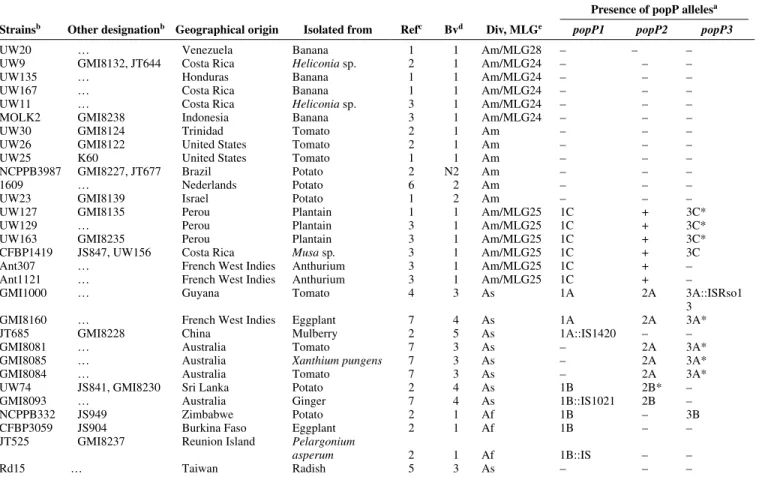
Table 1. Characteristics of the Ralstonia solanacearum strains used in this study and summary of the distribution of the allelic forms of popP genes present
in these strains
Presence of popP allelesa Strainsb Other designationb Geographical origin Isolated from Refc Bvd Div, MLGe popP1 popP2 popP3
UW20 … Venezuela Banana 1 1 Am/MLG28 – – –
UW9 GMI8132, JT644 Costa Rica Heliconia sp. 2 1 Am/MLG24 – – –
UW135 … Honduras Banana 1 1 Am/MLG24 – – –
UW167 … Costa Rica Banana 1 1 Am/MLG24 – – –
UW11 … Costa Rica Heliconia sp. 3 1 Am/MLG24 – – –
MOLK2 GMI8238 Indonesia Banana 3 1 Am/MLG24 – – –
UW30 GMI8124 Trinidad Tomato 2 1 Am – – –
UW26 GMI8122 United States Tomato 2 1 Am – – –
UW25 K60 United States Tomato 1 1 Am – – –
NCPPB3987 GMI8227, JT677 Brazil Potato 2 N2 Am – – –
1609 … Nederlands Potato 6 2 Am – – –
UW23 GMI8139 Israel Potato 1 2 Am – – –
UW127 GMI8135 Perou Plantain 1 1 Am/MLG25 1C + 3C*
UW129 … Perou Plantain 3 1 Am/MLG25 1C + 3C*
UW163 GMI8235 Perou Plantain 3 1 Am/MLG25 1C + 3C*
CFBP1419 JS847, UW156 Costa Rica Musa sp. 3 1 Am/MLG25 1C + 3C
Ant307 … French West Indies Anthurium 3 1 Am/MLG25 1C + –
Ant1121 … French West Indies Anthurium 3 1 Am/MLG25 1C + –
GMI1000 … Guyana Tomato 4 3 As 1A 2A 3A::ISRso1
3
GMI8160 … French West Indies Eggplant 7 4 As 1A 2A 3A*
JT685 GMI8228 China Mulberry 2 5 As 1A::IS1420 – –
GMI8081 … Australia Tomato 7 3 As – 2A 3A*
GMI8085 … Australia Xanthium pungens 7 3 As – 2A 3A*
GMI8084 … Australia Tomato 7 3 As – 2A 3A*
UW74 JS841, GMI8230 Sri Lanka Potato 2 4 As 1B 2B* –
GMI8093 … Australia Ginger 7 4 As 1B::IS1021 2B –
NCPPB332 JS949 Zimbabwe Potato 2 1 Af 1B – 3B
CFBP3059 JS904 Burkina Faso Eggplant 2 1 Af 1B – –
JT525 GMI8237 Reunion Island Pelargonium
asperum 2 1 Af 1B::IS – –
Rd15 … Taiwan Radish 5 3 As – – –
a popP: presence of the genes popP1, popP2, or popP3 in the strains; – indicates that no signal was obtained in Southern hybridization with the corresponding popP probe; + indicates that a signal was detected in Southern hybridization with the corresponding popP probe but no amplicon of the gene or part of it could be produced. 1A, 1B, 1C, 2A, 2B, 3A, 3B, and 3C correspond respectively to the different allelic groups of popP1, popP2, and
popP3 genes defined on the basis of phylogenetic analysis that were found in the strains. The presence of an insertion sequence in certain gene is indicated
by ::IS; * indicates strains where multiple copies of the corresponding gene are detected based on Southern hybridization.
b Strain designation: CFBP, Collection Française de Bactéries Phytopathogènes, INRA Angers, France; NCPPB, National Collection of Plant Pathogenic Bacteria, Harpenden, U.K.; UW, D. Cook and L. Sequeira, Department of Plant Pathology, University of Wisconsin–Madison, U.S.A.; GMI, CNRS-INRA, Auzeville, Castanet Tolosan Cedex, France; JS and JT, laboratoire de phytopathologie, CIRAD-FLHOR, Saint-Pierre, La Réunion, France.
c References: 1, Cook et al. 1989 ; 2, Poussier et al. 2000b; 3, P. Prior and M. Fegan, personal communication; 4, Salanoubat et al. 2002; 5, Deslandes et al. 1998; 6, Van Elsas et al. 2000 ; 7, this study.
d Biovars.
e Division (Div): As, Asiaticum; Am, Americanum; and Af, African as defined by Cook and associates (1989) and Poussier and associates (1999). Multilocus genotype (MLGs) as defined by Cook and Sequeira (1994).
A similar analysis was performed on the full-length popP2 genes (Fig. 2B) allowing the definition of two clearly distinct groups of alleles referred to as 2A and 2B.
These two groups can be distinguished on the basis of 13 specific base substitutions resulting in six amino acid changes in the proteins (Table 2). Proteins from group 2A are identical and share 98% of identity with group 2B proteins. The two proteins from group 2B differ from each other by one single amino acid change, Q81H.
No amplicon could be obtained for popP2 in strains from MLG25. However, Southern hybridizations performed under high-stringency conditions reproducibly gave a clear hybridi-zation pattern on these strains. The pattern observed for the six MLG25 strains is not enlightened by a popP1 or a popP3 probe (data not shown) and clearly is distinct from the patterns obtained with the popP2 probe on the other strains (Fig. 4). Altogether, this is a good indication that a popP2 gene might
be present in MLG25 strains and that these genes might form a distinct allelic group referred to as 2C.
Concerning PopP3, the neighbor-joining tree obtained after alignment of the 1,137 bp located at the 3′ end of the popP3 open reading frame identifies three groups of alleles (3A, 3B, and 3C) with a bootstrap value of above 99% (Fig. 2C). Strains from group 3A harbor popP3 genes whose sequences are identical to the sequence of the allele found in strain GMI1000, except that these genes are not interrupted by inser-tion of ISRso13. Group 3B is composed of a single gene origi-nating from an African isolate. The corresponding protein sequences (379 aa) share 88 and 92% identity with the PopP3 sequences from groups 3A and 3C, respectively.
Group 3C corresponds to four identical popP3 genes present in strains from the MLG25 subdivision of the Americanum phylum. The corresponding protein sequences share 88% iden-tity with the protein sequences from group 3A.
Fig. 2. Neighbor-joining tree established upon alignments of the nucleotide sequences of A, partial popP1, B, full-length popP2, and C, partial-length
popP3 genes showing the grouping in allelic forms. The numbers at the branch points correspond to the percentages of bootstrap replicates in which the clusters are found.
Table 2: Positions and identity of discriminative amino acids between
alleles 1A and 1B of the PopP1 proteins and between alleles 2A and 2B of the PopP2 proteins
Amino acids at allelic forms
PopP1 PopP2 Positions 1A 1B 2A 2B 71 M A … … 90 S A … … 97 H S … … 132 Q E … … 156 G A … … 239 A V … … 280 H R … … 282 Y Q … … 285 H Y … … 326 R S … … 13 … … S N 115 … … L P 146 … … R H 156 … … G D 288 … … S N 396 … … G E
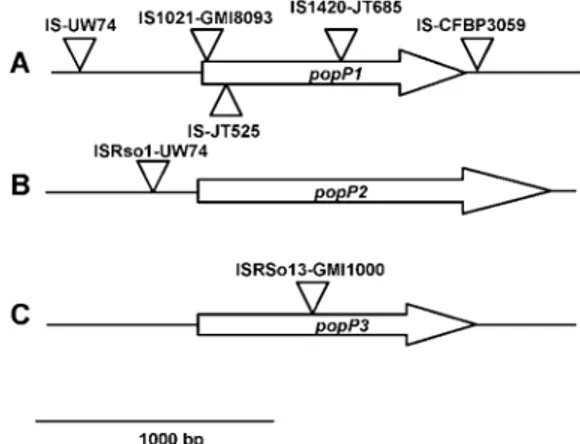
Fig. 3. Schematic representation of the locations of the insertions
sequences present within or in the vicinity of A, popP1, B, popP2, and C,
popP3 genes in the Ralstonia solanacearum strains studied. The positions
of insertion sequence (IS) insertions are indicated by an arrow; the name of the corresponding IS and the strain in which the insertion was present are indicated next to the arrow.
Taken all together, these results confirm the presence of at least one popP gene in all but one of the strains from the Asi-aticum division (Table 1). They also confirm that MLG25 strains form a specific group among Americanum strains be-cause they are the only strains from this phylum to harbor
popP genes. However, the popP alleles found in these strains
are clearly distinct from the alleles identified in Asiaticum strains. In addition, our data show that, when more than one
popP gene is present in a strain, no recombination is observed
between the three loci because the same combination of alleles are always found (i.e., a popP1 allele from group 1A exclu-sively is found associated with a group 2A allele of popP2 and with a group 3A allele of popP3). The same is true for popP genes from allelic groups B and C.
Occurrence of popP gene duplications.
In several cases, the banding patterns obtained in Southern blots of EcoRI-digested genomic DNA suggested that more than one copy of the gene was present in a single strain. For example, strain UW74, which carries a 2B allele of popP2, harbors four fragments that hybridize with the popP2 (Fig. 5A). Two of these fragments are shared with strain GMI8093, which carries a 2B allele of popP2. This allele harbors two internal EcoRI restriction site generating three restriction frag-ments with only two that are large enough to be visualized after hybridization. Therefore, the two additional hybridizing restriction fragments that are detected in strain UW74 corre-spond to at least one additional copy of a popP2 gene. As far as the 3C alleles of popP3 are concerned, even though a single hybridizing band was found for the strain CFB1419, additional bands with variable hybridizing intensity were detected in strains UW127, UW163 (Fig. 5B), and UW129 (data not
shown). No EcoRI site was present in the sequence of the cor-responding 3C allele of popP3 amplicons; this demonstrates the presence of multiple copies of popP3-related genes in strains UW127, UW163, and UW129. The same reasoning was applied for the popP3 genes that are present in two copies in the strains GMI8160, GMI8081, and GMI8085 (Fig. 5B) and GMI8084 (data not shown). This data is summarized in Table 1.
When thermal asymmetric interlaced (TAIL) PCR amplifi-cation was conducted on the upstream and downstream re-gions of popP in the corresponding strains, no difference was detected in the sequence generated at each locus. This estab-lished that the duplication covered a region of at least 170 bp on each side of PopP coding sequence. However, the differ-ence in the size of the restriction fragments of the hybridizing fragments strongly suggests that the different copies of the du-plicated genes are located within different genetic environ-ments. Alternatively, differences in hybridization intensities of these additional bands may be an indication that the different copies of popP gene within a strain might be too different to be amplified with a common set of primers.
Allelic variations
of popP1 and popP2 do not affect their avirulence function.
In strain GMI1000, the popP1 and popP2 genes behave as avirulence determinants on Petunia St40 and A. thaliana eco-type Nd-1, respectively (Deslandes et al. 2003; Lavie et al. 2002). To test whether the allelic variations observed within these genes actually affected their avirulence function, the
popP1 1B allele from strain UW74, the popP2 2B allele from
strain GMI8093, and the popP2 2B allele from UW74 were tested individually for avirulence activity following introduc-tion of pML1, pML2, and pML3 plasmids into strain Rd15 that is virulent on petunia St40 and A. thaliana Nd-1. The 1B allele used in these experiments was chosen as being the most divergent from the popP1 gene present in strain GMI1000. The 2B PopP2 proteins from strains UW74 and GMI8093 dif-fer by six and seven amino acid changes, respectively, from
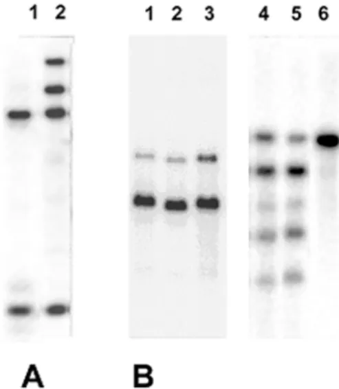
Fig. 5. Southern blot hybridization profiles of EcoRI-digested genomic
DNA obtained with A, a popP2 probe and B, a popP3 probe establishing the existence of popP gene duplication in several strains of Ralstonia
solanacearum. Hybridization probes used correspond to A, popP2 and B, popP3. Genomic DNA originate from stains UW74 (A, lane 2), GMI8160
(B, lane 1), GMI8085 (B, lane 2), GMI8081 (B, lane 3), UW127 (B, lane 4), UW163 (B, lane 5), and CFBP1419 (B, lane 6). The hybridization profiles for strains GMI8093 (A, lane 1) and CFBP1419 (B, lane 6), which carry a single copy of the popP2 and popP3 genes, respectively, also are presented for comparison.
Fig. 4. Hybridization patterns of a popP2 probe against EcoRI digests of
genomic DNA from selected positive strains of Ralstonia solanacearum belonging to the phyla Americanum and Asiaticum. Individual strains are: Lanes 1 and 10, GMI1000; lane 2, UW163; lane 3, Ant1121; lane 4, Ant307; lane 5, CFBP1419; lane 6, UW129; lane 7, UW127; lane 8, UW74; lane 9, GMI8093; lane 11, GMI8160; lane 12, GMI8085; lane 13, GMI8084; and lane 14, GMI8081.
GMI1000 PopP2. These tests were conducted as previously described using popP1 and popP2 genes from strain GMI1000 as controls (Deslandes et al. 2003; Lavie et al. 2002). In each case, no difference in avirulence activity was observed be-tween the new allelic forms tested and the controls.
DISCUSSION
We previously characterized two genes in strain GMI1000 of R. solanacearum that encode proteins belonging to the AvrRxv/YopJ family of cysteine proteases that are secreted by type-III protein secretion machinery from bacterial pathogens (Deslandes et al. 2003; Lavie et al. 2002). In this study, we identified a third gene, called popP3, encoding a new member of this gene family. In addition, we show that occurrence of multiple members of this gene family is a common feature among R. solanacearum strains. This is a rather unusual situa-tion among plant pathogens because the occurrence of several members of this gene family in a single strain has been re-ported only in X. campestris pv. vesicatoria (XcvT, race 1 75-3), in which the two closely related genes AvrRxv and AvrBsT are present (Ciesiolka et al. 1999).
In strain GMI1000, the structural genes for these PopP effec-tors are located at two distant regions of the chromosome har-boring most of the features characteristic of pathogenicity is-lands. The popP1 gene encodes a protein closely related to other members from this family that have been characterized in plant pathogens. The two others genes, popP2 and popP3, encode proteins that do not cluster with the other members of this family identified in either plant or mammal pathogens.
We also have established that the three popP genes are not evenly distributed among the three phyla identified in this species. They are found almost exclusively in strains that be-long to the divisions Asiaticum and African and usually are absent from strains classified as Americanum. Considering the wide conservation of this gene family among bacteria pathogenic to plants and mammals (Orth 2002), it is surpris-ing that such genes were absent from most Americanum strains. Therefore, this raises the question of whether other members of this gene family that remain to be identified are present in these strains. We know, however, that these genes are not essential for pathogenicity because a mutant of strain GMI1000 in which both popP1 and popP2 have been inacti-vated (in addition to popP3 that already is nonfunctional in this strain) retained the ability to cause disease on tomato and
A. thaliana (data not shown).
Several features indicate that the three PopP proteins might not be functionally equivalent. It has been shown that PopP2 is addressed to the plant nucleus (Deslandes et al. 2003), whereas PopP1 and PopP3 are devoid of a predicted nuclear localization signal. A potential myristoylation site is present at the N-terminus of PopP3, suggesting that this protein could be addressed to the plant cell membrane as established for several Avr proteins from P. syringae (Nimchuk et al. 2000; Shan et al. 2000). In addition, PopP1 is predicted to remain in the cytoplasm after translocation into plant cell. This is a strong indication that the three effectors have different targets in the plant and, therefore, act differently in controlling plant–bacte-ria interactions.
We have shown that allelic variations of popP genes in R.
solanacearum are rather limited and that they might have little
effect on the functions of the corresponding proteins because the popP1 and popP2 variant alleles tested retained the aviru-lence function of the corresponding alleles in strain GMI1000. In contrast, great variability exists concerning the presence or absence of each individual gene and the number of copies of each popP gene present in a particular strain. Moreover, we
found that, when present, expression of these genes frequently might be affected by the presence of IS elements within or in close vicinity to the coding sequence (Fig. 3). In addition to the popP3 gene that is disrupted in strain GMI1000, 3 of the 14 popP1 genes sequenced were disrupted by an insertion se-quence. Three IS also were found in popP flanking regions (one located 34 bp after the popP1 stop codon of strain CFBP3059, one at 500 bp before the popP1 start codon of strain UW74, and one 180 bp upstream of the popP2 start codon of strain UW74). In addition, several DNA duplications have been identified within the popP1–popP2 and popP3 patho-genicity islands and several strains were identified in which individual popP genes have been duplicated. Taken together, these results indicate that genomic rearrangements may play a major role in the control of expression of the pathogenicity functions encoded at these loci.
In this study, a strong correlation between the distribution of
popP genes and the phylogenic position of strains was
estab-lished. Taking into account the large background of evidence that suggests that these genes have been acquired relatively re-cently through horizontal gene transfer, this result is rather un-expected. Contrary to what would be expected in case of inde-pendent horizontal acquisition of the popP1–popP2 and
popP3 pathogenicity islands, recombination between popP1– popP2 and popP3 was never observed. Therefore, these
obser-vations favor a model in which a common ancestor to the Afri-can and Asiaticum phyla acquired popP genes through hori-zontal gene transfers. In addition, these genes would have been acquired more recently by a common ancestor of the subset of MLG25 strains from the phylum Americanum. According to such a model, one would predict that the long co-evolution of the popP genes with the core genome of Asiaticum and Afri-can strains should have resulted in adjustment of popP genes base composition to the average base composition of the ge-nome. This clearly is different from what we observe.
An alternative scenario to explain the correlation between
popP gene distribution and strain phylogeny would be that, in
order to acquire or stably maintain the popP genes, the host strains require particular functions encoded only in the core genome of the Asiaticum and African strains. Such functions could include, for example, phage receptors, restriction or modi-fication enzymes, other functions required for heterologous recombination of the incoming DNA, or alternatively, patho-genicity functions such as specific type-III effectors such as PopA, PopB, or PopW (Arlat et al. 1994; Guéneron et al. 2000; Salanoubat et al. 2002) that are encoded by genes indis-tinguishable from the core genome. However, again, the ab-sence of recombination between the popP genes is in contra-diction with this hypothesis.
In conclusion, the present study reveals that the mechanisms governing ACUR acquisition by R. solanacearum might be much more complex than we originally thought. Further work, based on the analysis of the distribution of a large number of effectors from ACUR, will be needed in order to understand the mechanisms which govern distribution and evolution of these genes in R. solanacearum and how these genes might be involved in adaptation of this versatile pathogen to its wide range of potential hosts.
MATERIALS AND METHODS Bacterial strains and growth conditions.
Strain TG1 of Escherichia coli grown in Luria-Bertani medium (Sambrook et al. 1989) at 37°C was used for cloning experiments. R. solanacearum strains used in this study are listed in Table 1. All R. solanacearum strains were grown at 28°C in B medium (Boucher et al. 1985). When required,
anti-biotics were used at the following concentrations: tetracycline at 10 mg liter–1 and ampicillin at 50 mg liter–1.
R. solanacearum electroporation.
Plasmids were introduced into R. solanacearum strains by electroporation (2.5 kV, 200 ohms, 25 µFd, 0.2-cm cuvette gap).
Molecular biology techniques.
Unless stated otherwise, standard methods were used (Ausubel et al. 1990). Genomic DNA was extracted from R.
solanacearum strains as described by Arlat and associates
(1992). PCR amplifications were done using the Expand Long Template PCR System (Roche Applied Science, Indianapolis, IN, U.S.A.).
For Southern analysis, approximately 3 µg of EcoRI-digested genomic DNA was loaded per lane on an agarose gel. DNA was transferred to Hybond N+ membranes (Amersham Biosciences, Piscataway, NJ, U.S.A.) and hybridizations were performed in 0.5 M sodium phosphate, pH 7.2; 1 mM EDTA, pH 8; 7% sodium dodecyl sulfate (SDS); and salmon sperm DNA at 20 µg/ml at 65°C. Membrane were washed at 65°C in 0.1× SSC (1× SSC is 0.15 M NaCl plus 0.015 M sodium citrate) and 0.1% SDS buffer. The DNA probes were labeled with [α-P32]dCTP using the kit Ready to Go (Amersham
Bio-sciences). popP1 and popP2 probes were obtained by PCR amplifications of the popP1 and popP2 full gene sequences from strain GMI1000 using primers Fw4-P1/Rev1-P1 and Fw3-P2/Rev3-P2, respectively. The popP3 probe was obtained by PCR amplification of the coding sequence of popP3 from strain GMI8084 using primers Fw1-P3 and Rev1-P3.
DNA amplifications and sequencing.
popP gene flanking sequences were isolated using
TAIL-PCR performed according to the method described by Liu and Whittier (1995), except that the Expand Long Template PCR System (Roche) and corresponding elongation conditions were used for amplifications. Two different degenerate primers were tested for each amplification. The largest products obtained were approximately 2.5 kb long.
popP gene amplification.
Several methods were used to amplify popP genes prior to sequencing.
In most of the cases, primers located in the flanking re-gions of popP genes in strain GMI1000 genome were used successfully to amplify popP genes from other strains. When no product could be obtained in this way, primers internal to the popP coding sequences were tested. In such cases, the missing sequences corresponding to the 5′ and 3′ ends of the corresponding genes were determined on TAIL-PCR amplifi-cation products.
TAIL-PCR and PCR products were analyzed by gel electro-phoresis and purification of amplicons was performed by pas-sage through a S400 column (Amersham Biosciences) when a single amplicon was present in the reaction mixture. When several amplicons were present, individual amplicon products were purified by extraction of excised gel fragments contain-ing each individual replicons. This extraction was performed using the Wizard SV gel and PCR clean-up system (Promega Bioscience, San Luis Obispo, CA, U.S.A.). The products thus obtained were used directly for sequencing.
Plasmid constructions.
pML2 and pML3 were constructed by cloning the popP2 genes from strains GMI8093 and UW74, respectively, be-tween the restriction sites BamHI and HindIII of the pLAFR3 vector (Lindgren et al. 1986). popP2 genes were obtained by
PCR amplification of a 1,489-bp DNA fragment, extending from the ATG of the popP2 gene down to 23 bp downstream of the stop codon, using the primers Fw3-P2 and Rev3-P2 that introduced the BamHI and HindIII restriction sites at the end of the amplicon (Table 2). In the resulting constructs, gene expression was under the control of the plac promoter of pLAFR3.
To construct pML4, a fragment EcoRI-HindIII of 1.4 kb, containing the popP1 gene from strain GMI1000 together with 307 bp of its 5′ flanking region and 35 bp of its downstream sequence, was PCR amplified with the primers Fw2-P1/Rev2-P1 that carry EcoRI and HindIII restriction sites, respectively, at their 5′ extremities. This fragment then was cloned between the EcoRI and HindIII restriction sites of pLAFR3.
pML1 is a pLAFR3 derivative in which a 1.4-kb
EcoRI-EcoRI fragment containing the popP1 gene from strain UW74
was cloned into the EcoRI restriction site of the pLAFR3. In strain GMI1000, the 5′ extremities of primers Fw3-P1 and Rev4-P1 are located 48 bp upstream of the popP1 start codon and at 17 bp downstream of the popP1 stop codon, respec-tively. These primers allow the amplification of a 1.2-kb DNA fragment. Using strain UW74 genomic DNA as template, the PCR amplification with Fw3-P1 and Rev4-P1 produced a DNA fragment of 2.5 kb. The partial sequencing of this frag-ment showed that the entire popP1 gene was contained be-tween the Fw3-P1 primer and an EcoRI site present within the amplified DNA fragment. Hence, EcoRI digestion of the 2.5-kb amplicon allowed isolation of a 1.4-2.5-kb DNA fragment that contained a full-length popP1 gene. This fragment was cloned under the control of the plac promoter in pLAFR3.
Plant tests.
Pathogenicity assays on Arabidopsis (ecotypes Col-5 and Nd-1) and petunia plants were performed as previously de-scribed by Marenda and associates (1998) and Deslandes and associates (1998).
Phylogenetic analysis.
popP gene sequences were analyzed using the
PHYLO-WIN software package (Galtier et al. 1996). Sequences were aligned with the Clustal X 1.8 software (Thompson et al. 1997). Phylogenetic trees were constructed from the align-ments by using the neighbor-joining method (Saitou and Nei 1987) with a correction of the evolutionary distances for mul-tiple mutations with the Kimura formula. Bootstrapping was performed with 1,000 replicates. The resulting phylogenetic trees were visualized using the Treeview 1.6.6 software.
ACKNOWLEDGMENTS
We thank P. Boistard and S. Genin for valuable discussions and B. Hogg for editing of the manuscript.
LITERATURE CITED
Alfano, J. R., Charkowski, A. O., Deng, W. L., Badel, J. L., Petnicki-Ocwieja, T., van Dijk, K., and Collmer, A. 2000. The Pseudomonas
syrin-gae Hrp pathogenicity island has a tripartite mosaic structure composed
of a cluster of type III secretion genes bounded by exchangeable effector and conserved effector loci that contribute to parasitic fitness and patho-genicity in plants. Proc. Natl. Acad. Sci. U.S.A. 97:4856-4861.
Arlat, M., Gough, C. L., Zischek, C., Barberis, P. A., Trigalet, A., and Boucher, C. A. 1992. Transcriptional organization and expression of the large hrp gene cluster of Pseudomonas solanacearum. Mol. Plant-Microbe Interact. 5:187-193.
Arlat, M., Van Gijsegem, F., Huet, J. C., Pernollet, J. C., and Boucher, C. A. 1994. PopA1, a protein which induces a hypersensitivity-like re-sponse on specific Petunia genotypes, is secreted via the Hrp pathway of Pseudomonas solanacearum. EMBO (Eur. Mol. Biol. Organ.) J. 13:543-553.
Ausubel, F. M., Brent, R., Kingston, R. E., Moore, D. D., Seidman, J. G., Smith, J. A., and Struhl, K. 1990. Current Protocols in Molecular Microbiology. Green Publishing Associates and Wiley Interscience, New York.
Bertolla, F., Van Gijsegem, F., Nesme, X., and Simonet, P. 1997. Condi-tions for natural transformation of Ralstonia solanacearum. Appl. Environ. Microbiol. 63:4965-4968.
Boucher, C. A., Barberis, P., Trigalet, A. P., and Démery, D. A. 1985. Transposon mutagenesis of Pseudomonas solanacearum: Isolation of Tn5-induced avirulent mutants. J. Gen. Microbiol. 131:2449-2457. Buchrieser, C., Rusniok, C., Kunst, F., Cossart, P., and Glaser, P. 2003.
Comparison of the genome sequences of Listeria monocytogenes and
Listeria innocua: clues for evolution and pathogenicity. FEMS (Fed.
Eur. Microbiol. Soc.) Immunol. Med. Microbiol. 35:207-213.
Buddenhaggen, I., Sequeira, L., and Kelman, A. 1962. Designation of races in Pseudomonas solanacearum. (Abstr.) Phytopathology 52:726. Buell, C. R., Joardar, V., Lindeberg, M., Selengut, J.. Paulsen, I. T.,
Gwinn, M. L., Dodson, R. J., Deboy, R. T., Durkin, A. S., Kolonay, J. F., Madupu, R., Daugherty, S., Brinkac, L., Beanan, M. J., Haft, D. H., Nelson, W. C., Davidsen, T., Zafar, N., Zhou, L., Liu, J., Yuan, Q., Khouri, H., Fedorova, N., Tran, B., Russell, D., Berry, K., Utterback, T., Van Aken, S. E., Feldblyum, T. V., D’Ascenzo, M., Deng, W. L., Ramos, A. R., Alfano, J. R., Cartinhour, S., Chatterjee, A. K., Delaney, T. P., Lazarowitz, S. G., Martin, G. B., Schneider, D. J., Tang, X., Bender, C. L., White, O., Fraser, C. M., and Collmer, A. 2003. The complete genome sequence of the Arabidopsis and tomato pathogen
Pseudomonas syringae pv. tomato DC3000. Proc. Natl. Acad. Sci.
U.S.A. 100:10181-10186.
Charity, J. C., Pak, K., Delwiche, C. F., and Hutcheson, S. W. 2003. Novel exchangeable effector loci associated with the Pseudomonas syringae hrp pathogenicity island: evidence for integron-like assembly from transposed gene cassettes. Mol. Plant-Microbe Interact. 16:495-507. Ciesiolka, L. D., Hwin, T., Gearlds, J. D., Minsavage, G. V., Saenz, R.,
Bravo, M., Handley, V., Conover, S. M., Zhang, H., Caporgno, J., Phen-grasamy, N. B., Toms, A. O., Stall, R. E., and Whalen, M. C. 1999. Regulation of expression of avirulence gene avrRxv and identification of a family of host interaction factors by sequence analysis of avrBsT. Mol. Plant-Microbe Interact. 12:35-44.
Cook, D., Barlow, E., and Sequeira, L. 1989. Genetic diversity of
Pseudo-monas solanacearum: detection of Restriction Fragment Length
Poly-morphisms with DNA probes that specify virulence and the Hypersen-sitive Response. Mol. Plant-Microbe Interact. 2:113-121.
Cook, D., and Sequeira, L. 1994. Strain differentiation of Pseudomonas
solanacearum by molecular genetics methods. Pages 77-93 in:
Bacterial Wilt: The Disease and Its Causative Agent Pseudomonas
solanacearum. A. C. Hayward and G. L. Hartman, eds. CAB
Interna-tional, Wallingford, U.K.
Cunnac, S., Occhialini, A., Barberis, P., Boucher, C., and Genin, S. Inven-tory and functional analysis of the large Hrp regulon in Ralstonia
so-lanacearum: identification of novel effector proteins translocated to
plant host cells through the Type III secretion system. Mol. Microbiol. in press.
Da Silva, A. C., Ferro, J. A., Reinach, F. C., Farah, C. S., Furlan, L. R., Quaggio, R. B., Monteiro-Vitorello, C. B., Van Sluys, M. A., Almeida, N. F., Alves, L. M., do Amaral, A. M., Bertolini, M. C., Camargo, L. E., Camarotte, G., Cannavan, F., Cardozo, J., Chambergo, F., Ciapina, L. P., Cicarelli, R. M., Coutinho, L. L., Cursino-Santos, J. R., El Dorry, H., Faria, J. B., Ferreira, A. J., Ferreira, R. C., Ferro, M. I., Formighieri, E. F., Franco, M. C., Greggio, C. C., Gruber, A., Katsuyama, A. M., Kishi, L. T., Leite, R. P., Lemos, E. G., Lemos, M. V., Locali, E. C., Machado, M. A., Madeira, A. M., Martinez-Rossi, N. M., Martins, E. C., Meidanis, J., Menck, C. F., Miyaki, C. Y., Moon, D. H., Moreira, L. M., Novo, M. T., Okura, V. K., Oliveira, M. C., Oliveira, V. R., Pereira, H. A., Rossi, A., Sena, J. A., Silva, C., de Souza, R. F., Spinola, L. A., Takita, M. A., Tamura, R. E., Teixeira, E. C., Tezza, R. I., Trindade dos, S. M., Truffi, D., Tsai, S. M., White, F. F., Setubal, J. C., and Kitajima, J. P. 2002. Comparison of the genomes of two Xanthomonas pathogens with differing host specificities. Nature 417:459-463.
Deslandes, L., Olivier, J., Peeters, N., Feng, D. X., Khounlotham, M., Boucher, C., Somssich, I., Genin, S., and Marco, Y. 2003. Physical interaction between RRS1-R, a protein conferring resistance to bacte-rial wilt, and PopP2, a type III effector targeted to the plant nucleus. Proc. Natl. Acad. Sci. U.S.A. 100:8024-8029.
Deslandes, L., Pileur, F. , Liaubet, L., Camut, S., Can, C., Williams, K., Holub, E., Beynon, J., Arlat, M., and Marco, Y. 1998. Genetic charac-terization of RRS1, a recessive locus in Arabidopsis thaliana that con-fers resistance to the bacterial soilborne pathogen Ralstonia
solanacea-rum. Mol. Plant-Microbe Interact. 11:659-667.
Ezra, D., Barash, I., Valinsky, L., and Manulis, S. 2000. The dual function in virulence and host range restriction of a gene isolated from the
pPATH (Ehg) plasmid of Erwinia herbicola pv. gypsophilae. Mol. Plant-Microbe Interact. 13:683-692.
Galtier, N., Gouy, M., and Gautier, C. 1996. SEAVIEW and PHYLO_WIN: two graphic tools for sequence alignment and molecu-lar phylogeny. Comput. Appl. Biosci. 12:543-548.
Gillings, M., Fahy, P., and Davies, C. 1993. Restriction analysis of an am-plified polygalacturonase gene fragment differentiates strains of the phytopathogenic bacterium Pseudomonas solanacearum. Lett. Appl. Microbiol. 17:44-48.
Gogarten, J. P., Doolittle, W. F., and Lawrence, J. G. 2002. Prokaryotic evolution in light of gene transfer. Mol. Biol. Evol. 19:2226-2238. Gophna, U., Ron, E. Z., and Graur, D. 2003. Bacterial type III secretion
systems are ancient and evolved by multiple horizontal-transfer events. Gene 312:151-163.
Guéneron, M., Timmers, A. C., Boucher, C., and Arlat, M. 2000. Two novel proteins, PopB, which has functional nuclear localization signals, and PopC, which has a large leucine-rich repeat domain, are secreted through the hrp-secretion apparatus of Ralstonia solanacearum. Mol. Microbiol. 36:261-277.
Hacker, J., Hentschel, U., and Dobrindt, U. 2003. Prokaryotic chromo-somes and disease. Science 301:790-793.
Hacker, J., and Kaper, J. B. 2000. Pathogenicity islands and the evolution of microbes. Annu. Rev. Microbiol. 54:641-679.
Hayward, A. C. 1964. Characteristics of Pseudomonas solanacearum. J. Appl. Bacteriol. 27:265-277.
Hayward, A. C. 1991. Biology and epidemiology of bacterial wilt caused by Pseudomonas solanacearum. Annu. Rev. Phytopathol. 29:65-87. Hayward, A. C. 1994. Systematics and phylogeny of Pseudomonas
so-lanacearum and related bacteria. Pages 123-135 in: Bacterial Wilt: The
Disease and Its Causative Agent, Pseudomonas solanacearum. A. C. Hayward and G. L. Hartman, eds. CAB International, Wallingford, U.K.
He, L. Y., Sequeira, L., and Kelman, A. 1983. Characteristics of strains of
Pseudomonas solanacearum. Plant Dis. 67:1357-1361.
Hutcheson, S. W. 1999. The hrp cluster of Pseudomonas syringae: A pathogenicity island encoding a type III protein translocation complex? Pages 309-329 in: Pathogenicity Islands and Other Mobile Virulence Elements. J. B. Kaper and J. Hacker, eds. American Society of Microbi-ology, Washington, D.C.
Jain, R., Rivera, M. C., Moore, J. E., and Lake, J. A. 2002. Horizontal gene transfer in microbial genome evolution. Theor. Popul. Biol. 61:489-495.
Jain, R., Rivera, M. C., Moore, J. E., and Lake, J. A. 2003. Horizontal gene transfer accelerates genome innovation and evolution. Mol. Biol. Evol. 20:1598-1602.
Johnson, D. R., Bathnagar, R. S., Knoll, L. J., and Gordon, J. I. 1994. Ge-netic and biochemical studies of protein N-myristoylation. Annu. Rev. Biochem. 63:869-914.
Lavie, M., Shillington, E., Eguiluz, C., Grimsley, N., and Boucher, C. 2002. PopP1, a new member of the YopJ/AvrRxv family of type III effector proteins, acts as a host-specificity factor and modulates aggres-siveness of Ralstonia solanacearum. Mol. Plant-Microbe Interact. 15:1058-1068.
Li, X., Dorsch, M., Del Dot, T., Sly, L. I., Stackebrandt, E., and Hayward, C. 1993. Phylogenetic studies of the rRNA group II pseudomonads based on 16S rRNA gene sequences. J. Appl. Bacteriol. 74:324-329. Lindgren, P. B., Peet, R. C., and Panopoulos, N. J. 1986. Gene cluster of
Pseudomonas syringae pv. “phaseolicola” controls pathogenicity of
bean plants and hypersensitivity of non-host plants. J. Bacteriol. 168:512-522.
Liu, Y. G., and Whittier, R. F. 1995. Thermal asymmetric interlaced PCR: automatable amplification and sequencing of insert end fragments from P1 and YAC clones for chromosome walking. Genomics 25:674-681. Marenda, M., Brito, B., Callard, D., Genin, S., Barberis, P., Boucher, C.
and Arlat, M. 1998. PrhA controls a novel regulatory pathway required for the specific induction of Ralstonia solanacearum hrp genes in the presence of plant cells. Mol. Microbiol. 27:437-453.
Nimchuk, Z., Marois, E., Kjemtrup, S., Leister, R. T., Katagiri, F., and Dangl, J. L. 2000. Eukaryotic fatty acylation drives plasma membrane targeting and enhances function of several type III effector proteins from Pseudomonas syringae. Cell 101:353-363.
Ochman, H., Lawrence, J. G., and Groisman, E. A. 2000. Lateral gene transfer and the nature of bacterial innovation. Nature 405:299-304. Orth, K. 2002. Function of the Yersinia effector YopJ. Curr. Opin.
Micro-biol. 5:38-43.
Pegg, K. G., and Moffett, M. 1971. Host range of the ginger strain of
Pseudomonas solanacearum in Queensland. Aust. J. Exp. Agric. Anim.
Husb. 11:696-698.
Perna, N. T., Plunkett, G., III, Burland, V., Mau, B., Glasner, J. D., Rose, D. J., Mayhew, G. F., Evans, P. S., Gregor, J., Kirkpatrick, H. A., Posfai,
G., Hackett, J., Klink, S., Boutin, A., Shao, Y., Miller, L., Grotbeck, E. J., Davis, N. W., Lim, A., Dimalanta, E. T., Potamousis, K. D., Apodaca, J., Anantharaman, T. S., Lin, J., Yen, G., Schwartz, D. C., Welch, R. A., and Blattner, F. R. 2001. Genome sequence of enterohaemorrhagic
Es-cherichia coli O157:H7. Nature 409:529-533.
Poussier, S., Prior, P., Luisetti, J., Hayward, C., and Fegan, M. 2000a. Par-tial sequencing of the hrpB and endoglucanase genes confirms and ex-pands the known diversity within the Ralstonia solanacearum species complex. Syst. Appl. Microbiol. 23:479-486.
Poussier, S., Trigalet-Demery, D., Vandewalle, P., Goffinet, B., Luisetti, J., and Trigalet, A. 2000b. Genetic diversity of Ralstonia solanacearum as assessed by PCR-RFLP of the hrp gene region, AFLP and 16S rRNA sequence analysis, and identification of an African subdivision. Microbiology 146:1679-1692.
Poussier, S., Vandewalle, P., and Luisetti, J. 1999. Genetic diversity of African and worldwide strains of Ralstonia solanacearum as deter-mined by PCR-restriction fragment length polymorphism analysis of the hrp gene region. Appl. Environ. Microbiol. 65:2184-2194.
Saitou, N., and Nei, M. 1987. The Neighbor joining method: a new method for reconstructing phylogenetic trees. Mol. Biol. Evol. 4:406-425. Salanoubat, M., Genin, S., Artiguenave, F., Gouzy, J., Mangenot, S., Arlat,
M., Billault, A., Brottier, P., Camus, J. C., Cattolico, L., Chandler, M., Choisne, N., Claudel-Renard, C., Cunnac, S., Demange, N., Gaspin, C., Lavie, M., Moisan, A., Robert, C., Saurin, W., Schiex, T., Siguier, P., Thebault, P., Whalen, M., Wincker, P., Levy, M., Weissenbach, J., and Boucher, C. A. 2002. Genome sequence of the plant pathogen
Ralsto-nia solanacearum. Nature 415:497-502.
Sambrook, J., Fritsch, E. F., and Maniatis, T. 1989. Molecular Cloning: A
Laboratory Manual. 2nd ed. Cold Spring Harbor Laboratory Press, Cold Spring Harbor, NY, U.S.A.
Seal, S. E., Jackson, L. A., Young, J. P. W., and Daniels, M. J. 1993. Dif-ferentiation of Pseudomonas solanacearum, Pseudomonas syzygii,
Pseudomonas pickettii and the Blood Disease Bacterium by partial 16S
rRNA sequencing: construction of oligonucleotides primers for sensi-tive detection by polymerase chain reaction. J. Gen. Microbiol. 139:1587-1594.
Shan, L., He, P., Zhou, J. M., and Tang, X. 2000. A cluster of mutations disrupt the avirulence but not the virulence function of AvrPto. Mol. Plant-Microbe Interact. 13:592-598.
Taghavi, M., Hayward, C., Sly, L. I., and Fegan, M. 1996. Analysis of the phylogenetic relationships of strains of Burkholderia
solana-cearum, Pseudomonas syzygii, and the blood disease bacterium of
banana based on 16S rRNA gene sequences. Int. J. Syst. Bacteriol. 46:10-15.
Thompson, J. D., Gibson, T. J., Plewniak, F., Jeanmougin, F., and Higgins, D. G. 1997. The CLUSTAL_X windows interface: flexible strategies for multiple sequence alignment aided by quality analysis tools. Nucleic Acids Res. 25:4876-4882.
AUTHOR-RECOMMENDED INTERNET RESOURCE
North Carolina State UniversityBioedit biological sequence alignment editor website: www.mbio.ncsu.edu/BioEdit/bioedit.html
INRA web server Ralstonia solanacearum page: sequence.toulouse.inra.fr/R.solanacearum.html