En vue de l'obtention du
DOCTORAT DE L'UNIVERSITÉ DE TOULOUSE
Délivré par :
Institut National Polytechnique de Toulouse (Toulouse INP)
Discipline ou spécialité :
Pathologie, Toxicologie, Génétique et Nutrition
Présentée et soutenue par :
Mme SU LUO le lundi 23 septembre 2019
Titre :
Unité de recherche : Ecole doctorale :
Individual and combined toxicity of a mycotoxin, the deoxynivalenol and a
trace metal, the cadmium on the intestine
Sciences Ecologiques, Vétérinaires, Agronomiques et Bioingénieries (SEVAB) Toxicologie Alimentaire (ToxAlim)
Directeur(s) de Thèse : MME ISABELLE OSWALD
Rapporteurs :
M. GUNNAR SUNDSTOL ERIKSEN, NORWEGIAN VETERINARY INSTITUTE Mme NOLWENN HYMERY, UNIVERSITE DE BRETAGNE OCCIDENTALE
Membre(s) du jury :
M. FRANCOIS BLACHIER, INRA PARIS, Président Mme ISABELLE OSWALD, INRA TOULOUSE, Membre
M. PHILIPPE PINTON, INRA TOULOUSE, Membre
1
Résumé Français
Le déoxynivalénol (DON) est une mycotoxine de type trichothécène de type B, principalement produite par le genre Fusarium. C'est l'une des mycotoxines les plus répandues, elle est largement trouvée dans les céréales et les produits dérivés des céréales. Le cadmium est un composant de la croûte terrestre et un polluant environnemental courant. C'est un métal trace non essentiel et toxique pour la santé des humains et des animaux. Bien que la toxicité individuelle du DON et du Cd ait été bien étudiée, leur effet combiné est peu étudié. L'intestin étant le premier organe ciblé par les contaminants alimentaires, le but de cette étude est d'explorer l'effet combiné du DON et du Cd sur la fonction de barrière intestinale à l'aide de modèles in vitro, in vivo et ex vivo.
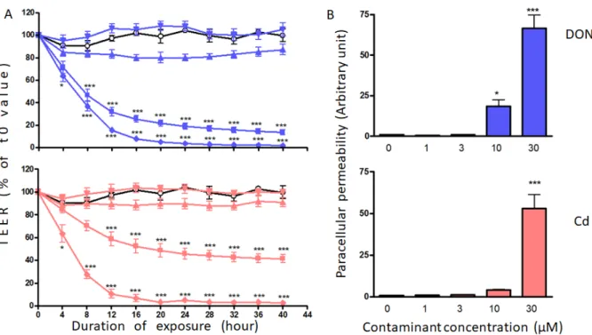
In vitro, des cellules épithéliales intestinales humaines Caco-2 ont été traitées avec une série de concentrations de DON et de Cd (0-30 µM) seules ou en combinaison. La fonction de barrière des cellules Caco-2 a été évaluée par la mesure de la résistance électrique transépithéliale (TEER), de la perméabilité paracellulaire et des protéines jonctionnelles. Le mélange DON, Cd et DON+Cd a diminué le TEER et augmenté la perméabilité paracellulaire de manière dépendante de la concentration. L'abondance des protéines jonctionnelles E-cadhérine et occludine a été considérablement réduite dans les cellules exposées au DON, au Cd et au DON+Cd, tandis que l'expression de ZO-1 et de claudine-3 et -4 est restée inchangée. Le mélange DON Cd a eu des effets légèrement supérieurs ou similaires à ceux du contaminant le plus toxique.
In vivo, les rats ont été exposés à des aliments contaminés par du DON (8,2 mg / kg), et à de l'eau de boisson contaminée par du Cd (5 mg / L) ou au mélange DON+Cd pendant 4 semaines. Les résultats n'ont montré aucun effet sur la prise de poids corporel au cours de l'expérience. Des dommages morphologiques légers caractérisés par un œdème au niveau de la lamina propria et un aplatissement et une fusion des villosités ont été découverts chez le rat exposé à chaque contaminant. Le score lésionnel du jéjunum était plus élevé chez tous les animaux traités que chez les animaux témoins. Une diminution significative de la profondeur de la crypte jéjunale a été observée chez les rats exposés au DON, au Cd et au DON+Cd, alors que la hauteur des villosités n'était pas affectée. Une immunomarquage plus faible de l'E-cadhérine dans le jéjunum de rats exposés à des contaminants seuls ou en association a également été observée, alors que l'occludine n'a diminué que chez les rats exposés au DON et au DON+Cd. Comme
2 indiqué in vitro, l'exposition in vivo au DON et au Cd a induit des effets similaires à ceux du contaminant le plus toxique.
Des explants jéjunaux de porcs ex vivo ont été exposés au DON (0-24 µM), au Cd (0-96 µM) et à la combinaison de DON+Cd. Le DON seul et le mélange DON Cd ont stimulé la réponse immunitaire chez le jéjunum en régulant positivement l'expression d'ARNm de IL-1β, IL-1α, IL-8 et TNF-α de manière dose-dépendante, tandis que le Cd seul n'a pas affecté ces gènes. L'expression génique des métallothionéines (MT), y compris MT1A et MT2A, était régulée positivement de manière dose-dépendante par le Cd seul et le mélange, mais n'était pas affectée par le DON seul. La régulation à la hausse des gènes de cytokines et de MT induite par le DON+Cd était similaire à celle obtenue par le DON ou le Cd seul.
En conclusion, le DON et le Cd modifient tous deux la fonction de barrière intestinale et l'effet combiné est similaire avec leur effet individuel. L'effet du mélange n'a démontré aucune synergie, ce qui suggère que la réglementation sur chaque contaminant protège suffisamment les consommateurs exposés aux mélanges de DON et de Cd.
Mots clés: déoxynivalénol, cadmium, effet combiné, fonction de barrière intestinale, protéines jonctionnelles, expression génique
3
Summary
Deoxynivalenol (DON) is a type B trichothecene mycotoxin mainly produced by Fusarium genus. It is one of the most prevalent mycotoxins widely found in cereals and cereal-derived products. Cadmium is a component of earth’s crust and also a common environmental pollutant. It is a non-essential trace metal and toxic for humans and animals health. Although the individual toxicity of DON and Cd has been well investigated, their combined effect is poorly studied. As intestine is the first organ targeted by food contaminants, the aim of this study is to explore the combined effect of DON and Cd on the intestinal barrier function using in vitro, in vivo and ex vivo models.
In vitro, the human intestinal epithelail cells Caco-2 were treated with a series of concentrations of DON and Cd (0-30 µM) alone or in combination. The barrier function of Caco-2 cells was assessed through the measurement of transepithelial electrical resistance (TEER), paracellular permeability and junctional proteins. DON, Cd and DON+Cd mixture decreased the TEER and increased the paracellular permeability in a concentration-dependent manner. The abundance of junctional proteins E-cadherin and occludin was considerably reduced in cells exposed to DON, Cd and DON+Cd, while the expression of ZO-1, and claudin-3 and -4 remained unchanged. The mixture DON+Cd induced slightly higher or similar effects than the most toxic contaminant.
In vivo, rats were exposed to DON-contaminated feed (8.2 mg/kg feed), and Cd-contaminated drinking water (5 mg/L) or to the mixture DON+Cd for 4 weeks. The results showed no effect on body weight gain during the experiment. Mild morphological damage characterized by edema in lamina propria and villi flattening and fusion was found in rat exposed to each contaminant. The lesional score of jejunum was higher in all the treated animals than that in control animals. A significant decrease of jejunal crypt depth was observed in rats exposed to DON, Cd and DON+Cd, whereas villi height remained unaffected. A lower immunostaining of E-cadherin in the jejunum of rats exposed to contaminants alone or in combination was also observed, whereas occludin was only decreased in rats exposed to DON and DON+Cd. As shown in vitro, in vivo exposure to both DON and Cd induced similar effects than the most toxic contaminant.
Ex vivo, jejunal explants of pigs were exposed to DON (0-24 µM), Cd (0-96 µM) and in combination DON+Cd. DON alone and mixture DON+Cd stimulated immune response in jejunum by upregulating mRNA expression of IL-1β, IL-1α, IL- 8 and TNF-α in a
dose-4 dependent manner, while Cd alone did not affect these genes. Gene expression of metallothioneins (MTs) including MT1A and MT2A was dose-dependently upregulated by Cd alone and mixture, but not affected by DON alone. The upregulation of cytokine and MTs genes induced by DON+Cd was similar than by DON or Cd alone.
In conclusion, both DON and Cd alter intestinal barrier function and the combined effect is similar with their individual effect. The effect of the mixture did not demonstrate any synergy, suggesting that regulation on individual contaminant is protective enough for consumers exposed to DON and Cd mixtures.
Keywords:Deoxynivalenol, cadmium, combined effect, intestinal barrier function, junctional
5
Acknowledgements
Firstly, I would like to express my deep gratitude to my thesis supervisors Dr. Isabelle Oswald and Dr. Philippe Pinton, for their continuous support of my doctoral study, for their patient guidance, enthusiastic encouragement and valuable critiques of this research work and thesis writing. Their continued support promoted me keep moving forward. Without their guidance and help, this work could not be carried out.
Besides my supervisors, my sincere thanks also goes to the other members of my thesis committee, Dr. Laurent Ferrier and Dr. El Hassan AJANDOUZ, for their insightful comments and questions, helpful suggestions and kind encouragement on my study.
I would also like to thank Professor Ana-Paula Brcarense for her help in tissue section
preparation and immunohistochemical analysis. My grateful thanks are also extended to Dr. Chloe Terciolo for her precious advice and assistance in immunofluorescence analysis, to Dr. Delphine Payros for her kind help in sampling in vivo experiments.
My particular thanks also goes to professor Hymery, professor Eriksen who are also my rapporteurs and professor Blachier, my jury members, for their constructive suggestions and comments on my thesis manuscript, which help a lot to improve this manuscript.
I thank my friends and colleagues, Manon Neves, Joelle Laffitte, Sylvie Puel for their collaboration and technical support for my research. Also I thank other members in our team, Laura Soler-Vasco, Le Thanh Huong, Sorphie Lorber, Christelle El Hajj Assaf, Amaranta Carvajal-Campos, Olivier Puel, Ophélie Rocher, Chrystian Zetina who helped me a lot in my lab life.
Finally, I would like to thank my husband, my parents and my brothers for their understanding and unconditional support for my study!
6
Contents
Introduction ... 9
1. Intestine ... 9
2. DON ... 11
2.1. Occurrence, exposure and regulation of DON in food and feed ... 12
2.2. Toxicokinetics of DON ... 13
2.3. Mode of action of DON ... 14
2.4. Toxicity of DON ... 15
2.4.1. Immunotoxicity ... 16
2.4.2. Genotoxicity ... 16
2.4.3. Reproductive and developmental toxicity ... 17
2.4.4. Neurotoxicity ... 17
2.4.5. Intestinal toxicity ... 17
2.4.5.1. DON-induced morphological lesions in intestine ... 17
2.4.5.2. DON-induced modification of intestinal immunity ... 18
2.4.5.3. DON-induced oxidative stress on intestine ... 19
2.4.5.4. Effects of DON on the intestinal barrier function ... 20
2.4.5.5. Effect of DON on the intestinal microbiota ... 25
3. Cadmium (Cd) ... 26
3.1. Occurrence, exposure and regulation of Cd in food and feed ... 26
3.2. Toxicokinetics of Cd ... 28
3.3. Mode of action of Cd ... 29
3.4. Toxicity of Cd ... 31
3.4.1. Immunotoxicity ... 31
3.4.2. Genotoxicity ... 32
3.4.3. Reproductive and developmental toxicity ... 32
3.4.4. Neurotoxicity ... 32
3.4.5. Intestinal toxicity ... 33
3.4.5.1. Cd-induced morphological lesions in intestine ... 33
3.4.5.2. Cd-induced modification of intestinal immune response ... 33
3.4.5.3. Cd-induced oxidative stress on intestine ... 34
3.4.5.4. Effects of Cd on the intestinal barrier function ... 35
3.4.5.5. Effect of Cd on the intestinal microbiota ... 36
7 1. In vitro and in vivo effects of a mycotoxin, deoxynivalenol, and a trace metal,
cadmium, alone or in mixture on the intestinal barrier ... 41
2. Specific intestinal toxicity of deoxynivalenol and cadmium: analysis on pig jejunal explants ... 68
General Discussion and conclusion ... 81
Perspectives ... 93
References ... 98
8
Abbreviations list
DON: Deoxynivalenol FUM: fumonisins Afla: Aflatoxins OTA: ochratoxin A PAT: Patulin TCT: trichotehcenes ZEN: ZearalenoneMAPKs: Mitogen-activated protein kinases ERK: Extracellular signal-regulated
protein kinase
JNK: Jun N-terminal kinase PKR: protein kinase R
Hck: Hematopoietic cell kinase
ROS: Reactive oxygen species RNS: Reactive nitric species
iNOS: Inducible nitric oxide synthase NO: Nitric oxide
GSH: Glutathione
SOD: Superoxide dismutase CAT: Catalase
GPx: Glutathione peroxidase XOR: Xanthine oxidoreductase HIF1α: Hypoxia-inducible factor 1 HMOX: Heme-oxigenase
IFN-γ: Interferon gamma
TNF-α: Tumor necrosis factor alpha
IL: Interleukin
NF-κB: Nuclear factor-kappa B
Ig: Immunoglobulin
EFSA: European Food Safety Authority ASTDR: Agency for Toxic Substances and Disease Registry
TDI: Tolerable daily intake TWI: Tolerable weekly intake DNA: Deoxyribonucleic acid RNA: Ribonucleic acid
qPCR: Quantitative polymerase chain reaction
IEC: Intestinal epithelial cells
Caco-2: Human colon carcinoma cells MDCK: Madin-Darby Canine Kidney cells
IPEC-1/IPEC-J2: intestinal porcine epithelial cells
TEER: transepithelial electrical resistance TJs: tight junction proteins
AJs: adherens junction proteins ZO: Zonula occludens
kD: kilo Dalton µM: Micromole
µg/kg b.w.: Microgram per kilogram of body weight
Ω: Ohms
FITC: Fluorescein isothiocyanate MLC: Myosin light chain
Cd: Cadmium
9
Introduction
1. Intestine
The gastrointestinal tract (GT) is the interface for the interaction of external and internal environment. Its primary functions include food digestion, nutrient absorption, hormone secretion and immune response. These functions are supported by the unique structure of layered GT, which is divided into four layers from the inside to the outside, including the mucosa, the submucosa, the muscularis propria and the serosa (Rao and Wang 2010) (Fig.1). Mucosa is the deepest layer being consisted of three layers. The first layer is composed of a single layer of epithelial cells covering the second layer, the lamina propria, which consists of connective tissue and lymph nodes, and the third layer is muscularis mucosae. Below the mucosa is the submucosa, which contains inflammatory cells, lymphatics, autonomic nerve fibers, and ganglion cells, as well as arteries and small venous channels. Beneath the submucosa is the muscularis propria, which rests on the outermost layer called serosa. These different layers are not isolate from each other but combined by connective tissue, nerves and vessels and function together (Rao and Wang 2010).
Among these layers, the first layer of mucosa named as epithelial cells or epithelium is structurally and functionally the pivotal part. This single-cell layer lining the gut lumen, efficiently provides a selective filter, preventing the permeation of harmful luminal molecules and affording the appropriate absorption of nutrients and water (Groschwitz and Hogan 2009; Suzuki 2013). The intestinal epithelium is renewed rapidly and frequently. The mature and absorptive enterocytes on the top of villi die and exfoliate in 3-5 days, which is beneficial to remove the damaged cells. The stem cells on the base of intestinal crypt continually divide and provide proliferating cells, which differentiate into mature enterocytes during migrating upward to replace the dead cells in villi (Wells et al. 2017). The balance between cell apoptosis and proliferation plays an important role in maintenance of intestinal homeostasis and barrier function.
The barrier function of intestinal epithelium is achieved by tight and adherens junction proteins (Fig.2) , which connect to the cellular cytoskeleton linking the adjacent cells to tightly seal the intercellular space (Groschwitz and Hogan 2009; Suzuki 2013). The tight junction proteins (TJs) located in apical side consist of transmembrane proteins including claudins, junctional adhesion molecules (JAMs) and occludin and intracellular scaffold proteins such as zonula occludens (ZO-1, -2 and -3). ZOs and occludin play an important role in regulation and
10 maintenance of TJ assembly (Suzuki 2013), while claudins act as both tightening (Claudin3, -4, -7 ect.) and pore-forming (Claudin-2 and -15) protein in TJ, which positively and negatively impact the paracellular permeability (Schumann et al. 2012; Lu et al. 2013). The adherens junction proteins (AJs) beneath the TJs like E-cadherin initiate and maintain the cell-cell adherion and support TJ formation (Garcia et al. 2018). In addition, junctional proteins interact with the cytoskeleton network not only to regulate paracellular solute and water flux but also to integrate such diverse processes as gene transcription, tumor suppression, cell proliferation, and cell polarity (Schneeberger and Lynch 2004). Disruption of these proteins, followed by increased paracellular permeability, induces activation of intestinal immune system and tissue injury leading to disturbance of homeostasis and systemic diseases (Suzuki 2013). Therefore, understanding of the alteration of junctional proteins is important on revealing the dysfunction of intestinal barrier caused by food contaminants.
The gut is the first barrier line to prevent organisms from the external harmful substances, which also makes it the first target of food contaminants, such as pathogens, toxins and other chemicals (Pinton and Oswald 2014; Payros et al. 2017). These contaminants can change intestinal morphology, modulate intestinal immunity, and disturb intestinal microbial homeostasis, consequently leading to dysfunction of intestine. Among these toxic food contaminants, mycotoxins and heavy metals are very common and the most studied ones. In the present study, we are going to investigate the toxicity of a mycotoxin, Deoxynivalenol, and a trace metal, cadmium or their combination on intestinal function.
11
Figure 1 The structure of gastrointestinal tract (Rao and Wang 2010)
Figure 2 Molecule structure of the intercellular junction of intestinal epithelial cells (Suzuki
2013)
2. DON
Fungi like Aspergillus, Fusarium and Penicillium are common pathogens infecting crops in the fields or during storage. They can produce various toxic secondary metabolites known as
12 mycotoxins, including zearalenone (ZEN), fumonisins (FUM), aflatoxins (Afla) , patulin (PAT), ochratoxin A (OTA) and trichothecenes (TCT) (Pinton and Oswald 2014; Payros et al. 2016). Among them, TCT mainly produced by Fusarium are a big family of over 150 toxins classified into A, B, C and D groups according to their molecular structure (McCormick et al. 2011; Proctor et al. 2018). Deoxynivalenol (DON) belongs to type B TCT and has a worldwide occurrence in cereals and cereals-derived products (Maresca 2013; Pierron et al. 2016a). Due to its property of emesis induction following acute exposure, DON is also called vomitoxin (Maresca 2013). DON possesses heat-resistance making it persistent throughout the food chain and it raises a worldwide concern (Pinton and Oswald 2014).
2.1. Occurrence, exposure and regulation of DON in food and feed
Data collected from 21 European countries have shown that 44.6%, 43.5% and 75.2% of unprocessed grains of undefined end-use, food and feed were contaminated by DON, with the highest concentration in maize, wheat and oat grains and derived products (EFSA 2013). Among these grains, wheat is more sensitive to DON accumulation. In a survey on the worldwide occurrence of mycotoxins, up to 55% wheat samples from northern and central Europe were positive for DON (Rodrigues and Naehrer 2012). The latest Biomin global mycotoxin survey showed that in 2895 tested finished feed and raw cereal samples, 64% of them was positive with DON in Europe. In each part of Europe the contamination of DON appears similar. DON is also the most prevalent mycotoxins in North and South America, as well as in Asia and Africa (BIOMIN 2019). The main contributors of DON exposure for human are grain-based products as bread, rolls and pasta. The average chronic exposure to DON ranges from 0.2 to 2.0 μg / kg body weight (b.w.) / day (EFSA 2017). The tolerable daily intake (TDI) of DON was set at 1μg / kg b.w./day (EFSA 2017). However, the exposure level for young children is close to or even higher than the TDI. For example, in Germany the high consumers in the group of 4-6 years old children are chronically exposed to DON at a level of 2.7 fold higher than the TDI (EFSA 2013). Chronic and acute exposure to DON for animals range from 3.9 to 43.3μg / kg b.w./day and from 11.6 to 317.9μg / kg b.w. (EFSA 2013).
To protect human and animal health, many countries established the maximum concentration of DON in food and feed. European Commission regulated the level of DON in food for human direct consumption ranges from 0.2 to 0.75 mg / kg depending on the food category and age groups of exposed population, in complementary and complete feedstuffs from 0.9 to 5 mg / kg depending on the groups of exposed animal population and species (Table 1 ) (European Union 2006a, b). The US Food and Drug Administration has determined an
13 advisory level of 1 ppm of DON on finished wheat products intended for human consumption, 10 ppm on grains and grain by-products used for cattle and chicken, 5 ppm in grain-based feed for pig and other animals (Food and Drug Administration 2010).
Table 1. Recommendation for DON in foodstuffs/feedstuffs in Europe (European Union
2006a, b)
Foodstuffs Maximum levels
(mg/kg) Unprocessed cereals other than durum wheat, oats and maize 1.25
Unprocessed durum wheat and oats 1.75
Unprocessed maize 1.75
Cereals intended for direct human consumption, cereal flour (including maize flour, maize meal and maize grits) bran as end product marketed for direct human consumption and germ, with the exception of foodstuffs
0.75
Pasta dry 0.75
Bread (including small bakery wares), pastries, biscuits, cereals snacks and breakfast cereals
0.5 Processed cereal-based foods and baby foods for infants and young children 0.2
Feedstuffs Maximum content in
mg/kg relative to a feed with a moisture content of 12 %
Feed materials
- Cereals and cereal products with the exception of maize by-products 8
- Maize by-products 12
Complementary and complete feedstuffs with the exception of: 5 - complementary and complete feedstuffs for pigs 0.9 - complementary and complete feedstuffs for calves (< 4 months), lambs
and kids
2
2.2. Toxicokinetics of DON
How DON is transported into the intestinal epithelial cells is still unclear. It is likely to be transported by passive diffusion as the cell entry of DON was not saturated when Caco-2 cells were exposed to 5 to 30 µM of DON (Videmann et al. 2007; Maresca 2013). Pig is the most sensitive farm animal to the toxicity of DON and its absorption in pig is rapid. In vitro intestinal absorption of DON analyzed by a dynamic in vitro GI-model is 51%, and most absorption take place in the jejunal compartment (Avantaggiato et al. 2004). In another in vivo
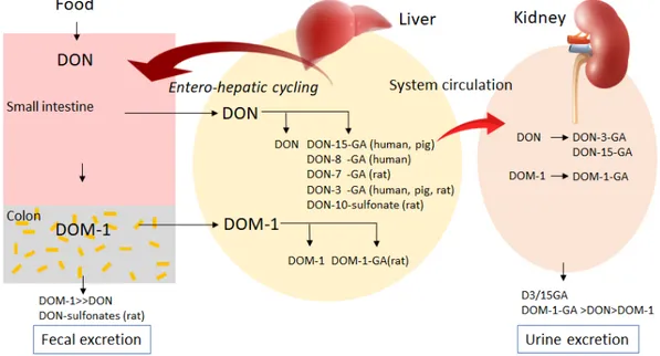
14 study, chronic exposure to DON (5.7 mg/kg diet, 5-8 weeks) showed a higher oral bioavailability with 89% than acute exposure (5.7 mg/kg diet, single bolus) with 54% in pig. DON was detected in serum samples as early as 15 min after chronic and actute exposure (Goyarts and Dänicke 2006). After feeding pigs with 4.2 mg DON/kg feed, the peak of DON concentration in serum was observed at 4.1 h. (Dänicke et al. 2004; EFSA 2017). DON can be transformed into less toxic metabolite DOM-1 by the microflora in colon of human and other monogastric animals, then excreted with feces. However, human microflora has a lower transformation efficiency than rodents, such as mouse and rat. Large amounts of ingested DON is conjugated to glucuronides or sulfonates in the liver and is excreted with urine or feces (Fig.3) (Payros et al. 2016; EFSA 2017). DON is rapidly excreted within 24 h and the metabolite DON-15-glucuronide is the main urinary biomarker in human (Vidal et al. 2018).
Figure 3 Absorption, metabolism and excretion of DON in human and monogastric animals.
DOM-1, de-epoxide metabolite of DON; DON-GA, glucuronide derivetives of DON; DOM-1-GA, glucuronide derivetives of DOM-1 (Payros et al. 2016).
2.3. Mode of action of DON
The most frequent reported modes of action on DON are ribotoxic stress and oxidative stress. It is known that via its epoxy group DON binds to the 60s ribosomal subunit and target on the peptidyl transferase. This interaction inhibits the initiation and elongation of peptide chains of protein synthesis resulting in an inhibition of RNA, DNA and protein synthesis, which called ‘ribotoxic stress’ (Waśkiewicz et al. 2014; EFSA 2017). Upon DON binding to ribosomes, the ribosome-associated double-stranded RNA-dependent protein kinase (PKR) and
15 the hematopoietic cell kinase (Hck) are activated within 5 minutes. Subsequently the downstream sensor of PKR eukaryotic initiation factor 2α (eIF2α) is phosphorylated, thereby inhibiting translation (Zhou et al. 2003b, 2014). On the other hand, DON is able to trigger ER stress by decreasing the glucose regulated protein 78 (GRP78) (Shi et al. 2009), followed by the activation of the PKR-like ER kinase (PERK), one of the sensors under ER stress, which induces phosphorylation of eIF2α leading to a global translation inhibition (Wek et al. 2006; Tsuru et al. 2016).
Following the ribotoxic stress, various downstream events occur. The DON-induced PKR and Hck activation triggers activation of the mitogen-activated protein kinases (MAPKs), including p38; extracellular signal regulated protein kinase 1 and 2 (ERK 1/2) and c-Jun N-terminal kinase 1 and 2 (JNK 1/2) (Pestka 2008). MAPKs activation induces cell apoptosis at high dose and mRNA expression of pro-inflammatory cytokine genes at low dose (Zhou et al. 2003a, 2005). In addition, recent reports demonstrated that DON induces rRNA cleavage by PKR/Hck-driven p-38 activation in the RAW264.7 cells, which could impair ribosome function and leading to cell apoptosis (Li and Pestka 2008; He et al. 2012).
Since the past several years, oxidative stress has also been considered as a potential toxicity mechanism of DON toxicity. Oxidative stress is triggered by over production of
reactive oxygen species (ROS) including hydroxyl radical (HO-), superoxide anion (O-2) and
perhydroxyl radical (HOO-) or reactive nitrogen species (RNS), such as nitric oxide (NO)
(Mishra et al. 2014). Several in vitro studies illustrated that DON exposure markedly induced DNA damage by inducing ROS production, inhibited activity of the antioxidant system including glutathione (GSH), superoxide dismutase (SOD), catalase (CAT), glutathione reductase (GR) and glutathione peroxidase (GPx), and increased lipid peroxidation leading to cell apoptosis (Zhang et al. 2009b; Dinu et al. 2011; Lautert et al. 2014). In vivo experiments using broiler chickens showed that DON (7.54±2.20 mg/kg feed) dietary exposure for 10 days induced mRNA expression of Xanthine oxidoreductase (XOR, associated with cellular defense enzyme systems) up-regulation, whereas that of hypoxia-inducible factor 1 (HIF1α) and Heme-oxigenase (HMOX) downregulation in the liver, both of which are sensitive markers of oxidative injury. In the jejunum the mRNA of HMOX and XOR were up-regulated, while in the ileum, only XOR was up-regulated (Osselaere et al. 2013). These data suggest that oxidative stress is an important mechanism of action of DON toxicity.
16 Toxicity of DON depends on its capacity to break through such biological barriers as intestinal or blood brain barriers and to impact the functions and viability of the cells forming such organic systems (Maresca 2013). DON is able to impair the intestinal, immune and nervous systems thus exerts its immunotoxicity, genotoxicity, reproductive and developmental toxicity and neurotoxicity (EFSA 2017). The symptoms of DON poisoning, in the case of acute exposure, include diarrhea, emesis, abdominal stress, headache and dizziness; while in the case of chronic dietary exposure, include anorexia, decrease of body weight gain and low nutrition efficiency (Pestka 2008).
2.4.1. Immunotoxicity
DON has either an immunostimulatory or immunosuppressive effect depending on the duration and doses of exposure (Pestka et al. 2004; Liao et al. 2018). For instance, an increased
IgA concentration in the serum and lower mRNA expression of IFN-γ and TGF-β in mesenteric
lymph node of pigs exposed to DON (2.2-2.5 mg / kg feed) was observed. Meanwhile, DON exposure altered vaccinal immune response in animals (Pinton et al. 2008). DON exposure (0.5-2mg/kg b.w.) for 14 days increased the population of CD8+ cells in the spleen and CD4+ T cells in mesenteric lymph node (MLN) of BALB/c mice. The expression of TLR4 in spleen and TLR2-4 in MLNs was decreased. In addition, the level of IgA and IgE in serum was reduced and increased respectively, while the mucosal IgA was significantly increased in the duodenum.
The level of inflammatory cytokines as IFN-γ, IL-2, IL-4 and IL-6 was increased in the serum
in DON-exposed animals compared to control animals (Islam et al. 2013). A higher concentration of DON at 11.4 mg / kg feed exposure for 30 days, induced significant lymphocyte apoptosis and immunosuppression in the lymphoid organs of rats (Bracarense et al. 2016). These results indicate that the immune system in different species is extremely sensitive to the toxicity of DON.
2.4.2. Genotoxicity
DON exerts its genotoxicity by inducing oxidative damage to DNA. Human peripheral blood lymphocytes were exposed to DON (6.25-500 ng/mL, which equal to 0.02-1.7 µM) for 6, 12 and 24h, lipid peroxidation, DNA damage and protein expression of DNA repair genes were observed (Yang et al. 2014). DON (12.5-50 µM) exposure for 8h was also reported to induce genotoxicity in intestinal cells IEC-6 indicated by phosphorylated histone H2AX, a marker of DNA double-strand breaks (Payros et al. 2017). In human liver cell line HepG2, DON ranging from 3.75 to 30 µM induced DNA strand breaks after 1h exposure via ROS production (Zhang et al. 2009b). These in vitro studies demonstrated that DON possesses
17 genotoxicity properties. Nevertheless, no correlation was found between DON and tumor or cancer thus it was classified in group 3 by the International Agency for Research on Cancer (IARC 1993).
2.4.3. Reproductive and developmental toxicity
Recent studies in different animal models have highlighted the link between DON exposure and the deficiency of maternal reproduction and fetus development (Yu et al. 2017). An in vitro study performed on pig oocytes and zygotes demonstrated that 1.88 µM of DON markedly inhibited the meiotic progression and maturation of pig oocytes and delayed the development of pig zygotes (Alm et al. 2002). DON (4.2 mg/ kg) exposure of sows at 63 and 70 days of gestation, is able to pass placental barrier entering into placenta, which may change the population of leukocytes and lymphocytes of fetus (Goyarts et al. 2010). Oral exposure of female rats to DON (2.5 and 5 mg/ kg b.w/d) by daily gavage at 6-19d of gestation, significantly decreased the body weight gain, crown-rump length, and vertebral ossification of fetus (Collins et al. 2006). These in vivo data indicate that DON pass through the placental barrier, subsequently induces restriction of fetus development.
2.4.4. Neurotoxicity
There is a potential link between DON exposure and neuronal diseases. It was reported that DON (1-100 µM) exposure for 48h altered the functions and decreased the viability of microglia and enterocytes, which are responsible for brain homeostasis (Razafimanjato et al. 2011). In addition, DON is capable of reducing the viability of brain endothelial cells and destroying the blood-brain barrier. DON at 1 and 10 µM significantly decreased the TEER and increased the paracellualer permeability of primary porcine brain capillary endothelial cells, with a decreased viability at 10 µM after 48h exposure (Behrens et al. 2015). This process is contribute to entrance of DON into the central nervous system and alteration of brain homeostasis, thereby inducing neurotoxicity. Low dose of DON (0, 0.125, 0.25, 0.5, 1 and 2 µg/mL) is also able to inhibite proliferation and induce apoptosis of PC12 cells by mitochondrial apoptosis pathway after 24h of exposure (Wang et al., 2016).
2.4.5. Intestinal toxicity
Due to its special function, intestine is the first target organ of many food contaminants after ingestion. Here, we are going to stress the general toxicity of DON on the intestine and its specific effect on intestinal barrier function.
18 The gut is the first barrier line to prevent organisms from the external harmful substances, such as antigens, toxins and other chemicals (Pinton and Oswald 2014; Payros et al. 2016). Thus, it is also the first target that can be exposed to a higher concentration of DON than other organs. On the other hand, DON is a protein synthesis inhibitor making it more toxic to such tissues with a rapid renewal as intestinal epithelium (Wells et al. 2017). The main morphological lesions in the intestine are multifocal atrophy, villi fusion and apical necrosis, which resulted in shorter and thicker villi (Bracarense et al. 2012; Pinton et al. 2012; Gerez et al. 2015). This suggests that DON induces an imbalance between proliferation and apoptosis of enterocytes. The impairment caused by DON on the intestinal morphology contributes to the decrease of nutrient absorption, consequently resulting in growth deficiency in animals (Alizadeh et al. 2015).
2.4.5.2. DON-induced modification of intestinal immunity
As described above, DON-induced inflammatory cytokine expression is mediated by MAPKs activation, which is associated with the phosphorylation of PKR and Hck. MAPKs plays a key role in signal transduction in the immune response. It has been proved that DON-induced p-38 and ERK activation modulate the gene expression involving in pro-inflammatory or inflammatory cytokines in neutrophils or macrophages (Chung et al. 2003; Islam et al. 2006; Gauthier et al. 2013). The intestine has its own immune network, which protects it from infections or other harmful injuries, thus inflammatory or immune response will occur in the intestine to counteract DON-induced damage.
Different intestinal cell lines and animal species have been used to investigate DON-induced inflammatory or immune response. In the human epithelial intestine 407 cells, DON (25, 500 and 1000ng/mL) exposure for 12h promoted IL-8 expression via ERK1/2 activation (Moon et al. 2007). The expression of IL-8 also observed in the DON-treated Caco-2 cells with
absent changes in the expression levels of IL-1β and TNF-α (Kadota et al. 2013). However, in
the DON-exposed porcine epithelial cells IPEC-1, the mRNA expression of IL-8, IL-1α, IL-1β
and TNF-α significantly increased (Cano et al. 2013). Several ex vivo studies constructed by jejunum explants demonstrated that the DON-mediated up-regulation of inflammatory genes was associated with MAPKs regulation (Cano et al. 2013; Alassane-Kpembi et al. 2017a; García et al. 2018). In vivo studies carried out in piglets further supported the obtained conclusions of in vitro and ex vivo investigations. Swine exposed to DON exhibited a potent inflammatory response in the intestine. The regulated dose of 0.9 mg DON/kg feed, induced
19
pigs (Alizadeh et al. 2015). Chronic ingestion of DON caused an up-regulation of IL-1β, IL-6,
MIP-1β and IL-2 mRNA in jejunum of piglets. In addition, the mRNA of IL-1β, TNF-α and
IL-6 was up-regulated in the ileum (Bracarense et al. 2012). 2.4.5.3. DON-induced oxidative stress on intestine
Oxidative stress is a non-specific response to cell injury caused by toxins or inflammation. The production of ROS can degrade the membrane phospholipids of cells or organelles, which is responsible for damage of intestinal integrity and increase of intestinal barrier permeability (Wu et al. 2014).
Several intestinal cell lines were used to test the capacity of DON to induce oxidative stress. Human HT-29 cells exposed to DON (250 and 500ng/mL) for 24h, exhibited a higher level of ROS and nitrite production compared to the non-treated cells. The higher production of ROS and nitrite was co-occurrence with NF-κB activation (Krishnaswamy et al. 2010). DON (1, 2.5 and 5μM) exposure of the non-tumorigenic rat intestinal epithelial cell line IEC-6, induced an evident increase of ROS generation, which could be exacerbated by nivalenol (Del
Regno et al. 2015). The application of NF-κB inhibitor showed that NF-κB activation was
involved in DON-mediated ROS release (Del Regno et al. 2015), which could explain the NF-κB activation in the previous study (Krishnaswamy et al. 2010). The two studies demonstrated
that NF-κB plays an important role in DON-mediated oxidative stress. In the latter study,
NF-κB activation mediated the increase of iNOS (inducible nitric oxide synthase) protein expression leading to the release of NO, which reacted with superoxide anion resulting in peroxynitrite formation. Peroxynitrite can nitrate tyrosine residues in proteins, an alternative to phosphorylation, thereby changing the structure and function of proteins, consequently altering the cytoskeletal organization and impairing cell signal transduction (Del Regno et al. 2015). All these reactions contribute to intestinal barrier dysfunction.
On the other hand, DON induced Nif2 activation, an oxidative stress sensor, increasing the expression of NQO1 and HO-1, which are the main products of Nif2 activation for protecting cells from oxidative stress injuries (Del Regno et al. 2015). However, another study performed on the human Caco-2 cell model lead to different result in terms of iNOS expression. DON (0.1 and 1μM) exposure for 3, 6 and 12h induced a time- and dose-dependent increase in iNOS mRNA expression, whereas the iNOS protein and the NO production were undetectable. These were caused by proteasome-dependent degradation of iNOS protein, in which the PKR, MAPKs (p-38 and ERK1/2) and NF-κB signal pathways were involved (Graziani et al. 2015). These contradictory conclusions suggest that DON could use different mechanisms to exert its
20 toxicity in different conditions depending on cell types or the doses and duration of exposure. The oxidative injury was also observed in DON-treated (0.9mg DON/kg feed treated for 10 days) piglets, with upregulated and downregulated mRNA expression of HMOX1, one of the most sensitive indicator of oxidative stress, in the colon and jejunum (Alizadeh et al. 2015). These data revealed that low dose of DON efficiently induces oxidative stress in the intestine and intestinal cells.
2.4.5.4. Effects of DON on the intestinal barrier function
The intestinal barrier is an epithelium monolayer with the tight junction proteins (TJs), adherens junction proteins (AJs) and desmosomes connecting the adjacent cells, which are responsible for sealing the intercellular space. The intestinal epithelium functions as a selective permeable barrier allowing the dietary nutrients, electrolytes and water absorption and against bacteria, virus and other toxic molecules entrance, thereby maintaining the intestinal homeostasis (Groschwitz and Hogan 2009). Due to their important role in maintaining the barrier function, TJs and AJs can be targets of the food contaminants, such as DON. DON is able to damage the intestinal barrier by disturbance and redistribution of TJs, AJs and the cytoskeleton structure (Pinton et al. 2012; Akbari et al. 2014).
A study carried out on Caco-2 cells demonstrated that the transepithelial electrical resistance (TEER) of the monolayer and the expression of TJs(claudin-1, -3 and -4) were dose-dependently decreased in cells exposed to DON (1.39, 4.17 and 12.5μM), whereas the expression level of occludin and ZO-1 remained unchanged (Akbari et al. 2014). Treated piglets with 3mg DON/kg feed for 35d, induced a significant decrease of the expression of AJ protein E-cadherin and TJ protein occludin in the ileum (Bracarense et al. 2012) Nevertheless, a later study showed that DON elevated occludin expression in the duodenum, jejunum and colon of DON-fed (0.9mg/kg feed) pigs (Alizadeh et al. 2015). This distinction may be caused by different dose and duration of exposure.
The underlying mechanisms of adverse effects on intestinal barrier function caused by DON have been investigated using various cell lines. The porcine intestinal epithelial cell line (IPEC-1) is highly sensitive to DON. DON (30 μM) significantly declined the expression of claudin-4 and the TEER of the monolayer, and raised the permeability to 4kD-dextrant after 48h exposure. When treated for 1h, an obvious augmentation of ERK phosphorylation was observed. Interestingly, when the cells were pretreated with ERK inhibitor U0126 for 2h before DON addition, the alteration of claudin-4 expression, TEER and permeability was restored. They therefore demonstrated that ERK activation mediated decrease of claudin-4 expression
21 (Pinton et al. 2010). This conclusion was supported by Lei et al. (Lei et al. 2014), who pointed out that ERK activation reduced the expression of TJ proteins in the LPS-exposed Caco-2 cells. However, this is contrast with another study executed on the Caco-2 cells, which showed that claudin-4 decrease was independent of ERK activation by DON and U0126 simultaneous exposure (De Walle et al. 2010). The different results may due to the routes of ERK inhibitor
U0126 application. A recent study has reported that DON (20 μM) induced intestinal barrier
dysfunction in IPEC-J2 cells by reduction of claudin-1 and claudin-3 expression. The decrease of claudin-3 not claudin-1 expression partially caused by ERK activation (Springler et al. 2016).
In contrast, Zhang et al. demonstrated that 20 μM DON exposure of IPEC-J2 caused a reduction
of TEER and expression of claudin-4, and promoted p-38 activation and inhibited ERK activation (Zhang et al. 2016). This clearly suggests that different concentrations of DON selectively affects specific TJs in different cell line, and the activation of MAPKs is not the only mechanism of intestinal barrier damage.
On the other hand, ERK activation was reported to increase the expression of ZO-1 and occludin and decrease the permeability to FITC-dextran in the intestinal epithelial cells IEC-6 (Yang et al. 2005). Basuroy demonstrated that ERK activation inhibited H2O2-induced redistribution of ZO-1 and occludin in Caco-2 cells thereby maintain the normal barrier function (Basuroy et al. 2006). An interesting study conducted by Aggarwal et al. revealed that ERK activation in under-differentiated Caco-2 cells promoted tight-junction disruption, whereas in the differentiated cells enhanced tight junction integrity (Aggarwal et al. 2011). The results indicate that the effects of ERK on intestinal tight junction integrity depends on differentiation state of cells.
It is notable that DON has different sensitive target TJ and AJ proteins in different cell lines. Thus, DON may impede intestinal barrier function via alternative mechanisms depending on cell types. Although these studies obtained different or even discrepant conclusions, it still can be seen that MAPKs/ERK plays an important role in DON-induced intestinal barrier dysfunction. Due to the role of ERK in regulation of intestinal barrier function is still controversial, further studies are needed to illustrate its mechanisms of toxic actions.
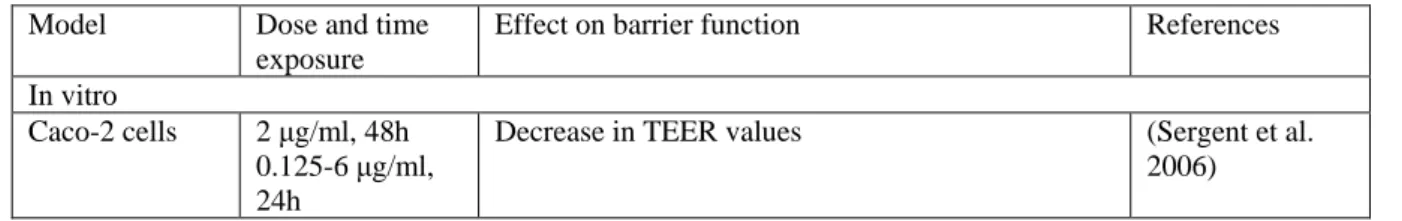
Table 3 Effect of DON on intestinal barrier function
Model Dose and time exposure
Effect on barrier function References In vitro
Caco-2 cells 2 μg/ml, 48h 0.125-6 μg/ml, 24h
Decrease in TEER values (Sergent et al. 2006)
22
Caco-2 cells 1.39–12.5 µM 24 h
Decrease in TEER values Decrease in horizontal impedance
Increase in permeability of LY and 4 kDa FITC-dextran
Increase in transcript level of claudin3, claudin4, occludin and ZO-1
Decrease in protein expression of claudin1, claudin3 and claudin4
Redistribution of claudin1, claudin3, claudin4, occludin and ZO-1.
(Akbari et al. 2014)
Caco-2 cells 0.16–16 µM 24 h
Decrease in TEER values
Increase in permeability of mannitol Increase in transcript level of claudin4 and occludin
Decrease in protein expression of claudin4
(De Walle et al. 2010)
Caco-2 cells 5–100 µM 48 h
Decrease in TEER values
Increase in permeability of 4 kDa FITC-dextran Increase in translocation of pathogenic Escherichia coli (strain 28C)
Decrease in protein expression of claudin4
(Pinton et al. 2009)
Caco-2 cells 1–100 µM 12 h
Decrease in TEER values
Increase in permeability of HRP and 4 kDa FITC-dextran
Increase in translocation of commensal Escherichia coli (strain k12)
(Maresca et al. 2008)
Caco-2 cells 0.37–1.5 µM 6–120 h
Decrease in horizontal impedance value of undifferentiated cells (Manda et al. 2015) Caco-2 cells T84 cells 0.16–0.67 µM 14 days
Decrease in TEER values Increase in permeability of LY
Kasuga et al. (1998) HT-29-D4 cells 0.001–100 µM
48 h
Decrease in TEER values (Maresca et al. 2002)
IPEC-1 cells 30 µM 48 h
Decrease in TEER values
Increase in permeability of 4 kDa FITC-dextran Decrease in protein expression of claudin3, claudin4 Redistribution of claudin4 (Pinton et al. 2010, 2012) IPEC-1 cells 5–50 µM 48 h
Decrease in TEER values
Increase in permeability of 4 kDa FITC-dextran Decrease in protein expression of claudin3 and claudin4 (Pinton et al. 2009) IPEC-1 cells IPEC-J2 cells 0.67–6.7 µM 48 h
Decrease in protein expression of ZO-1 Redistribution of ZO-1
(Diesing, et al., 2011 b) IPEC-J2 cells 6.74 µM
48 h
Decrease in TEER values
Decrease in protein expression of claudin3, occludin and ZO-1
Redistribution of ZO-1
(Gu et al. 2014)
IPEC-J2 cells 0.67–13.4 µM 24–72 h
Decrease in TEER values
Decrease in protein expression of claudin3 and ZO-1 Redistribution of claudin3 (Diesing, et al., 2011 a) IPEC-J2 cells 1.68–33.7 µM 72 h
Decrease in TEER values
Increase in permeability of doxycycline and paromomycin
(Goossens et al. 2012)
23
IPEC-J2 cells 0.33–3.3 µM 24 h
Increase in translocation of pathogenic Salmonella typhimurium (strain 112910a)
(Vandenbroucke et al. 2011) IPEC-J2 cells 4 µM
12 h
Decrease in TEER values
Increase in permeability of 4 kDa FITC-dextran Increase in translocation of commensal Escherichia coli (strain ATCC® 25922™)
Increase in transcript level of claudin1, claudin4, occludin and ZO-1
Decrease in protein expression of claudin3 and claudin4
(Ling et al. 2016)
IPEC-J2 cells 20 µM 72h
Decrease in protein expression of claudin1 and claudin4
(Springler et al. 2016)
IPEC-J2 cells 20 µM 1h
Decrease in TEER values
Decrease in protein expression of claudin4
(Zhang et al. 2016) IPEC-J2 cells 0.2 – 2 μg / mL
6, 12, 24h
Decrease in mRNA expression of ZO-1, occludin and claudin1 (Liao et al. 2017) Ex vivo Pig jejunal explants 5–50 µM 2 h
Increase in permeability of 4 kDa FITC-dextran (Pinton et al. 2009) Pig jejunal
explants
10 µM 4h
Decrease in E-cadherin expression (Basso et al. 2013) Pig jejunal
explants
10 µM 3h
Increase in permeability of fluorescein sodium salt (García et al. 2018) Pig jejunal
explants
10 µM 4h
Decrease in E-cadherin expression (Da Silva et al. 2019)
In vivo
Piglet 3 mg/kg feed 5 weeks
Decrease in protein expression of E-cadherin and occludin in ileum
(Bracarense et al. 2012) pig 2.85 mg/kg feed
5 weeks
Decrease in protein expression of claudin4 in jejunum
(Pinton et al. 2009) pig 3.5 mg/kg feed
6 weeks
Decrease in transcript level of claudin3, claudin 4 and occludin in ileum
(Lessard et al. 2015) pig 0.9 mg/kg feed
10 days
Increase in transcript level of CLDNs (cecum), OCLD (duodenum, ileum, cecum and colon) and ZOs (duodenum and colon)
Decrease in transcript level of claudin 4, occludin, ZO-1 and ZO-2 in jejunum
Increase in protein expression of occludin in duodenum, jejunum and colon
(Alizadeh et al. 2015)
Mouse 25 mg/kg bw 6 h
Increase in permeability of 4 kDa FITC-dextran Increase in transcript level of CLDN2, claudin 3 and claudin 4 in distal small intestine
Redistribution of claudin 1-3 in distal small intestine
(Akbari et al. 2014)
Mouse 5 mg/kg bw 24 h
Increase in transcript level of CLDN2 and claudin 3 in duodenum
Decrease in protein expression of claudin 3 in duodenum
(Bol-Schoenmakers et al. 2016, p.) Broiler chicken 7.5 mg/kg feed
3 weeks
Increase in transcript level of CLDN5 in jejunum and claudin 1, CLDN5, ZO-1 and ZO-2 in ileum
(Osselaere et al. 2013)
Broiler chicken 10 mg/kg feed 3 weeks
Decrease in mRNA level of jejunal occludin and claudin1 (Wu et al. 2018) Atlantic Salmon 5.5 mg/kg feed 8 weeks
Decrease in mRNA level of claudin25b, occludin and tricellulin in middle and distal intestine
(Moldal et al. 2018)
24
From the description above, it is not difficult to imagine that DON-induced
inflammatory injury, oxidative stress or other potential mechanisms of toxicity may also involve in DON-mediated damage on intestinal barrier function.
In the intestine of patients suffering inflammation bowel disease (IBD), Crohn’s disease or ulcerative colitis, the intestinal barrier is always defective. This aroused the interest to investigate the link between inflammation and intestinal barrier dysfunction. The interferon gamma (IFN-γ) was the first inflammatory cytokine that was observed to induce TJ disruption in human derived intestinal cells (Madara and Stafford 1989). Since then, researchers continue
to explore the effects of IFN-γ on the intestinal barrier and the underlying mechanisms. They
found that IFN-γ induced occludin endocytosis from the TJ complexes leading to TJ barrier
disruption. This process requires myosin light chain (MLC) phosphorylation and Rho kinase
activation. Both NF-κB and PI-3 kinase inhibition restored the adverse effects caused by
IFN-γ indicating that the two pathways are involved in IFN-IFN-γ induced increase of intestinal permeability and decrease of occludin expression (Al-Sadi et al. 2009).
In addition to IFN-γ, other cytokines’ effects on intestinal barrier were also investigated. The permeability of Caco-2 monolayer was evidently increased by a physiological
concentration of IL-1β via NF-κB pathway. This was further proved by NF-κB p65 depletion,
which completely abolished IL-1β induced increase of TJ permeability (Al-Sadi and Ma 2007).
Further, TNF-α was reported to promote opening of Caco-2 TJ barrier via MLCK expression
mediated by NF-κB activation (Ye et al. 2006). Later, TNF-α was proved to active MAPK/ERK
signaling pathway to promote transaction factor Elk-1binding to MLCK, leading to MLCK activation and expression. These reactions resulted in a decreasd TEER on Caco-2 monolayer and an increased intestinal permeability in mouse (Al-Sadi et al. 2013). Since NF-κB activation is the downstream event of MAPKs activation, it is reasonable to hypothesize a main pathway for TNF-α induced intestinal barrier disruption: TNF-α - MAPKs/ERK - NF-κB – MLCK - TJ disruption. These data indicate that DON induced inflammation in intestine is contribute to barrier dysfunction.
Oxidative stress is also able to trigger intestinal injury and barrier disruption. In the intestinal cells, oxidative stress caused an increase of permeability of Caco-2 monolayer by reorganization of occludin-ZO-1 and E-cadherin-β-catenin complexes from the intercellular junctions and dissociation occludin from ZO-1 (Rao et al. 2002; Basuroy et al. 2006).
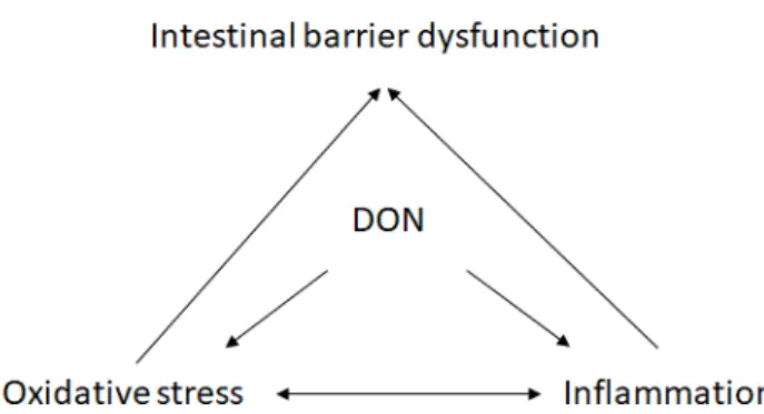
25 In fact, inflammation and oxidative stress can affect one another (Fig.4). Oxidative stress-mediated tissue damage could induce overexpression of inflammatory cytokines in human intestine who suffered intestinal inflammation disease (Alzoghaibi 2013; Moret et al.
2014; Oyinloye et al. 2015; Vitali et al. 2015). It is reported that hydrogen peroxide H2O2
upregulated the mRNA expression of 8 and TNF-α and increased protein expression of IL-8 in IPEC-J2 cells (Paszti-Gere et al. 2012). NO, one of the reactive nitrogen species, could induce inflammatory cytokines production by activation of NF-κB or AP-1(Korhonen et al. 2005). In turn, inflammatory cytokines also mediate ROS or NOS production in the inflammatory or epithelial cells (Federico et al. 2007).
Figure 4 The relationship between DON-induced inflammation, oxidative stress and intestinal
barrier dysfunction
2.4.5.5. Effect of DON on the intestinal microbiota
Gut microbiota have a marked influence on the the physiology functions of hosts and dysbiosis induces malnutrition, systematic diseases and chronic inflammatory diseases (Guinane and Cotter 2013; Thursby and Juge 2017). The effects of DON on the intestinal microbiota have been investigated in various animal models, such as rodents, pigs and chickens. Germ-free rats inoculated with human fecal flora were daily exposed to 100μg DON/kg b.w. for four weeks. It was found that the abundance of Bacteroides/Prevotella was significantly increased, while the concentration of Escherichia coli was decreased (Saint-Cyr et al. 2013). DON (10 μg/kg b.w./day) exposure of mice for 9 months caused an elevation of Proteobacteria and a reduction of Bacteroidetes (Vignal et al. 2018). DON dietary challenge altered the cecal microbiota of broiler chickens with a decrease of Proteobacteria and an increase of Firmicutes (Lucke et al. 2018). In pigs, it was reported that DON exposure increased the number of aerobic mesophilic bacteria and did not affect the abundance of anaerobic sulfite-reducing bacteria at 28d post exposure (Waché et al. 2009). It is worth noting that the decrease of the number of
26 Bacteroidetes and the increase of Proteobacteria was frequently observed in the IBD and Crohn’s diseases, where are involved in intestinal inflammation diseases. The study of Wang et al. demonstrated that intestinal dysbacteriosis induced losses of barrier function and increased bacteria translocation, which can induce systematic inflammation (Wang et al., 2014). It suggests that DON-induced alteration of microbiota causes breaking of intestinal barrier that favor bacteria translocation leading to inflammation in intestine, which in turn, having a negative effect on the intestine defection of barrier function.
3. Cadmium (Cd)
Cadmium (Cd) is a toxic and carcinogenetic heavy metal widely distributed in the environment. Both natural (volcanic eruption, weathering and erosion) and anthropogenic activities contribute to Cd contamination and Cd-related health problems (EFSA 2009; ATSDR 2012).
3.1. Occurrence, exposure and regulation of Cd in food and feed
Cd naturally exists in the earth’s crust as a consequence of volcano eruption, minerals and rock exfoliation. Industrial emission is the main source of Cd pollution. Cd from nature and industries can directly enter into air, water and soil and absorbed by the roots of cereals or vegetables, consequently arriving and accumulating in the edible part (EFSA 2009; ATSDR 2012). A survey performed between 2003 and 2007 in 20 European countries on 137, 200 samples of raw cereals, vegetables and meat indicated that 66% were contaminated with Cd. While 5% of them even exceed the maximum level of 0.2 mg/kg for bran, germ, wheat and rice and of 0.1 mg/kg for other raw cereals specified by European Commission (European Union
2006a; EFSA 2009). In the 73rd report of JECFA, all the 1503 wheat samples tested from 19
European countries and 11 other countries worldwide were contaminated with Cd (JECFA 2011). The smokers and the occupational workers are exposed to Cd via smoking and inhalation during work process (ATSDR 2012). Cd-contaminated food and water are the primary sources for the general population (EFSA 2009). Grain and cereal products, as well as fish and offal were the major contributor for human Cd dietary exposure (Fig.5) (Filipic et al. 2006; ATSDR 2012). A tolerable weekly intake (TWI) for Cd of 2.5μg/ kg b.w. was established (EFSA 2009, 2011). The mean dietary exposure to Cd for adults across European countries was close to or slightly exceeding this TWI, and subgroups, such as vegetarians, children, smokers exceed the TWI by about 2-fold (EFSA 2009).
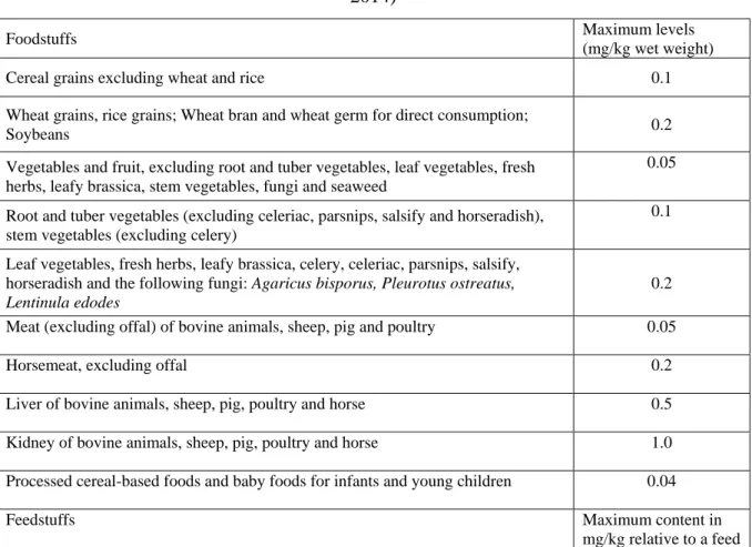
27 In Europe, the guidance concentration in cereal grains used for food ranges from 0.1 to 0.2 mg / kg depending on the grain species, no more than 0.5 mg / kg in the complete feed, and 0.005 mg / L in water (Table 2) (European Union 2013, 2014).
Figure 5 Sources of human exposure to cadmium (EFSA 2009)
Table 2. Recommendation for Cd in foodstuffs/feedstuffs in Europe (European Union 2013,
2014)
Foodstuffs Maximum levels
(mg/kg wet weight)
Cereal grains excluding wheat and rice 0.1
Wheat grains, rice grains; Wheat bran and wheat germ for direct consumption;
Soybeans 0.2
Vegetables and fruit, excluding root and tuber vegetables, leaf vegetables, fresh herbs, leafy brassica, stem vegetables, fungi and seaweed
0.05 Root and tuber vegetables (excluding celeriac, parsnips, salsify and horseradish),
stem vegetables (excluding celery)
0.1 Leaf vegetables, fresh herbs, leafy brassica, celery, celeriac, parsnips, salsify,
horseradish and the following fungi: Agaricus bisporus, Pleurotus ostreatus, Lentinula edodes
0.2 Meat (excluding offal) of bovine animals, sheep, pig and poultry 0.05
Horsemeat, excluding offal 0.2
Liver of bovine animals, sheep, pig, poultry and horse 0.5 Kidney of bovine animals, sheep, pig, poultry and horse 1.0 Processed cereal-based foods and baby foods for infants and young children 0.04
Feedstuffs Maximum content in
28
with a moisture content of 12 %
Feed materials of vegetable origin 1
Feed materials of animal origin 2
Complete feed with the exception of: 0.5
- complete feed for cattle (except calves), sheep (except lambs), goats (except kids) and fish;
1
- complete feed for pet animals 2
3.2. Toxicokinetics of Cd
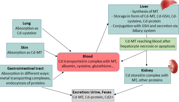
Since Cd is not an essential element for human or animals, a specific transporter for Cd does not exist. Some transport proteins for essential metals, such as iron, zinc and calcium are reported to be responsible for Cd absorption. Divalent metal transpoter 1 (DMT 1) was highly induced by Fe deficient diet in rat duodenum, which was associated with the higher tissue Cd concentration (Park et al. 2002; Ryu et al. 2004). DMT1 knockdown Caco-2 cells displayed a 50% reduction of Cd uptake (Bannon et al. 2003). Knockdown of Zinc transporters Zrt/Irt-related protein 8 (ZIP8) and ZIP14 significantly decreased Cd uptake in mouse kidney proximal tubule cells (Fujishiro et al. 2012). Calbindin D9k, the major calbindin isoform in the enterocyte is also considersred as a candidate transporter for Cd. These transporters are mainly expressed in duodenum and jejunum where most Cd is absorbed (Vesey 2010). The absorption rate for Cd through gastrointestinal tract was estimated between 3% and 7% in adult human (Vesey 2010). The ingested Cd entries into the blood and binds to proteins such as albumin and metallothioneins (MTs). Then, Cd firstly arrives in the liver where it induces synthesis of liver MTs. The Cd-MT complexes are released to blood again after hepatocytes necrosis or apoptosis. Meanwhile, some parts of these complexes may be excreted with bile and reenter the small intestine through the entro-hepatic cycle. The Cd-MT complexes released by hepatocytes then reach the kidney via blood circulation. These complexes are filtrated in the glomerulus and reabsorbed in the proximal tubules. It then remains in tubule cells for a long time (Godt et al. 2006). Cd was mainly excreted with urine and feces even though the excretion of Cd is very scarce (Fig.6).
29
Figure 6 Metabolism, storage and excretion pf Cd in human body (Godt et al. 2006)
3.3. Mode of action of Cd
Oxidative stress is considered as the effective molecular basis of Cd toxic actions. Cd exposure induces a significant increase of ROS production in vivo (rat) and in vitro (primary rat hepatocytes), which contributes to hepatocyte apoptosis (Liu et al. 2011; Wang et al. 2014b). In fact, Cd can neither participate directly in redox reaction nor generate ROS, as it is a redox-stable metal. However, Cd is able to substitute with copper, zinc and iron in various cytoplasmatic and membrane proteins, thereby increasing the components of free redox-active metal that can induce oxidative stress (Dorta et al. 2003; Liu et al. 2009). These metals induce ROS generation through Fenton-type chemical reaction (Desurmont 1983; Lee et al. 2012). Although Cd is redox-stable, the production of hydroxyl radical has an extremely strong oxidizing ability, which is highly toxic to human health.
metal n+ + H2O2 metal n+1 + -OH + OH
-On the other hand, Cd interferes with antioxidant enzymes activity, such as superoxide dismutase (SOD), catalase (CAT), glutathione reductase (GR) and glutathione peroxidase (GPx) through binding their thiol groups (Filipic et al. 2006; El-Boshy et al. 2015). Oral exposure to Cd (40mg/L) for 30 days decreased the content of GSH, SOD, CAT and GPx in serum (El-Boshy et al. 2015), meanwhile intraperitoneal injection of Cd (6.5mg Cd/kg bw / d) for 5 days markedly reduced the activity of these antioxidant enzymes in hepatic and renal tissues (Dkhil
30 et al. 2014). These studies suggest that Cd exposure through different routes induces oxidative stress by inducing ROS production in an indirect way and compromises the antioxidant system by directly binding to their thiol groups.
Cd-induced ROS generation is responsible for the downstream events as DNA damage, apoptosis, inflammation and genomic instability.
Heavy metals interact directly with DNA or nuclear protein, causing DNA damage or conformational changes that may lead to carcinogenesis (Wang and Shi 2001; Beyersmann and Hartwig 2008; Qin et al. 2016). However, Cd did not induce DNA breaks on the pure DNA extracted from liver cells of mice, suggesting Cd may not be able to directly attack on DNA (Valverde et al. 2001), while in the intact cells exposed to Cd, the free radicals increased significantly (Valverde et al. 2001). In addition, ROS production especially the highly reactive
hydroxyl radical (OH-) can directly attack on DNA bases inducing DNA damage (Dizdaroglu
and Jaruga 2012; Cadet and Wagner 2013), as well as hydrogen peroxide H2O2 (Gafter-Gvili et al. 2013). These data suggest that ROS plays an important role in Cd-induced DNA damage.
Cd not only induces DNA damage but also inhibits the DNA repair systems, including excision repair (base excision repair (BER) and nucleotide excision repair (NER)), mismatch repair (MMR) and recombination repair (homologous recombination (HR) and non-homologous end-joining (NHEJ)), the last system is involved in DNA double-strand breaks (DSBs) (Giaginis et al. 2006; Lei et al. 2015; Li et al. 2015). Together with inhibition of DNA repair, DNA damage is associated with genotoxicity, genomic instability and carcinogenesis of Cd contamination. Since the oxidative stress and the inflammation caused by Cd were repeatedly observed in literature (Liu et al. 2015; Ghosh and N 2018), it promotes to speculate that there may be a link between the two processes. In the human umbilical vein endothelial cells (HUVECs), Cd increased the expression and secretion of inflammatory cytokine TNF-α via p38/ MAPK activation (Dong et al. 2014). These results indicate that Cd-induced inflammation isregulated by oxidative stress-mediated MAPKs activation (Cd-ROS-MAPKs/p38-inflammation).
Many reports demonstrated that acute Cd-induced apoptosis is p53- and MAPKs-dependent (Yu et al. 2011). Cadmium exposure upregulated p53 gene expression and increased p53 protein level in vitro, leading to apoptosis(Achanzar et al. 2000; Al-Assaf et al. 2013; Lee et al. 2016). In addition siRNA-induced p53 silencing effectively inhibited cell apoptosis (Aimola et al. 2012), indicating that p53 plays a critical role in Cd-induced cell apoptosis. Other hypothesis proposed that Cd induces apoptosis via MAPK/JNK-mediated mitochondrial pathway, as the JNK inhibitor or JNK-specific siRNA interference protected cells from
Cd-31 induced apoptosis (Chang et al. 2013; Jiang et al. 2014; Yuan et al. 2015). This reveals that MAPK/JNK pathway is involved in Cd-mediated cell apoptosis. Nevertheless, JNK inhibition did not decrease the ROS generation, while the antioxidant N-acetylcysteine (NAC) restored induced events, including JNK activation (Chang et al. 2013). This clearly implies that Cd-induced events associated to apoptosis are mediated by Cd-generated ROS. These data guide to speculate that p53-and MAPKs-dependent ways of apoptosis may be in the same signaling pathway (Cd-ROS-MAPKs/JNK-p53-Bax-Mitochodria-caspases-apoptosis).
Apoptosis is an important process in organisms to eliminate transformed and mutated cells from the body. Thus, cancer cells have to develop various efficient mechanisms against apoptosis to keep survival. Chronic low-dose exposure of Cd to rat liver cells and human prostate cells can induce cell transformation with an apoptotic resistance property, which is due to the inhibition of JNK activation associated with MT or Bcl-2 overexpression (Qu et al. 2006, 2007). The abnormal proliferation of these DNA-damaged cells could elicit malignant transformation that finally develop to cancer.
3.4. Toxicity of Cd
Cd has various target organs, such as kidney, liver, lung, reproductive and nervous system. In these organs Cd is able to induce genotoxicity, cancer, immunotoxicity, neurotoxicity, reproductive and developmental toxicity (ATSDR 2012; Rani et al. 2014).
3.4.1. Immunotoxicity
There is still controversy on pro- or anti-inflammatory properties of Cd. Some studies demonstrated that Cd stimulate immune system and promote inflammatory reactions; while other studies suggested that Cd has non-inflammatory or anti-inflammatory properties (Olszowski et al. 2012). In vivo, Cd (40 mg/L drinking water) treated for 30 days evidently
increased the secretion of inflammatory cytokines IL-1β, TNF α, IL-6 and IL-10 and decreased
IFN-γ and the content of lymphocytes in serum of rats (El-Boshy et al. 2015). Similarly, the
gene expression of TNF-α, IL-6 and IL-1β in larval zebrafish was up-regulated by Cd (1, 3, 10
µM) after 96h exposure (Jin et al., 2015). While, in adult zebrafish, the release of TNF-α was
increased in the liver, brain and ovary, the mRNA level of NF-κB was upregulated in the liver
and ovary at 24h exposure of Cd (1 mg / L). By contrast, in Cd (3 µM) treated rat primary lung
cells and rat alveolar macrophages, the mRNA expression of TNF-α and IL-1β was reduced
after 20h exposure, while their protein levels were not affected. The expression and release of IL-6 in alveolar macrophages was downregulated and unaltered (Låg et al. 2010). These data
32 revealed both positive and negative effects of cadmium on immune modulation. To improve understanding of the immunotoxicity of Cd, more studies in mammals should be done.
3.4.2. Genotoxicity
Cd induces genotoxicity characterized by DNA damage through induction of ROS production, resulting in cell apoptosis or malignant transformation that may develop to cancer
(Achanzar et al. 2001; Chang et al. 2013). In human bronchial epithelial cells, 5 μM of Cd
exposed for several weeks triggered genomic instability, DNA breaks and down-regulation of DNA repair genes (hMSH2, ERCC1, XRCC1, and hOGG1). Meanwhile, the mutation of exons on these genes was also observed in tumorgenetic cells of nude mice (Zhou et al. 2013).
Occupational exposure to Cd (blood concentration 5.4-30.8 µg/L) significantly increased the
frequency of sister chromatid exchanges (SCEs) as well as the leukocytes with DNA fragmentation in lymphocytes compared with controls (Palus et al. 2003). This study is consistent with a previous study performed on mice, which demonstrated that single intravenous exposure of Cd 5.7 and 7.6 mg /kg body weight, evidently increased the percentage of polychromatic erythrocytes with micronuclei, the frequency of SCEs and the portion of chromosomal aberrations in mouse bone marrow (Fahmy and Aly 2000). These gene toxic effects caused by Cd are necessary prerequisites for cadmium-induced cancer.
3.4.3. Reproductive and developmental toxicity
Reproductive system is a key site of Cd toxicity both in male and female. Cd is able to inhibit oocyte development in ovary and decrease sperm motility. In addition, it induces implantation delay, consequently inhibiting embryo development (Thompson and Bannigan
2008; Zhao et al. 2017). A study performed with bovine oocytes proved that Cd (2 and 20 μM)
exposure for 24h, efficiently decreased the oocytes maturation, as well as the post-fertilization cleavage rate in zygotes and blastocyst development (Akar et al. 2018). Another research illustrated that the motility of human and mouse sperm were significantly reduced by Cd (2.5 – 10 μg/ml) exposure for 6-24h in a dose- and time-dependent manner. Exposure to these doses of Cd for 30 min did not alter sperm motility but significantly decreased the fertilization rate of sperm in vitro. Besides, exposure to low dose of Cd at 0.625 and 1.25 μg/ml for 12-84h dramatically declined the blastocyst formation rate of embryo (Zhao et al. 2017). Cd is also reported to pass through placental barrier and accumulate in embryo, resulting in degeneration and decompaction in blastocysts or early pregnancy loss (Thompson and Bannigan 2008; Gundacker and Hengstschläger 2012).