Open Archive TOULOUSE Archive Ouverte (OATAO)
OATAO is an open access repository that collects the work of Toulouse researchers and
makes it freely available over the web where possible.
This is an author-deposited version published in :
http://oatao.univ-toulouse.fr/
Eprints ID : 18548
To link to this article : DOI:10.1007/s00330-016-4413-4
URL :
http://dx.doi.org/10.1007/s00330-016-4413-4
To cite this version : Faruch-Bilfeld, Marie and Lapegue, Franck and
Chiavassa-Gandois, Hélène and Bayol, Marie - Aurélie and
Bonnevialle, Nicolas and Sans, Nicolas Ultrasound of the
coracoclavicular ligaments in the acute phase of an acromioclavicular
disjonction: Comparison of radiographic, ultrasound and MRI
findings. European Radiology, vol. 27 (n°2). pp. 483-490. ISSN
0938-7994
Any correspondence concerning this service should be sent to the repository
administrator:
staff-oatao@listes-diff.inp-toulouse.fr
Ultrasound
of the coracoclavicular ligaments in the acute phase
of
an acromioclavicular disjonction: Comparison of radiographic,
ultrasound
and MRI findings
Marie Faruch Bilfeld1 & Franck Lapègue1 & Hélène Chiavassa Gandois1 &
Marie Aurélie Bayol1 & Nicolas Bonnevialle2 & Nicolas Sans1
Abstract
Objectives Acromioclavicular joint injuries are typically diag-nosed by clinical and radiographic assessment with the Rockwood classification, which is crucial for treatment plan-ning. The purpose of this study was to describe how the ultra-sound findings of acromioclavicular joint injury compare with radiography and MRI findings.
Methods Forty-seven patients with suspected unilateral acromioclavicular joint injury after acute trauma were enrolled in this prospective study. All patients underwent digital radi-ography, ultrasound and 3T MRI. A modified Rockwood clas-sification was used to evaluate the coracoclavicular ligaments. The classifications of acromioclavicular joint injuries diag-nosed with radiography, ultrasound and MRI were compared. MRI was used as the gold standard.
Results The agreement between the ultrasound and MRI find-ings was very good, with a correlation coefficient of 0.83 (95 % CI: 0.72–0.90; p < 0.0001). Ultrasound detected coracoclavicular ligament injuries with a sensitivity of 88.9 %, specificity of 90.0 %, positive predictive value of 92.3 % and negative predictive value of 85.7 %. The agreement be-tween the ultrasound and radiography findings was poor, with a correlation coefficient of 0.69 (95 % CI: 0.51–0.82; p < 0.0001).
Conclusion Ultrasound is an effective examination for the diagnostic work-up of lesions of the coracoclavicular liga-ments in the acute phase of an acromioclavicular injury. Key Points
• Ultrasound is appropriate for acute acromioclavicular trau-ma due to its accessibility.
• Ultrasound contributes to the diagnostic work-up of acute lesions of the coracoclavicular ligaments.
• Ultrasound is appropriate in patients likely to benefit from surgical treatment.
• Ultrasound could be a supplement to standard radiography in acute acromioclavicular trauma.
Keywords Acromioclavicular joint . Rookwood
classification . Coracoclavicular ligament . Ultrasound . MRI
Acromioclavicular joint injuries represent about 10 % of shoulder injuries and are more frequently encountered in con-tact sports [1]. The most common aetiology is a fall with direct impact to the shoulder, with the arm adducted. Depending on the severity of the trauma, the coracoclavicular ligaments can be stretched, partially torn or completely torn. Rockwood's scoring system is currently the most widely used and reliable means of classifying such injuries; it describes six grades of increasing injury severity [2,3].
The coracoclavicular ligaments (trapezoid and conoid) are known as the‘suspensory ligaments of the shoulder’; they are responsible for acromioclavicular stability in the vertical and horizontal planes. Several studies have demonstrated the ad-vantages of MRI for the diagnosis of acromioclavicular inju-ries [4,5]. Unlike standard radiographs, which analyse the dis-placement of bony structures, MRI allows direct analysis of the soft tissues that stabilise this joint, namely the acromioclavicular ligaments.
* Marie Faruch Bilfeld mariefaruch@hotmail.com
1
Service de Radiologie, CHU Toulouse-Purpan, place du docteur Baylac, 31059 Toulouse Cedex 9, France
2 Service d’Orthopédie, CHU Toulouse-Purpan, place du docteur
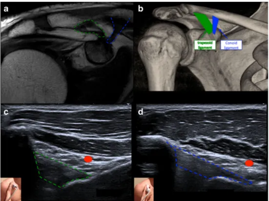
Very few published studies have evaluated the contribution of ultrasound to the diagnosis of acromioclavicular joint inju-ries. The earliest work on ultrasound-based evaluation of acromioclavicular joint injuries looked at acromioclavicular and coracoclavicular diastasis, and found good correlation of the ultrasound and radiology findings [6–8]. Peetrons demon-strated the advantage of ultrasound with dynamic manoeuvres for the diagnosis of low-grade sprains, differentiating between Rockwood grade 1 and grade 2 injuries [9]. Heers showed how ultrasound was a useful addition to standard radiography for the diagnosis of lesions of the trapezodeltoid fascia [10]. The coracoclavicular ligaments can also be visualised with ultrasound. They are located just beneath the acromial vascu-lar pedicle running along the deep aspect of the deltoid mus-cle, which serves as a landmark (Fig.1) [11].
It is essential that these acromioclavicular joint inju-ries be treated, otherwise the patient may be left with d i s a b l i n g r e s i d u a l p a i n a n d a n u n s i g h t l y acromioclavicular deformity. The treatment of these le-sions depends on the extent of the instability, which is usually assessed clinically and with radiography [12]. The most widely used classification system for acromioclavicular injuries is the Rockwood classification [2]. Currently, surgical treatment is reserved for grade 4, 5 and 6 lesions, for which numerous techniques have been described [13]. Grades 1 and 2 are treated conser-vatively initially [14]. The management of grade 3 le-sions is the subject of debate [15]. They are evaluated on a case-by-case basis, taking into account the patient’s
age and their sports activities. Since the Rockwood clas-sification is focussed on clavicle displacement, it only provides an indirect assessment of ligament damage. Ultrasound imaging provides a direct view of the coracoclavicular ligaments. In cases of Rockwood grade 3 injury where surgical treatment may be needed, diag-nostic ultrasound could contribute information that will help guide the treatment decision [3].
The aim of this study was to assess the value of ultrasound in the diagnosis of coracoclavicular ligament lesions in the acute phase of acromioclavicular joint injury.
Materials and method
Between January 2013 and July 2014, 47 patients presenting with an acromioclavicular injury at the emergency room of our hospital were prospectively enrolled in the study. The diagnosis of acromioclavicular joint injury was made using the clinical criteria of a suggestive injury mechanism com-bined with deformity and pain upon palpation of the acromioclavicular joint. The diagnosis was made by an emer-gency physician or orthopaedic surgeon. Oral informed con-sent was obtained from each patient. The hospital’s institution-al review board approved the study.
The following diagnostic imaging protocol was used for each patient. On the day of the injury, standard radio-graphs were taken comprising one true anteroposterior view with an ascending 15° beam. The week following
Fig. 1 The coracoclavicular ligaments. a)MRI T1 coronal view showing the
coracoclavicular ligaments: trapezoid ligament outlined in green and conoid ligament outlined in blue. b)VRT CT reconstruction with a schematic representation of the
coracoclavicular ligaments. c)Ultrasound appearance of the coracoclavicular ligaments. Longitudinal view of the trapezoid ligament. Red dot shows the acromial vascular pedicle. d)Ultrasound appearance of the coracoclavicular ligaments. Longitudinal view of the conoid ligament
the injury, the patient was seen again for MRI and ultra-sound. These two examinations were performed in a blinded manner by two senior radiologists with 8 and 12 years’ experience, respectively: one radiologist interpreted the MRI and the other performed the ultrasound without having knowledge of the clinical diagnosis or radiographic appearance. These examinations could not be performed on the same day as the patient was admitted to the emer-gency ward because the MRI unit was not always available.
The MRI was performed with a 3T unit (Philips Achieva) according to the following protocol:
& Coronal T1-weighted (TE = 15 ms, TR = 640 ms, FOV = 100, slice thickness 3 mm) in the plane of the acromioclavicular joint, parallel to a line drawn from the coracoid process to the lesser tuberosity. Imaging was per-formed with the patient’s arm in neutral position [4]. & Sagittal T1, axial, coronal and sagittal in proton density
with fat saturation (TE = 15 ms, TR = 1939 ms, FOV = 100, slice thickness 3 mm).
For the ultrasound examination (Aplio 400 Toshiba, 14–18 MHz probe), the patient was seated facing the operator, with their arm hanging down alongside their body. The coracoclavicular ligaments are challenging to analyse because of their deep position. They are located immediately below the acromial neurovascular bundle, which courses on the deep aspect of the deltoid, serving as a landmark. These ligaments are taut during scapular retraction (hand flat on buttocks with elbow pointing back), making them easier to discern. Since this position cannot be achieved by a patient with an acute acromioclavicular injury, we decided to evaluate them in neutral position, with the arm hanging down. The ultrasound probe was placed on the coracoid process along the axial plane, and then rotated toward the clav-icle to view the major axis of the trapezoid ligament. The probe was then translated slightly medially and ro-tated clockwise 20–30° to view the conoid ligament. The view was parallel to the major axis of the ligament. Both ligaments were scanned longitudinally (Fig. 1).
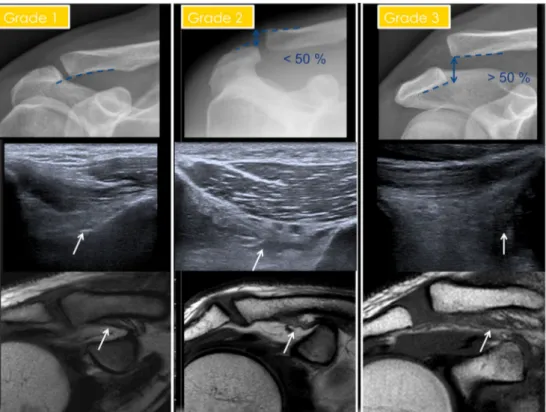
We analysed and scored the following elements using a modified Rockwood classification [2] (Fig. 2). Since the vertical stability of the acromioclavicular joint is ensured by the coracoclavicular ligaments, we specifi-cally assessed the position of the clavicle in the frontal plane on radiographs, as this provides indirect evidence of the integrity of the coracoclavicular ligaments: & grade 1: no displacement
& grade 2: displacement of < 50 % of the height of the acromioclavicular joint
& grade 3: displacement of > 50 % of the height of the acromioclavicular joint
In Rockwood grade 4, 5 and 6 injuries, the trapezius and deltoid muscles are damaged, along with the coracoclavicular ligaments. This results in more than 50 % clavicle displacement. In grade 4 injuries, the clavicle is displaced posteriorly into trapezius; in grade 5 injuries, the clavicle is displaced more than 100 %; in grade 6 injuries, the clavicle is displaced inferiorly and anteriorly. Since the goal of our study was to use ultra-sound to evaluate the coracoclavicular ligaments, we used a simplified three-level classification system to de-scribe the integrity of the coracoclavicular ligaments. All radiological grade 4, 5 and 6 injuries were consid-ered as grade 3 injuries in our modified classification, as the clavicle had moved at least 50 % of the height of the acromioclavicular joint.
Using the MRI and ultrasound images, we classified the injuries into three grades of increasing severity to analyse the integrity of the coracoclavicular ligaments [16]. If the score differed between the conoid and trap-ezoid ligaments, the most severe grade was retained. & Grade 1: normal coracoclavicular ligaments.
& Grade 2: distended coracoclavicular ligaments. Ligaments are enlarged with loss of echogenic appearance on ultra-sound, diffuse abnormal changes in signal intensity on MRI with ligament tissue swelling.
& Grade 3: ruptured coracoclavicular ligaments. Normal lig-ament anatomy is disrupted. Liglig-ament stump located at the bone insertion.
MRI was used as the gold standard for this study; it has been shown to be accurate when describing lesions of the coracoclavicular ligaments [4,5,16].
Statistical analysis
We performed the statistical analysis using standard sta-tistical software (Medcalc®). The percentage of injuries of each grade identified by radiography, ultrasound and MRI was calculated. Using MRI as the gold standard for injury detection, we then calculated the sensitivity, specificity, positive predictive value and negative predic-tive value of ultrasound. We compared the results of the MRI analysis with those of the ultrasound analysis using Spearman’s coefficient of rank correlation (rho) and 95 % confidence intervals. We also compared the results of the radiography analysis with those of the ultrasound analysis using Spearman’s coefficient of rank correlation (rho) and 95 % confidence intervals.
Results
The study included 47 patients (three women and 44 men) with a mean age of 36 years (22–60 years). Thirty-five of the 47 patients (74.5 %) had right-sided injuries and 12 (25.5 %) had left-sided injuries.
Analysis of radiographs using the modified Rockwood classification
The radiographic analysis found 16 (34 %) patients in grade 1, 22 (47 %) patients in grade 2 and nine (19 %) patients in grade 3 (Table1).
Analysis of coracoclavicular ligaments on ultrasound images using our classification
The ultrasound analysis found 15 (32 %) patients in grade 1, 6 (13 %) patients in grade 2 and 26 (55 %) patients in grade 3 (Table1).
Analysis of MRI images using an MRI-specific version of the Rockwood classification [16].
The MRI analysis found 16 (34 %) patients in grade 1, four (8.5 %) patients in grade 2 and 27 (57.5 %) patients in grade 3 (Table1).
Comparison of the scores obtained with MRI and ultrasound
Of the 47 patients in our study, ultrasound led to correct clas-sification of the coracoclavicular ligament injuries in 41 cases with a Spearman rho of 0.83 (95 % CI: 0.725–0.907, p < 0.0001). Of the six cases incorrectly classified with ultra-sound, ultrasound overestimated the grade three times (i.e. a higher grade was found on ultrasound than MRI) and underestimated it three times (i.e. a lower grade was found on ultrasound than MRI). Ultrasound detected injuries of the coracoclavicular ligaments with a sensitivity of 88.9 %, spec-ificity of 90.0 %, positive predictive value of 92.3 % and negative predictive value of 85.7 %.
Comparison of ultrasound and radiographic findings Comparison of the ultrasound and radiography scores re-vealed agreement in 25 cases, with a Spearman rho of 0.697 ( 9 5 % C I : 0 . 5 1–0.82, p < 0.0001). Radiography underestimated the grade in 20 cases (17 cases of grade 2 on radiographs were classified as grade 3 with ultrasound) and
Fig. 2 Representative images for radiography, ultrasound and MRI scoring. Grade 1: Normal radiograph. Trapezoid ligament (arrow) appears normal on ultrasound and MRI. Grade 2: Radiograph with less than 50 % acromioclavicular displacement. Trapezoid ligament (arrow) is distended on ultrasound and MRI. Grade 3: Radiograph with more than 50 % acromioclavicular displacement. Trapezoid ligament (arrow) is ruptured on ultrasound and MRI
Table 1 Distribution of the acromioclavicular joint injuries into the three grades for the 47 patients who were evaluated using radiographs, ultrasound and MRI
Grade 1 Grade 2 Grade 3 Radiographs 16/47 (34 %) 22/47 (47 %) 9/47 (19 %) Ultrasound 15/47 (32 %) 6/47 (13 %) 26/47 (55 %) MRI 16/47 (34 %) 4/47 (8.5 %) 27/47 (57.5 %)
overestimated the grade in two cases (two cases of grade 3 on radiographs were classified as grade 1 and grade 2 with ultrasound).
Comparison of the radiography, ultrasound, and MRI results
To assess the contribution of ultrasound to the management of acromioclavicular joint injuries, the results obtained with each imaging modality were compared for each patient. The ultra-sound, MRI and radiography results were in agreement in 24 of the 47 cases. In the other 23 cases, one of the three imaging techniques had a different grade than the others. These were distributed as follows (Table2):
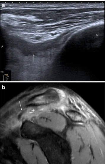
– In two cases, radiography showed a grade 3 injury, sug-gestive of ruptured coracoclavicular ligaments (subse-quently confirmed on MRI), whilst ultrasound had found one grade 2 and one grade 1 (Fig.3).
– In one case, radiography showed a grade 1 injury, which was confirmed on MRI, whilst ultrasound had found grade 2.
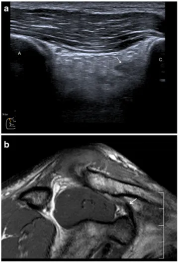
– In one case, radiography showed a grade 1 injury, MRI grade 2, whilst ultrasound had found grade 3 (Fig.4). – In one case, radiography showed a grade 2 injury, MRI
grade 1, whilst ultrasound had found grade 3.
– In 17 cases, ultrasound showed a grade 3 injury sugges-tive of ruptured coracoclavicular ligaments, which was confirmed on MRI, whilst radiography had given grade 2 (Fig.5).
– In one case, radiography and ultrasound suggested a grade 2 injury whilst MRI confirmed grade 3.
Ultrasound was therefore wrong in six cases and radiogra-phy in 24 cases.
Discussion
Our study population is consistent with epidemiological data for acromioclavicular injuries, as these injuries primarily oc-cur in young male patients [1]. Our study population did not include more severe injuries (grades IV, V and VI in the Rockwood classification). This can be explained by the fact that patients were recruited in a clinical setting following low-energy trauma. The prevalence of coracoclavicular ligaments damage is consistent with several studies on acromioclavicular joint injury [1,16]. One of the biases of this study is that each examination was performed by a single operator, making it im-possible to assess the reproducibility of ultrasound. Variations in the inter-observer findings for coracoclavicular ligaments in the acute phase of an acromioclavicular joint injury will need to be evaluated to determine the reproducibility of this method.
Table 2 Distribution of radiography, ultrasound and MRI grades Number of patients Radiography grade Ultrasound grade MRI grade Consistent classification− 24/47 cases
14/47 1 1 1
3/47 2 2 2
7/47 3 3 3
Inconsistent classification− 23/47 cases
17/47 2 3 3 2/47 3 1 3 1/47 1 3 2 1/47 2 3 1 1/47 1 2 1 1/47 2 3 2
Fig. 3 Discrepancy between ultrasound and MRI. a) Ultrasound shows grade 2: the trapezoid ligament in the longitudinal view (arrow) is thickened, distended, but continuous. Acromion (A), clavicle (c), acromial vascular pedicle (arrowhead). b) MRI, on a sagittal DP FAT SAT weighted sequence, shows grade 3: acromial detachment can be seen. Arrows shows the trapezoid ligament stump within diffuse soft tissue oedema (star)
The Rockwood classification, which is based on radio-graphic analysis of the acromioclavicular joint, is the most used classification for managing this acromioclavicular joint injury [2,3,17,18]. However, radiographs can only be used to indirectly evaluate the coracoclavicular liga-ments through the clavicle’s position; direct analysis of the condition of the coracoclavicular ligaments is impossi-ble with radiographs. In this study, we found excellent agreement between the ultrasound and MRI results (41 of 47 cases), by directly looking at the coracoclavicular ligaments, whereas the same injury grade was found on radiographs and ultrasound in only 25 of 47 cases. This can be explained by the fact that a ligament can be dam-aged (stretched, partially or completely torn) without caus-ing joint diastasis. This is reflected in our findcaus-ings: in 17 cases, a grade 3 injury was found on MRI and ultrasound, while a grade 2 injury was found on radiographs. Our findings are in agreement with those of Schaefer et al., who showed that 20 % more grade III injuries were found
on MRI than on radiographs [19]. Similarly, Barnes et al. found a poor agreement between the radiological outcomes and MRI [20].
Fig. 4 Discrepancy between ultrasound and MRI. a)The ultrasound shows grade 3: clavicular stump of the trapezoid ligament visible in the longitudinal view (arrow) with diffuse soft tissue oedema. Acromion (A), clavicle (c). b)MRI shows grade 2 with a continuous trapezoid ligament (sagittal T1 weighted sequence)
Fig. 5 Discrepancy between radiography, ultrasound and MRI findings: a grade 3 injury was found on ultrasound and MRI, but this was classified as grade 2 on radiographs. a)Grade 2 injury on radiographs (diastasis < 50 %). b)Ultrasound shows coracoclavicular ligament rupture; ligament stump indicated by arrow. c)MRI (T2-weighted fat-sat sequence, coronal oblique plane) shows coracoclavicular ligament rupture
While the treatment of grade 1 and 2 injuries (conservative) and grade 4, 5 and 6 injuries (surgical) has been validated, the treatment of grade 3 injuries is still being debated [3]. There are insufficient published data to justify any of the treatments for grade 3 injuries [15]. Minimally invasive and arthroscopic techniques are advocated by certain surgical teams for grade 3 injuries, especially in manual workers and athletes, as they allow for early resumption of activity [3,15,21].
Analysis of the condition of the coracoclavicular lig-aments is vital to the management of grade 3 injuries. Radiographs alone are not sufficient to evaluate the in-tegrity of the coracoclavicular ligaments. Bossart et al. found that grade 2 injuries are difficult to differentiate from grades 3 injuries on radiographs [22]. Our results support Bossart et al.’s findings: in 17 of the 47 cases, both ultrasound imaging and MRI found grade 3 inju-ries that may require surgical treatment, while these cases were classified as grade 2 injuries on radiographs. Ultrasound is relevant in this indication as it allows direct evaluation of the coracoclavicular ligaments, just like with MRI. Its low cost and accessibility are major assets of ultrasound imaging. When performed in the acute phase, MRI can be difficult to interpret because of movement artefacts caused by the patient being un-able to stay still for a long period to time due to pain. Ultrasound performed in the neutral position is better tolerated by patients. Ultrasound could be combined with radiography for the diagnostic work-up of acromioclavicular injuries in the acute phase.
This was a preliminary study that needs to be followed by a study with a larger number of patients and an evaluation of inter-observer agreement.
Conclusion
Ultrasound is an effective examination for the diagnostic work-up of coracoclavicular ligament damage in the acute phase of an acromioclavicular joint injury. It could be per-formed in addition to standard radiography, particularly in patients likely to benefit from surgical treatment of grade 3 lesions.
Acknowledgments The authors acknowledge the editorial assistance of Joanne Archambault, PhD. The scientific guarantor of this publication is Marie Faruch Bilfeld. The authors of this manuscript declare no rela-tionships with any companies, whose products or services may be related to the subject matter of the article. The authors state that this work has not received any funding.
One of the authors has significant statistical expertise. Institutional Review Board approval was obtained. Written informed consent was waived by the Institutional Review Board. This was is a prospective study performed at one institution.
References
1. Melenevsky Y, Yablon CM, Ramappa A, Hochman MG (2011) Clavicle and acromioclavicular joint injuries: a re-view of imaging, treatment, and complications. Skelet Radiol 40(7):831–42
2. Rockwood C, William G, Toung D (1996) Acromioclavicular injuries. Fractures in adults. p. 1341-1413
3. Tauber M (2013) Management of acute acromioclavicular joint dislocations: current concepts. Arch Orthop Trauma Surg 133(7): 985–95
4. Alyas F, Curtis M, Speed C, Saifuddin A, Connell D (2008) MR imaging appearances of acromioclavicular joint disloca-tion. Radiographics 28(2):463–479
5. Antonio GE, Cho JH, Chung CB, Trudell DJ, Resnick D (2003) Pictorial essay. MR imaging appearance and classifi-cation of acromioclavicular joint injury. AJR Am J Roentgenol 180(4):1103–10
6. Heer G, Götz J, Anders S, Grifka J, Hedtmann A (2006) Ultrasound evaluation of the acromioclavicular joint–a corre-lation of anatomical and sonographical findings. Ultraschall Med 27(6):549–52
7. Iovane A, Midiri M, Galia M, Bartolotta TV, Abate M, Sorrentino F et al (2004) Acute traumatic acromioclavicular joint lesions: role of ultrasound versus conventional radiography. Radiol Med 107(4): 367–75
8. Kock HJ, Jurgens C, Hirche H, Hanke J, Schmit-Neuerburg KP (1996) Standardized ultrasound examination for evalua-tion of instability of the acromioclavicular joint. Arch Orthop Trauma Surg 115(3-4):136–40
9. Peetrons P, Bédard JP (2007) Acromioclavicular joint injury: enhanced technique of examination with dynamic maneuver. J Clin Ultrasound 35(5):262–7
10. Heers G, Hedtmann A (2005) Correlation of ultrasonograph-ic findings to Tossy’s and Rockwood’s classification of acromioclavicular joint injuries. Ultrasound Med Biol 31(6): 725–32
11. Testut L, Jacob O (1921) Traité d’anatomie topographique avec applications médicochirurgicales. Doin. Paris; p. 756-74 12. Walton J, Mahajan S, Paxinos A, Marshall J, Bryant C, Shnier R et al (2004) Diagnostic values of tests for acromioclavicular joint pain. J Bone Joint Surg Am 86-A(4):807–12
13. Mazzocca AD, Arciero RA, Bicos J (2007) Evaluation and treat-ment of acromioclavicular joint injuries. Am J Sports Med 35(2): 316–29
14. Mikek M (2008) Long-term shoulder function after type I and II acromioclavicular joint disruption. Am J Sports Med 36(11):2147–50
15. Smith TO, Chester R, Pearse EO, Hing CB (2011) Operative versus non-operative management following Rockwood grade III acromioclavicular separation: a meta-analysis of the current evidence base. J Orthop Traumatol 12(1):19–27 16. Nemec U, Oberleitner G, Nemec SF, Gruber M, Weber M, Czerny
C et al (2011) MRI versus radiography of acromioclavicular joint dislocation. AJR Am J Roentgenol 197(4):968–73
17. Beris A, Lykissas M, Kostas-Agnantis I, Vekris M, Mitsionis G, Korompilias A (2013) Management of acute acromioclavicular joint dislocation with a double-button fixation system. Injury 44(3):288–92
18. Stucken C, Cohen SB (2015) Management of acromioclavicular joint injuries. Orthop Clin N Am 46(1):57–66
19. Schaefer FK, Schaefer PJ, Brossmann J, Hilgert RE, Heller M, Jahnke T (2006) Experimental and clinical evaluation of acromioclavicular joint structures with new scan orientations in MRI. Eur Radiol 16(7):1488–93
20. Barnes CJ, Higgins LD, Major NM, Basamania CJ (2004) Magnetic resonance imaging of the coracoclavicular ligaments: its role in defining pathoanatomy at the acromioclavicular joint. J Surg Orthop Adv 13(2):69–75
21. Boileau P, Old J, Gastaud O, Brassart N, Roussanne Y (2010) All-arthroscopic Weaver-Dunn-Chuinard procedure with double-button
fixation for chronic acromioclavicular joint dislocation. Arthroscopy 26(2):149–60
22. Bossart PJ, Joyce SM, Manaster BJ, Packer SM (1988) Lack of efficacy of « weighted » radiographs in diagnosing acute acromioclavicular separation. Ann Emerg Med 17(1):20–4