Fibrillation auriculaire chez des patients à haut risque:
Du diagnostic précoce à la prévention
thromboembolique
Atrial fibrillation in high-risk patients: From early
diagnosis to thromboembolic prevention
Thèse
Luis Asmarats Serra
Doctorat en sciences cliniques et biomédicales
Philosophiæ doctor (Ph. D.)
Fibrillation auriculaire chez des patients à haut risque :
Du diagnostic précoce à la prévention thromboembolique
Atrial fibrillation in high-risk patients:
From early diagnosis to thromboembolic prevention
Thèse
Lluis Asmarats Serra
Sous la direction de :
Josep Rodés-Cabau, directeur de recherche
François Philippon, codirecteur de recherche
RÉSUMÉ
La fibrillation auriculaire (FA) est la plus fréquente des arythmies cardiaques. La FA est associée à un risque accru d’accident vasculaire cérébral, d’insuffisance cardiaque et de mortalité, constituant un problème de santé publique majeur. L’avènement de nouvelles technologies permettant une surveillance électrocardiographique a démontré une haute prévalence de FA subclinique ou silencieuse chez les patients âgés à haut risque. Récemment, plusieurs efforts et essais thérapeutiques ont été dirigés vers une identification et un traitement plus précoces de la FA chez ces patients. L’anticoagulation orale a bien prouvé son efficacité dans la prévention thromboembolique chez les patients qui présentent un haut risque thromboembolique, mais au prix d’une augmentation significative des événements hémorragiques, un risque qui s’élève régulièrement chez les patients âgés et avec une comorbidité importante.
Au cours des dernières années, des nouvelles alternatives non-pharmacologiques dans la prévention thromboembolique ont été développées. La fermeture percutanée de l’auricule gauche, site de formation de la majorité (~90%) des thrombus, est progressivement devenue une alternative valable à l’anticoagulation chez des patients avec FA non valvulaire à haute risque hémorragique. L’expérience des opérateurs et les innovations technologiques ont permis une amélioration remarquable des résultats en ce qui concerne la sécurité et l’efficacité. Cependant, quelques questions restent sans réponse. Les préoccupations les plus débattues suite à la fermeture de l’auricule gauche sont la prise en charge de l’anticoagulation postprocédure et la prévention/gestion de la thrombose de dispositif.
Les objectifs de ce travail de recherche sont : (i) évaluer la charge arythmique silencieuse chez des patients à haut risque à l’aide de l’utilisation de nouveaux systèmes d’enregistrement électrocardiographique prolongé, et (ii) analyser l'impact hémodynamique et thrombogénique de la fermeture percutanée de l'auricule gauche avec les dispositifs actuels et émergents.
ABSTRACT
Atrial fibrillation (AF) is the most common cardiac arrhythmia. AF is associated with an increased risk of stroke, heart failure and mortality, posing a major public health problem. The advent of new technologies for continuous electrocardiographic monitoring has demonstrated a high incidence of subclinical or silent AF in elderly high-risk patients. Recently, several therapeutic efforts and studies have been directed towards earlier identification and treatment of AF in these patients. Oral anticoagulation has proven to be effective in preventing thromboembolism in patients at high thromboembolic risk, albeit at the expense of a significant increase in hemorrhagic events; a risk that increases steadily in elderly patients with high comorbidity burden.
In recent years, novel non-pharmacological alternatives have been developed for thromboembolic prevention. Percutaneous left atrial appendage (LAA) closure, site of origin of the vast majority (~ 90%) of thrombi, has progressively become a valid alternative to anticoagulation in patients with non-valvular AF at high bleeding risk. Increasing operators' experience and technological innovations have led to remarkable improvements in the safety and efficacy of the procedure. However, some issues remain unanswered or controversial. Two of the most debated concerns are post-procedural antithrombotic management and device-related thrombosis (DRT) following LAA closure.
The aims of the present research study are: (i) to evaluate the silent arrhythmic burden in high-risk patients using novel prolonged continuous electrocardiographic monitoring systems, and (ii) to assess the hemodynamic and thrombogenic impact of percutaneous LAA closure using current and emerging devices.
TABLE OF CONTENTS
RÉSUMÉ ... ii
ABSTRACT ... iii
TABLE OF CONTENTS ... iv
LIST OF TABLES ... viii
LIST OF FIGURES ... x
LIST OF ABBREVIATIONS ... xii
ACKNOWLEDGMENTS ... xv
FOREWORD ... xvii
INTRODUCTION ... 1
1.1. ATRIAL FIBRILLATION ... 2
1.1.1. Epidemiology ... 2
1.1.2. Atrial fibrillation and stroke ... 3
1.1.3. Pathophysiology: Thrombogenic mechanisms in atrial fibrillation ... 3
1.1.4. Diagnosis ... 6
1.1.5. Clinical risk stratification for stroke prevention ... 10
1.2. PHARMACOLOGICAL STRATEGIES FOR STROKE PREVENTION ... 13
1.2.1. Vitamin k antagonist anticoagulants... 13
1.2.2. Non-vitamin k antagonist anticoagulants ... 14
1.2.3. Antiplatelet agents ... 16
1.2.3.1. Single antiplatelet therapy ... 16
1.2.3.2. Dual antiplatelet therapy (DAPT) ... 17
1.3. NON-PHARMACOLOGICAL STRATEGIES FOR STROKE PREVENTION .... 18
1.3.1. Rationale for left atrial appendage closure ... 18
1.3.2. Embryology, anatomy and function of the LAA ... 19
1.3.3. Indications for LAA closure ... 22
1.3.4. Surgical left atrial appendage closure ... 25
1.3.5. Percutaneous left atrial appendage closure ... 28
1.3.5.1. Devices ... 28
1.3.5.2. Implantation technique ... 30
1.3.5.3. Procedural complications and management ... 33
1.3.5.4. Safety and efficacy ... 35
1.3.5.5. Post-procedural management ... 40
HYPOTHESIS AND OBJECTIVES ... 45
I. HYPOTHESIS ... 46
I.I. General hypothesis ... 46
I.II. Specific hypothesis ... 46
II. OBJECTIVES ... 47
II.II. Specific objectives... 47
CHAPTER 1. Prolonged Continuous ECG Monitoring Prior to Transcatheter Aortic Valve Replacement. The PARE Study ... 48
1.1. RÉSUMÉ ... 49
1.2. ABSTRACT ... 50
1.3. INTRODUCTON ... 51
1.4. METHODS ... 51
1.4.1. Study Design and Patients. ... 51
1.4.2. The CardioSTAT® device ... 52
1.4.3. Outcomes. ... 52
1.4.4. Statistical analysis ... 53
1.5. RESULTS ... 54
1.5.1. Incidence and type of arrhythmic events during 7-day CEM ... 56
1.5.2. Factors associated with arrhythmic events ... 58
1.5.3. Arrhythmic events post-TAVR ... 60
1.6. DISCUSSION ... 63
1.7. CONCLUSIONS ... 67
1.8. PERSPECTIVES ... 67
1.9. ACKNOWLEDGMENTS ... 68
CHAPTER 2. Percutaneous Left Atrial Appendage Closure: Current Devices and Clinical Outcomes ... 69
2.1. RÉSUMÉ ... 70
2.2. ABSTRACT ... 71
2.3. INTRODUCTION ... 72
2.4. DEVICE CHARACTERISTICS AND TECHNICAL ASPECTS ... 73
2.4.1. Plaato ... 73
2.4.2. Watchman and Watchman FLX ... 73
2.4.3. Amplatzer Cardiac Plug and Amulet ... 75
Table 2.1. Percutaneous LAAC Devices ... 77
2.4.4. Other emerging devices ... 79
2.4.4.1. WaveCrest ... 79
2.4.4.2. Occlutech LAA Occluder ... 79
2.4.4.3. LAmbre LAA Closure System ... 79
2.4.4.4. Sideris Patch ... 80
2.4.4.5. Ultraseal ... 80
2.4.4.6. Pfm ... 80
2.4.4.7. LARIAT ... 81
2.4.4.8. Sierra Ligation System ... 81
2.5. PROCEDURAL AND IN-HOSPITAL OUTCOMES ... 84
2.5.1. Watchman device ... 84
2.5.2. Amplatzer Cardiac Plug and Amulet ... 85
2.5.3. Lariat ... 87
2.6. LATE CLINICAL OUTCOMES ... 90
2.6.1. Randomized controlled trials: PROTECT-AF and PREVAIL ... 90
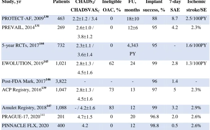
2.6.2. Registries ... 92
2.7.1. Optimal post-procedure antithrombotic therapy ... 97
2.7.2. LAAC vs. DOAC ... 97
2.7.3. Head-to-head comparison between LAAC devices... 98
2.7.4. Pre- and procedural imaging ... 98
2.7.5. Combined procedures ... 98
2.8. CONCLUSIONS ... 102
2.9. FUNDING SOURCES ... 102
2.10. CONFLICT OF INTEREST DISCLOSURES ... 102
CHAPTER 3. Percutaneous Left Atrial Appendage Closure with the Ultraseal Device: Insights from the Initial Multicenter Experience ... 103
3.1. RÉSUMÉ ... 104
3.2. ABSTRACT ... 105
3.3. INTRODUCTION ... 106
3.4. METHODS ... 106
3.4.1. Study population ... 106
3.4.2. Device characteristics and implantation ... 107
3.4.3. Statistical analysis ... 110 3.5. RESULTS ... 110 3.5.1. Procedural results ... 110 3.5.2. In-hospital outcomes... 113 3.5.3. Follow-up ... 114 3.6. DISCUSSION ... 116 3.7. CONCLUSIONS ... 120 3.8. CLINICAL PERSPECTIVES ... 120
CHAPTER 4. Hemodynamic Impact of Percutaneous Left Atrial Appendage Closure in Patients with Paroxysmal Atrial Fibrillation... 121
4.1. RÉSUMÉ ... 122 4.2. ABSTRACT ... 123 4.3. INTRODUCTION ... 124 4.4. METHODS ... 124 4.4.1 Patient selection ... 124 4.4.2 Echocardiographic examination ... 125
4.4.3. LAA volume assessment by cardiac computed tomography ... 125
4.4.4. Statistical analysis ... 126
4.5. RESULTS ... 126
4.5.1. LA and left ventricular function determined by 2D echocardiography ... 126
4.5.2. Cardiac 3D-CT data ... 130 4.6. DISCUSSION ... 131 4.7. CONCLUSIONS ... 134 4.8. AKNOWGLEDGMENTS ... 134 4.9. CONFLICT OF INTEREST ... 134 4.10. ETHICAL APPROVAL ... 134
CHAPTER 5. Short-Term Oral Anticoagulation Versus Antiplatelet Therapy Following Transcatheter Left Atrial Appendage Closure ... 135
5.2. ABSTRACT ... 137
5.3. INTRODUCTION ... 138
5.4. METHODS ... 138
5.4.1. Study Design. ... 138
5.4.2. Procedures and follow-up ... 139
5.4.3. Blood sample collection. ... 139
5.4.4. Outcomes ... 139
5.4.5. Statistical analysis ... 140
5.5. RESULTS ... 140
5.5.1. Changes in the markers of coagulation activation after LAAC... 141
5.5.2. Factors associated with enhanced prothrombotic status post-LAAC ... 145
5.5.3. DRT following LAAC ... 145
5.5.4. Clinical outcomes after LAAC ... 148
5.6. DISCUSSION ... 149
5.6.1. Antithrombotic therapy and hemostatic markers ... 149
5.6.2. Enhanced coagulation activation: associated factors and DRT ... 150
5.6.3. Study limitations ... 151
5.7. CONCLUSIONS ... 151
5.8. ACKNOWLEDGMENTS ... 152
5.9. DICLOSURES ... 152
5.10. SUPPLEMENTAL MATERIAL ... 153
CHAPTER 6. Recurrence of Device-Related Thrombus Following Percutaneous Left Atrial Appendage Closure ... 159
6.1. RÉSUMÉ ... 160
6.2. ABSTRACT ... 161
6.3. RESEARCH LETTER ... 162
6.4. DATA SHARING ... 165
6.5. FOUNDING SOURCES ... 165
6.6. CONFLICT OF INTEREST DISCLOSURES ... 166
DISCUSSION, CLINICAL PERSPECTIVES AND CONCLUSIONS ... 167
7.1. DISCUSSION ... 168
7.1.1. Role of continuous AF monitoring in high-risk populations ... 168
7.1.2. Evolution of transcatheter LAAC ... 169
7.1.3. Impact of transcatheter LAAC on cardiac function... 172
7.1.4. Antithrombotic therapy and DRT ... 174
7.2. CLINICAL IMPLICATIONS ... 178
7.3. FUTURE PERSPECTIVES ... 181
7.4. CONCLUSIONS ... 182
LIST OF TABLES
Table 1. Classification of atrial fibrillation ... 7
Table 2. Types of ambulatory cardiac monitoring devices ... 8
Table 3. Stroke risk prediction algorithms and antithrombotic recommendations... 11
Table 4. Bleeding risk scores ... 12
Table 5. Randomized data on Pharmacological Stroke Prevention Therapies in AF... 16
Table 6. Society guideline recommendations for left atrial appendage closure ... 23
Table 7. Comparison of surgical left atrial appendage closure techniques ... 26
Table 8. Major procedural complications from the PROTECT-AF and PREVAIL randomized trials, and largest LAAC registries ... 33
Table 9. Overview of the largest randomized trials and registries using Watchman and Amulet ... 37
Table 1.1. Clinical characteristics according to the occurrence of arrhythmic events during 7-day CEM ... 55
Table 1.2. New-onset arrhythmic events observed during 1-week continuous ECG monitoring with Cardiostat before transcatheter aortic valve replacement ... 57
Table 1.3. Procedural and 30-day outcomes in patients undergoing TAVR, overall and according to the occurrence of AEs during 7-day continuous electrocardiographic monitoring pre-TAVR ... 61
Table 2.2. Technical Success and Procedure-Related Complications Associated with Percutaneous LAAC ... 88
Table 2.3. Long-Term Clinical Outcomes Following Percutaneous LAAC ... 95
Table 2.4. Main Ongoing and Future Studies on Percutaneous LAAC ... 100
Table 3.1. Baseline clinical characteristics ... 111
Table 3.2. Procedural findings ... 112
Table 3.3. In-hospital outcomes ... 113
Table 3.4. Follow-Up Clinical and TEE Findings ... 115
Table 4.1. Baseline clinical characteristics ... 127
Table 4.2. Echocardiographic volumetric indexes of left atrial function ... 128
Table 4.3. Hemodynamic and echocardiographic parameters of left ventricular systolic function ... 128
Table 4.4. Echocardiographic parameters of left ventricular diastolic function. ... 129
Table 4.5. Three-Dimensional computed tomography volumetric measurements ... 130 Table 5.1. Baseline, Procedural and In-Hospital Characteristics of the Study population . 142
Table 5.2. Degree of activation of the coagulation markers, according to baseline and procedural variables in patients with antiplatelet therapy (n=48) ... 146 Table 5.3. Follow-up outcomes (after hospital discharge) ... 148 Supplementary Table 5.1. Characteristics of patients with device-related thrombus. ... 154 Supplementary Table 5.2. Degree of activation of the coagulation markers, according to baseline and procedural variables in patients with oral anticoagulation therapy (n=30) ... 155 Table 7.1. Studies on Long-Term Follow-up After Left Atrial Appendage Closure ... 171 Table 7.2. Changes in cardiac function following percutaneous LAAC ... 172 Table 7.3. Recent studies on LAAC using different antithrombotic regimens ... 176
LIST OF FIGURES
Figure 1. Projected number of persons with atrial fibrillation in the United States and in
Europe ... 2
Figure 2. Virchow’s triad components for thrombogenesis in atrial fibrillation. ... 4
Figure 3. Atrial fibrillation ECG findings. ... 6
Figure 4. Screening tools for atrial fibrillation diagnosis. ... 9
Figure 5. Mechanism of action of vitamin K antagonists and direct oral anticoagulants .... 14
Figure 6. Localization of left atrial thrombi in patients with non-valvular atrial fibrillation or flutter. ... 19
Figure 7. The left atrial appendage and surrounding structures. ... 20
Figure 8. Multimodality imaging morphological classification of the LAA assessed by transesophageal imaging, angiography and computed tomography ... 21
Figure 9. The AtriClip device. ... 27
Figure 10. Most commonly used percutaneous LAAC devices. ... 29
Figure 11. Step-by-step implantation of the Watchman and Amulet devices ... 32
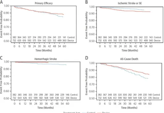
Figure 12. 5-year efficacy outcomes of the PROTECT-AF and PREVAIL trials. ... 36
Figure 13. Likely mechanisms for peri-device leaks for endocardial and epicardial LAAC devices. ... 42
Figure 14. Illustration of a pedunculated and a large, laminar device-related thrombus attached to the Watchman device on transesophageal echocardiography. ... 43
Figure 15. Kaplan-Meier curves for thromboembolic events according to the presence of thrombus on the device. ... 44
Figure 1.1. Patient Flowchart. ... 54
Figure 1.2. Incidence of bradyarrhythmic events during 7-day ambulatory cardiac monitoring pre-TAVR according to pre-existing conduction disturbances at baseline electrocardiogram ... 59
Figure 1.3. New-onset atrial fibrillation and need for pacemaker according to the occurrence of previously unknown arrhythmic events during 7-day cardiac monitoring pre-TAVR ... 62
Figure 2.1. Percutaneous left atrial appendage closure devices ... 83
Figure 2.2. Temporal trends in procedural complications following left atrial appendage closure. ... 86
Figure 2.3. Long-term outcomes in randomized trials. ... 91
Figure 3.1. The Cardia Ultraseal device. ... 108
Figure 3.2. Ultraseal device implantation. ... 109
Figure 3.4. Peridevice leak postimplantation and at 6-month follow-up. ... 116 Fig. 4.1. Stroke volume before and after LAA closure. ... 129 Fig. 4.2. Relationship between LAA/LA ratio and stroke volume. ... 130 Fig. 4.3. Three-dimensional computed tomography reconstruction of the left atrium and left atrial appendage. ... 131 Figure 5.1. Changes in coagulation system activation within the 6 months post-LAAC, according to antithrombotic therapy (antiplatelet versus anticoagulation therapy) ... 144 Figure 5.2. Changes in coagulation system activation within the 6 months post-LAAC, according to antithrombotic therapy (SAPT vs DAPT vs OAC therapy) ... 144 Figure 5.3. Changes in coagulation system activation post-LAAC, according to the occurrence of device-related thrombus... 147 Supplementary Figure 5.1. Flow chart of the different antithrombotic strategies during the first year following LAAC. ... 157 Supplementary Figure 5.2. Changes in coagulation system activation within the 6 months post-LAAC in patients with device-related thrombus ... 158 Figure 6.1. Flowchart of Study Population ... 163
LIST OF ABBREVIATIONS
ACP: Amplatzer Cardiac Plug AF: Atrial fibrillationAPT: Antiplatelet therapy AT: Atrial tachycardia AVB: Atrioventricular block CEM: Continuous ECG monitoring
CHADS2: Congestive heart failure, hypertension, age ≥75 years, diabetes mellitus, prior
stroke or transient ischemic attack
CHA2DS2-VASC: Congestive heart failure, hypertension, age ≥75 years, diabetes mellitus,
prior stroke or transient ischemic attack, vascular disease, age 65-74 years, sex category
CT: Cardiac computed tomography DAPT: Dual antiplatelet therapy DOAC: Direct oral anticoagulants DRT: Device-related thrombosis
ECG: Electrocardiographic
ESVEA: Excessive supraventricular ectopic activity
F1+2: Prothrombin fragments 1+2
HAS-BLED: Hypertension, abnormal renal/liver function, stroke, bleeding history or predisposition, labile INR, elderly, drugs/alcohol
HAVB: High-degree atrioventricular block LA: Left atrium
LAA: Left atrial appendage
LAAC: Left atrial appendage closure LBBB: Left bundle branch block MAE: Major adverse event
NSVT: Non-sustained ventricular tachycardia OAC: Oral anticoagulation
PET: polyethylene terephthalate PPM: Permanent pacemaker RBBB: Right bundle branch block
SAPT: Single antiplatelet therapy SCAF: Subclinical atrial fibrillation TAT: thrombin-antithrombin III complex
TAVR: Transcatheter aortic valve replacement TEE: Transesophageal echocardiography
“Medicine is a science of uncertainty and an art of probability”
ACKNOWLEDGMENTS
“Difficult roads often lead to beautiful destinations” Zig Ziglar (1926 – 2012)
The debts of gratitude incurred in a work of this nature are countless. My deep gratitude goes first to Josep Rodés-Cabau, my mentor, the person who opened the door for me without even knowing me, who trusted in me since the very beginning and taught me the beauty of research and the distinctive value of a rigorous scientific method. It has been a tremendous privilege and pleasure to be one of your fellows, and hopefully only the beginning of a lasting relationship and friendship.
To François Philippon, for sharing your expertise and knowledge in the field of arrhythmology, your wise and constructive advises throughout the elaboration of the present work have been of major relevance.
To my old colleagues in Mallorca, particularly those who encouraged me to embark on the path of interventional cardiology (Jaume Maristany) and undertaking the Quebecoise experience (Jaume Pons, José Ignacio Sáez de Ibarra).
To my current and future teammates, especially Dabit Arzamendi and Antoni Serra, for allowing me to continue learning and growing as a physician and researcher in the field of interventional and structural heart disease.
To all the members of the Cath Lab of the Quebec Heart and Lung Institute, with particular mention to the LAAC team (Drs Jean-Michel Paradis, Gilles O’Hara, Jean Champagne; Sonia Bérubé, Maryse Lemyre), the TAVR team (Drs Robert de Larochellière,
Jean-Michel Paradis, Siamak Mohammadi, Eric Dumont, Dimitri Kalavrouziotis; Julie Demers, Martine Page) and the Mitral/Tricuspid team (Drs François Dagenais, Eric Dumont, Mathieu Bernier, Élisabeth Bédard and Caroline Gravel).
I owe tremendous gratitude to Dr Rodés’ research team. A very sincere mention to Dominique Lachance, Mélanie Côté and Émilie Pelletier, three of the most competent and marvelous people I have ever worked with. You are undoubtedly the pillar of this incredible research team, both personally and professionally.
I want to thank my fellow labmates in the IUCPQ. It has been a pleasure to share these years with you. It has been much more than just a team-work, but a true family during this journey, who have become lifelong friends.
None of these achievements would doubtless have been possible without the support and priceless advices of my parents and my sister Carla. Thank you for your constant encouragement, stimulus and unwavering support.
Last, but most importantly, my biggest gratitude goes to my wife Ana, whom this work is dedicated. It has been a long journey, which I would have never accomplished without your unconditional support and loving understanding. A road filled with challenges and joys along the way, that has led to the beautiful destination of our growing family.
FOREWORD
The research works embedded in this PhD Thesis were carried out in the research group of Structural Heart Disease Interventions at the Quebec Heart and Lung Institute (IUCPQ, Laval University, Québec, City, Québec), led by Dr Josep Rodés-Cabau.
Throughout the research project, the student received a research grant from the Fundación Alfonso Martin Escudero (Madrid, Spain), effective between January 2017 and December 2018; and Dr Rodés-Cabau holded the Research Chair “Fondation Famille Jacques Larivière” for the Development of Structural Heart Disease Interventions.
This thesis is composed by 6 research articles, which have been published in high-impact peer-review cardiovascular journals.
The first article included in this thesis is entitled: “Prolonged Continuous ECG Monitoring Prior to Transcatheter Aortic Valve Replacement. The PARE Study”. It has been published in JACC: Cardiovascular Interventions, and the student is the first author. The study evaluated the prevalence of subclinical atrial fibrillation and other cardiac arrhythmias in elderly patients with aortic stenosis screened for transcatheter aortic valve replacement at the IUCPQ. The student was the first author, and Dr Josep Rodés-Cabau, the senior author. Drs François Philippon and Isabelle Nault participated in the conception and design of the study, as well as in the interpretation of the data. All other authors approved the manuscript and contributed with their critical review of the manuscript.
The second article entitled: “Percutaneous Left Atrial Appendage Closure: Current Devices and Clinical Outcomes” was published in Circulation: Cardiovascular Interventions. This review analyzed the temporal trends in procedural complications since the beginning of the percutaneous left atrial appendage closure era, as well as the available evidence on mid- and long-term outcomes following left atrial appendage closure. The
student was the first author of this article and participated in the review of the literature, drafting and revision of the manuscript, under the supervision of Dr Josep Rodés-Cabau.
The third article of this thesis is entitled: “Percutaneous Left Atrial Appendage Closure with the Ultraseal Device: Insights from the Initial Multicenter Experience”. This work was presented as an oral communication at the Transcatheter Cardiovascular Therapeutics meeting in September 2018 (TCT 2018, San Diego, USA) with simultaneous publication in JACC: Cardiovascular Interventions. It was also presented as an oral communication at the meeting from the Spanish Society of Cardiology (Seville, Spain, October 2018). This study included the initial worldwide experience with the Ultraseal LAAC device, with 15 centers from Canada and Europe. The student was responsible for the collection of the data at the IUCPQ, the management, analysis and interpretation of the data from the worldwide centers, and drafting of the manuscript. Dr Josep Rodés-Cabau was the principal investigator and senior author of the study, and supervised each of the steps. All other authors contributed with their comments and constructive review of the manuscript.
The fourth article, which was entitled: “Hemodynamic Impact of Percutaneous Left Atrial Appendage Closure in Patients with Paroxysmal Atrial Fibrillation” was published in the Journal of Interventional Cardiac Electrophysiology, and evaluated non-invasively the acute hemodynamic impact of percutaneous left atrial appendage occlusion. The student is the first author of the study, and participated in the conception of the study, analysis and interpretation of the data, and drafting of the manuscript, under the supervision of Dr Josep Rodés-Cabau. All other authors approved and revised the manuscript.
The fifth article of this thesis is entitled: “Short-Term Oral Anticoagulation Versus Antiplatelet Therapy Following Transcatheter Left Atrial Appendage Closure” and has been published in Circulation: Cardiovascular Interventions. This work compared the activation of the coagulation system following left atrial appendage occlusion between patients receiving antiplatelet therapy or 45-day oral anticoagulation therapy after the
procedure at our institution. The student was responsible for the collection and interpretation of the data, drafting and revision of the manuscript. The statistical analysis was performed by Mélanie Côté, with all steps supervised by the senior author, Dr Josep Rodés-Cabau. All other authors approved the manuscript and revised it for relevant intellectual content.
The last and sixth article presented in this thesis was entitled: “Recurrence of Device-Related Thrombus After Percutaneous Left Atrial Appendage Closure”. It was published in the Circulation Journal. The student was the first author, developed the database for the study, and was responsible for the collection, analysis and interpretation of the data, as well as drafting of the manuscript. Dr Josep Rodés-Cabau supervised all of these steps. All other authors approved the manuscript and contributed with their critical review of the manuscript.
1.1. ATRIAL FIBRILLATION 1.1.1. Epidemiology
Atrial fibrillation (AF) is the most common cardiac arrhythmia worldwide1-4 with an incidence projected to double to more than 12 million in the United States by 2050 and 17.9 million in Europe by 2060 (Figure 1).1, 2 It is noteworthy that this prevalence is largely underestimated due to underdiagnosis in elderly asymptomatic patients or when associated with only transient symptoms. Of note, age is the most important risk factor for AF. Compared to individuals aged 50 to 59, the risk of occurrence is 4.98-, 7.35- and 9.33-fold higher in patients aged 60-69 years, 70-79 years and 80-89 years, respectively.5 Two other traditional cardiovascular risk factors, diabetes (odds ratio: 1.4 for men, 1.6 for women) and hypertension (odds ratio: 1.5 for men, 1.4 for women) have been identified as independent risk factors for AF.6
Figure 1. Projected number of persons with atrial fibrillation in the United States (top) and in Europe (bottom).
The solid curve in the top figure indicates assuming no further increase in age-adjusted incidence, and the dotted curve assuming a continued increase in incidence as observed between 1980 and 2000. From Miyasaka et al.1 and Krijthe et al.2 with permission.
AF represents a major public health concern, significantly increasing the risk of stroke, morbidity, mortality and healthcare costs.7 Indeed, AF has been associated with an increased risk of frailty (HR: 4.1), heart failure (HR: 1.2 – 23.2), hospitalization (HR: 1.1), sudden death (HR: 3.3) and increased risk of cardiac and total mortality (HR: 1.7 – 2.1).3, 8-10
AF has substantial socioeconomic implications mainly derived from AF-related hospitalisations, with exponential increase in hospital costs by 24% to 468% over the last decade.4, 11 Hence, AF constitutes a growing epidemic with enormous economic and public health burden. A compelling clinical need exists for improved healthcare measures including novel screening technologies to optimize early identification of asymptomatic individuals, and enhancement of stroke prevention measures to decrease AF-related healthcare expenses.
1.1.2. Atrial fibrillation and stroke
AF confers a 5-fold increase in the risk of stroke,12 and 17-fold increase in patients with rheumatic valve disease.13 Importantly, AF-related strokes are associated with greater morbidity - >50% greater disability, handicap and recurrence -, mortality and costs compared with non-cardioembolic strokes.12 The risk of ischemic stroke in non-valvular AF patients averages 5% per year (8% in patients aged 75 years-old or older). About 15-20% of all ischemic strokes are attributed to AF.14 This proportion increases steadily with age, accounting for up to 40% of strokes in patients over the age of 80 years.15, 16 Noteworthy, the prevalence of AF increased by 22% and by 38% among patients admitted in the United States for acute ischemic stroke and transient ischemic attack respectively between 2003 and 2014.17 Up to 50-70% of all strokes in AF patients are thought to be cardioembolic, the main source of these cardioembolic events lying within the left atrial appendage (LAA, >90% of cases in non-rheumatic AF, 57% in rheumatic AF).18
1.1.3. Pathophysiology: Thrombogenic mechanisms in atrial fibrillation
Thrombogenesis in AF is a complex and multifactorial process, with several mechanisms promoting a prothrombotic or hypercoagulable state. Interestingly, the triad postulated by Rudolf Virchow in 1856 to explain thrombus formation - endothelial or endocardial
damage; abnormal blood stasis; abnormal blood constituents - is also fulfilled in AF (Figure 2).19
Figure 2. Virchow’s triad components for thrombogenesis in atrial fibrillation.
From Watson et al.19 and Glikson et al.20 with permission.
Abnormalities of the vessel wall. The LAA is the most common site of intra-atrial thrombus formation. Because of AF, the atrial wall and endocardium experience
progressive dilatation, endocardial denudation with thrombotic aggregation, myocytic hypertrophy, and edematous or fibro-elastic infiltration of the extracellular matrix. Importantly, the disruption of the extracellular matrix observed in AF patients (with abnormal concentrations of matrix metalloproteinases) may not only lead to conduction defects potentially perpetuating AF, but also induce fibrosis and infiltration of the endocardium, thus promoting thrombogenesis. Altogether, such cardiac stunning underscores the importance of an adequate thromboembolic stroke prevention strategy even after restoration of sinus rhythm.19
Abnormal blood stasis. The structural changes associated with AF along with the loss of atrial systole, contribute to the increased stasis in the left atrium and diminished flow velocities within the LAA. This phenomenon is even more remarkable in the presence of mitral stenosis, with greater left atrial dilatation and further stasis. Of note, atrial size has been identified as an independent risk factor for stroke.21 In normal sinus rhythm, a
quadriphasic pattern of blood flow can be noted in the LAA, with none or minimal blood stasis. In AF, this pattern disappears and spontaneous echo contrast can be visualized by transesophageal echocardiography, which is thought to be linked to an interaction between fibrinogen and erythrocytes, being a risk factor for both thrombus formation and thromboembolism.22
Abnormal blood constituents. Platelets and coagulation cascade, along with other blood constituents (inflammatory cytokines and growth factors), constitute the chief intravascular promoters of thrombogenesis in AF patients. AF constitutes a prothrombotic state itself, with increased fibrin turnover and abnormally high levels of prothrombotic markers (prothrombin fragments 1+2, thrombin-antithrombin complex).19 Furthermore, in non-valvular AF, there is a significant correlation between prothrombin fragments 1+2, fibrinopeptide A, thrombin-anti-thrombin complex and the presence of spontaneous echo contrast at transesophageal echocardiography.23 Similarly, AF patients exhibit greater levels of D-dimer and β-thromboglobulin, the former potentially predicting the presence of LAA thrombus, and subsequent thromboembolic events.24 Von Willebrand factor, a well-established marker of endothelial dysfunction, has been demonstrated to be a predictor of stroke and vascular events, improving clinical risk stratification for stroke.25 However, its non-specificity may probably hamper its applicability in AF. The potential role of platelets in the hypercoagulable state associated with AF remains controversial. Platelets seem to interact with the endocardium, proteins of the coagulation cascade, and inflammatory cells to increase the thrombogenecity in AF. CD40 ligand is present on the platelet surface only after activation and then cleaved, generating the soluble biologically active fragment sCD40L. In AF, sCD40L is slightly elevated, but the stimulus for this is unclear.26 P-selectin and CD63, well-validated markers of platelet activation, have been related to the embolic status of patients with non-valvular AF.27 Finally, abnormal changes in systemic inflammation have been suggested to drive the prothrombotic state in AF. Levels of interleukin-6 and C-reactive-protein are abnormally elevated in AF, which could increase platelet production and sensitivity to thrombin. Likewise, vascular endothelial growth factors have been shown to alter in AF, acting as potent stimulants for tissue factor
expression, which in turn acts as a cofactor to factor VIIa, a well-known trigger to thrombin formation.19
1.1.4. Diagnosis
The diagnosis of AF requires documentation by 12-lead electrocardiography or a single-lead ECG tracing showing absolutely irregular RR intervals and no discernible, distinct P waves (Figure 3). By accepted convention, an episode lasting at least 30 seconds is diagnostic.28
Figure 3. Atrial fibrillation ECG findings.
(A) Electrocardiogram in sinus rhythm. Orange arrows indicate normal p waves and regular RR intervals (orange bracket).
(B) Electrocardiogram in atrial fibrillation. Blue arrows indicate abnormal atrial fibrillation waves and irregular RR intervals (blue bracket).
Five patterns of AF have been described according to the clinical presentation, duration, and termination of AF episodes (Table 1).
Table 1. Classification of atrial fibrillation
AF pattern Definition
First diagnosed AF AF not diagnosed before, irrespective of the duration or symptoms
Paroxysmal AF Self-terminating, in most cases within 48 hours (for up to 7 days)
Persistent AF Lasting longer than 7 days, including episodes terminated by cardioversion
Long-standing persistent AF Lasting for ≥1 year when it is decided to adopt a rhythm
control strategy
Permanent AF Accepted by the patient (and physician). Rhythm control interventions are not pursued in those patients.
AF: Atrial fibrillation. From Hindricks et al.28
Undiagnosed AF is frequent, particularly among elderly patients. Data from the GLORIA-AF (Global Registry on Long-Term Antithrombotic Treatment in Patients with GLORIA-AF) registry showed that AF may be asymptomatic or minimally symptomatic in up to 70% of patients.29 Of note, subclinical AF (SCAF) has been associated with a greater risk of stroke
given the potential delay in oral anticoagulation (OAC) prescription in the absence of symptoms.30 Opportunistic screening for SCAF has proved cost-effective in elderly populations >65 years of age.31 Current guidelines recommend opportunistic AF screening (during routine interactions with the healthcare system) by pulse palpation or ECG rhythm strip in patients ≥65 years (I-B), with systematic screening (at some predefined time point outside routine medical care) considered to detect AF in patients aged ≥ 75 years, or those at high stroke risk (IIa-B).28 However, recent data from the ARIC (Atherosclerosis Risk in Communities), which identified a 2.5% prevalence of SCAF in elderly populations using 2 weeks continuous ambulatory ECG monitoring, casted doubts on the role of pulse palpation or single lead ECG for screening, since the majority of SCAF cases (75%) were intermittent and had low AF burden (≤10%).32 In fact, effectiveness of screening has been
strongly related to its duration, with longer continuous ECG monitoring having a higher diagnostic yield to detect SCAF.
With newer ambulatory monitoring technologies enabling extended continuous electrocardiographic monitoring,33, 34 a high prevalence of previously unknown AF has been detected in high-risk populations – elderly,35 with comorbid disorders,36 or in patients undergoing transcatheter aortic valve replacement (TAVR) (Table 2, Figure 4).37 Prompt identification of SCAF, classically undiagnosed, and early initiation of directed therapies, including OAC or non-pharmacologic stroke prevention therapies, is essential to optimize our management of patients with AF.
Table 2. Types of ambulatory cardiac monitoring devices
AF: Atrial fibrillation; ECG: electrocardiogram; ICD: implantable cardioverter defibrillator. Adapted from Calkins et al.38
Type of recorder Monitoring duration
Continuous recording
Unique features Sensitivity
Holter monitor 24-48 hs Yes -Short term
-Data on arrhythmic burden
44-60 %
Patch monitor 1-3 weeks Yes -Intermediate term
-Improved patient compliance without leads
N/A
External loop recorder
1 month Yes -Good correlation between symptoms and arrhythmias
39-68 %
Mobile cardiac telemetry
1 month Yes -Real-time central monitoring -Relatively expensive
N/A
Implantable loop recorder
≤3 years Yes -Improved patient compliance -AF needs confirmation by ECG
45-88 %
Smartphone monitor Indefinite No -Inexpensive long-term
-Dependent on patient compliance
98.5 %
Pacemaker or ICD Indefinite Yes -Good documentation of burden -AF needs confirmation by electrogram tracing (in the absence of an atrial lead)
Figure 4. Screening tools for atrial fibrillation diagnosis. From Mairesse et al.39 with permission.
1.1.5. Clinical risk stratification for stroke prevention
The prevention of AF-related ischemic stroke is based on a balance between benefit and harm from a specific strategy using probability calculation tools. In fact, stroke and bleeding risk factors often overlap, posing a major clinical challenge for decision making. Since the first stroke risk-stratification schemes developed in the late 1990s, the CHADS2 score (congestive heart failure, hypertension, age >75 years, diabetes and previous stroke [doubled]), with a C-statistic of 0.82, emerged in 2001 as the simplest and most precise predictor of stroke.40 Although widely used for many years, the CHADS2 score showed several shortcomings (age treated as a binary variable, exclusion of important risk factors, and poor ability to identify low-risk patients who do not benefit from stroke prevention therapy). To overcome this limitations, Lip et al.41 created in 2010 a new risk-scoring scheme by adding additional risk factors (female gender, vascular disease, and age categories [65-74 years, and ≥75 years]): the extended CHA2DS2-VASC score. Given its superiority over CHADS2 score in quantifying stroke risk, the CHA2DS2-VASC score is the recommended stroke risk prediction tool for patients with nonvalvular AF by the European Society of Cardiology and American College of Cardiology / American Heart Association guidelines, since 2010 and 2014 respectively. Thresholds for OAC recommendation vary slightly between guidelines (Table 3).
The European and American guidelines for the management of AF advise OAC for patients with AF and a CHA2DS2-VASC ≥ 2 (class I recommendation, level A).28, 42 Patients with a CHA2DS2-VASC =1 (which represent about 10-15% of patients), constitute a gray area of uncertainty. However, a net clinical benefit from OAC has recently been shown even in the presence of 1 non-sex-related risk factor,43 and latest guidelines recommend the possibility of stroke prevention in those patients, taking patient values and preferences into consideration (IIa-B and IIb-C for European and American guidelines, respectively). Finally, Canadian guidelines recommend OAC in all AF patients aged > 65 or with CHADS2 score ≥ 1.44 Importantly, most elements of these scores are dynamic, requiring periodic risk reassessment.
Table 3. Stroke risk prediction algorithms and antithrombotic recommendations
2019 AHA/ACC/HRS42
2020 ESC28 2018 CCS44
CHA2DS2-VASC CHADS-65
Congestive heart failure 1 1
Hypertension 1 1
Age ≥ 75 years 2 1
Diabetes Mellitus 1 1
Stroke, TIA, embolism 2 1
Vascular Disease 1 -
Age 65-74 years 1 1
Female Sex 1 -
0 No (IIa) No (III) No (conditional)*
1 NOAC > VKA (IIb) NOAC > VKA (IIa) NOAC (strong) ≥ 2 VKA > NOAC (I) NOAC > VKA (I) NOAC (strong)
*ASA for patients aged <65 years with a CHADS2 score=0 with arterial vascular disease (coronary,
aortic, or peripheral)
ACC: American College of Cardiology; AHA: American Heart Association; ASA: Aspirin; CCS: Canadian Cardiovascular Society; ESC: European Society of Cardiology; HRS: Heart Rhythm Society; NOAC: novel oral anticoagulant; OAC: Oral anticoagulant; TIA: transient ischemic attack; VKA: vitamin K antagonist.
Several bleeding risk scores have been developed to date, albeit generally with a modest predictive ability (Table 4). The most commonly used risk score for assessing the risk of bleeding in AF patients is the HAS-BLED score, which takes into account seven factors: hypertension >160 mmHg, abnormal renal or liver function (creatinine >200 µmol/l, dialysis or kidney transplant, cirrhosis or bilirubin >2x normal or AST/ALT >3x normal), stroke, bleeding, labile INR (time in therapeutic range <60%), >65 years old and consumption of anti-inflammatory or anti-platelet drugs or alcohol.45 A score ≥ 3 represents a high risk of bleeding which may translate into close monitoring, but in general should not result in withholding OAC. Other bleeding risk scores include the ORBIT (older age ≥75 years, reduced hemoglobin or anemia, bleeding history, insufficient kidney function, treatment with antiplatelets), the ATRIA (anemia, severe renal disease, age, any prior hemorrhage, diagnosed hypertension), or the HEMORR2HAGES (hepatic or renal dysfunction, ethanol abuse, malignancy, older age, reduced platelet count or function, rebleeding risk, hypertension, anemia, genetic factors, excessive fall risk, stroke).46-49
Table 4. Bleeding risk scores
ATRIA HAS-BLED ORBIT HEMORR2HAGES
Anemia 3
Hypertension-uncontrolled
1 Older age (>75yo) 1 Hepatic or renal disease 1 Severe renal disease 3 Abnormal renal or liver function 1 or 2 Reduced hemoglobin, hematocrit or anemia 2 Ethanol abuse 1
Age 2 Stroke 1 Bleeding history 2 Malignancy 1
Any prior hemorrhage
1 Bleeding history 1 Insufficient kidney
function
1 Older age 1
Hypertension 1 Labile INR 1 Treatment with
anti-platelets
1 Reduced platelet count or function
1
Elderly 1 Rebleeding risk 2
Drugs or alcohol 1 or 2 Hypertension 1
Anemia 1
Genetic factors 1 Excessive fall risk 1
Stroke 1
Maximum score: 10 Maximum score: 9 Maximum score: 7 Maximum score: 12
High risk ≥5 High risk ≥3 High risk ≥4 High risk ≥4
The HAS-BLED score is the most often used to estimate bleeding risk given its ease of use and having proven to be superior to other scores in predicting bleeding risk.50, 51 Interestingly and unlike other scores, HAS-BLED includes modifiable risk factors that can easily be addressed such as uncontrolled high blood pressure, concomitant treatment with an antiplatelet therapy (which can generally be withdrawn after one year of an acute coronary syndrome), alcohol abuse or poor anticoagulation control. Furthermore, its predictive value for bleeding has been validated in several scenarios (e.g. direct OAC, bridging therapy in patients undergoing percutaneous coronary intervention).52, 53 However, some important limitations should be acknowledged. First, it has modest risk discrimination ability, with C statistics ranging from 0.50 to 0.80.54 Second, because of the parallel nature of bleeding and stroke risk scores, many of the risk factors encountered in HAS-BLED overlap with those included in stroke risk scores, lacking ability to discriminate bleeding risk from stroke risk. Third, in a recent substudy of the Amulet Observational Study (Abbott, Plymouth, Minnesota), HAS-BLED did not predict major bleeding events in patients undergoing left atrial appendage closure (LAAC) treated by antiplatelet therapy, especially in those with a history of previous gastrointestinal bleeding.55
More recently, the biomarker-based ABC-bleeding risk score (age, biomarkers [GDF-15, cTnT-hs, hemoglobin], clinical history [prior bleeding]) has been proposed with the potential to overcome some of the limitations of previous scores by including tailored blood biomarker guidance, while avoiding risk factors overlap (other than age) with other ischemic stroke risk scores.56 However, ABC-bleeding failed to show long-term advantage over HAS-BLED score in a recent“real-world”validation study (the latter performing better in identifying low-bleeding risk patients [HAS-BLED 0-2]), and will require further validation in larger populations outside of clinical trials.57 Overall and most importantly, most bleeding risk factors should be perceived as potentially correctable factors to be revisited periodically, rather than contraindicate OAC initiation or continuation per se.
1.2. PHARMACOLOGICAL STRATEGIES FOR STROKE PREVENTION
Oral anticoagulation therapy is the mainstay of treatment for stroke prevention in patients with AF, providing a stroke risk reduction >60%, and a 26% reduction in all-cause mortality compared with control or placebo.58 Within the last decade, the introduction of four direct OAC and the development of minimally invasive non-pharmacologic strategies have emerged as alternative therapeutic options for AF stroke prevention.
1.2.1. Vitamin k antagonist anticoagulants
For more than 50 years and until 2009, vitamin K antagonists were the only available OAC class, with a large body of evidence supporting effectiveness of warfarin in thromboembolic prevention.58-60 Vitamin K antagonists act by inhibiting the enzyme vitamin K epoxide reductase, thereby inhibiting carboxylation activation of coagulation factors II, VII, IX and X, and proteins C and S (Figure 5). Vitamin K antagonists are metabolized by C-P450 enzymes, and interact with a broad range of drugs and foods, requiring regular international normalized ratio (INR) monitoring and dose adjustment. The pharmacological characteristics of vitamin K antagonists – warfarin, phenprocoumon, acenocumarol – particularly their narrow therapeutic window requiring close coagulation monitoring, frequent dose adjustments,61 and drug and food interactions, have led to
reluctance to prescription among physicians (<50% prescription prevalence even in high-risk patients),62, 63 and high discontinuation rates.64-68
1.2.2. Non-vitamin k antagonist anticoagulants
Direct Factor Xa inhibitors – apixaban, rivaroxaban and edoxaban – as well as the direct thrombin inhibitor dabigatran, have been developed to overcome the limitations of vitamin K antagonists therapy. The formers are competitive, selective and potent direct inhibitors of the Factor Xa, that determine a strong inhibition of Factor-Xa binding to its active site both when free and when pro-thrombin (Factor II) bound. Dabigatran is a potent, competitive direct thrombin inhibitor that binds specifically and in a reversible manner both clot-bound and free thrombin, inhibiting thrombin-induced platelet aggregation (Figure 5). Importantly, direct Factor Xa inhibitors are not pro-drugs and do not require activation, whereas Dabigatran is administered as a pro-drug (dabigatran etexilate) which is rapidly activated by carboxylesterases, with no hepatic metabolism by cytochrome P450.
Figure 5. Mechanism of action of vitamin K antagonists (left) and direct oral anticoagulants (right).
Glu: glutamic acid; Gla: -carboxyglutamic acid. With permission from De Caterina et al.69 and Nutescu et al.70
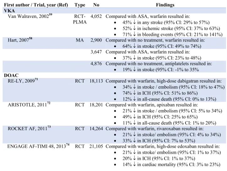
Of note, these therapies yielded favorable safety and efficacy profiles compared with warfarin in 4 large randomized controlled trials (Table 5),71-74 with consistent reduction in intracranial and fatal/life-threatening bleeding compared to warfarin,75 emerging as the preferred choice for stroke prevention in AF patients, particularly in those newly started on OAC.28, 76 In a meta-analysis including data from all four direct OAC studied in the pivotal phase 3 clinical trials (vs warfarin), direct OAC significantly reduced stroke or systemic embolic events by 20% compared with warfarin (RR 0.81, 95% CI 0.73-0.91, p<0.0001), all-cause mortality by 10% (0.90, 0.85-0.95, p=0.0003) and intracranial hemorrhage by
50% (0.48, 0.39-0.59, p<0.0001), but slightly increased the risk of gastrointestinal bleeding (RR 1.25, 1.01-1.55, p=0.04).75 Despite slight increase in OAC use linked to the advantageous characteristics of direct OAC over vitamin K antagonists (improved efficacy/safety ratio, predictable anticoagulant effect with no need for routine monitoring, fewer drug and food interactions), up to 40% of AF patients at high stroke risk still fail to receive appropriate thromboembolic prophylaxis in contemporary practice.77
Importantly, there seems to be no “class effect” of direct OAC, since the potential risk of bleeding from different direct OACs is not necessarily the same. The AVERROES (Apixaban versus Aspirin to Prevent Stroke in Atrial Fibrillation Patients Who Have Failed or Are Unsuitable for Vitamin K Antagonist Treatment) showed similar rates of major bleeding (1.4% vs 1.2% per year, p=0.57) and intracranial hemorrhage (0.4% vs 0.4%, p=0.69) between apixaban and aspirin, the former being much more effective in stroke prevention (1.6% vs 3.7% per year, HR=0.45, p<0.001).78 In the recently published ELDERCARE AF trial (Edoxaban Low-Dose for Elder Care Atrial Fibrillation Patients), which randomized once-daily 15-mg dose of edoxaban or placebo in nearly 1,000 very elderly Japanese patients (mean age 87±4 years) unsuitable for direct OAC (mean creatinine clearance 36±14 ml/min, low body weight 51±11 kg, 1/3 on non-steroidal anti-inflamatory drugs, >50% on antiplatelet agents), low-dose edoxaban did not increase the risk of intracranial bleeding (0.3% vs 0.6%), although it tripled the rate of gastrointestinal bleeding (2.3% vs 0.8%) compared to placebo.79 Altogether, the efficacy and safety of direct OAC is generally consistent across different studies regardless of having received prior vitamin K antagonists, but care should be taken to avoid gastrointestinal bleedings.
Table 5. Randomized data on Pharmacological Stroke Prevention Therapies in AF
Adapted from Alkhouli et al80 with permission.
ARISTOTLE: Apixaban for Reduction in Stroke and Other Thromboembolic Events in Atrial Fibrillation; ASA: aspirin; CI: confidence interval; DOAC: direct oral anticoagulants; ENGAGE AF-TIMI 48: Effective Anticoagulation with Factor Xa Next Generation in Atrial Fibrillation–Thrombolysis In Myocardial Infarction 48; HR:hazard ratio; ICH: intracranial hemorrhage; MA: analysis; OAC: Oral anticoagulation; PLMA: patient-level meta-analysis; RCT: randomized controlled trial; RE-LY: Randomized Evaluation of Long-Term Anticoagulation Therapy; ROCKET AF: Rivaroxaban Once Daily Oral Direct Factor Xa Inhibition Compared with Vitamin K Antagonism for Prevention of Stroke and Embolism Trial in Atrial Fibrillation; VKA: vitamin K antagonists
1.2.3. Antiplatelet agents
1.2.3.1. Single antiplatelet therapy
Aspirin monotherapy has been shown to be less effective than warfarin for stroke prevention in AF patients,58 and its benefit compared with no therapy remains
First author / Trial, year (Ref) Type No Findings VKA
Van Walraven, 200259
RCT-PLMA 4,052 Compared with ASA, warfarin resulted in: 45% in any stroke (95% CI: 29% to 57%)
52% in ischemic stroke (95% CI: 37% to 63%)
71% in bleeding events (95% CI: 21% to 141%) Hart, 200758 MA 2,900 Compared with no treatment, warfarin resulted in:
64% in stroke (95% CI: 49% to 74%) 3,647 Compared with ASA, warfarin resulted in:
37% in stroke (95% CI: 23% to 48%) 4,876 Compared with no treatment, antiplatelets resulted in:
19% in stroke (95% CI: -1% to 35%
DOAC
RE-LY, 200971 RCT 18,113 Compared with warfarin, high-dose dabigatran resulted in:
34% in stroke / embolism (95% CI: 18% to 47%)
74% in ICH (95% CI: 51% to 86%)
12% in all-cause death (95% CI: 0% to 13%) ARISTOTLE, 201172 RCT 18,201 Compared with warfarin, apixaban resulted in:
21% in stroke / embolism (95% CI: 5% to 34%)
49% in ICH (95% CI: 25% to 65%)
11% in all-cause death (95% CI: 1% to 20%) ROCKET AF, 201173 RCT 14,264 Compared with warfarin, rivaroxaban resulted in:
21% in stroke/ embolism (95% CI: 4% to 34%)
33% in ICH (95% CI: 7% to 53%)
ENGAGE AF-TIMI 48, 201374 RCT 21,105 Compared with warfarin, high-dose edoxaban resulted in:
21% in stroke/ embolism (95% CI: 1% to 37%)
20% in ICH (95% CI: 1% to 37%)
controversial. A meta-analysis by Hart et al.58 showed a 19% nonsignificant reduction in stroke with aspirin vs no therapy (95% CI: 1.0 to 35.0), but a large observational study in Sweden showed a higher incidence of ischemic stroke and thromboembolic events with aspirin monotherapy compared with no antithrombotic therapy.81 Overall, bleeding rates on aspirin monotherapy are non-inferior to those on OAC,82 and should not be recommended for stroke prevention in AF patients.
1.2.3.2. Dual antiplatelet therapy (DAPT)
The ACTIVE W and ACTIVE A (Atrial Fibrillation Clopidogrel Trial With Irbesartan for Prevention of Vascular Events) randomized trials evaluated the potential benefit of adding clopidogrel to aspirin for stroke prevention in AF patients. The ACTIVE W trial (DAPT vs warfarin) was stopped prematurely due to a significantly lower annual rate of stroke, systemic embolism, myocardial infarction or vascular death with warfarin compared to DAPT (3.9% vs 5.6%, RR: 0.69, 95% CI, 0.57-0.85).83 In the ACTIVE A trial (DAPT vs aspirin in patients ineligible for OAC), DAPT was associated with a lower rate of stroke, systemic embolism, myocardial infarction or vascular death (RR: 0.89, 95% CI, 0.81-0.98) but at the expense of increased major bleeding (RR: 1.57, 95% CI, 1.29-1.92).84 Overall, a pooled analysis of these trials showed that the addition of clopidogrel translated into a modest net clinical benefit in patients unsuitable for OAC (0.57 ischemic stroke prevented [95% CI: 0.12-1.24] per 100 patients-year of treatment).85 However, bleeding risk on DAPT is similar to that on OAC, and should be avoided as stroke prevention therapy in AF patients.
1.3. NON-PHARMACOLOGICAL STRATEGIES FOR STROKE PREVENTION
1.3.1. Rationale for left atrial appendage closure
Despite the benefits and increasing use of direct OAC, close to one in ten patients have a contraindication to OAC and 2% have an absolute contraindication (major intracranial pathology or end-stage liver disease).86, 87 In contemporary clinical practice, up to 40% of patients at high risk for stroke do not receive OAC due to fear of serious bleedings, and approximately 20% of patients discontinued direct OAC therapy in randomized clinical trials.77 The pivotal role of the LAA in AF-related thrombogenesis constitutes the rationale for mechanical LAAC, as an alternative stroke-prevention therapy for AF patients deemed not suitable for OAC.
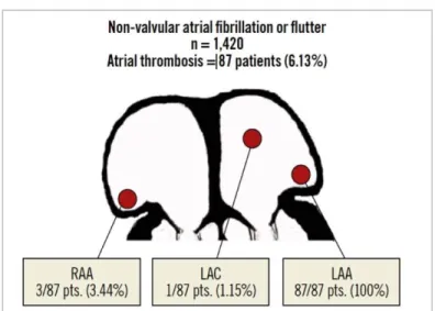
Autopsy and surgical data suggest that LAA is the most common source of thrombus formation in AF,18, 88 since the fibrillating LAA creates a favorable milieu for blood stagnation and thrombus formation. Furthermore, transesophageal echocardiography suggests that most AF-related strokes result from LAA thromboembolism.89 A meta-analysis from Blackshear et al.18 pointed out that 91% of thrombi in nonvalvular AF patients were located in the LAA. In a recent, large-scale study including >1,400 patients with nonvalvular AF or atrial flutter undergoing transesophageal echocardiography before electrical cardioversion, the localization of atrial thrombosis was inside the LAA in 100% of the cases, and 4.6% of the patients (0.28% of the overall study population) had concomitant extra-LAA thrombus (3.4% in the right atrial appendage and 1.2% in the left atrial cavity) (Figure 6).90 The low prevalence of extra-LAA thrombus observed in this study reinforces the potential role of LAAC for preventing thromboembolic events.
Figure 6. Localization of left atrial thrombi in patients with non-valvular atrial fibrillation or flutter.
From Cresti et al90 with permission.
Finally, there may also be a role for LAAC as an adjunctive therapy to OAC in patients with recurrent stroke despite optimal OAC, after exclusion of other plausible causes (by cerebral computed tomography, echocardiography, carotid Doppler). Although this strategy may apply to a limited proportion of LAAC candidates (<5%), recent data suggest feasibility and safety of such an approach at 2-year follow-up.91, 92
1.3.2. Embryology, anatomy and function of the LAA
Embryologically, the LAA is a remnant of the primary atrial tube which develops during the third week of fetal cardiac development, whereas the remaining smooth left atrium derives from the primordial pulmonary veins. At week 4, right-handed looping of the primary endocardial tube takes place, bringing the caudal and cranial ends in close proximity. At this stage, the appendages and the atrium differentiate (balloon outward) laterally from the superolateral wall of the primary heart, whereas the ventricles balloon out in a more anterior-posterior fashion. The outpouching of the superior and left sided aspect of the primary atrial tube finally constitutes the LAA, with subsequent trabeculae formation around the fifth week of gestation.93
The LAA varies in size, shape and in its relationship with surrounding structures (Figure 7). There are three components: an ostium (or os), neck and body. The ostium connects the left atrium and the LAA, generally running at an oblique angle to the mitral valve annulus, and is the distance from the limbus to the mitral annulus. The neck is the narrowest part of the LAA and overlays the left circumflex artery. The body is the most variable part, often multilobulated (range: 1 to 4), with 2 lobes in up to 54% of the patients.94 The LAA is an anterolateral structure that extends parallel to the left pulmonary veins, with the tip directed anteriorly and cranially, overlapping the pulmonary trunk and adjacent to the origin of the left descending coronary artery. The superior aspect is related to the pulmonary trunk, separated by the transverse sinus. The inferior aspect is closely related to the left circumflex artery and the great cardiac vein, that run beneath the neck of the LAA and along the atrioventricular groove and the mitral valve. Anteriorly, the lobes run parallel to the obtuse margin of the left ventricle and the left phrenic nerve courses posterolaterally. The posterior and superior aspects of the ostium are well-delimitated by a ridge separating the ostium from the left upper pulmonary vein, which corresponds epicardially to the ligament (or vein) of Marshall. The left phrenic nerve courses posterolaterally.95
Figure 7. The left atrial appendage and surrounding structures.
From Naksuk et al.95 with permission.
GCV: Great cardiac vein; LAA: Left atrial appendage; LCX: Left circumflex; LIPV: Left inferior pulmonary vein; LLL: Left lateral ridge; LOM: Ligament of Marshall; LPN: Left phrenic nerve; LSPV: Left superior pulmonary vein; PA: Pulmonary artery; SVC: Superior vena cava
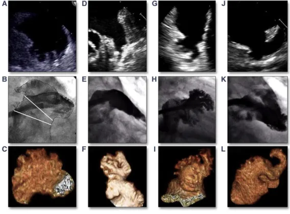
Several LAA shapes and variants have been described (Figure 8): chicken wing (dominant lobe with a bend in the middle part, folding back on itself with a secondary lobe or twig), windsock (one dominant lobe larger than the distal portion of the LAA), cactus (dominant central lobe with secondary lobes extending superiorly and inferiorly) and cauliflower (complex, irregular, multilobed anatomy with no dominant lobe). In a study by Biase et al.96 using computed tomography and cardiac magnetic resonance, the chicken wing morphology was the most common (48%) and less thrombogenic anatomy, followed by cactus (30%), windsock (19%) and cauliflower (3%), the latter associated with the highest risk of embolic event.
Figure 8. Multimodality imaging morphological classification of the LAA assessed by transesophageal imaging (top), angiography (middle) and computed tomography (bottom).
Cauliflower (A-C), windsock (D-F), cactus (G-I), chicken wing (J-L). From Beigel et al.97 with permission.
The LAA has several physiologic unique functions, mainly reservoir, neurohormonal and electrical. First, the LAA is the most compliant structure within the left atrial chamber, acting as a reservoir and decompression chamber during left ventricular systole and in conditions of left atrial pressure and/or volume overload (eg. exercice, atrial arrhythmias or heart failure).98 In patients with AF and/or increased filling pressures, LAA remodeling takes place, resulting in reduced contractile function and distensibility, and greater risk of thrombus formation. Second, the LAA is responsible for about 30% of all atrial natriuretic peptide production, modulating the left atrial pressure by activating stretch receptors, with effects on heart rate, diuresis and natriuresis.99 Finally, chronic AF may result into remodeling, inflammation and fibrosis of both the left atrium and the LAA, leading to focal triggers and re-entry arrhythmias, thus constituting a vicious cycle.100
1.3.3. Indications for LAA closure
OAC therapy remains the standard of care for patients with nonvalvular AF and a CHA2DS2-VASC score 2. However, long-term OAC may chronically expose patients to
increased risk of hemorrhagic complications, a concerning issue among frail, elderly patients, with prior bleeding history or predisposition, considered poor candidates for OAC. Over the last 2 decades, LAAC has emerged as a valid alternative to OAC for patients with contraindication to long-term OAC. Current American and European guidelines state that percutaneous LAAC may be considered for high-risk AF patients who are deemed unsuitable for OAC and consider surgical LAA excision for AF patients undergoing cardiac surgery (Table 6).28, 42
Table 6. Society guideline recommendations for left atrial appendage closure
Guide Recommendation Grade LOE
2019 AHA/ACC/HRS42 Percutaneous LAAC may be considered in patients with AF at increased risk of stroke who have contraindications to long-term OAC
IIb B
Surgical LAAC may be considered in patients with AF undergoing cardiac surgery, as a component of an overt heart team approach to the management of AF
IIb B
2020 ESC28 Percutaneous LAAC may be considered in patients with AF and contraindications for long-term anticoagulant treatment
IIb B
Surgical excision of the LAA may be considered in patients with AF undergoing cardiac surgery
IIb C
ACC: American College of Cardiology; AF: Atrial fibrillation; AHA: American Heart Association; ESC: European Society of Cardiology; HRS: Heart Rhythm Society; LAAC: Left atrial appendage closure; LOE: Level of evidence; OAC: Oral anticoagulation
Additionally, the latest consensus statement on catheter-based LAAC considers potential indications for transcatheter LAAC in the following 5 scenarios:20
Patients with a contraindication for OAC. Those patients represent the most accepted clinical indication and the vast majority of patients currently undergoing LAAC. Whereas no specific definition for “absolute”contraindication to OAC exists, conditions generally contraindicating long-term OAC include risk for major or life-threatening bleeding (intracranial/intraspinal bleeding, severe gastrointestinal bleeding, untreatable pulmonary or urogenital bleeding) or severe side effects under vitamin K antagonists or direct OAC. Although no randomized data targeting this specific group of patients is available so far, safety and efficacy of this strategy have been widely demonstrated in several observational studies and registries, and are currently being evaluated in ongoing randomized trials (ASAP-TOO, NCT0292828497; CLOSURE-AF, NCT03463317).