HAL Id: hal-03018580
https://hal.archives-ouvertes.fr/hal-03018580
Submitted on 23 Nov 2020HAL is a multi-disciplinary open access archive for the deposit and dissemination of sci-entific research documents, whether they are pub-lished or not. The documents may come from teaching and research institutions in France or abroad, or from public or private research centers.
L’archive ouverte pluridisciplinaire HAL, est destinée au dépôt et à la diffusion de documents scientifiques de niveau recherche, publiés ou non, émanant des établissements d’enseignement et de recherche français ou étrangers, des laboratoires publics ou privés.
Spectroscopic study of GdVO4: Yb + Er crystals
S Klimin, P Loiseau, Daniel Caurant, M Popova
To cite this version:
S Klimin, P Loiseau, Daniel Caurant, M Popova. Spectroscopic study of GdVO4: Yb + Er crystals. Quantum Electronics, Turpion, 2020. �hal-03018580�
Quantum Electronics 50 (3) 259 – 262 (2020)
Spectroscopic study of GdVO
4: Yb + Er crystals
S.A. Klimin, P. Loiseau, D. Caurant, M.N. Popova
S.A. Klimin, M.N. Popova Institute for Spectroscopy, Russian Academy of Sciences, Fizicheskaya ul. 5, Troitsk, 108840 Moscow, Russia; e-mail: [email protected]
P. Loiseau, D. Caurant Chimie ParisTech, PSL Research University, CNRS, Institut de Recherche de Chimie Paris, 75005, Paris, France
Abstract. GdVO4 crystals doped with different erbium and ytterbium concentrations have been
grown in order to study their spectroscopic and kinetic properties and identify the nature of active centres in them. Their polarised transmission and luminescence spectra have been used to construct energy level diagrams of the Er3+ ion. The lifetime of the lower level of the 4I11/2
excited-state multiplet has been shown to decrease markedly with increasing ytterbium concentration. Inequivalent erbium centres have been observed to form in the crystals with increasing ytterbium concentration.
Keywords: GdVO4 :Yb + Er, Stark levels, luminescence, lifetime, inequivalent centres.
1. Introduction
Yb3+ and Er3+ ions are widely used as a pair in photonics in multilayer film light sources [1],
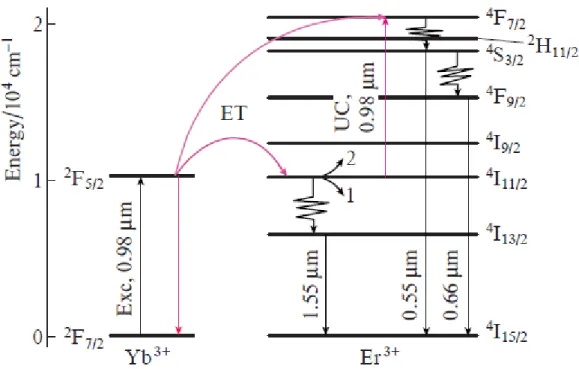
microchip lasers [2], novel compact ‘eye-safe’ fibre lasers [3], efficient up-conversion sources [4, 5], optical memory [6], etc. The Yb3+ and Er3+ trivalent ions, a sensitiser and an activator, respectively, serve different functions: owing to its rich system of energy levels, erbium allows one to obtain emission in different spectral ranges, whereas ytterbium ensures strong pump absorption and efficient energy transfer to erbium (Fig. 1). RVO4 (R = Gd, Y) vanadate crystals
(zircon structure) doped with erbium and ytterbium optically active ions have excellent optical characteristics [7 – 14]. They are commonly known to be promising and widely used laser materials owing to their unique properties: good chemical stability, excellent thermal conductivity and large absorption and emission cross sections [15 – 19].
Gadolinium vanadate has the zircon (ZrSiO4 mineral) structure. It has the form of colourless
crystals with tetragonal symmetry: space group I41/amd (Z = 4), unit-cell parameters a = 0.7212
nm and c = 0.6346 nm [20]. Its crystal structure is schematised in Fig. 2. Each gadolinium atom is coordinated by eight O2– oxygen ions. The gadolinium site has point symmetry D2d. The
GdO8 polyhedra form a three-dimensional network. Each polyhedron shares one edge with each
of the four adjacent GdO8 polyhedra. In addition, the GdO8 polyhedra are linked through VO4
tetrahedra isolated from each other. The Er3+ ion has a rich set of optical transitions, among which the 4I13/2 → 4I15/2 transition falls in the very important spectral range around l = 1.5 mm.
This spectral range is of great interest for telecommunications because here silica fibre has a minimum absorption. Besides, the spectral range 1.5 – 1.6 mm is known to be eye-safe and is used in telemetry. The ability to improve the performance of emitters operating in this range, including laser sources, has been the subject of extensive studies (see e.g. Refs [21 – 24]). In the Er – Yb codoped samples, process 1 (Fig. 1: relaxation from the 4I11/2 multiplet to 4I13/2,
followed by emission at = 1.55 mm) competes with process 2 – up-conversion through energy transfer from ytterbium to excited erbium, which is thus brought to the 4F7/2 state, following
which visible emission channels appear. In practice, processes 1 [3, 12] and 2 [7, 8, 10, 11, 14, 16] occur simultaneously. Despite the extensive studies of the optical properties of erbium- and/or ytterbium-doped GdVO4 crystals and nanopowders, information about the energy level
structure of Er3+ and Yb3+ ions in this host is still limited. In this paper, we report a systematic study of the energy level structure of optically active erbium and ytterbium ions in GdVO4
crystals. GdVO4 single crystals doped with different erbium and ytterbium concentrations were
grown by the Czochralski technique. Their spectroscopic properties were studied by optical absorption and luminescence spectroscopies. We determined the lifetime of the Er3+4I11/2 level
in different crystals and examined how it was influenced by the sensitiser concentration. 2. Experimental
We grew GdVO4 : Er (1 at %), GdVO4 : Yb (2 at %), GdVO4 : Yb (10 at %) + Er (0.5 at %)
and GdVO4 : Yb (37 at %) + Er (0.5 at %) single crystals. For measurements, we prepared
plane-parallel plates whose faces were parallel to the c axis. Transmission spectra were obtained at temperatures from 4.5 to 300 K and frequencies from 5 000 to 22 500 cm–1 on a Bruker IFS 125HR high-resolution Fourier transform spectrometer. To gain information about the symmetry of levels, spectra were measured for two light polarisations: (k c, E c, H || c) and (k c, E || c, H | c). Luminescence spectra were excited by the radiation from an argon laser (Coherent Innova 90) pumped Ti:sapphire laser (Coherent Ring Model 899) and synchronously detected using a monochromator fitted with a PbS detector. In lifetime measurements, excitation was provided by a parametric oscillator at the absorption wavelength of the Yb3+ ion. The oscillator was pumped by the BMI Nd: YAG third harmonic. The excitation pulse duration was 10 ns. The emission from the sample was detected at the monochromator output using an InGaAs detector and 150-MHz digital oscilloscope. The signal was averaged over 256 measurements.
3. Results and discussion
In the crystal structure of GdVO4, the Gd3+ trivalent ions and rare-earth (RE) substituents
occupy sites of tetragonal symmetry (point group D2d). The wave functions of an RE ion having
an odd number of electrons (as in Er3+ and Yb3+) transform according to the
6 and 7
irreducible representations of point group D2d. Table 1 presents nonzero components of the
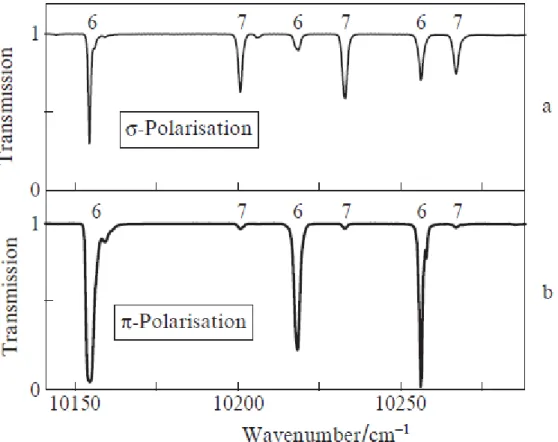
electric and magnetic dipole moments for optical transitions in this symmetry group. It is seen that the 7 6 electric dipole (ED) transitions are allowed for both the- and -polarisations.
At the same time, the 7 7 and 6 6 ED transitions can only be observed in the case of
-polarisation. Figure 3 shows transmission spectra of the GdVO4 : Er (1 at %) crystal in the
spectral range of the Er3+ 4I15/2 4I11/2 intermultiplet transition for both - and -polarisations.
This multiplet consists of six doublets, three of each symmetry: 36 + 37. We assume the
ground state of the Er3+ ion to have 7 symmetry, as in YVO4 : Er [25] and the isostructural
compound YPO4 : Er [26]. Since the energy of the first excited state of the Er3+ ion in the crystal
temperature of 4.5 K, so just six lines, corresponding to transitions from the ground state (of 7
symmetry) to the six levels of the 4I15/2 4I11/2 multiplet, should be seen in the spectrum of
the 4I11/2 transition. According to selection rules, three lines of 7 6 ED transitions are
allowed for -polarisation, which we observe as strong lines in Fig. 3b. The 7 7 transitions
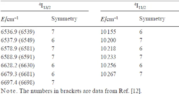
for -polarisation can show up only as MD transitions. In the case of -polarisation, all transitions are allowed in the ED approximation. Thus, we were able to determine not only the energy but also the symmetry of the Stark levels of the 4I11/2 multiplet (Table 2). Also indicated
in Table 2 are the present data for the 4I13/2 multiplet. The energies of the 4I13/2 multiplet levels
determined in this study agree well with those reported by Ter-Gabrielyan et al. [12].
Figure 4 shows absorption spectra of the GdVO4 : Er (1 at %), GdVO4 : Yb (2 at %) and GdVO4
: Yb (10 at %) + Er (0.5 at %) crystals. It is seen that the Er3+ and Yb3+ ions in the GdVO4
host have identical lower level energies of the 4I
11/2 (Er) and 2F5/2 (Yb) multiplets, which is a
critical condition for efficient energy transfer. The lines at 10155 cm–1 in stpectra 1 and 2 are seen to have weak satellites, which are probably due to RE pair centres [27, 28].
Figure 5a shows luminescence spectra in the region of the 4I13/2 4I15/2 transition at
liquid-nitrogen and room temperatures. At the higher temperature, the emission lines are broad. Cooling markedly reduces their width and leads to an intensity redistribution due to changes in level populations in the upper multiplet, which are determined by the Boltzmann distribution. Analysis of the spectra with the use of the data in Table 2 makes it possible to identify all spectral lines with certainty and reliably determine the energies of the upper Stark levels 6, 7 and 8 of the 4I15/2 multiplet in Fig. 5b: 245, 265 and 300 cm–1, respectively. The measured
lifetime of the lower level of the 4I11/2 multiplet in the GdVO4 : Er (1 at %) crystal is 230 ms.
In the codoped Yb – Er samples, the fluorescence around 1 mm originates from the thermalised 2F5/2 (Yb) and 4I11/2 (Er) multiples and the measured lifetime decreases to 195 ms in the case
of GdVO4 : Yb (10 at %) + Er (0.5 at %) and is even shorter (79 ms) in the GdVO4 : Yb (37 at
%) + Er (0.5 at %) crystal. At the same time, the lifetime of the 4I13/2 state is essentially
independent of the ytterbium concentration in the crystals: ~2.76 ms. The decrease in the lifetime of the 4I11/2 state with increasing ytterbium concentration is likely due to the increase
in up-conversion probability (Fig. 1, process 2).
Figure 6 shows the line shapes in the low-temperature transmission spectra for the same transitions in two GdVO4 crystals: one singly doped with erbium and the other co-doped with
erbium and ytterbium. In the former case, the lines are rather narrow and their width is determined by inhomogeneous broadening. In the latter, each line has a complex shape and is a combination of a few lines. A similar line shape in spectra of RE ions was observed by us previously for other compounds co-doped with RE ions [29 – 31]. It occurs when the nearest neighbour environment of an RE ion contains another RE ion having a different ionic radius. As shown in Fig. 2, each gadolinium ion in GdVO4 has four gadolinium ions as nearest
neighbours. The GdVO4 : Yb (10 at %) + Er (0.5 at %) crystal can contain different erbium
centres: a usual centre, surrounded by four gadolinium ions; a centre in which one nearest neighbour site is occupied by ytterbium instead of gadolinium; etc. Thus, there are inequivalent centres. Since the Gd3+ and Yb3+ ions differ in ionic radius, the GdO8 and YbO8 polyhedra are
distorted because they share one edge, or in other words two oxygens. The crystal field distortion for such a centre entails corrections to its Hamiltonian and, accordingly, corrections to its Stark level energies. As shown earlier [30, 31], the integrated intensities of envelopes constituting a composite line are proportional to the probabilities (P) of formation of inequivalent centres. Such probability can be found as
P = Cn4x4(1 – x)4 – n, (1)
where x is the fraction of an impurity ion producing defects and n is the number of nearest neighbour impurity atoms. The situation can also be described in terms of clusters. The main cluster has the form of ErGd4; a centre with one defect is an ErYbGd3 cluster. Using relation
(1), it is easy to obtain that, in the case of GdVO4 :Yb (10 at %) + Er (0.5 at %), the probability
of formation of the main cluster is ~0.65 and that of the first defect cluster is 0.29. At first glance, such relations qualitatively describe the complex line shapes in the spectra in Fig. 6. 4. Conclusions
Polarised transmission spectra of erbium- and ytterbium doped GdVO4 crystals have been
measured for the first time, and information about the energies and symmetries of the Stark levels of the 4I11/2 and 4I13/2 multiplets has been obtained. Varying the ytterbium doping level
(ytterbium concentration) changes the probabilities of phonon relaxation and up-conversion from the 4I11/2 multiplet. The formation of inequivalent erbium centres with increasing
ytterbium concentration leads to complex, multicomponent shapes of the spectral lines corresponding to Er3+ intermultiplet transitions.
Acknowledgements. This work was supported through the Photonic Technologies in Probing
Inhomogeneous Media and Biological Objects Program. References
1. Bai G., Yang Z., Lin H., Jie W., Hao J. Nanoscale, 10, 9261 (2018). 2. Du T., Luo Z., Yang R., Huang Y., et al. Opt. Lett., 42, 462 (2017).
3. Galagan B.I., Denker B.I., Egorova O.N., Kamynin V.A., Ponosova A.A., Sverchkov S.E., Semjonov S.L., Tsvetkov V.B. Quantum Electron., 48, 550 (2018) [ Kvantovaya Elektron., 48, 550 (2018)].
4. Du K., Xu X., Yao S., Lei P., Dong L., Zhang M., Feng J., Zhang H. CrystEngComm, 40, 1945 (2018).
5. Zhang Q., Yue S., Sun H., Wang X., Hao X., An S. Mater. Chem. C, 5, 3838 (2017).
6. Wolter B., Pullen M.G., Baudisch M., Sclafani M., Hemmer M., Senftleben A., Schröter C.D., et al. Phys. Rev. X, 5, 021034 (2015).
7. Min B.H., Jung K.Y. RSC Adv., 9, 20002 (2019).
8. Gavrilovié T.V., Jovanovié D.J., Lojpur V., Dramiéanin M.D. Sci. Sintering, 47, 221 (2015). 9. Gavrilovié T.V., Jovanovié D.J., Lojpur V., Nikolié A., Dramiéanin M.D. Sci. Rep., 4, 4209 (2014).
10. Khrushchalina S.A., Ryabochkina P.A., Kyashkin V.M., Vanetsev A.S., Gaitko O.M., Tabachkova N.Yu. JETP Lett., 103, 302 (2016) [ Pis’ma Zh. Eksp. Teor. Fiz., 103, 342 (2016)]. 11. Petrov V.V., Pestryakov E.V., Trunov V.I., et al. Proc. SPIE, 6731, 67310I (2007).
12. Ter-Gabrielyan N., Fromzel V., Ryba-Romanowski W., Lukasiewicz T., Dubinskii M. Opt.
Express, 20, 6080 (2012).
13. Savchuk O.A., Carvajal J.J., Cascales C., Aguiló M., Díaz F. ACS Appl. Mater. Interfaces, 8, 7266 (2016).
14. Mahata M.K., Tiwari S.P., Mukherjee S., Kumar K., Rai V.K. J. Opt. Soc. Am. B, 31 (8), 1814 (2014).
15. Sokolska I., Heumann E., Kiick S., et al. Adv. Solid-State Lasers, 50, 378 (2001). 16. Zhuang N.F., Hu X.L., et al. Appl. Phys. B, 82, 607 (2006).
17. Dimitrov D., Rafailov P., Marinova V., et al. J. Phys. Conf. Ser., 794, 012029 (2017). 18. Huber G., Kränkel C., Petermann C. J. Opt. Soc. Am. B, 27, B93 (2010).
19. Gorbachenya K.N., Kisel’ V.E., Yasyukevich A.S., Matrosov V.N., Tolstik N.A., Kuleshov N.V. J. Appl. Spectrosc., 82, 208 (2015) [ Zh. Prikl. Spektrosk., 82, 2214 (2015)]. 20. Mullica D.F., Sappenfield E.L., Abraham A.A., Chakoumakos B.C., Boatner L.A. Inorg.
Chim. Acta, 248, 88 (1996).
21. Trikshev A.I., Kamynin V.A., Tsvetkov V.B., Itrin P.A. Quantum Electron., 48, 1109 (2018) [ Kvantovaya Elektron., 48, 1109 (2018)].
22. Gorlachuk P.V., Ivanov A.V., Kurnosov V.D., Kurnosov K.V., Marmalyuk A.A., Romantsevich V.I., Simakov V.A., Chernov R.V. Quantum Electron., 48, 495 (2018) [Kvantovaya Elektron., 48, 495 (2018)].
23. Dyudelev V.V., Mikhailov D.A., Andreev A.D., et al. Quantum Electron., 49, 1158 (2019) [Kvantovaya Elektron., 49, 1158 (2019)].
24. Ponosova A.A., Azanova I.S., Mironov N.K., Yashkov M.V., Riumkin K.E., Kel’ O.L., Sharonova Yu.O., Melkumov M.A. Quantum Electron., 49, 693 (2019) [Kvantovaya Elektron., 49, 693 (2019)].
25. Capobianco J.A., Kabro P., Ermeneux F.S., Moncorge R., Bettinelli M., Cavalli E. Chem.
Phys., 214, 329 (1997).
26. Popova M.N., Klimin S.A., Moiseev S.A., Gerasimov K.I., Minnegaliev M.M., Baibekov E.I., Shakurov G.S., Bettinelli M., Chou M.C. Phys. Rev. B, 99, 235151 (2019).
27. Pytalev D.S., Klimin S.A., Popova M.N. Phys. Lett. A, 372, 3506 (2008). 28. Pytalev D.S., Klimin S.A., Popova M.N. Phys. Lett., A, 372, 2332 (2008).
29. Sattayaporn S., Loiseau P., Aka G., Klimin S., Boldyrev K., Mavrin B. J. Lumin., 219, 116895 (2020).
30. Popova M.N., Klimin S.A., Higel P., Dhalenne G. Phys. Lett. A, 354, 487 (2006).
Table 1. Nonzero components of the electric and magnetic dipole moments, di and i (i = x, y, z), respectively, of transitions for point group D2d and ions having an odd number of electrons.
The subscripts e (electric dipole, ED) and m (magnetic dipole, MD) indicate allowed transitions for the corresponding polarisation.
Figure 1. Energy level diagram of the Yb3+ and Er3+ ions, showing multiplets and the wavelengths of some transitions; Exc, excitation; ET, energy transfer; process 1: nonradiative relaxation and emission; process 2: up-conversion (UC).
Figure 2. Partial crystal structure of GdVO4. Right side: GdO8 polyhedron projected along the
Figure 3. Transmission spectra of the GdVO4 : Er (1 at %) sample for the Er3+ 4I15/2 → 4I11/2
transition at a temperature of 4.5 K in polarised light for (a) - and (b) -polarisations. Also indicated are representations of the symmetry group D2d: 6 (6) and 7 (7) of the 4I11/2 multiplet
levels.
Figure 4. Absorption spectra of the ( 1 ) GdVO4 : Er (1 at %), ( 2 ) GdVO4 : Yb (2 at %) and ( 3 ) GdVO4 : Yb (10 at %) + Er (0.5 at %) samples in the region of the Yb3+ 0 – 0 transition at a
Figure 5. (a) Luminescence spectra of the GdVO4 : Yb (37 at %) + Er (0.5 at %) crystals at
temperatures of 77 and 300 K; (b) energy level diagram of the 4I15/2 ground-state and 4I13/2
excited-state multiplets, with some spectral transitions indicated.
Figure 6. Spectral lines of the 4I15/2 4I9/2, 4I15/2 4S3/2 and 4I15/2 4I13/2 transitions for the
( 1 ) GdVO4 : Er (1 at %) and ( 2 ) GdVO4 : Yb (10 at %) + Er (0.5 at %) samples at a temperature