HAL Id: hal-03023067
https://hal.archives-ouvertes.fr/hal-03023067
Submitted on 25 Nov 2020
HAL is a multi-disciplinary open access
archive for the deposit and dissemination of
sci-entific research documents, whether they are
pub-lished or not. The documents may come from
teaching and research institutions in France or
abroad, or from public or private research centers.
L’archive ouverte pluridisciplinaire HAL, est
destinée au dépôt et à la diffusion de documents
scientifiques de niveau recherche, publiés ou non,
émanant des établissements d’enseignement et de
recherche français ou étrangers, des laboratoires
publics ou privés.
laser ablation in liquids
Hongli Du, Victor Castaing, Dongcai Guo, Bruno Viana
To cite this version:
Hongli Du, Victor Castaing, Dongcai Guo, Bruno Viana.
Rare-earths doped-nanoparticles
prepared by pulsed laser ablation in liquids.
Ceramics International,
Elsevier,
2020,
UNCORRECTED
PROOF
Ceramics International xxx (xxxx) xxx-xxx
Contents lists available at ScienceDirect
Ceramics International
journal homepage: http://ees.elsevier.com
Rare-earths doped-nanoparticles prepared by pulsed laser ablation in liquids
HongliDu
a,b,∗∗, VictorCastaing
a, DongcaiGuo
b, BrunoViana
a,∗aPSL Research University, CNRS, Institut de Recherche de Chimie Paris, Chimie ParisTech, Paris, France bCollege of Chemistry and Chemical Engineering, Hunan University, Changsha, China
A R T I C L E I N F O
Keywords Laser ablation Nanoparticles Persistent luminescence Rare-earthsA B S T R A C T
This feature article highlights the recent advances on the lanthanide-activated nanoparticles prepared by pulsed laser ablation in liquids (PLAL). In the past decade, rare-earths-activated nanoparticles have been recognized from various applications including lots of high-tech products, green technologies, bioimaging and medical uti-lization. To obtain inorganic nanoparticles with different morphologies and sizes, pulsed laser ablation in liquids is a green and versatile technique. In this paper, a survey of the nanoparticle's formation during the laser abla-tion in the liquids is first introduced. We focus on the control of the size and morphology through proper laser parameters, choice of the reaction solution and proper surfactants. In the last part of the paper, we successfully prepared persistent luminescent SrAl2O4: Eu2+, Dy3+by laser ablation in liquids and their optical features are
investigated.
1. Introduction
With abundant and unique energy level structures arising from 4fn
inner shell configurations, rare-earths-activated inorganic materials can exhibit emission mainly via intra-4f or 5d-4f transitions. When 4f-4f transitions are concerned, the luminescence features of trivalent lan-thanides (RE3+) will present high photo stability, high luminescence
quantum yield, narrow bandwidth, long-lived emission, large Stokes shifts, and have received high interest because of the increasing de-mand for photoluminescence related applications, including lighting, electronic display, anti-counterfeiting, biological labeling, imaging [1], theranostic [2]etc. Among them, persistent luminescence [3–5] is a spe-cial property of RE3+or transition metal doped materials with
signifi-cant interest. Based on the nanoengineering development, the prepara-tion of nano-sized lanthanide-activated materials is an important chal-lenge [6]. Wet chemical methods, including sol-gel procedure [7], com-bustion synthesis [8], hydro-(solvo-) thermal route [9], coprecipitation [10], microwave synthesis [11], template method [12], and thermal decomposition [13] are commonly used routes to obtain nano-sized materials. Most of these chemical techniques require organic solvents during the synthesis which may cause toxic residues, highly undesir-able in view of applications and for green chemistry. Pulsed laser ab-lation in liquids (PLAL) has demonstrated to be an efficient and ver-satile technique to produce high-quality nanoparticles (NPs) of a wide range of materials [14]. PLAL technique possesses a number of ad-vantages over traditional methods previously cites. First, laser ablation process is considered as “green chemistry” process. Second, the nano
materials with size dispersion below 100 nm can be produced in faster and cleaner ways, even if sometimes the compounds quantity is at a lim-ited value. Third, as long as there is the related laser equipment, experi-mental set up is minimal, and chemical precursors are replaced by bulk materials obtained by solid state reaction, so PLAL could be considered as a low-cost method. Finally, PLAL can be used to construct some com-plex structure compounds or modify the surface of nanoparticles, such as, inorganic nanoparticles coated with organic molecules that can be obtained in one step, in-situ or ex-situ [15]. Due to such advantages, the research on nanoparticles elaboration using PLAL has been of some interest during the last ten years. Looking at the topic literature, (see Fig. 1(a)), one can observed the increase interest for nanoparticles elab-orated with PLAL technique and a large number of elements of the pe-riodic table have been investigated as marked in Fig. 1 (b). As a sum-mary in Fig. 1 (c), the nanoparticles obtained by PLAL methods with size ranged between 5 and 100 nm have been proposed in biological, optical devices or as photocatalyst.
Laser ablation technique could deliver large amounts of energy highly concentrated into one point of a material. Laser ablation of mate-rials has been carried out in vacuum, in air and in liquids. Because of the unique confinement effect from liquid environment, there is many ad-vantages when the laser ablation occurs in liquid. First, the work piece will be immersed into a liquid or be employed with a liquid film. The liquid environment could lower the heat load on the work piece, con-fines the vapor and plasma, and increases the shock pressure on the sur-face [16,17]. At the same time, the main fractions of the particles ob-tained by PLAL are often preserved with the same chemical composi
∗Corresponding author.
∗∗Corresponding author. PSL Research University, CNRS, Institut de Recherche de Chimie Paris, Chimie ParisTech, Paris, France.
E-mail addresses: hongli.du@chimieparistech.psl.eu (H. Du); bruno.viana@chimieparistech.psl.eu (B. Viana)
https://doi.org/10.1016/j.ceramint.2020.04.291
Received 18 December 2019; Received in revised form 14 April 2020; Accepted 30 April 2020 Available online xxx
UNCORRECTED
PROOF
Fig. 1. (a) Citation per year in the past decade determined by a Web of Science search using the keyword “pulsed laser ablation in liquids” and “nanoparticles”. (b) Library of materials
available for PLAL is summarized in the periodic table. (c) Materials synthesized through PLAL technique and applications.
tion as the bulk targets. This has been demonstrated with alloys [18], and various oxides-based compounds [19]. Therefore, PLAL is gener-ally considered to combine top-down (micro-sized solid targets) and bot-tom-up (the formed plasma, atoms and clusters) methods with major processes controlled by laser plasma and physical cavitation [20]. How-ever, as other chemical synthesis processes, several parameters have to be optimized to get the highest possible quality of materials. In case of PLAL techniques, laser parameters (such as wavelength, pulse duration, laser energy) and the liquid environment (solvent, temperature, surfac-tant for instance) are the key points to optimize.
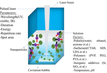
The possible factors that could influence the formation of crystalline nanoparticles with controlled size and morphology are summarized in Fig. 2 [21]. In the following part, we will briefly report how these fac-tors can control the nanoparticles formation.
2. Nanoparticles formation mechanism under laser ablation
During the whole laser ablation in liquids, the nanoparticles forma-tion process can be described as follows: when the laser pulses irradi-ate the bulk target in liquids, breakdown process and plasma genera-tion take place. Then, because of the liquid environment, a stronger con-finement of the plasma and a fast energy transfer from the plasma to the surrounding liquid occur. Later, a cavitation bubble, which is a dy-namic system with an expansion phase and a collapse phase, is formed. Finally, the cavitation bubbles collapse when the bubble reaches its maximum radius [20], and at the same time, NPs are released in the liquid to form a stable colloidal solution. So, the whole laser ablation process could also be considered as the plasma formation and cavita-tion bubbles accavita-tion as well as the nanoparticles formacavita-tion. The formed
UNCORRECTED
PROOF
H. Du et al. Ceramics International xxx (xxxx) xxx-xxx
Fig. 2. Main factors affecting the nanoparticles formation by pulsed laser ablation in
liq-uids media (adapted from Ref. [20]).
particles always present bimodal size distribution. The following part describes the main processes and bimodal size particles formation.
2.1. Plasma formation, cavitation bubbles and nanoparticles formation
The main process with the laser-target-liquid system can be summa-rized as following, when the laser is switched on, the microscopic dy-namic action on the surface of the bulk target could go through three stages: plasma formation, cavitation bubbles expansion, and bubbles col-lapse with particles release (see Fig. 3 (a), (b) and (c) respectively) [22].
When laser pulses interact with a target immersed in a liquid, a plasma composed of highly ionized or atomic species is formed imme-diately. The plasma's temperature and pressure can be as high as thou-sands of Kelvin and hundreds of Pascal. This high pressure, high tem-perature, and high density of the plasma could provide a favorable ther-modynamic environment for the formation of some NPs which are dif-ficult to obtain by conventional chemical methods. But as Park's group reported [23], the duration of the plasma is generally in the range of tens of ns to few μs for each laser pulse. So, it is difficult to determine the chemical reactions in the plasma. During the plasma decay, the en-ergy is transferred to the liquid, producing a layer of vapor with roughly the same volume as the plasma, comparable to shockwaves. Shockwaves favor the chemical reaction of the ionized species in the expanded cavi-tation bubbles. Gilmore model [24] describes the cavicavi-tation bubble dy-namics occurring during PLAL. In contrast, during bubble expansion and compression, the temperature inside the cavitation bubbles is only few hundreds of Kelvin. Recently, simulation results were experimentally verified [20].
2.2. Bimodal size particles formation
The particles obtained by PLAL technique often present bimodal size distribution. It has been reported that there is primary and secondary formation of NPs during the laser ablation. Small nanoparticles forma-tion is based on thermal vaporizaforma-tion mechanism and larger particles formation is attributed to explosive boiling mechanism [25,26]. Soli-man et al. have investigated the bubble action to give insights on the possible processes of primary and secondary NPs formation [27]. They concluded that primary particles are uniformly dispersed from the tar-get surface toward the top of the cavitation bubble, and that their mass varies according to the bubble's shape. The secondary particles are dom-inant particularly in the center of the cavitation bubble, and they cannot penetrate into the liquid before the bubble collapses. Larger particles are formed after the collapsed of the bubble. In addition, Bonse et al. come up with the pulse duration-related mechanisms, and they proposed that size distribution could have a relationship with the beam intensity dis-tribution on the target surface [28].
3. Control of size, morphology and yield of the process
To prepare nanoparticles with good performances, one should take into consideration the size, the morphology and the productivity or namely the quantitative yield of the process. For most laser systems, multiple tunable parameters may influence the preparation of the nanoparticles. Herein, some laser parameters and the choice of the solu-tion are considered.
3.1. Laser parameters
For the laser experimental set up, there are many parameters that should be taken into account, the uppermost being the following: laser fluence, pulse duration, repetition rate, and laser wavelengths. Yield and size of nanoparticles (NPs) vary with main laser parameters summarize in Table 1. Among these laser parameters, each parameter could af-fect the others. For example, larger NPs could be formed with high-en-ergy laser pulses [29], because high-enhigh-en-ergy laser pulses cause higher NPs formation yield and performed strong explosive boiling. This en-hances aggregation and coalescence of the particles. On the contrary, it has been reported that reducing laser energy [30,31] could decrease both size and size-distribution of PLAL-synthesized particles. However, smaller NPs can be obtained with low energy laser pulses but unfortu-nately with lower yield [32]. In contrast, when laser pulse fluence in-creases, smaller sizes NPs can be generated with narrow size distrib-ution [33]. Also, as Hamad et al. reported, when increasing the laser pulse duration, TiO2NPs synthesized by PLAL in deionized water could
lead to smaller particles [34]. Furthermore, considerations on the rep-etition rate, types of laser pulses, absorption by particles with fs, ps
Fig. 3. Main processes of laser ablation occurring in solution (figure adapted from Ref. [22]).
UNCORRECTED
PROOF
Table 1Yield and size of nanoparticles (NPs) vary with main laser parameters (“↑↓” means increase and decrease, respectively).
Main laser
parameters variation yield Size Size distribution laser fluence [37] ↑ ↑ ↑ smaller NPs↓; larger NPs ↑ pulse duration [36] ↑ ↑ ↓ ns, ↓; fs and ps, ↑ repetition rate [38] ↑ ↑ ↓ ↑
Wavelength [39] UV→NIR ↑ ↑ ↑
and ns laser systems have been reported. Tsuji et al. defined: (i) “in-ter-pulse” energy absorption by the ablated particles with fast repeti-tion rates and ultrafast lasers. (ii) “intra-pulse” absorprepeti-tion for ns laser systems. Another important laser parameter is the laser wavelength. It is reported that, under the same pulse duration, shorter-wavelengths (such as, 532 or 355 nm) are a good choice to generate smaller size NPs [35,36].
Taking all the laser parameters into consideration, it can be con-cluded that, the complexes and mutual cross-effects of laser parameters make difficult to develop a versatile system to control the size, and yield of the obtained nanoparticles. Then, one should take care of all these parameters based on the properties of the materials to get high-quality nanoparticles by PLAL technique.
3.2. PLAL solutions
For PLAL technique, the liquid or solution plays a very important role on the formation of nanoparticles. Solution is not only considered as a liquid medium to suspend the source particles but also strongly affects the size and stability of the obtained NPs as well as the production yield. In addition, possible chemical reactions such as oxidation or reduction [40] could occur. Then, solution with or without additives could have a large effect on the NPs size distribution and properties.
It has been reported that the nanoparticles can be prepared in vari-ous solutions. Maneeratanasarn et al. reported the synthesis of ɤ-Fe2O3
in ethanol and acetone liquid-phase by pulsed laser ablation technique, while amorphous α-Fe2O3was formed in deionized water [41]. Gökce
et al. investigated plasmonic nanoparticles (copper, silver and gold) pre-pared in different solvents (water, acetone, ethanol and ethyl acetate). They found that the polarity of the solvents have a significant impact on the size, quantitative yield and growth dynamics of laser-generated nanoparticles, and concluded that smaller particles in a single growth regime can be obtained in more polar solvents [42].
The temperature of the solution could also have some influence on the property of obtained particles. With different temperatures, different sizes and morphologies of ZnO and Zn(OH)2nanoparticles are formed
[43]. ZnO columnar crystals were formed from Zn under laser pulses in deionized water at 80 °C. ZnO small nanoparticles are possibly pro-duced by ablation even at a higher temperature than room temperature [44]. Haram et al. took silver and gold nanoparticles formation as ex-ample, and they found that increasing the temperature of the distilled water promoted the fusion of nanoparticles leading to the formation of nano-chains, nano-networks and super clusters [45].
In practice, the formed particles in solution are always charged. The ablated species formed during the laser ablation process have charged interfaces and different pH media result in nanoparticles with various degrees of aggregation. Palazzo et al. reported the stability of gold nanoparticles prepared by laser ablation in different pH media and con-cluded that a strong negative zeta-potential assures the stability against aggregation [46].
Some others parameters such as liquid depth above the target could also play significant role in the laser ablation process and therefore in
the size of nanoparticles. Mahdied et al. investigated this parameter with nanosecond laser ablation in various water depths and concluded that optimized water depth above the target can result in the produc-tion of smaller size and uniform nanoparticles with narrow size distribu-tions [47]. The dynamics of nanosecond pulsed laser ablation in liquids (ns-PLAL) could be significantly altered in the immediate vicinity of a free boundary. When the liquid layer thickness approximates the plasma size that is induced in bulk-liquid ablation, part of the plasma plume is formed outside the liquid [48]. The authors concluded that the liquid layer should be thicker than the plasma plume size to ensure laser-in-duced stresses which are useful for formation of the particles [48].
Furthermore, in order to improve the properties of nanoparticles ob-tained by PLAL, different kinds of additives, such as surfactants, salts, polymers, biomolecules, and organic solvents, can be applied to tune the NPs' size and modify the NPs’ property. For instance, Abdi et al. used CTAB as cation surfactant to improve the degree of crystallinity of ZnO nanoparticles [49]. SDS as anion surfactant has often been used to control the size of the particles prepared with laser ablation in wa-ter [50]. In addition, for biological applications, biopolymers [51,52], such as albumin, starch, gelatin and chitosan are used in the laser abla-tion technique because they have a salting-out effect on the formaabla-tion of NPs, in addition to their excellent biocompatibility and biodegradabil-ity. Concerning the shape of NPs, Mafuné et al. successfully designed core-shell structure to control the Ni NPs size during their formation by PLAL in water [53]. Silver nanoparticles can be loaded on silica support (SBA-15). Because of the stable silica support, smaller and more homo-geneously dispersed nanoparticles can be elaborated.
4. Rare-earths doped-nanoparticles
With the increasing need of nanoparticles to be used in semicon-ductor and bio-applications, more and more researches focus on the rare-earth doped nanoparticles. Rare-earths doped nanoparticles pre-sent good photostability and wide possible optical features including the well-known up-conversation, down-conversation and persistent lumines-cence processes [54]. To obtain desired NPs sizes and properties with green and effective technique, PLAL method is one of the most effec-tive method but not extensively used up to now. For optical nanopar-ticles, Park et al. successfully prepared the well-known Y3Al5O12:Ce3+
nanoparticles by PLAL with spherical shape and size ranging from 5 to 25 nm. The obtained colloidal YAG:Ce3+nanocrystals present good
lu-minescence properties and similar optical features than their bulk coun-terparts [55]. In a domain of wide expansion, up-conversion nanoparti-cles have been prepared by PLAL and present good crystallinity and op-tical properties (see Table 2). When the nanoparticles are prepared in liquid, special solutions or additives could protect the NPs from defects on the surface, improve the properties of nanoparticles and favor their good dispersion. Then, researchers used surfactant [56] or inorganic ad-ditives [57] to improve the crystallinity, morphology and to limit size dispersion. Some of the researches on the lanthanide-activated nanopar-ticles prepared by PLAL have been summarized in Table 2.
5. Focus on SrAl2O4: Eu2+, Dy3+NPs persistent phosphor and challenges
For the abundant applications of lanthanide-activated nanoparticles in various fields, such as, lighting, display, energy and biomedicine, it is necessary to obtain nanoparticles with high crystal quality, good morphology and uniform nano-sizes. Among the wet chemical synthe-sis methods, PLAL is one of the best ways to prepare lanthanide-acti-vated nanoparticles with high crystallinity and multi-element composi-tions. Lots of lanthanide-activated nanoparticles have been synthesized by PLAL, for instance, lanthanide-based down-conversation [64] and up-conversation nanoparticles [61,66]. Concerning the persistent phos-phors, the number of works is very limited. Recently, with transition
UNCORRECTED
PROOF
H. Du et al. Ceramics International xxx (xxxx) xxx-xxx
Table 2
Lanthanide-activated nanoparticles prepared by PLAL technique.
Materials Solution & surfactant Morphology Size(nm) Garnets
Y3Al5O12:Ce3+[55] Deionized water spherical 5–25
Y3Al5O12:Ce3+
(YAG:Ce) [31] deionized water &2-[2-(2-methoxyethoxy)ethoxy]acetic acid (MEEAA)
nanorod 1.9–4.7 Y3Al5O12(YAG:Ce)
[58] deionized water string-like 9; 100
Silicates
Gd2SiO5:Ce3+ Deionized water / 5–10
Sr2MgSi2O7: Eu2+,
Dy3+[57] deionized water + SiO2 ellipticalnanorod /
Ca2Si5N8:Eu2+,
Tm3+[59] Water & NaOH and acetate web-like 3.5
Ca-α-SiAlON:Eu2+
[60] deionized water aggregation 90–110
Fluorides NaYF4:Yb3+/Er3+
[14,61] deionized water/2-[2-(2-Methoxyethoxy)ethoxy]acetic acid (MEEAA)
spherical 10-20; 40-60 YOF: Er3+, Yb3+
[62] deionized water nanorod 20–200
NaGdF4:Nd3+[63] DMSO not
completely spherical 20–30 Other Oxides Y2O3:Eu3+, Lu2O2S:Eu3+, Lu3TaO7:Gd3+/Tb3+ [64] Deionized water / 5–10
Tb3Al5O12:Ce3+[65] deionized water & lauryl
dimethylaminoacetic acid betain (LDA)
/ /
Y2O3:Eu3+,
Gd2O3:Eu3+,
deionized water &
2-[2-(2-methoxyethoxy)ethoxy]acetic acid (MEEAA)
nanorod 1.9–4.7 Y2O3: Er3+, Yb3+
[66] deionized water / Tens,hundred
CaMoO4:Tm3+/Yb3+
[67] ethanol spherical 15–30
NaYF4:Yb3+/Er3+
[14,61] deionized water/2-[2-(2-Methoxyethoxy)ethoxy]acetic acid (MEEAA)
spherical 10-20; 40-60 YVO4:Eu3+
[19,56,68] deionized water/sodium dodecylsulfate Ovoid-like 30–50
metal cations, nano-sized deep red persistent luminescent materials (ZnGa2O4:Cr3+) were successfully prepared by the pulsed laser ablation
in liquids without any surfactant [69]. The nanoscale particles present excellent deep red long persistent luminescence that may be use in the future as new probe in the first biological window. Indeed, in our lab-oratory in Paris, we have demonstrated the strong interest for this ma-trix for bio-imaging applications by a pioneer work published in 2011 [70] followed by important breakthroughs in the field [71–78]. Based on the previous works, Ca2Si5N8:Eu2+, Tm3+is one pioneer nice
exam-ple of the capability of PLAL technique to obtain RE doped NPs with persistent luminescence, and we further performed the first real-time in vivo imaging with Ca2Si5N8:Eu2+, Tm3+ NPs obtained with PLAL
method [59]. PLAL elaborated Sr2MgSi2O7: Eu2+, Dy3+nano-phosphors
can also present interesting persistent luminescence properties [57]. Fol-lowing these pioneering works, the preparation of rare-earth doped per-sistent phosphors such as SrAl2O4: Eu2+, Dy3+is encouraged through
pulsed laser ablation in liquids.
SrAl2O4: Eu2+, Dy3+compound, which is the most developed
per-sistent phosphor due to its outstanding long afterglow time and inten-sity, has never been prepared by this technique even if previous elabora-tion under laser are presented in the literature [78,79]. Laser synthesis techniques yield nanosized particles but the annealing at high tempera-ture required for the reduction of europium results in the formation of larger micro-sized agglomerates. The persistent luminescence researches on this material began more than twenty years ago, with the discov-ery of SrAl2O4doped with Eu2+, then through co-doping with trivalent
dysprosium, the afterglow was enhanced and reached more than 10 h [80]. Up to now, it is widely used in various identification and warn-ing signs on buildwarn-ings or highways. Furthermore, its high brightness and long-lasting time make this material interesting for further applica-tions in biomedicine, very recently, Terraschke et al. coated magnetic nanoparticles with SrAl2O4:Eu2+,Dy3+to open the path to combination
of imaging and in situ manipulation of relevant biomolecules [81]. How-ever, the elaboration of NPs still remains a challenge [82], which pro-pels to prepare nano-sized SrAl2O4: Eu2+, Dy3+phosphor.
5.1. SrAl2O4: Eu2+, Dy3+preparation and characterization
The preparation of the SrAl2O4: Eu2+, Dy3+persistent phosphor was
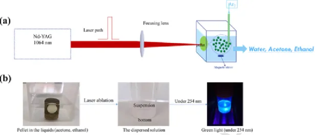
realized through pulsed laser ablation in liquids under the following conditions. The sintered pellets are used as targets for preparation of SrAl2O4: Eu2+, Dy3+nanophosphor. The experimental process to
pre-pare pellets is introduced in Supporting Information. During the ablation process, the pellets are immersed in 50 mL of solution in a quartz cell, and irradiated with 1064 nm-ns pulsed Nd:YAG laser at 10 Hz (see Fig. 4).
Different solutions such as water, ethanol, and acetone, different laser wavelengths (355 nm, 1064 nm) and different laser powers (10 mJ, 400 mJ) were used for the material preparation. The experi-mental conditions are summarized in Supporting Information, Fig. S1. Firstly, with the laser ablation in water, it appears impossible to obtain the SrAl2O4: Eu2+, Dy3+NPs but mainly SrCO3was formed [57] as seen
by XRD presented in Supporting Information, Fig. S2. This is due to the reaction between the Sr2+ion and CO2dissolved in water. In order to
remove the CO2from the solution, one should keep the process under Ar
gas bubbling all along the synthesis. In ethanol and acetone solutions, as shown in Fig. 5 presenting the XRD patterns, the obtained particles pre-sent SrAl2O4phase. As already reported, SrAl2O4has a tridymite
struc-ture constructed by corner sharing AlO4tetrahedra tilted with respect to
each other [83]. The occupation of Al3+cations in the compound
in-duced a charge deficiency that is compensated by Sr2+cation
incorpo-ration in channels created within rings of corner shared AlO4tetrahedra,
in the so-called stuffed tridymite structure [84]. There are two possible positions for Sr2+cations in this matrix.
When laser energy value is fixed at 10 mJ, with the near-infrared wavelength (1064 nm), the yield of the obtained particles appears larger than that with the UV wavelength (355 nm). For the same wavelength (1064 nm), but with much higher energy (400 mJ), higher yield and larger particles are obtained. This can be explained by the pulsed laser ablation mechanism in the solution as presented before. During the pulsed laser ablation on the pellet, cavitation bubbles will be formed on the surface of the pellet continuously, and with laser power increase, the cavitation bubbles lifetime increases. As a result, this will enhance the quantity of particles formed during the laser pulse. At the same time, high laser power may cause mechanical stress, as the tempera-ture and pressure inside the plasma plume are increased. This could further result in a collapse of the cavitation bubbles, which thereby intensify the collision frequency among particles, leading to coales-cence and larger particles formation [85,86]. Considering large pro-ductivity and good crystalline phase, SrAl2O4: Eu2+, Dy3+NPs were
UNCORRECTED
PROOF
Fig. 4. (a) Schematic diagram of the PLAL experimental setup for SrAl2O4: Eu2+, Dy3+synthesis; (b) pictures of different states of the target pellet during the reaction process.
Fig. 5. XRD spectra of the synthesized particles in different solutions.
prepared in either ethanol or acetone with laser wavelength at 1064 nm. Size distribution and optical feature are presented in the following part of the paper.
Fig. 6 shows images of the collected particles. The obtained parti-cles present bimodal size distribution. Parts of the partiparti-cles show mi-cro-size (Fig. 6 (a)) while TEM image in Fig. 6 (b) presents nanopar-ticles with sizes around 7.5 nm. As reported before, during the laser ablation process, particle size distributions could be composed of two modes. Small nanoparticles nucleate and grow in the target-solution mixing region based on thermal vaporization mechanism, while larger particles are formed by the fragmentation of the superheated molten
metal layer attributed to explosive boiling mechanism [26,87]. The size distributions of the thermal modes for each laser condition are very sim-ilar, only second fragmentation mode appears to vary with laser para-meters. The micro size particles are formed like a “top-down” method, while nano size particles are formed by the thermal nucleation in the cavitation bubbles.
5.2. Optical properties
The photoluminescence spectra of the particles obtained by laser ab-lation in ethanol and acetone (laser wavelength 1064 nm) are presented in Fig. 7.
The typical emission spectra of SrAl2O4:Eu2+, Dy3+at 20 K with
two emission bands located at 520 nm and 445 nm [88] are obtained for the laser ablation particles either in ethanol or acetone as shown in Fig. 7 (a). One could clearly observe an intensity variation between the two emission bands according to the elaboration medium. The two crystallographically different sites for Sr2+have different coordination
numbers, similar average Sr–O distances (i.e., 2.886 Å and 2.947 Å, re-spectively) [89], but slightly different individual Sr–O distances. Indeed, the two environments differ by a slight distortion [90]. Considering the two slightly different Sr2+ sites, one can expect Eu2+ to occupy the
sites more or less equally due to similar ionic radii. Non-radiative en-ergy transfers occur at room temperature between these two sites as already reported in the same host [89] or in others hosts [91]. Eu2+
cations in Sr II sites should present broad emission band at higher en-ergy (445 nm) while the Eu2+in the Sr I sites should be responsible for
the lower energy band (520 nm) (see insert Fig. 7). At higher tempera-ture, the thermal energy promotes Sr II to Sr I energy transfer as shown the insert in Fig. 7(b), which leads to a strong decrease of the 445 nm emission band in favor to the 520 nm band. Thus, one observed redis-tribution of emission intensities from both sites at different tempera
Fig. 6. Size distribution of the particles obtained in ethanol with 1064 nm: (a), micro-sized particles shown by Keyence VHX microscope; (b) nano-sized particles shown by TEM.
UNCORRECTED
PROOF
H. Du et al. Ceramics International xxx (xxxx) xxx-xxx
Fig. 7. PL spectra of the synthesized materials at different temperatures. (a) at 20 K; (b) at 300 K. Insert presents the energy transfer between two Eu2+sites occurring at RT. (UV
Excita-tion wavelength, 10 Hz).
tures [83,89,92,93]. Furthermore, the intensity of the emission band at 445 nm from particles obtained in acetone is higher than that of pellet and particles obtained in ethanol, keeping the intensity at 520 nm con-stant, revealing different energy transfer efficiencies between the two sites according to the synthesis conditions. As Gökce et al. reported, through PLAL process, larger particles will be formed in ethanol in re-gard to acetone due to agglomeration occurring in less polar solvents [42]. Therefore, one can assume that the smaller particles obtained in acetone present larger energy transfer efficiency probably due to their crystal field variation around the Eu2+ions [94,95].
SrAl2O4:Eu2+, Dy3+bulk compounds present excellent long
persis-tent luminescence. This phenomenon can be explained by the follow-ing processes: firstly, under UV light excitation, Dy3+stabilized
elec-trons trapping and enhanced the persistent luminescence; secondly, the electrons could be slowly released at room temperature and recombined
with the excited state of Eu2+. Finally, when return to Eu2+ ground
state, they give rise to persistent luminescence [80].
Fig. 8 shows the persistent luminescence decay profiles of the ab-lated SrAl2O4: Eu2+, Dy3+NPs in acetone and ethanol. After
excita-tion at 365 nm for 5 min at room temperature, the particles present per-sistent luminescence, but much shorter than that of their bulk coun-terpart compounds. Indeed, after about 5 min, the luminescence of the SrAl2O4:Eu2+, Dy3+NPs is too weak to be catched by eyes. The main
possible reason is probably related to the ultra-small size of particles and surface effects that occur in the non-optimized process, but also may be due to a reduction of the effective trap density as already re-ported [96]. As can be seen in Fig. 8, the persistent luminescence of the particles obtained in acetone present slightly longer persistent de-cay than that in ethanol. As already known, persistent luminescence is controlled by the defects, which are not only associated with the parti
UNCORRECTED
PROOF
Fig. 8. Persistent decay curve of the ablated particles in acetone (a) and in ethanol (b) at300 K, after being excited at 365 nm for 5 min.
cles size but also with the crystalline quality. Castaing et al. have demon-strated, in the ZnGa2O4:Cr3+nano-glass ceramics, the strong effect of
the crystalline quality on the persistent properties [97]. Cristoforetti et al. also reported that for particles prepared in acetone, enolate anions are formed on the particles surface and that ensures more stability [98]. Moreover, during the plasma creation, according to the solution, differ-ent reactive species can be formed. All that will have an influence on the defects and therefore on the persistent luminescence properties.
6. Conclusions
There is a lot of materials successfully prepared by PLAL. In or-der to elaborate nanoparticles with limited size, good morphology and outstanding properties through PLAL technique, various laser parame-ters, multiple solutions, and many others factors have been investigated. However, due to the complex inter-mutual-effects between these para-meters, it is difficult to develop a standard in the experimental proce-dure, especially to manage the bimodal sized nanoparticles formation by PLAL, even if some experimental devices, such as filters, are known to help controlling the particles size. Furthermore, upscaling of the quanti-tative yield of nanoparticles is another important issue that still need to be solved.
In the present work, while difficult in aqueous medium we success-fully synthesize SrAl2O4:Eu2+, Dy3+ NPs with PLAL in acetone and
ethanol solutions. At this time, control of uniform nano-size remains dif-ficult while the intensity of the persistent luminescence is far from those obtained in the micro size compounds. In the following steps, one should further better control the laser parameters and use various additives in the solution to enhance the optical properties. In case of success, this should open the path to new applications and new modality for instance as bio-label or in catalysis applications.
Declaration of competing interest
The authors declare that they have no known competing financial in-terests or personal relationships that could have appeared to influence the work reported in this paper.
Acknowledgments
China Scholarship Council (CSC) Grant and ANR-18-CE08-0012 PER-SIST for their support.
Appendix A. Supplementary data
Supplementary data to this article can be found online athttps://doi. org/10.1016/j.ceramint.2020.04.291.
References
[1] H H Gorris, O S Wolfbeis, Photon-upconverting nanoparticles for optical encoding and multiplexing of cells, biomolecules, and microspheres, Angew. Chem. 52 (2013) 3584–3600.
[2] J H Liu, T Lecuyer, J Seguin, N Mignet, D Scherman, B Viana, C Richard, Imaging and therapeutic applications of persistent luminescence nanomaterials, Adv. Drug Deliv. Rev. 138 (2019) 193–210.
[3] A Abdukayum, J T Chen, Q Zhao, X P Yan, Functional near infrared-emitting Cr3+/Pr3+co-doped zinc gallogermanate persistent luminescent nanoparticles
with superlong afterglow for in vivo targeted bioimaging, J. Am. Chem. Soc. 135 (2013) 14125–14133.
[4] Y Zhuang, L Wang, Y Lv, T-L Zhou, R-J Xie, Optical data storage and multicolor emission readout on flexible films using deep-trap persistent luminescence materials, Adv. Funct. Mater. 28 (2018) 1705769.
[5] Y Li, M Gecevicius, J R Qiu, Long persistent phosphors-from fundamentals to applications, Chem. Soc. Rev. 45 (2016) 2090–2136.
[6] X Qin, X Liu, W Huang, M Bettinelli, X Liu, Lanthanide-activated phosphors based on 4f-5d optical transitions: theoretical and experimental aspects, Chem. Rev. 117 (2017) 4488–4527.
[7] F Gao, H Liu, F Ren, K Wang, X Li, Y Wang, C He, Y Wei, Tunable structure and intensive upconversion photoluminescence for Ho3+-Yb3+codoped bismuth
titanate composite synthesized by sol-gel-combustion (SGC) method, Ceram. Int. (2019).
[8] H Chang, J Xie, B Zhao, B Liu, S Xu, N Ren, X Xie, L Huang, W Huang, Rare earth ion-doped upconversion nanocrystals: synthesis and surface modification, Nanomaterials 5 (2014) 1–25.
[9] C Wang, T Zhou, J Jiang, H Geng, Z Ning, X Lai, J Bi, D Gao, Multicolor tunable luminescence based on Tb3+/Eu3+doping through a facile hydrothermal route,
ACS Appl. Mater. Interfaces 9 (2017) 26184–26190.
[10] M N Luwang, R S Ningthoujam, S K Srivastava, R K Vatsa, Disappearance and recovery of luminescence in Bi3+, Eu3+codoped YPO4nanoparticles due to the
presence of water molecules up to 800 degrees C, J. Am. Chem. Soc. 133 (2011) 2998–3004.
[11] M Yang, G Deng, T Hou, X Jia, Y Wang, Q Wang, B Li, J Liu, X Liu, Facile microwave-assisted synthesis of Zn2GeO4:Mn2+, Yb3+uniform nanorods and
near-infrared down-conversion properties, Opt. Mater. 64 (2017) 152–159. [12] L S Lin, J Song, H H Yang, X Chen, Yolk-shell nanostructures: design, synthesis,
and biomedical applications, Adv. Mater. (2018) 30.
[13] J C Boyer, F Vetrone, L A Cuccia, J A Capobianco, Synthesis of colloidal upconverting NaYF4 nanocrystals doped with Er3+, Yb3+and Tm3+, Yb3+via
thermal decomposition of lanthanide trifluoroacetate precursors, J. Am. Chem. Soc. 128 (2006) 7444–7445.
[14] W J Parak, M Osinski, X-J Liang, L Gemini, M-C Hernandez, R Kling, Formation of upconversion nanoparticles of 18%Yb:1%Er:NAYF4by ultra-short pulse laser
ablation in water, 2016, p. 9722 972205.
[15] J Lee, S Mahendra, P J Alvarez, Nanomaterials in the construction industry: a review of their applications and environmental health and safety considerations, ACS Nano 4 (2010) 3580–3590.
[16] H W Kang, H Lee, A J Welch, Laser ablation in a liquid-confined environment using a nanosecond laser pulse, J. Appl. Phys. 103 (2008) 083101. [17] D J Hwang, T Y Choi, C P Grigoropoulos, Liquid-assisted femtosecond laser
drilling of straight and three-dimensional microchannels in glass, Appl. Phys. A 79 (2004) 605–612.
[18] K Y Niu, J Yang, S A Kulinich, J Sun, X W Du, Hollow nanoparticles of metal oxides and sulfides: fast preparation via laser ablation in liquid, Langmuir: ACS J. Surf. Colloid. 26 (2010) 16652–16657.
[19] H Wang, O Odawara, H Wada, Facile and chemically pure preparation of YVO4:Eu3+colloid with novel nanostructure via laser ablation in water, Sci. Rep.
6 (2016) 20507.
[20] M DellʼAglio, R Gaudiuso, O De Pascale, A De Giacomo, Mechanisms and processes of pulsed laser ablation in liquids during nanoparticle production, Appl. Surf. Sci. 348 (2015) 4–9.
[21] V Amendola, M Meneghetti, What controls the composition and the structure of nanomaterials generated by laser ablation in liquid solution?, Phys. Chem. Chem. Phys.: Phys. Chem. Chem. Phys. 15 (2013) 3027–3046.
[22] S I Kudryashov, A A Samokhvalov, A A Nastulyavichus, I N Saraeva, V Y Mikhailovskii, A A Ionin, V P Veiko, Nanosecond-laser generation of nanoparticles in liquids: from ablation through bubble dynamics to nanoparticle yield, Materials 12 (2019) 15.
[23] K K Kim, M Roy, H Kwon, J K Song, S M Park, Laser ablation dynamics in liquid phase: the effects of magnetic field and electrolyte, J. Appl. Phys. 117 (2015) 074302.
[24] F R Gilmore, The Growth or Collapse of a Spherical Bubble in a Viscous Compressible Liquid, 1952.
UNCORRECTED
PROOF
H. Du et al. Ceramics International xxx (xxxx) xxx-xxx
[25] J P Sylvestre, A V Kabashin, E Sacher, M Meunier, Femtosecond laser ablation of gold in water: influence of the laser-produced plasma on the nanoparticle size distribution, Appl. Phys. A 80 (2005) 753–758.
[26] W T Nichols, T Sasaki, N Koshizaki, Laser ablation of a platinum target in water. II. Ablation rate and nanoparticle size distributions, J. Appl. Phys. 100 (2006) 114912.
[27] W Soliman, N Takada, K Sasaki, Growth processes of nanoparticles in liquid-phase laser ablation studied by laser-light scattering, Appl. Phys. Exp. 3 (2010) 035201. [28] J Bonse, S Baudach, J Krüger, W Kautek, M Lenzner, Femtosecond laser ablation
of silicon–modification thresholds and morphology, Appl. Phys. A 74 (2014) 19–25.
[29] M H Mahdieh, B Fattahi, Size properties of colloidal nanoparticles produced by nanosecond pulsed laser ablation and studying the effects of liquid medium and laser fluence, Appl. Surf. Sci. 329 (2015) 47–57.
[30] S B, A V K, F M W, M Meunier, Ultrafast laser based “green” synthesis of non-toxic nanoparticles in aqueous solutions, Appl. Phys. A 93 (2008).
[31] D Amans, C Malaterre, M Diouf, C Mancini, F Chaput, G Ledoux, G Breton, Y Guillin, C Dujardin, K Masenelli-Varlot, P Perriat, Synthesis of oxide nanoparticles by pulsed laser ablation in liquids containing a complexing molecule: impact on size distributions and prepared phases, J. Phys. Chem. C 115 (2011) 5131–5139. [32] P Chewchinda, O Odawara, H Wada, The effect of energy density on yield of
silicon nanoparticles prepared by pulsed laser ablation in liquid, Appl. Phys. A 117 (2014) 131–135.
[33] E Solati, L Dejam, D Dorranian, Effect of laser pulse energy and wavelength on the structure, morphology and optical properties of ZnO nanoparticles, Optic Laser. Technol. 58 (2014) 26–32.
[34] A Hamad, L Li, Z Liu, A comparison of the characteristics of nanosecond, picosecond and femtosecond lasers generated Ag, TiO2and Au nanoparticles in
deionised water, Appl. Phys. Mater. Sci. Process 120 (2015) 1247–1260. [35] T Tsuji, K Iryo, Y Nishimura, M Tsuji, Preparation of metal colloids by a laser
ablation technique in solution: influence of laser wavelength on the ablation efficiency (II), J. Photochem. Photobiol. Chem. 145 (2001) 201–207. [36] A Hamad, L Li, Z Liu, A comparison of the characteristics of nanosecond,
picosecond and femtosecond lasers generated Ag, TiO2and Au nanoparticles in
deionised water, Appl. Phys. A 120 (2015) 1247–1260.
[37] R A Ismail, A M Mousa, M H Amin, Effect of laser fluence on the structural, morphological and optical properties of 2H-PbI2 nanoparticles prepared by laser ablation in ethanol, J. Inorg. Organomet. Polym. Mater. 28 (2018) 2365–2374. [38] A Menéndez-Manjón, S Barcikowski, Hydrodynamic size distribution of gold
nanoparticles controlled by repetition rate during pulsed laser ablation in water, Appl. Surf. Sci. 257 (2011) 4285–4290.
[39] K Zhang, D S Ivanov, R A Ganeev, G S Boltaev, P S Krishnendu, S C Singh, M E Garcia, I N Zavestovskaya, C Guo, Pulse duration and wavelength effects of laser ablation on the oxidation, hydrolysis, and aging of aluminum nanoparticles in water, Nanomaterials (2019) 9.
[40] T Tsuji, T Hamagami, T Kawamura, J Yamaki, M Tsuji, Laser ablation of cobalt and cobalt oxides in liquids: influence of solvent on composition of prepared nanoparticles, Appl. Surf. Sci. 243 (2005) 214–219.
[41] P Maneeratanasarn, T V Khai, S Y Kim, B G Choi, K B Shim, Synthesis of phase-controlled iron oxide nanoparticles by pulsed laser ablation in different liquid media, Phys. Status Solidi 210 (2013) 563–569.
[42] B Gökce, D D van’t Zand, A Menéndez-Manjón, S Barcikowski, Ripening kinetics of laser-generated plasmonic nanoparticles in different solvents, Chem. Phys. Lett. 626 (2015) 96–101.
[43] G G Guillén, M I M Palma, B Krishnan, D Avellaneda, G A Castillo, T K D Roy, S Shaji, Structure and morphologies of ZnO nanoparticles synthesized by pulsed laser ablation in liquid: effects of temperature and energy fluence, Mater. Chem. Phys. 162 (2015) 561–570.
[44] Y Ishikawa, Y Shimizu, T Sasaki, N Koshizaki, Preparation of zinc oxide nanorods using pulsed laser ablation in water media at high temperature, J. Colloid Interface Sci. 300 (2006) 612–615.
[45] N Haram, N Ahmad, formation of gold and silver nanochains and nanonetworks by liquid assisted laser ablation at elevated temperature, J. Cluster Sci. 26 (2014) 713–725.
[46] G Palazzo, G Valenza, M Dell’Aglio, A De Giacomo, On the stability of gold nanoparticles synthesized by laser ablation in liquids, J. Colloid Interface Sci. 489 (2017) 47–56.
[47] M H Mahdieh, B Fattahi, Effects of water depth and laser pulse numbers on size properties of colloidal nanoparticles prepared by nanosecond pulsed laser ablation in liquid, Optic Laser. Technol. 75 (2015) 188–196.
[48] T T P Nguyen, R Tanabe-Yamagishi, Y Ito, Impact of liquid layer thickness on the dynamics of nano- to sub-microsecond phenomena of nanosecond pulsed laser ablation in liquid, Appl. Surf. Sci. 470 (2019) 250–258.
[49] S Abdi, D Dorranian, Effect of CTAB concentration on the properties of ZnO nanoparticles produced by laser ablation method in CTAB solution, Optic Laser. Technol. 108 (2018) 372–377.
[50] S I Alnassar, E Akman, B G Oztoprak, E Kacar, O Gundogdu, A Khaleel, A Demir, Study of the fragmentation phenomena of TiO2nanoparticles produced by
femtosecond laser ablation in aqueous media, Optic Laser. Technol. 51 (2013) 17–23.
[51] C Rehbock, V Merk, L Gamrad, R Streubel, S Barcikowski, Size control of laser-fabricated surfactant-free gold nanoparticles with highly diluted electrolytes and their subsequent bioconjugation, Phys. Chem. Chem. Phys.: Phys. Chem. Chem. Phys. 15 (2013) 3057–3067.
[52] M Barry, B Ding, Y Jung, B V K Reddy, T X Phuoc, M K Chyu, Pulsed nanosecond laser ablation of gold in deionized water and aqueous chitosan solution, Optic Laser. Eng. 55 (2014) 59–68.
[53] F Mafuné, T Okamoto, M Ito, Surfactant-free small Ni nanoparticles trapped on silica nanoparticles prepared by pulsed laser ablation in liquid, Chem. Phys. Lett. 591 (2014) 193–196.
[54] D Jaque, C Richard, B Viana, K Soga, X G Liu, J G Sole, Inorganic nanoparticles for optical bioimaging, Adv. Optic Photon 8 (2016) 1–103.
[55] G S Park, K M Kim, S W Mhin, J W Eun, K B Shim, J H Ryu, N Koshizaki, Simple route for Y3Al5O12:Ce3+colloidal nanocrystal via laser ablation in deionized
water and its luminescence, Electrochem. Solid State Lett. 11 (2008) J23. [56] H Wang, O Odawara, H Wada, One-step preparation of YVO4:Eu3+nanoparticles
by pulsed laser ablation, J. Alloys Compd. 683 (2016) 1–6.
[57] M Ishizaki, T Katagiri, T Sasagawa, Y Kitamoto, O Odawara, H Wada, Preparation of SiO2-capped Sr2MgSi2O7:Eu,Dy nanoparticles with laser ablation in liquid, J.
Nanotechnol. 2012 (2012) 1–6.
[58] N Tsuruoka, T Sasagawa, T Yodo, M Yoshimoto, O Odawara, H Wada, Facile preparation of YAG:Ce nanoparticles by laser irradiation in water and their optical properties, SpringerPlus 5 (2016) 325.
[59] G S T Maldiney, B Viana, D Gourier, C Richard, D Scherman, M Bessodes, K Van den Eeckhout, D Poelman, P F Smet, In vivo optical imaging with rare earth doped Ca2Si5N8persistent luminescence nanoparticles, Opt. Mater. Express 2
(2012).
[60] H Wang, T Tomiya, T Takeda, N Hirosaki, O Odawara, H Wada, Fabrication of nanoscale Ca-α-SiAlON:Eu2+phosphor by laser ablation in water, Appl. Phys.
Exp. 8 (2015) 115001.
[61] L Gemini, T Schmitz, R Kling, S Barcikowski, B Gokce, Upconversion nanoparticles synthesized by ultrashort pulsed laser ablation in liquid: effect of the stabilizing environment, ChemPhysChem: Eur. J. Chem. Phys. Phys. Chem. 18 (2017) 1210–1216.
[62] R Anjana, K M Kurias, M K Jayaraj, Clean synthesis of YOF:Er3+, Yb3+
upconversion colloidal nanoparticles in water through liquid phase pulsed laser ablation for imaging applications, Opt. Mater. 72 (2017) 730–736.
[63] J Dai, G Feng, S Wang, H Zhang, S Dai, S Zhou, Transparent NaGdF4:Nd3+colloid
prepared by femtosecond laser ablation as a liquid laser medium, Optic Laser. Technol. 92 (2017) 202–205.
[64] G Ledoux, D Amans, C Dujardin, K Masenelli-Varlot, Facile and rapid synthesis of highly luminescent nanoparticles via pulsed laser ablation in liquid,
Nanotechnology 20 (2009) 445605.
[65] S W Mhin, J H Ryu, K M Kim, G S Park, H W Ryu, K B Shim, T Sasaki, N Koshizaki, Pulsed-laser-induced simple synthetic route for Tb3Al5O12:Ce colloidal
nanocrystals and their luminescent properties, Nanoscale Res. Lett. 4 (2009) 888–895.
[66] T Ikehata, Y Onodera, T Nunokawa, T Hirano, S-i Ogura, T Kamachi, O Odawara, H Wada, Photodynamic therapy using upconversion nanoparticles prepared by laser ablation in liquid, Appl. Surf. Sci. 348 (2015) 54–59.
[67] K Cho, J Choi, J-I Lee, J H Ryu, Pulsed-laser-assisted synthesis of a Tm3+/Yb3+
co-doped CaMoO4colloidal nanocrystal and its upconversion luminescence, J.
Kor. Phys. Soc. 68 (2016) 22–27.
[68] H Wang, O Odawara, H Wada, Morphology and optical properties of YVO4:Eu 3+
nanoparticles fabricated by laser ablation in ethanol, Appl. Surf. Sci. 425 (2017) 689–695.
[69] M S Relvas, M R N Soares, S O Pereira, A V Girão, F M Costa, T Monteiro, Trends in Cr3+red emissions from ZnGa2O4nanostructures produced by pulsed laser
ablation in a liquid medium, J. Phys. Chem. Solid. (2019).
[70] A Bessiere, S Jacquart, K Priolkar, A Lecointre, B Viana, D Gourier, ZnGa2O4:Cr3+:
a new red long-lasting phosphor with high brightness, Optic Express 19 (2011) 10131–10137.
[71] T Maldiney, A Bessiere, J Seguin, E Teston, S K Sharma, B Viana, A J Bos, P Dorenbos, M Bessodes, D Gourier, D Scherman, C Richard, The in vivo activation of persistent nanophosphors for optical imaging of vascularization, tumours and grafted cells, Nat. Mater. 13 (2014) 418–426.
[72] N Basavaraju, K R Priolkar, D Gourier, S K Sharma, A Bessiere, B Viana, The importance of inversion disorder in the visible light induced persistent luminescence in Cr3+doped AB
2O4(A = Zn or Mg and B = Ga or Al), Phys.
Chem. Chem. Phys.: Phys. Chem. Chem. Phys. 17 (2015) 1790–1799. [73] N Basavaraju, S Sharma, A Bessière, B Viana, D Gourier, K R Priolkar, Red
persistent luminescence in MgGa2O4: Cr3+; a new phosphor for in vivo imaging, J.
Phys. Appl. Phys. 46 (2013) 375401.
[74] T Lecuyer, E Teston, G Ramirez-Garcia, T Maldiney, B Viana, J Seguin, N Mignet, D Scherman, C Richard, Chemically engineered persistent luminescence nanoprobes for bioimaging, Theranostics 6 (2016) 2488–2524.
[75] B Viana, S K Sharma, D Gourier, T Maldiney, E Teston, D Scherman, C Richard, Long term in vivo imaging with Cr3+doped spinel nanoparticles exhibiting
persistent luminescence, J. Lumin. 170 (2016) 879–887.
[76] A Bessière, A Lecointre, R A Benhamou, E Suard, G Wallez, B Viana, How to induce red persistent luminescence in biocompatible Ca3(PO4)2, J. Mater. Chem. C
1 (2013) 1252–1259.
[77] J Xu, J Ueda, Y Zhuang, B Viana, S Tanabe, Y3Al5−xGaxO12:Cr3+: a novel red
persistent phosphor with high brightness, Appl. Phys. Exp. 8 (2015) 042602. [78] S K Sharma, D Gourier, E Teston, D Scherman, C Richard, B Viana, Persistent
luminescence induced by near infra-red photostimulation in chromium-doped zinc gallate for in vivo optical imaging, Opt. Mater. 63 (2017) 51–58.
UNCORRECTED
PROOF
[79] R Aroz, V Lennikov, R Cases, M L Sanjuán, G F de la Fuente, E Muñoz, Laser synthesis and luminescence properties of SrAl2O4:Eu2+, Dy3+phosphors, J. Eur.
Ceram. Soc. 32 (2012) 4363–4369.
[80] T Matsuzawa, A new long phosphorescent phosphor with high brightness, SrAl2O4:Eu2+,Dy3+, J. Electrochem. Soc. 143 (1996) 2670.
[81] H Terraschke, M Franzreb, C Wickleder, Magnetism and afterglow united: synthesis of novel double core-shell Eu2+-doped bifunctional nanoparticles, Chemistry (2020).
[82] H Zhang, Z Xue, B Lei, H Dong, H Zhang, S Deng, M Zheng, Y Liu, Y Xiao, A top-down method to fabricate SrAl2O4: Eu2+,Dy3+nanosheets from commercial
blocky phosphors, Opt. Mater. 36 (2014) 1802–1807.
[83] M Nazarov, M G Brik, D Spassky, B Tsukerblat, A Nor Nazida, M N Ahmad-Fauzi, Structural and electronic properties of SrAl2O4:Eu2+from density functional
theory calculations, J. Alloys Compd. 573 (2013) 6–10.
[84] A R Schulze, H Ml Buschbaum, Zur Verbindungsbildung von MeO: M2O3. IV. Zur
Struktur von monoklinem SrAl2O4, Z. Anorg. Allg. Chem. 475 (1981) 205–210.
[85] S A Al-Mamun, R Nakajima, T Ishigaki, Effect of liquid level and laser power on the formation of spherical alumina nanoparticles by nanosecond laser ablation of alumina target, Thin Solid Films 523 (2012) 46–51.
[86] M Abbasi, D Dorranian, Effect of laser fluence on the characteristics of Al nanoparticles produced by laser ablation in deionized water, Optic Spectrosc. 118 (2015) 472–481.
[87] C-Y Shih, R Streubel, J Heberle, A Letzel, M V Shugaev, C Wu, M Schmidt, B Gökce, S Barcikowski, L V Zhigilei, Two mechanisms of nanoparticle generation in picosecond laser ablation in liquids: the origin of the bimodal size distribution, Nanoscale 10 (2018) 6900–6910.
[88] J Botterman, J J Joos, P F Smet, Trapping and detrapping in SrAl2O4:Eu,Dy
persistent phosphors: influence of excitation wavelength and temperature, Phys. Rev. B 90 (2014).
[89] M Nazarov, M G Brik, D Spassky, B Tsukerblat, Crystal field splitting of 5d states and luminescence mechanism in SrAl2O4:Eu2+phosphor, J. Lumin. 182 (2017)
79–86.
[90] D Dutczak, T Justel, C Ronda, A Meijerink, Eu2+luminescence in strontium
aluminates, Phys. Chem. Chem. Phys.: Phys. Chem. Chem. Phys. 17 (2015) 15236–15249.
[91] A Bessiere, R A Benhamou, G Wallez, A Lecointre, B Viana, Site occupancy and mechanisms of thermally stimulated luminescence in Ca9Ln(PO4)7(Ln =
lanthanide), Acta Mater. 60 (2012) 6641–6649.
[92] P Dorenbos, Energy of the first 4f7→4f65d transition of Eu2+in inorganic
compounds, J. Lumin. 104 (2003) 239–260.
[93] J Bierwagen, S Yoon, N Gartmann, B Walfort, H Hagemann, Thermal and concentration dependent energy transfer of Eu2+in SrAl
2O4, Opt. Mater. Express
6 (2016) 793.
[94] B Cheng, H Liu, M Fang, Y Xiao, S Lei, L Zhang, Long-persistent phosphorescent SrAl2O4:Eu2+, Dy3+nanotubes, Chem. Commun. (2009) 944–946.
[95] Z Fu, S Zhou, S Zhang, Study on optical properties of rare-earth ions in nanocrystalline monoclinic SrAl2O4: Ln (Ln = Ce3+, Pr3+, Tb3+), J. Phys. Chem.
B 109 (2005) 14396–14400.
[96] Y Lin, Z Zhang, F Zhang, Z Tang, Q Chen, Preparation of the ultrafine
SrAl2O4:Eu,Dy needle-like phosphor and its optical properties, Mater. Chem. Phys.
65 (2000) 103–106.
[97] V Castaing, A D Sontakke, A J Fernández-Carrión, N Touati, L Binet, M Allix, D Gourier, B Viana, Persistent luminescence of ZnGa2O4:Cr3+transparent glass
ceramics: effects of excitation wavelength and excitation power, Eur. J. Inorg. Chem. 2017 (2017) 5114–5120.
[98] G Cristoforetti, E Pitzalis, R Spiniello, R Ishak, F Giammanco, M Muniz-Miranda, S Caporali, Physico-chemical properties of Pd nanoparticles produced by Pulsed Laser Ablation in different organic solvents, Appl. Surf. Sci. 258 (2012) 3289–3297.