Université de Montréal
Escherichia coli STb toxin induces apoptosis in intestinal epithelial cell lines
par
Hamida Claudia Syed
Département de pathologie et microbiologie Faculté de médecine vétérinaire
Mémoire présenté à la Faculté de médecine vétérinaire en vue de l’obtention du grade de maître ès sciences (M.Sc.)
en sciences vétérinaires option microbiologie
Décembre 2011
Université de Montréal
Faculté de médecine vétérinaire
Ce mémoire intitulé :
Escherichia coli STb toxin induces apoptosis in intestinal epithelial cell lines
présenté par : Hamida Claudia Syed
a été évalué par un jury composé des personnes suivantes :
Mario Jacques, président-rapporteur J. Daniel Dubreuil, directeur de recherche
La toxine stable à la chaleur de type b (STb) est une des toxines produites par les souches Enterotoxigenic Escherichia coli (ETEC) impliquée dans le développement de la diarrhée. Une étude antérieure par Goncalves et al. (2009) a démontré que les cellules ayant internalisé la toxine STb démontraient une morphologie qui rappelle l’apoptose. Le changement du potentiel membranaire observé par Goncalves et al. (2009) nous a incité à vérifier la capacité de la toxine STb à induire l’apoptose des cellules HRT-18 et IEC-18 par la voie intrinsèque. Les cellules HRT-18 et IEC-18 ont été traitées avec de la toxine purifiée pour une durée de 24 heures puis ells ont été récoltées et examinées pour des caratéristiques de l’apoptose. L’activation des caspases-9 et -3, mais pas de la caspase-8, a été observée dans les deux lignées cellulaires à l’aide des substrats fluorescents spécifiques pour chaque caspase. L’ADN extrait des cellules HRT-18 et IEC-18 a révélé une fragmentation lorsque migré sur gel d’agarose. La condensation et la fragmentation des noyaux ont été observées en microscopie à fluorescence suite à une coloration de l’ADN au Hoechst 33342. Les indices apoptotiques des cellules HRT-18 et IEC-18 traitées avec des quantités croissantes de STb montrent une dose-réponse pour les deux lignées. L’activation de la caspase-9 est une indication que la voie intrinsèque de l’apoptose est activée dans les cellules HRT-18 et IEC-18. L’absence de l’activation de la caspase-8 démontre que la voie extrinsèque n’est pas impliquée dans la mort cellulaire médiée par STb.
Mots clés: apoptose, caspase, mort cellulaire, fragmentation de l’ADN, ETEC, fragmentation nucléaire, toxine STb.
Heat-stable toxin b (STb) is one of the toxins produced by Enterotoxigenic
Escherichia coli (ETEC) strains implicated in the development of diarrhea. A
previous study conducted by Goncalves et al. (2009) showed that cells having internalized STb toxin demonstrated apoptotic-like morphology. The change in the mitochondrial membrane potential observed by Goncalves et al. (2009) prompted us to verify the ability of STb toxin to induce apoptosis via the intrinsic pathway in HRT-18 and IEC-18 cells. Both cell lines were treated with purified STb toxin for a period of 24 hours, harvested, and examined for apoptotic features. Activation of caspases-9 and -3, but not -8, was observed in HRT-18 and IEC-18 cells as determined with the use of fluorescent substrates specific to each caspase. Extracted DNA revealed DNA laddering when migrated on agarose gels. Nuclear condensation and fragmentation of Hoechst 33342 stained DNA of HRT-18 and IEC-18 cells were visualized by fluorescence microscopy. Apoptotic indexes of HRT-18 and IEC-18 cells treated with increasing amounts of STb toxin revealed dose-dependent responses in both cell lines. The activation of caspase-9 is an indication of the intrinsic pathway being activated in HRT-18 and IEC-18 cells by STb toxin. The lack of caspase-8 activation demonstrates that the extrinsic pathway of apoptosis is not involved in the programmed cell death mediated by STb.
Key words: apoptosis, caspase, cell death, DNA laddering, ETEC, nuclear fragmentation, STb toxin.
TABLE OF CONTENTS RÉSUMÉ………..iii ABSTRACT……….vi TABLE OF CONTENTS………..viii LIST OF FIGURES……….xi LIST OF ABBREVIATIONS………xii ACKNOWLEDGMENTS………....xvii INTRODUCTION………1
REVIEW OF THE LITERATURE………...4
1. PATHOGENIC E. COLI………...5
2. ETEC………...8
2.1 ETEC VIRULENCE FACTORS………10
2.2 COLONIZATION FACTORS………10 2.2.1 FIMBRIAE……….10 2.2.2 AFIMBRIAE………...11 2.3 TOXINS……….12 2.3.1. LT TOXIN………...12 2.3.2 ST TOXINS………..14 2.3.2.1 EAST-1………..14 2.3.2.2 STa TOXIN………15 3. STb TOXIN………...16 3.1 GENETICS……….17 3.2 SYNTHESIS………19 3.3 BIOCHEMICAL STRUCTURE……….22 3.4 RECEPTOR OF STb TOXIN………24 3.5 INTERNALIZATION………26 3.6 MODE OF ACTION……….28 3.7 IMMUNOGENIC POTENTIAL……….32
4. APOPTOSIS………..33
4.1 GENETIC REGULATION OF APOPTOSIS……….36
4.2 BCL-2 FAMILY PROTEINS………..39
4.2.1 BH3 DOMAIN………40
4.3 CASPASES………..42
4.4 EXTRINSIC APOPTOSIS……….45
4.5 INTRINSIC APOPTOSIS………...46
5. IONS IMPLICATED IN APOPTOSIS………..49
5.1 POTASSIUM………...49
5.2 CHLORIDE……….51
5.3 CALCIUM………...54
6. TOXINS INDUCING APOPTOSIS………...56
6.1 ENTEROTOXINS………56
6.1.1 CLOSTRIDIUM DIFFICILE TOXIN A………56
6.1.2 ESCHERICHIA COLI LT TOXIN………..57
6.2 PORE-FORMING ENTEROTOXINS……….58
6.2.1 VIBRIO CHOLERAE CYTOLYSIN………58
6.2.2 CLOSTRIDIUM PERFRINGENS ENTEROTOXIN………58
6.3 PORE-FORMING TOXINS………...59
6.3.1 STAPHYLOCOCCUS AUREUS -TOXIN………...59
6.3.2 HELICOBACTER PYLORI VACA……….60
7. PROJECT DESCRIPTION……….60
METHODOLOGY AND RESULTS………62
Article: Escherichia coli STb toxin induces apoptosis in intestinal epithelial cell lines………63
Abstract……….64
Introduction………..65
Materials and methods……….69
Discussion……….76 Conclusions………...79 References……….80 Acknowledgments……….86 Figure Legends……….87 Figures………..89 DISCUSSION………..94
1. CELL-TYPE AND APOPTOSIS……….95
2. CASPASE INVOLVEMENT IN STB-MEDIATED APOPTOSIS………97
3. FRAGMENTATION OF DNA AND NUCLEI BY STB………99
4. APOPTOSIS INDUCED BY STB IS DOSE-DEPENDENT………...100
5. APOPTOSIS INDUCTION BY ENTERIC PATHOGENS……….101
CONCLUSION……….104
FUTURE DIRECTIONS……….107
1. CELL-CYCLE ANALYSIS………..108
2. INVOLVEMENT OF OLIGOMERIZATION IN APOPTOSIS INDUCTION…109 3. APOPTOSIS IN IN VIVO CONDITIONS………...110
4. DEVELOPMENT OF A LOCAL INHIBITOR OF APOPTOSIS…………...110
LIST OF FIGURES
REVIEW OF THE LITERATURE
Figure 1: Mode of action of ETEC bacteria………...9
Figure 2: Synthesis of STb toxin………..20
Figure 3: Tertiary structure of STb………...23
Figure 4: Proposed mode of action of STb………...29
Figure 5: Comparison of the cellular death pathway in C. elegans and mammals………...37
Figure 6: BCL-2 family proteins………..40
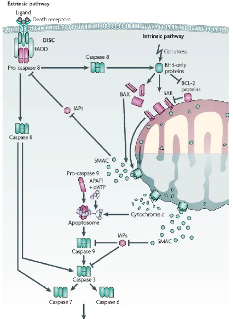
Figure 7: Extrinsic and intrinsic apoptosis………...48
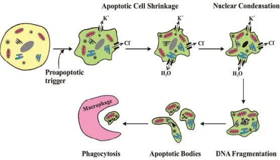
Figure 8: Cell shrinkage during apoptosis………53
ARTICLE Figure 1: Caspase activation in HRT-18 cells treated with STb………..89
Figure 2: Caspase activation in IEC-18 cells treated with STb………90
Figure 3: Gel electrophoresis DNA extracted from HRT-18 and IEC-18 cells treated with STb………...91
Figure 4: Condensed and fragmented nuclei in HRT-18 and IEC-18 cells….92 Figure 5: STb induces a dose-dependent apoptosis in HRT-18 and IEC-18 cells………93
LIST OF ABBREVIATIONS
AAA+ ATPase domain
AAF Aggregative adherence fimbriae AB toxin A: activity domain; B: binding domain
Ac Acetyl
ADN Acide déoxyribonucléique ADP Adenosine diphosphate A/E Attaching and effacing Afa afimbriae adhesins AGS Gastric epithelial cells
AIDA-I Adhesin Involved in Diffuse Adherence
Ala Alanine
AMP Adenosine monophosphate
APAF-1 Apoptosis Protease Activating Factor-1
Arg Arginine
Asp Aspartic acid
AVD Apoptotic Volume Decrease BAK Bcl-2 homologous antagonist/killer BAX Bcl-2–associated X protein
BCL-2 B-cell lymphoma 2
BH B-cell lymphoma 2 homology domain BID BH3 interacting-domain death agonist BIK Bcl-2-interacting killer
BIM Bcl-2-like protein 11 cAMP cylic AMP
CARD Caspase Activation and Recruitment Domain CD Circular Dichroism
CED-3 Cell Death Protein 3 CED-4 Cell Death Protein 4
CED-9 Cell Death Protein 9
CFA/I Colonization Factor Antigen I
CFTR Cystic fibrosis transmembrane conductance regulator cGMP cyclic GMP
CHAPS 3-[(3-cholamidopropyl)dimethylammonio]-1-propanesulfonate Cif Cycle inhibiting factor
CPE Clostridium perfringens enterotoxin
CS Coli Surface Antigen
CT Cholera toxin
DAEC Diffusely Adherent Escherichia coli DAF Decay Accelerating Factor
DEVD Aspartic Acid – Glutamic Acid – Valine – Aspartic Acid DFF DNA Fragmentation Factor
DMEM Dulbecco’s Modified Eagle Medium DNA Deoxyribonucleic acid
DsbA Disulfide-bond A oxidoreductase DsbC Disulfide-bond C oxidoreductase DISC Death-Inducing Signaling Pathway DTT Dithiothreitol
EAEC Enteroaggregative Escherichia coli EAST-1 Enteroaggregative heat-stable toxin 1 EDTA Ethylene diamine tetraacetic acid EGL-1 Egg-Laying Protein 1
EHEC Enterohemorrhagic Escherichia coli EIEC Enteroinvasive Escherichia coli ELISA Enzyme-linked immunosorbent assay EPEC Enteropathogenic Escherichia coli Esp E. coli secreted protein
ETEC Enterotoxigenic Escherichia coli FADD Fas Associated Death Domain FasL Fas Ligand
FasR Fas Receptor FBS Fetal Bovine Serum FITC Fluorescein isothiocyanate
G protein guanine nucleotide-binding protein GC-C guanylate cyclase type C
Gly Glycine
GM1 Monosialotetrahexosylganglioside GMP Guanosine monophosphate
GTP Guanosine triphosphate
HEPES 4-(2-hydroxyethyl)-1-piperazineethanesulfonic acid HRT-18 Human Colon Tumour Cells
IAP Inhibitor of Apoptosis Protein ICE Interleukin-1 Converting Enzyme IEC-18 Rat Ileum Epithelial Cells
Ile Isoleucine
IPTG Isopropyl-β-D-thio-galactoside IS Insertion Sequence
JC-1 5,5',6,6'-tetrachloro-1,1',3,3'-
tetraethylbenzimidazolylcarbocyanine iodide KLH Keyhole Limpet Hemacyanin
LEE Locus of enterocyte effacement
LExD Leucine – Glutamic Acid – Threonine or Histidine – Aspartic Acid
LT Heat-labile
Lys Lysine
MBP Maltose-binding Protein
MBP-STb Maltose-binding protein – Heat stable enterotoxin b
Met Methionine
mRNA messenger Ribonucleic acid NMDA N-methyl-d-aspartate
nmol nanomole
NMR Nuclear Magnetic Resonance
NMDGCl N-methyl-D-glucamine hydrochloride OMM outer mitochondrial membrane
OmpF Outer membrane protein F ORF Open Reading Frame
PARP-1 Poly [ADP-ribose] polymerase 1 PBS Phosphate Buffered Saline Pen/Strep Penicillin/Streptomcyin Pet Plasmid-encoded toxin
Phe Phenylalanine
PI Propidium Iodide
PMSF phenylmethylsulfonyl fluoride PTP permeability transition pore Rb Retinoblastoma protein
RP-HPLC Reverse Phase – High Performance Liquid Chromatography RPMI Roswell Park Memorial Institute
SDS-PAGE sodium dodecyl sulfate polyacrylamide gel electrophoresis SepA serine-protease autotransporter
ShET1 Shigella enterotoxin 1
SMAC Small mitochondria-derived activator of caspases
ST Heat-stable
STa Heat-stable enterotoxin a STb Heat-stable enterotoxin b
Stx Shiga toxin
tBID Truncated BID TLR-4 Toll-like receptor 4 TNF Tumour Necrosis Factor
TNFR Tumour Necrosis Factor Receptor TolC Outer membrane channel protein TRADD TNF receptor-associated death domain
TUNEL Terminal deoxynucleotidyl transferase dUTP nick end labeling T3SS Type 3 Secretion System
TxA Clostridium difficile toxin A
Tyr Tyrosine
UPEC Uropathogenic Escherichia coli
VacA Helicobacter pylori vacuolating toxin A
Val Valine
VCC Vibrio cholerae cytolysin
WD40 Sequence of ~ 40 amino acids ending with Tryptophan – Aspartic Acid
ACKNOWLEDGMENTS
I would like to thank first and foremost my director, Dr. J. Daniel Dubreuil, for welcoming me into his lab and giving me the opportunity to work on this project. Ce fut un véritable plaisir d’être étudiante sous ta direction, merci pour ta disponibilité, ton appui et ton implication dans mon projet. Merci de m’avoir donné la possibilité de présenter mes résultats à la CSM et de publier un chapitre de livre pour la SFET. Surtout merci pour avoir cru en moi tout au long de ma maîtrise.
I would like to thank members of my graduate committee, Dr. Michaël Mourez and Dr. Mariela Segura, for their advice and interest in my project, and for being there whenever I needed them. Thank you to Dr. Segura for accepting to correct my thesis as a jury member.
Thank you to my lab members Clément Mukiza, Cristina Paiva de Sousa, and Makrem Arfaoui for making life in the lab fun! As well, thank you to past student Marie-Astrid Albert for helping me find my footing in the Dubreuil lab.
Thank you to Samuel-Mohammed Chekabab (Simo) for encouraging me to broaden my horizons scientifically and for giving me the chance to work on his manuscript, but mostly, thank you for your friendship.
Thank you to Dr. Mario Jacques for accepting to act as my reporting president for the correction of this thesis.
Thank you to Dr. Carl Gagnon and his post-docs Dr. Chantal Provost and Dr. Christian Savard for their insightful discussions on apoptosis and for helping me learn the ins and outs of fluorescence microscopy, a technique so useful to my project.
Thank you to my parents and my brothers for understanding the demands of my life as a graduate student, their continued support through my Master’s degree, and for encouraging me during the difficult periods.
Enterotoxigenic Escherichia coli (ETEC) bacteria constitute an important cause of diarrhea in children under the age of 5 years old residing in developing countries. Tourists from industrialized nations visiting these countries are also susceptible to the diarrhea mediated by ETEC strains (Okoh
et al., 2008). In addition to inducing diarrhea in Man, ETEC causes diarrhea
in animals, particularly in swine (Nagy et al., 2005).
The virulence factors produced by ETEC are mainly adhesins and enterotoxins. Heat-stable enterotoxin b (STb) is one of the toxins produced by ETEC and is most commonly associated with porcine diarrhea (Dubreuil, 2008). Aside from causing diarrhea, STb induces morphological changes at the histological and cellular levels. Intestines exposed to STb toxin revealed shortening and atrophy of villi as well as a reduction of mucosal surfaces (Rose et al., 1987). The formation of non-specific pores has been observed in brush border membrane vesicles treated with pure toxin (Goncalves et al., 2007). Cultured cells having internalized STb displayed apoptotic-like morphology (Goncalves et al., 2009).
The induction of apoptosis of intestinal epithelial cells by toxins produced by enteric pathogens such as Clostridium perfringens (Chakrabarti et al., 2005),
Clostridium difficile (Carneiro et al., 2006), and Vibrio cholerae (Saka et al.,
Programmed cell death has been proposed as a mechanism to gain access to underlying mucosa (Hausmann, 2010) to promote colonization of the gut by enteric pathogens (Lupp et al., 2007), thus contributing to the development of infection mediated by enteric pathogens.
The thesis presented here describes the mechanism employed by STb toxin to induce apoptosis in intestinal epithelial cells and the implication of this phenomenon in the pathogenesis of STb+ ETEC strains.
1. PATHOGENIC E. COLI
Pathogenic E. coli are commensal strains which have acquired genes coding for virulence factors responsible for the induction of illnesses and the adaptation of E. coli to broader ecological niches. The virulence factors allowing E. coli to modulate the development of illnesses consist of adhesins, toxins, secretion systems, invasins, and iron acquisition systems (Johnson et
al., 2009). Genes coding for these virulence factors are usually carried on
mobile genetic elements such as transposons, plasmids, and pathogenicity islands which are transmitted by DNA transfer mechanisms such as conjugation, transformation, and transduction (Kaper et al., 2004). Pathogenic
E. coli constitute important causes of extra-intestinal and intestinal infections.
Meningitis, urinary infections, septicemia, and pneumonia are examples of the extra-intestinal ailments caused by extra-intestinal pathogenic E. coli Neonatal Meningitis Escherichia coli (NMEC) and Uropathogenic Escherichia coli (UPEC). Intestinal infections mediated by pathogenic E. coli consist in the development of diarrhea (Croxen et al., 2010). The six pathotypes implicated in the promotion of diarrhea are classified according to the combination of virulence factors they express and can be regrouped as follows: Enteroaggregative Escherichia coli (EAEC), Enteroinvasive Escherichia coli (EIEC), Enterohemorrhagic Escherichia coli (EHEC), Enteropathogenic
Escherichia coli (EPEC), Diffusely Adherent Escherichia coli (DAEC), and
EAEC possess the particularity of forming aggregates appearing as stacked bricks while adhering to intestinal cells. The formation of aggregates is mediated by the Aggregative adherence fimbriae (AAF), an adhesin type which is also implicated in the formation of biofilms with intestinal mucin. Aside from AAF, the EAEC pathotype secretes toxins such as Plasmid-encoded toxin (Pet), Shigella enterotoxin 1 (ShET1), and Enteroaggregative heat-stable toxin 1 (EAST-1), all of which are implicated in the secretion of electrolytes and water (Harrington et al., 2006).
The EIEC pathotype is similar to Shigella and distinguishes itself from the other intestinal pathotypes by its ability to invade host cells due to the expression of Ipa invasins, effector proteins secreted by the Type 3 Secretion System (T3SS). Invasion of host cells is followed by vacuole lysis, intracellular multiplication, and colonization of adjacent cells. In addition to invasins, EIEC possesses other virulence factors such as the serine-protease autotransporter (SepA) and ShET1 which contribute to the cytotoxicity caused by these bacteria. As EIEC is similar to Shigella, the diarrhea mediated by this pathotype can be likened to the shigellosis elaborated by Shigella sp. (Parsot, 2005).
EHEC bacteria produce attaching and effacing (A/E) lesions and carry the locus of enterocyte effacement (LEE) pathogenicity island similarly to the
EPEC pathotype. The production of Stx toxins by EHEC, however, is a characteristic which distinguishes this pathotype from EPEC. The bloody diarrhea development by EHEC is dependent on the expression of the AB Shiga-like toxin (also called verocytotoxin). EHEC bacteria are also responsible for the induction of hemolytic-uremic syndrome (Schmidt, 2010). Serotype O157 is the most common member of the EHEC pathotype implicated in the development of diarrhea (Pennington, 2010).
As previously stated, EPEC carry the LEE and produce A/E lesions in the intestine. The histopathology of A/E lesions is the result of intimate bacterial adherence, microvilli effacement, and the accumulation of polymerized actin beneath adherent bacteria giving rise to pedestal formation. Pedestal formation is mediated by the expression of intimin, which is implicated in intimate adherence of EPEC to host cells, Tir, the translocated receptor of intimin, and Escherichia coli secreted proteins (Esp), effector proteins secreted by T3SS responsible for disrupting the epithelium (Garmendia et al., 2005).
DAEC are characterized by the diffuse adherent pattern they exhibit on Hep-2 and HeLa cells (Nataro et al., 1987). Diffuse adherence is mediated by the expression of F1845, afimbriae adhesins (Afa), and Adhesin Involved in Diffuse Adherence (AIDA-I). The fimbriae F1845 interacts with its receptor Decay Accelerating Factor (DAF) causing the formation of long cellular extensions of the host cell around the bacteria (Servin, 2005).
2. ETEC
ETEC bacteria are an important cause of diarrhea in children under the age of 5 years residing in developing countries as well as in travelers from industrialized countries (Nagy et al., 2005). ETEC strains are responsible for 210 million cases of diarrhea annually in Man (Kaper et al., 2004). Poor sanitation practices and an inadequate clean water supply in developing countries are responsible for the high contamination level of water with pathogenic bacteria such as ETEC, thus explaining the prevalence of ETEC-mediated diarrhea in Man in these countries. Bangladesh, Mexico, Peru, Egypt, Argentina, and Nicaragua are among the developing countries plagued by ETEC-mediated diarrhea (Qadri et al., 2005). In addition to targeting Man, ETEC bacteria also induce diarrhea in farm animals such as swine and calves (Nagy et al., 2005). The susceptibility of swine to ETEC will vary according to age: piglets younger than one week experience higher mortality rates than piglets older than one week. Moreover, ETEC triggers weight loss and growth retards in piglets older than one week indicating the age of the animal influences on the outcome of the disease developed (Dubreuil, 2008). As the swine commonly infected with ETEC are destined to the porcine industry, morbidity and mortality in these animals caused by ETEC constitute an important source of financial losses in the porcine industry (Nagy et al., 1999).
The development of diarrhea by ETEC depends on the expression of both colonization factors and toxin. The ingestion of ETEC bacteria through
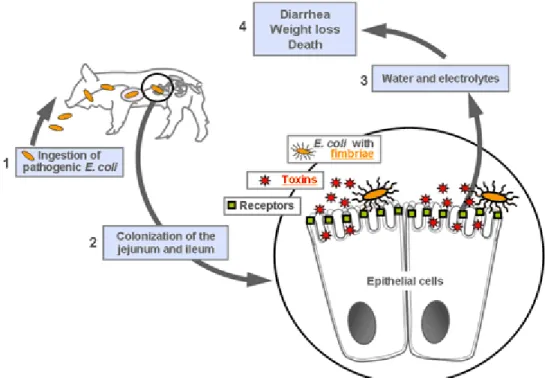
contaminated food and water is the first step in the mediation of diarrhea (Okoh et al., 2008), and is followed by bacterial adhesion to the intestine resulting from the interaction between colonization factors and their receptors at the surface of enterocytes. Bacteria colonize the intestine and produce, in proximity to enterocytes, heat-labile (LT) and heat-stable (ST) toxins responsible for the induction of diarrhea characterized by the secretion of electrolytes and water (Okoh et al., 2008) (Figure 1).
Figure 1: Mode of action of ETEC bacteria. 1. ETEC bacteria are ingested via contaminated food and water. 2. ETEC colonize the intestine with the help of adhesins, this is step is followed by the production of toxins. 3. Toxins stimulate the secretion of water and electrolytes. 4. Diarrhea, weight loss, and death of piglets ensue. (www.ecl-lab.ca)
2.1 ETEC VIRULENCE FACTORS
Colonization factors and enterotoxins are the virulence factors associated with the ETEC pathotype. The genes coding for these virulence factors are carried on plasmids which also carry antibiotic resistance genes (Fairbrother et al., 2005).
2.2 COLONIZATION FACTORS
Colonization factors are protein structures expressed at the bacterial surface which play a role in the development of the pathogenesis caused by ETEC. Abolishment of colonization factors results in bacteria being unable to attach and colonize the intestine suggesting that colonization factors are essential to the progression of the diarrhea mediated by ETEC. Fimbriae constitute the most common colonization factor expressed by ETEC bacteria, however, the presence of afimbriae colonization factors has also been reported (Gaastra et
al., 1996).
2.2.1 FIMBRIAE
ETEC fimbriae are proteinaecous filaments composed of minor and major subunits, the latter being of variable molecular masses and essential to the function of adhesion (Gaastra et al., 1996). The operons coding for fimbriae are generally carried on large plasmids and are composed of genes coding for
the major and minor subunits of the fimbriae as well as the proteins implicated in their transport and assembly (Turner et al., 2006). The expression of fimbriae is regulated by phase variation mechanisms reflecting changes in environmental conditions such as pH and temperature, thus, allowing ETEC bacteria to adapt to such changes (Nagy et al., 1999).
The naming nomenclature of colonization factors is based on host tropism: animal colonization factors are assigned “F” (Turner et al., 2006) while human colonization factors are called Colonization Factor Antigen I (CFA/I) and coli surface antigens (CS) (Gaastra et al., 1996). The fimbriae F4 (K88), F5 (K99), F6 (987P), F41, F42, F165, F17 and F18 are commonly associated with animal strains (Nagy et al., 1999), with F4 and F18 being most prevalent in post-weaning diarrhea in piglets (Fairbrother et al., 2005).
2.2.2 AFIMBRIAE
The Adhesin Involved in Diffuse Adhesion (AIDA-I) belongs to the family of autotransporters anchored to the external membrane of bacteria (Benz et al., 1992) and is responsible for diffuse adherence of bacteria to host cells (Benz et
al., 1989). AIDA-I has been identified as a potential adhesin in porcine ETEC
strains (Ravi et al., 2007) expressing the fimbriae F18 associated with post-weaning diarrhea (Niewerth et al., 2001). The gene coding for AIDA-I is prevalent in diarrheagenic strains carrying the gene coding for STb toxin
(Ngeleka et al., 2003). Thus far, AIDA-I has not been isolated from strains originating from animals other than swine (Niewerth et al., 2001).
2.3 TOXINS
Toxins elaborated by ETEC strains are classed according to their resistance to heat and their molecular masses. Heat-labile toxins (LT) are sensitive to treatments to heat and possess high molecular masses. Heat-stable toxins (ST), on the other hand, are resistant to treatments to heat and have low molecular masses. LT toxins are subdivided as LT-I and LT-II toxins while ST toxins consist of STa, STb, and EAST-1. Toxins are produced at a later stage in the development of diarrhea and are responsible for the secretion of water and electrolytes.
2.3.1 LT TOXIN
LT toxin was first isolated from a pathogenic porcine strain in 1969 by Gyles and Barnum (Gyles et al., 1969) and has been shown to induce diarrhea in Man (Qadri et al., 2005) and in animals (Nagy et al., 2005). LT has also been associated with severe diarrhea and septicemia in piglets (Berberov et al., 2004). LT toxin is an 84 kDa AB toxin composed of one A subunit and five B subunits linked to each other by a disulfide bonds (Sixma et al., 1991). The A subunit of the toxin is an ADP-ribosyl transferase implicated in the
development of diarrhea (Sixma et al., 1993). The B subunits are responsible for binding the ganglioside GM1 which constitutes the receptor of the toxin (Spangler, 1992). The LT-I and LT-II forms of the LT toxin differ in the B subunits they possess: a homology of 57% has been reported between the B subunits shared by LT-I and LT-II (Pickett et al., 1987). Of the two, LT-I is most commonly associated with animal and human ETEC strains. A homology of 55% has been reported between LT and cholera toxin (CT) suggesting they are conserved and possess similar modes of actions (Pickett et al., 1989).
In order to cause diarrhea, LT first binds the GM1ganglioside present at the surface of enterocytes via the B subunits (Griffiths et al., 1986, Tsuji et al., 1985). Receptor binding is followed by internalization of LT into vesicles which are retrograde transported to the Golgi apparatus and endoplasmic reticulum (Rappuoli et al., 1999). Proteolytic cleavage of the A subunit into A1 and A2 domains (Lencer et al., 1997) occurs and is followed by the
translocation of the A1 domain into the cytosol. The A1 domain
ADP-ribosylates the -unit of the Gs protein, the G protein regulating the activity of
adenylate cyclase, resulting in the permanent activation of adenylate cyclase (Rappuoli et al., 1999) which in turn causes abnormal increases in cyclic AMP (cAMP) levels (Nataro et al., 1998). Augmented cAMP levels are followed by the activation of cAMP-dependent protein kinase A and the phosphorylation of chloride channels such as Cystic fibrosis transmembrane conductance regulator (CFTR). This leads to chloride secretion from crypt epithelial cells
and the inhibition of sodium absorption by villous enterocytes resulting in secretory diarrhea (Spangler, 1992).
2.3.2 ST TOXINS
ST toxins are heat-stable toxins produced ETEC strains. EAST-1, STa, and STb are the toxins belonging to this group.
2.3.2.1 EAST-1
EAST-1 was first isolated from the strain EAEC 17-2 (Savarino et al., 1991) and later isolated from human and animal ETEC strains (Veilleux et al., 2006). EAST-1 causes an increase in cyclic GMP (cGMP) levels in enterocytes similarly to STa toxin (discussed below). As EAST-1 bears structural similarities to STa toxin, it is believed that EAST-1 possesses a similar mode of action (Savarino et al., 1993). The gene coding for EAST-1, astA, is frequently isolated from porcine ETEC strains harbouring the genes for F4 and STa toxin (Choi et al., 2001). The gene astA has also been isolated from healthy subjects, thus the ability of EAST-1 to induce diarrhea mediated by ETEC strains (Ngeleka et al., 2003) and to stimulate loss of electrolytes from intestines (Berberov et al., 2004) currently remains elusive.
2.3.2.2 STa TOXIN
The STa toxin is coded by the gene estA located on the transposon Tn1681 which is itself carried by plasmids (So et al., 1980). STa is first synthesized as a 72 amino acid pre-polypeptide which is translocated to the periplasm via the sec machinery (Okamoto et al., 1990). This pre-polypeptide then undergoes proteolytic cleavage yielding an intermediate 53 amino acid form of the toxin which is secreted in the extracellular milieu to be cleaved again, thus, giving rise to mature STa toxin, a toxin of 18 or 19 amino acids (Rasheed et al., 1990). STa is a structural analogue to guanyline, an endogenous peptide regulating the transport of water and electrolytes across the epithelium (Giannella, 1995).
The pathogenesis mediated by STa toxin begins by the binding of the toxin to the trans-membrane guanylate cyclase type C receptor (GC-C) resulting in the activation of the guanylate cyclase in enterocytes. Stimulated guanylate cyclase, in turn, synthesizes cGMP from Guanosine triphosphate (GTP) resulting in the rise of cGMP levels (Vaandrager, 2002). Increases in cGMP levels lead to cGMP-dependent protein kinase II phosphorylation of CFTR causing its activation. Secretion of chloride and water by osmosis and inhibition of sodium absorption ensues leading to the occurrence of diarrhea (Sears et al., 1996, Vaandrager, 2002).
3. STb TOXIN
STb toxin was isolated by Burgess et al. in 1978 from the porcine ETEC strain P16 (Burgess et al., 1978). Prior to the identification of STb, STa was the only known ST toxin associated with ETEC strains. Abnormalities observed during the purification process of the ST indicated the production of more than one ST toxin by the strain P16. Indeed, this toxin revealed itself to be insoluble in methanol, a feature which clearly distinguishes it from methanol-soluble STa toxin. The activity of this new toxin was then evaluated in ligated intestines of newborn piglets, weaned piglets, rabbits, and calves. This toxin was shown to be active in the intestines of weaned piglets and rabbits and inactive in the intestines of newborn piglets and calves, contrary to STa toxin. The differences in the activities of the ST toxins in the intestines of newborn piglets and weaned piglets further confirmed the production of a toxin other than STa by the strain P16. The discovery of this new toxin could explain the variability of ST activity in ligated rabbit intestines and in mice leading to the speculation that ETEC strains produce more than one ST toxin. As the variability observed arose from ST toxin, this new methanol-insoluble toxin was named STb enterotoxin (Burgess et al., 1978).
3.1 GENETICS
The gene estB which codes for STb toxin was first shown to be carried on a transposon by Lee et al. (1985). This transposon, which itself is carried on the P307 plasmid, is flanked by defective IS2 elements yet is capable of transposing from one plasmid to another. Transposition assays conducted by Lee et al. (1985) demonstrated that the transposon carrying the gene estB uses a simple transposition mechanism to transpose from the pBR322 plasmid to the F plasmid. As the sizes of pBR322, the F plasmid, and estB gene are, respectively, 14 kb, 4 kb, and 1 kb, the estimated the size of the transposon carrying the gene coding for STb was 9 kb (Lee et al., 1985). The transposition of the gene estB seems to be a mechanism by which virulence factors are disseminated from one ETEC strain to another. The gene coding for STb toxin from various clinical isolates appeared uniform in size, however, flanking sequences are heterogeneous suggesting that the estB gene can be found on heterogeneous transposons. Indeed, the plasmids carrying the transposon coding for STb are also heterogeneous as they carry genes coding for STa, LT, colonization factors, and colicin (Harnett et al., 1985).
The transposon carrying the estB gene was designated Tn4521 (Hu et al., 1987) and was further characterized in studies conducted by Hu et al. (1987; 1988). Terminal regions were shown to be composed of IS2 sequences in an inverted position (Hu et al., 1987). Mutations of different regions of Tn4521
resulted in the finding that the right terminal area was required for transposition. An ORF located within the right terminal area codes for a 159 amino acid protein which was shown by frameshift mutation to be essential for transposition. This suggested that this protein could be a possible transposase for the functional transposon Tn4521 (Hu et al., 1988).
As STb toxin produced by ETEC strains is relatively low, the strength of the promoter controlling the expression of estB gene was assessed by Spandau and Lee (1987) (Spandau et al., 1987). The promoter and the 5’ coding sequence of estB gene were fused to the lacZ gene such that the production of -galactosidase was under the control of the estB gene promoter. The strength of the promoter controlling estB gene expression was compared to the strength of the ompF and lac operons fused to the lacZ gene. The mRNA transcript of each promoter was analyzed by Northern blot and in vitro transcription. Low levels of the mRNA transcript of the lacZ gene were observed under the control of the promoter of the estB gene indicating a weak promoter. Quantification of the -galactosidase protein produced under the control of the
estB gene promoter yielded low protein levels further confirming a weak
3.2 SYNTHESIS
Nucleotide sequencing of the estB gene revealed STb toxin is synthesized as a 71 amino acid peptide. The presence of a peptide-like sequence in the amino terminal region suggests STb is synthesized as a pre-toxin and is subsequently transported across the bacterial membrane (Lee et al., 1983) (Figure 2). Indeed, amino acid sequencing of STb produced by ETEC cultures revealed a protein of 48 amino acids (Kupersztoch et al., 1990) while SDS-PAGE experiments determined the molecular mass of mature STb toxin to be 5.2 kDa (Lawrence et al., 1990). The first 23 amino acids present in the pre-toxin were not observed in the extracellular form of STb suggesting that proteolytic cleavage plays a role in the maturation of STb (Kupersztoch et al., 1990). Cellular fraction experiments demonstrated that the same form of STb, confirmed by amino acid sequencing, is present in the periplasm and extracellularly. According to the authors, this suggested proteolytic cleavage yielding mature STb occurs in the periplasm. As the secA gene is implicated in the conversion of periplasmic and outer membrane proteins from their precursor into the mature forms, the implication of secA in the conversion of STb pre-toxin into mature STb was investigated. Bacteria mutated in the secA gene did not express mature extracellular STb indicating that the conversion of STb to its mature form is dependent on secA gene expression (Kupersztoch et
Figure 2: Synthesis of STb toxin. 1. STb is first synthesized in the cytoplasm as a pre-polypeptide. 2. Proteolytic cleavage occurs in the periplasm giving rise to mature STb. 3. DsbA assures the formation of disulfide bonds in the periplasm. Mature STb is then secreted into the extracellular medium via TolC. (Taillon, 2010).
Proteolytic cleavage is followed by the formation of disulfide bonds in the periplasm (Dreyfus et al., 1992). Indeed, the presence of 4 cysteine residues observed by Burgess et al. (1978) pointed to the possibility of disulfide bonds being formed between these residues. Furthermore, the observation that treatment of STb toxin with a reducing agent caused a decrease in toxicity supported the hypothesis that disulfide bonds may be present in mature STb (Dreyfus et al., 1992). STb peptides which became unlinked following DTT treatment were sequenced in order to determine the cysteine residues implicated in the formation of the disulfide bonds. The cysteine residues at the
10-48 and 21-36 positions of mature STb formed disulfide bonds with each other. Oligonucleotide-directed mutagenesis eliminating one or both disulfide bonds resulted in the inability of STb to translocate from the periplasm to the extracellular environment and reduce the toxic activity of STb. Taken together, these findings indicate both of the disulfide bonds are essential for the translocation of STb from the periplasm to the extracellular environment and are necessary for STb to retain its toxic activity (Dreyfus et al., 1992). A subsequent study confirmed the role of disulfide bonds in STb toxicity (Arriaga et al., 1995).
The implication of Disulfide-bond A oxidoreductase (DsbA) in the formation of disulfide bonds of STb was verified by Okamoto et al. (1995). Cellular fraction of DsbA mutants revealed STb is not present in either the periplasm or in culture supernatants. This suggested that DsbA is implicated in the maturation of STb toxin. Indeed, complementation of dsbA- strains with wild-type strains resulted in the detection of STb in the periplasm and in culture supernatant. The role of DsbA in the formation of disulfide bonds in STb toxin was determined by substituting DsbA with Disulfide-bond C oxidoreductase (DsbC), another protein implicated in disulfide bond formation in E. coli. Substitution experiments revealed a lack of disulfide bonds in STb indicating that DsbA, but not DsbC, is involved in disulfide formation in STb.
The implication of Outer membrane channel protein (TolC) and DsbA in the secretion of mature STb from the periplasm to the extracellular environment has also been demonstrated (Foreman et al., 1995). Bacteria harboring TolC mutants were unable to secrete STb into the extracellular environment. This indicates that TolC could act as channel permitting the passage of STb from the periplasm into the extracellular medium. Although the protein DsbA is implicated in the formation of disulfide bonds between cysteine residues (Okamoto et al., 1995), Foreman et al. (1995) observed that STb was not detected in the extracellular environment of DsbA mutants. Indeed, the absence of disulfide bonds renders STb susceptible to proteolytic cleavage and, thus, results in the absence of STb in the extracellular medium.
3.3 BIOCHEMICAL STRUCTURE
Nuclear Magnetic Resonance (NMR) studies revealed that mature STb toxin is composed of two antiparallel alpha-helices separated by a glycine-rich loop (Figure 3) (Sukumar et al., 1995). The first -helix, located between the amino acid residues 10 to 22, is hydrophilic, explaining the exposure of the lateral chains of amino acids Asp8, His12, Gln15, Lys18, Glu19, Lys22, and Lys23
to the solvent. The second -helix ranges from amino acid residues 38 to 44 and is hydrophobic. The glycine-rich loop is located between residues 21 and 36 and contains a cluster of hydrophobic residues. The authors of this study also conducted Circular Dichroism (CD) experiments revealing loss of the
disulfide bridges results in loss of STb structure, which was associated with a loss of function (Sukumar et al., 1995), findings observed by Dreyfus et al. (1992) and Arriaga et al. (1995).
Figure 3: Tertiary structure of STb toxin. Mature STb is composed of an amphipathic -helix, a glycine-rich flexible loop, a hydrophobic -helix, and two disulfide bonds. (Sukumar et al., 1995)
The -helix in the carboxy-terminal region has been shown to be implicated in the formation of oligomers by STb toxin. Site-directed mutagenesis of residues located in this -helix identified the hydrophobic amino acids Phe37,
Ile41, and Met42 as being indispensable to the formation hexamers and
heptamers. The effect of temperature on the formation of oligomers was also evaluated and revealed that temperature has no effect on oligomerization of
STb. Treatment of STb with -mercaptoethanol, a reducing agent which prevents the formation of disulfide bonds, interferes with the formation of oligomers indicating proper structure is required for the formation of oligomers. The influence of the presence or absence of sulfatide, the toxin’s receptor, was also investigated and demonstrated that oligomerization occurs independently of the presence of sulfatide (Labrie et al., 2001b).
3.4 RECEPTOR OF STb TOXIN
The chemical nature of the receptor of STb was first determined by Rousset et al. (1998a) who observed the attachment of STb to microvilli of frozen porcine tissue cuts. Attachment assays conducted by Rousset et al. (1998a) revealed that attachment of STb to microvilli occurs rapidly with saturation occurring after 10 minutes. The optimum pH of this attachment has been reported to be 5.8 while temperature has no effect on attachment. Attachment of STb to all tissues tested (jejunum, duodenum, caecum, liver, spleen, and kidney) was reported. These observations suggest that the molecule acting as a potential receptor for STb is a surface molecule ubiquitously present on these tissues. In order to determine the chemical nature of STb’s receptor, tissues were subjected to enzymatic and chemical treatments prior to attachment assays conducted with STb, identifying a glycosphingolipid as a potential receptor (Rousset et al., 1998a).
In a subsequent study by Rousset et al. (1998b), attachment of STb to various commercial glycolipids was tested revealing strong attachments to acidic glycosphingolipids and to certain gangliosides. Amongst the glycolipids tested, attachment of STb to sulfatide was the strongest and occurred in a dose-dependent and saturable manner. A lipid extracted from jejunum brush border epithelial cells was analyzed by thin layer chromatography revealing the presence of a molecule having the same migration distance as commercial sulfatide, an acidic glycosphingolipid abundantly present at the surface of intestinal epithelial cells. In addition, this molecule was also recognized by an anti-sulfatide antibody. These results permitted the identification of sulfatide as a functional receptor for STb toxin (Rousset et al., 1998b). Indeed, pretreatment of ligated rat intestine segments with either laminine, known to interact specifically with sulfatide, or sulfatase resulted in a decrease of STb activity supporting the finding that sulfatide could be the receptor of STb (Rousset et al., 1999). Mass spectrometry would later confirm sulfatide as the receptor of STb toxin (Beausoleil et al., 2002b).
The affinity and physical characteristics of the binding of STb to sulfatide was determined by microplate binding assays. These experiments demonstrated that STb binds to sulfatide with a high specifity in a dose-dependent and saturable manner (Beausoleil et al., 2001). However, the affinity of this interaction was described as weak as demonstrated by the kd value of 2-6 +/- 1.5 and was partially inhibited by elevated concentrations of charged
carbohydrates. The kd value obtained by the authors is in accordance with the value obtained by Chao and Dreyfus (1997) who studied the interaction of STb with intestinal epithelial cells such as T84 and HT29 cells. The affinity of STb to sulfatide would be re-evaluated with the use of SPR (Surface Plasmon Resonance) technology obtaining a kd value of 2.4+/-0.6 nM indicating a higher affinity of STb to sulfatide (Goncalves et al., 2008) than that reported by Beausoleil and Dubreuil (2001). The kd value obtained by Goncalves et al. (2008) was in accordance with values obtained for other toxins possessing glycolipid molecules as functional receptors. Carrageenan was shown to inhibit the interaction between STb and sulfatide as well as permeabilization of cell membranes caused by STb (Goncalves et al., 2008). Thus, carrageenan could represent a molecule that could be used as a prophylactic agent to protect piglets against STb during the post-weaning period.
3.5 INTERNALIZATION
Internalization of STb toxin was first reported by Chao and Dreyfus (1997) who had observed that STb integrates within the membrane of cells following its attachment to epithelial cells. The internalization of STb occurred independently of temperature, cytoskeleton rearrangements, energy, and hypertonic conditions. These results suggested that the processes of clathrin-dependent and -inclathrin-dependent, caveolae internalization or micropinocytosis were not implicated in STb uptake. The authors of this study emitted the
hypothesis that the formation of a stable complex with lipids rather than internalization by a ligand could be the mode of STb internalization. As well, the authors suggested that the toxin associated to the membrane could directly penetrate the membrane to interact with regulatory proteins such as G proteins (Chao et al., 1997).
The fate of STb following uptake was studied by Labrie et al. (2002) in vivo in rat intestinal epithelial cells using an anti-STb gold labeled assay and transmission electron microscopy revealing that STb does not seem to target a particular organelle as gold particles were observed dispersed throughout cells. Internalization of wild-type and the mutant I41E-M42R characterized by decreased hydrophobicity were compared and revealed diminished uptake of the mutant indicating these amino acids are essential to proper internalization of STb (Labrie et al., 2002). Indeed, site-specific mutagenesis of the residues Phe37, Ile41 and Met42 demonstrated they are necessary to the binding of STb to
its receptor as these mutations resulted in reduced binding (Labrie et al., 2001a), thus supporting the decrease of the uptake of these mutants observed by Labrie et al. (2002). The toxicity of these mutants was evaluated in the rat loop assay revealing decreased toxicity compared to the wildtype. The contribution of the residues Gly22, Gly23, and Arg29 of the flexible loop was
evaluated and also exhibited decreased binding and toxicity of STb toxin. Overall, these data suggested that hydrophobic and electrostatic interactions are important for STb binding and toxicity (Labrie et al., 2001a).
3.6 MODE OF ACTION
A distinguishing characteristic of the colibacillosis mediated by STb in animals is the lack of adenylate cyclase and guanylate cyclase activation as observed in diarrhea induced by LT and STa toxins (Hitotsubashi et al., 1992, Peterson et al., 1995). The binding of STb toxin is followed by uptake of the toxin resulting in the stimulation of a pertussis-sensitive G protein and in an increase of intracellular calcium levels. Augmented calcium levels are the result of an influx of calcium ions inside cells as demonstrated by Dreyfus et al. (1993). Pretreatment of cells with inhibitors of calcium channels following STb treatment revealed a lack of increase of intracellular calcium levels indicating increases of calcium levels are the result of an influx from the extracellular medium. Calcium influx leads to the activation of a calmodulin-dependent kinase II protein as the activation was not observed in cells pretreated with calcium channel inhibitors (Fujii et al., 1997, Dreyfus et al., 1993). Calmodulin-dependent kinase II protein stimulates the opening of an ionic channel as well as the activation of a kinase C protein and of CFTR (Dreyfus et al., 1993).
Augmented calcium levels also regulate the activities of the phospholipases A2
and C and induce the release of arachidonic acid from membrane phospholipids which in turn leads to the production of prostaglandin E2 and
secretagogues occurs in a dose-dependent manner in response to increasing quantities of STb in the intestinal lumen. Indeed, treatment of rats with ketanserin, an antagonist of serotonin receptors, results in decreased intestinal secretion by STb implying a role of serotonin in the mode of action of STb (Harville et al., 1995). Moreover, Fujii et al. (1995) demonstrated serotonin release and fluid accumulation is proportional to the quantity of STb used to treat cells. These results are in accordance with the observations made by Harville and Dreyfus (1995).
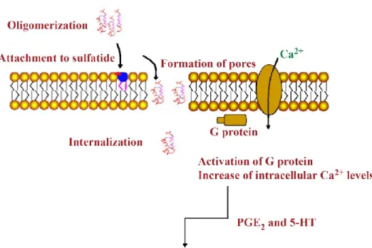
Figure 4: Proposed mode of action of STb toxin. Oligomerized STb toxin binds to its receptor, sulfatide (blue), and is internalized presumably through the formation of pores. Pores also permit an influx of calcium to occur resulting in the activation of a G protein and in an increase of intracellular
calcium levels. Augmented calcium levels stimulate prostaglandin E2 and
serotonin (5-HT) which in turn are responsible for stimulating the secretion of electrolytes and water. (Taillon, 2010).
Stimulation of secretagogues is followed by the secretion of electrolytes, which include Na+, Cl-, and HCO3-, and water in the intestinal lumen (Figure 4). Fluid accumulation caused by STb was evaluated in mouse intestinal loop assays by Hitotsubashi et al. (1992). The authors observed fluid accumulation occurs as rapidly as 30 minutes following STb treatment and with maximum accumulation occurring at 3 hours (Hitotsubashi et al., 1992). The diarrhea caused by STb was reported to be accompanied by histological damages characterized by shortening and atrophy of the villi as well as replacement of columnar epithelial cells with cuboidal or squamous cells (Whipp et al., 1986, Whipp et al., 1987). The shortening and atrophy of the villi were subsequently confirmed using morphometric techniques (Rose et al., 1987). Swine, lamb, and rabbit intestines exposed to STb-positive filtrates were measured and revealed reduction of villous epithelial surface area and mucosal volume in swine and lamb intestines. These data supports the hypothesis that a species ability to secrete in response to STb treatment is required for histological damages to occur (Whipp et al., 1986). The damages observed by Whipp et al. (1986) and Rose et al. (1987) have been attributed to loss of absorptive capacity as well as to the induction of net secretion caused by STb.
The ability to form pores by STb in swine brush border membrane vesicles was evaluated by Goncalves et al. (2007). Brush border membrane vesicles
were treated with pure toxin followed by a treatment to valinomcyin, a potent membrane potential generator, which in turn results in an efflux of potassium ions. The membrane potential sensitive probe revealed a decrease in the membrane potential of the vesicles treated with STb and valinomycin indicating STb permeabilizes the membrane to ions other than potassium. The lack of change in membrane potential of vesicles treated with STb but not valinomycin suggests the permeability of STb is nonspecific. The ability of STb to form pores was also assessed in this study. Vesicles treated with STb and valinomycin were placed in an isotonic solution of N-methyl-D-glucamine hydrochloride (NMDGCl). Fluorescence levels increased before leveling off with increasing quantities of STb toxin indicating pore formation depends on the interaction of STb with its receptor and occurs in a dose-dependent and saturable manner. The pores formed by STb could be partially responsible for the loss of electrolytes and water observed during diarrhea mediated by STb (Goncalves et al., 2007).
A subsequent study by Goncalves et al. (2009) evaluating the internalization mechanism of STb revealed STb induces apoptotic-like morphology of cultured cells characterized by cell shrinkage, membrane blebbing, granular cytoplasm, and enlarged nuclei. Cells labeled with PI revealed colocalization of FITC-labeled STb with mitochondrion which was observed by confocal microscopy after 6 hours and became more pronounced after 12 hours. Cells treated with STb demonstrated dose-dependent changes in mitochondrial
membrane potential, over time, as measured with the fluorescent probe JC-1 and flow cytometry. Though not proven by Goncalves et al., (2009) the results obtained by the authors suggest STb could possibly possess the ability to kill eukaryotic cells.
3.7 IMMUNOGENIC POTENTIAL
The capacity of STb to induce neutralizing antibodies was verified by immunizing rabbits with purified toxin isolated from ETEC porcine strains. ELISA assay was used to determine the level of antibodies present in the serum of immunized rabbits revealing low levels of antibodies. A poor level of antibody production was also observed following the administration of booster shots suggesting that STb is a poor immunogen (Dubreuil et al., 1991). The low yield in antibodies was attributed to the small size of the toxin. Subsequent studies examined the production of neutralizing antibodies against STb in rabbits and mice immunized with the fusion proteins ompF-STb--galacosidase (Lawrence et al., 1990) and STb-KLH (Urban et al., 1990), respectively, in an attempt to circumvent the small size of STb toxin. Immunization of animals with fusion proteins containing STb increased the production of antibodies against STb, measured by ELISA, in both of these studies.
Similarly, a subsequent study also revealed an augmentation in the production of anti-STb neutralizing antibodies in rabbits immunized with the fusion protein maltose binding protein-heat stable toxin b (MBP-STb) (Dubreuil et
al., 1996). The authors also evaluated the production of neutralizing
antibodies following the immunization of rabbits with a fusion protein comprised of MBP and truncated STb revealing a lack of production of antibodies. This suggested that the conformation and each amino acid influenced the immunogenic properties of STb toxin. However, neutralizing antibodies directed against STb are incapable of neutralizing STa or CT toxins indicating STb possesses distinctive immunogenic properties not shared with either STa or CT and pointing to the possibility of STb possessing a differing mode of action than STa or CT (Hitotsubashi et al., 1992).
4. APOPTOSIS
The term apoptosis was first coined by Kerr, Wyllie, and Currie in 1972 to describe a form of programmed cellular death distinctive from necrosis (Kerr
et al., 1972). The term apoptosis originates from Greek (“falling off” as leaves
fall from trees) and is used to describe a controlled physiological process of removing individual components of an organism without damage or destruction of the organism. Kerr and his colleagues observed that apoptosis of embryonic tissues was consistently accompanied by structural changes such as membrane blebbing, cell shrinkage, chromatin condensation, and nuclear
fragmentation which were visualized by electron microscopy. The structural changes of apoptosis occur in two phases: the first consisting of nuclear and cytoplasmic condensation followed by the breaking up of the cell into membrane-bound vesicles (Kerr, 1971), and the second consisting of the elimination of these apoptotic bodies by either phagocytosis or degradation by other cells (Kerr, 1972).
The formation of apoptotic bodies is characterized by nuclei and cytoplasmic condensation, nuclear fragmentation, and detachment of cells from tissues (Kerr, 1971). Apoptotic bodies are condensed cell fragments harboring condensed chromatin and tightly-packed organelles (Kerr, 1972). The exact composition of apoptotic bodies will vary and depend on the cellular constituents present at the time of the formation of these bodies. The varied composition of apoptotic bodies also influences their size: small apoptotic bodies are composed of nuclear chromatin whereas large ones are composed of cytoplasmic components. The degree of condensation of apoptotic bodies is thought to be the result of water exclusion which also influences their size (Kerr et al., 1972).
The presence of apoptotic bodies in intact cells suggested elimination of these bodies by phagocytosis. Kerr et al. (1972) believed these bodies were being engulfed by cells due to changes in the properties of their surface membranes. Ingested apoptotic bodies then undergo a process within phagosomes that is
similar to in vitro autolysis of whole cells (Trump et al., 1965). The membranes of the apoptotic bodies inside phagosomes as well as the membranes of the organelles located within these apoptotic bodies are degraded, ribosomes become swollen and undistinguishable, and cessation of metabolic activities of the apoptotic bodies occurs. Lysosomal enzymes are then acquired as a result of the fusion of the phagosomes with lysosomes and contribute to the degradation of apoptotic bodies. The process of phagocytosis and subsequent elimination of apoptotic bodies usually occurs within a 24-hour timeframe (Kerr, 1971).
According to Kerr et al., (1972) the appearance of apoptotic bodies in healthy tissues indicates apoptosis occurs in healthy tissues as a result of normal cell turnover. Indeed, apoptosis is considered as a form of controlled cell death occurring in healthy adult mammalian tissues contributing to the maintenance of the cell population within these tissues. The occurrence of apoptosis in embryonic tissues at specific time points during development supported the notion of apoptosis being a form of controlled death responsible for the maintenance of cellular populations (Saunders, 1966). Kerr et al. (1972) had observed that the susceptibility of embryonic cells to apoptosis varies depending on the developmental stage in which they are. The developmental timing of apoptosis and the consistent accompanying morphological changes highlighted the possibility of apoptosis being genetically regulated (Kerr et al., 1972).
4.1 GENETIC REGULATION OF APOPTOSIS
The genetic regulation of apoptosis was confirmed by studies conducted by the team led by Horvitz using Caenorhabditis elegans (Yuan et al., 1993, Yuan et
al., 1992, Conradt et al., 1998, Hengartner et al., 1992, Hengartner et al.,
1994) and will be discussed in greater detail below. The nematode C. elegans was chosen by Horvitz and his team to study cell death due to its small size, cellular simplicity, easy handling, and rapid generation time (Wood et al., 1988). Moreover, as C. elegans is transparent, death of individual cells is relatively easy to observe in the living organism. Horvitz and his team created mutants by transposon insertion in order to identify genes implicated in the process of apoptosis. Many of the genes subsequently identified were determined to possess a human counterpart and according to Horvitz and his team, suggested molecular mechanisms regulating cell death are conserved in nematodes and mammals (Figure 5).
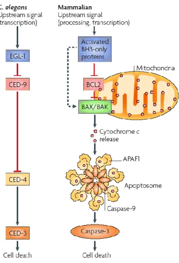
Figure 5: Comparison of the cellular death pathway in C. elegans and in mammals. Each protein implicated in cell death in C. elegans possesses a human homologue. (Degterev et al., 2008)
Studies conducted on 3 (Yuan et al., 1993), 4 (Yuan et al., 1992),
ced-9 (Hengartner et al., 1ced-9ced-92, Hengartner et al., 1ced-9ced-94), and egl-1 (Conradt et al.,
1998) genes resulted in the inhibition of apoptosis, suggesting all four genes are essential to programmed cell death and that apoptosis is an active process. The Cell Death Protein 3 (CED-3) was shown to be similar to the human cysteine protease Interleukin-1 Converting Enzyme (ICE), suggesting it could be a caspase-like protein. Indeed, both CED-3 and ICE are generated as
pro-proteins requiring proteolytic cleavage to be activated. CED-3 will then modulate cell death by activating downstream proteins responsible for cell death and by inactivating proteins which protect cells from death, similarly to caspase-3 in mammals (Yuan et al., 1993). Cleavage of CED-3 is mediated by Cell Death Protein 4 (CED-4), a protein bearing similar amino acid sequence to Apoptosis Protease Activating Factor-1 (APAF-1) (Yuan et al., 1992). The protein Egg-Laying Protein 1 (EGL-1) contains a B-cell lymphoma 2 homology domain 3-like domain (BH3) which allows it to interact with the B-cell lymphoma 2-like protein (BCL-2) Cell Death Protein 9 (CED-9) thereby suppressing the inhibition of apoptosis exerted by CED-9 (Conradt et al., 1998).
Decreased or elimination of CED-9 resulted in cells undergoing apoptosis instead of surviving. Conversely, over-expression of CED-9 causes cell survival (Hengartner et al., 1992). Amino acid sequencing of CED-9 revealed that it is similar to the human proto-oncogene BCL-2, a protein which suppresses cell death. Expression of cloned human BCL-2 in C. elegans caused cell survival in cells destined to die as well as in ced-9 deficient cells. These results suggest CED-9 and BCL-2 are functional homologues and support the idea of the conservation of molecular mechanisms regulating cell death in both nematodes and mammals (Hengartner et al., 1994).
4.2 BCL-2 FAMILY PROTEINS
B cell lymphoma 2 (BCL-2) family proteins have been shown to play essential roles in the regulation of apoptosis (Youle et al., 2008). The first protein discovered in this family was BCL-2 which was defined as the key oncogene in follicular lymphomas (Tsujimoto et al., 1985). Non-cancerous cells introduced with the BCL-2 protein demonstrated an increased capacity to survive in the absence of growth factors and showed a suppression of the expression of morphological features associated with apoptosis such as membrane blebbing, nuclear condensation, and DNA cleavage. These findings suggested that BCL-2 can both promote proliferation and actively block cell death in cancer cells which, unlike other known oncogenes at the time, were strictly attributed to the proliferation of cancer cells. Hence, BCL-2 was classed as an anti-apoptotic protein (Vaux et al., 1988, McDonnell et al., 1989).
Amino acid sequencing of BCL-2 family proteins revealed that these are structurally diverse yet share up to four BCL-2 homology domain (BH domains) and one transmembrane domain (TM) (Figure 6). Anti-apoptotic members possess all four BH domains while pro-apoptotic members possess up to three BH domains. Pro-apoptotic BCL-2 family proteins can be further subdivided into two categories: the first consisting of proteins harboring the BH3 domain in combination with BH1 and BH2 while the second class of proteins carry only the BH3 domain. The proteins carrying BH3 and another
BH domain are known as BH1-3 proteins whereas proteins carrying solely the BH3 domain are known as BH3-only proteins (Degterev et al., 2008, Lomonosova et al., 2008).
Figure 6: BCL-2 family proteins. Anti-apoptotic proteins are characterized by the presence of all four BH domains. Pro-apoptotic members harbor domains BH1-BH3 or only the BH3 domain. The BH3 domain is the core domain of all BCL-2 family proteins. (Lomonosova et al., 2008)
4.2.1 BH3 DOMAIN
The BH3 domain is the only domain present in all BCL-2 family proteins, and thus, considered the core-defining domain of this family of protein (Figure 6) (Youle et al., 2008). Consequently, this domain has been more extensively studied than the other domains associated with the BCL-2 family proteins. The confirmation that the BH3 domain is implicated in apoptosis comes from studies conducted on Bcl-2-interacting killer (BIK) and BCL-2 homologous antagonist/killer (BAK). Mutations of this domain in BAK and BIK resulted