I
DÉPARTEMENT OFBIOCHEMISTRY
THESIS
Presented by
KADDOUR Sabrina Manel
For the fulfillment of the requirements for the degree of
Doctorat 3 cycle
BIOLOGY
Special filed: BIOCHEMISTRY
TOPIC
Phytochemical, Antioxidant and Anti-inflammatory Effects of Medicinal
Plants Extracts
Presented publically in: 24 / 12 / 2020 MEMBERS OF JURY
President: KHENNOUF Seddik Prof. UFA Setif 1
Supervisor: BAGHIANI Abderrahmane Prof. UFA Setif 1
Examiners: BOURAS Mourad Prof. Univ. Batna
DJARMOUNI Meriam MCA UFA Setif 1
BOUAZIZ Amel MCA UFA Setif 1
Laboratory of applied biochemistry
N°……….…………..…….……/SNV/2019
I
ACKNOWLEDGEMENTS
First and foremost, praises and thanks to ALLAH subhana-Wa-Taala, the Almighty, for providing me the blessings throughout my research work to complete the research successfully.
I wish to express my sincere gratitude to my supervisor Pr. Baghiani Abderrahmane, for his valuable advices and guidance, timely suggestions and whole-hearted supports. His continuous interest and great generosity helped my work to be accomplished and also supported me during a course of this study.
I would like to thank my Committee members, Pr. Khennouf Seddik University Ferhat Abbas Setif 1, Pr. Bouras mourad University of Batna, Dr. Djarmouni Meriam University Ferhat Abbas Setif 1 and Dr. Bouaziz Amel University Ferhat Abbas Setif 1, for critical reading of the thesis, and for their valuable comments, discussions and suggestions.
I want to offer my special thanks to Pr. Arrar Lekhmissi and Dr. Guemaz Thoraya for their valuable advices, continuous support, assistance, and to all other technical supports. I am also thankful to Pr. Charef Noureddine, Pr. Khennouf Seddik, Pr. Dahamna Saliha for help and provision of research facilities in their laboratories and Dr. Safsaf University Hospital of Sétif for providing all the facilities to realise histological slides and interpreting them.
Many thanks send to all my colleagues in laboratory of applied biochemistry, who provided the most amicable and lively working environment.
This work carried out in laboratory of applied biochemistry and laboratory of phytotherapy applied to chronic diseases.
II
DEDICATE
I would like to thank my family, especially my mother and my husband and everyone who
stood by me who gave me strength to go on all the way of getting to the end. For all their
III
ﺺﺨﻠﻣ
ﻒﻠﺘﺨﻤﻟ ﺪﺒﻜﻟا ﺔﯾﺎﻤﺣ ﻰﻠﻋ ةرﺪﻘﻟا ﻰﻟا ﺔﻓﺎﺿﻻﺎﺑ بﺎﮭﺘﻟﻼﻟ ةدﺎﻀﻤﻟا و ةﺪﺴﻛﻸﻟ ةدﺎﻀﻤﻟا ﺔﯿطﺎﺸﻨﻟا ﻢﯿﯿﻘﺗ ﻰﻟا ﺔﺳارﺪﻟا هﺬھ فﺪﮭﺗ ﻲﺋاﻮﮭﻟا ءﺰﺠﻟا ﻦﻣ ةﺮﻀﺤﻤﻟا تﺎﺼﻠﺨﺘﺴﻤﻟا ﻦﯿﺗﺎﺒﻨﻠﻟ Halogeton sativus و Arthrophytum scoparium . ﺑﺪﻟا و تاﺪﯾﻮﻧﻮﻓﻼﻔﻟا و ﺔﯿﻠﻜﻟا لﻮﻨﯿﻔﻟا تاﺪﯾﺪﻋ ﺮﯾﺪﻘﺗ ﻢﺗ مﺎﺨﻟا ﻲﻟﻮﻧﺎﺜﯿﻤﻟا ﺺﻠﺨﺘﺴﻤﻟا ﻦﻣ ﻞﻛ ﻲﻓ غﺎ ﺔﺠﺗﺎﻨﻟا راﻮطﻷاو ﻦﯿﺘﺘﺒﻨﻠﻟ ﻰﺗﺎﺒﻛﺮﻤﻟا هﺬھ ﺮﯾﺪﻘﺗ ﻰﻟا ﺔﻓﺎﺿﻻﺎﺑ ﻲﺋﺎﻤﻟا ﺺﻠﺨﺘﺴﻤﻟا و ﻞﯿﺛﻻا تﺎﺘﯿﺳأ ﺺﻠﺨﺘﺴﻣ و ﻲﻣرﻮﻓورﻮﻠﻜﻟا ﺺﻠﺨﺘﺴﻤﻟﺎﻛ ﺎﮭﻨﻣ ﻦﯾرﻮﻛﺬﻤﻟا ﻦﯿﺗﺎﺒﻨﻟا . ﻰﻠﻐﻣ نأ ﺞﺋﺎﺘﻨﻟا ﺖﻨﯿﺑ A. Scoparium لﻮﻨﯿﻔﻟا تاﺪﯾﺪﻋ ﻦﻣ ﺔﯿﻟﺎﻋ ﺔﯿﻤﻛ ﻰﻠﻋ يﻮﺘﺤﯾ , و تاﺪﯾﻮﻧﻮﻓﻼﻔﻟا ﺔﺘﺒﻨﻟ تﺎﺘﯿﺳا ﻞﯿﺜﯾﻻا ﺺﻠﺨﺘﺴﻣ ىﻮﺘﺣا ﺎﻤﻨﯿﺑ غﺎﺑﺪﻟا H.sativus ةرﻮﻛﺬﻤﻟا تﺎﺒﻛﺮﻤﻠﻟ ﺔﺒﺴﻨﻟﺎﺑ ﻢﯿﻘﻟا ﻰﻠﻋأ ﻰﻠﻋ . ﺔﺘﺒﻨﻟ مﺎﺨﻟا ﺺﻠﺨﺘﺴﻤﻟا ﺮﮭظأ A. Scoparium و تﺎﺒﻨﻟ تﺎﺘﯿﺳأ ﻞﯿﺛﻻا ﺺﻠﺨﺘﺴﻣ H.sativus ةرﺪﻗ ﺔﺣازﻻ ﺔﯿﻟﺎﻋ روﺬﺟ ABTS, DPPH تﺎﻧﻮﯾﻸﻟ ﺔﯿﺒﻠﺨﻤﻟا ةرﺪﻘﻟا و ﺔﯿﻏﺎﺟرﻻا ةرﺪﻘﻟا ﻰﻟا ﺔﻓﺎﺿﻻﺎﺑ ﻞﯿﺴﻛورﺪﯿﮭﻟا و , ﻦﻣ ﻼﻛ ﺮﮭظأ ﺎﻤﻨﯿﺑ ﺔﺘﺒﻧ ﻰﻠﻐﻣ A.scoparium و ﺔﺘﯿﻨﻟ مﺎﺨﻟا ﺺﻠﺨﺘﺴﻤﻟا H.sativus ﻦﯿﺟورﺪﯿﮭﻟا ﺪﯿﺴﻛوﺮﯿﺑ ﺔﺣازﻻ ﺔﯿﻤﻈﻋﻷا ةرﺪﻘﻟا . ﺔﺘﺒﻨﻟ تﺎﺘﯿﺳأ ﻞﯿﺛﻻا ﺺﻠﺨﺘﺴﻣ ﻞﻤﻋ H.Sativus مﺎﺨﻟا ﺺﻠﺨﺘﺴﻤﻟاو ﺔﺘﺒﻨﻟ A.scoparium ﻦﯿﺗورﺎﻛﺎﺘﯿﺒﻟا ةﺪﺴﻛأ ﻂﯿﺒﺜﺗ ﻰﻠﻋ ﺔﺒﺴﻨﺑ 99.12 ± 0.41 % و 69.35 ± 0.32 % ﺐﯿﺗﺮﺘﻟا ﻰﻠﻋ . ﻦﯿﺘﺘﺒﻨﻟا ﺎﺘﻠﻜﻟ مﺎﺨﻟا ﺺﻠﺨﺘﺴﻤﻟا و ﻰﻠﻐﻤﻟا ﻦﻣ ﻞﻛ ﻞﻤﻋ ﺔﻄﺳاﻮﺑ ثﺪﺤﻤﻟا ءاﺮﻤﺤﻟا مﺪﻟا تﺎﯾﺮﻛ لﻼﺤﻧا ﻂﯿﺒﺜﺗ ﻰﻠﻋ AAPH . مﺎﺨﻟا ﺺﻠﺨﺘﺴﻤﻟا ﻦﻣ ﻞﻜﻟ نﺎﻛو ﺔﺘﺒﻨﻟ A.Scoparium و ﺔﺘﺒﻨﻟ تﺎﺘﯿﺳا ﻞﯿﺛﻻا ﺺﻠﺨﺘﺴﻣ H.sativus ﺔﻄﺳاﻮﺑ ثﺪﺤﻤﻟا ءاﺮﻤﺤﻟا مﺪﻟا تﺎﯾﺮﻛ لﻼﺤﻧا ﺪﺿ ﺎﯿﻄﯿﺒﺜﺗ اﺮﯿﺛﺄﺗ .FeSO4 و هﺬھ ﻦﻣ ﻦﯿﺘﻋﺮﺠﺑ ناذﺰﺠﻟا ﺔﻠﻣﺎﻌﻣ ﺖﻤﺗ ﻦﯿﺘﺘﺒﻨﻟا ﻼﻜﻟ مﺎﺨﻟا ﺺﻠﺨﺘﺴﻤﻠﻟ ﺪﺒﻜﻠﻟ ﺔﯿﻗاﻮﻟا ﺔﯿطﺎﺸﻨﻟا ﺔﺳارﺪﻟ تﺎﺼﻠﺨﺘﺴﻤﻟا ) 250 و 500 ﻎﻣ / ﻎﻛ ( ةﺪﻤﻟ 7 ﺪﻌﺑو ﺔﯿﻟﺎﺘﺘﺘﻣ مﺎﯾأ 24 ﻦﻣ ﺔﻋﺎﺳ ﺐﻛﺮﻣ ﻦﻘﺣ ﻢﺗ ﺔﻠﻣﺎﻌﻤﻟا CCl4 . ﺐﻛﺮﻤﺑ ﺔﻠﻣﺎﻌﻤﻟا تدأ CCl4 و ﻦﯿﻨﺗﺎﯾﺮﻜﻟا و ﺎﯾرﻮﯿﻟا تﺎﯾﻮﺘﺴﻣ ﻦﻣ ﻞﻛ ﻲﻓ سﻮﺴﺤﻣ عﺎﻔﺗرا ﻰﻟا ALT و AST , دﺎﮭﺟﻻا تاﺮﺷﺆﻣ ﺾﻌﺑ ﺖﻌﻔﺗرا ﺎﻤﻨﯿﺑ ﻞﺜﻣ يﺪﺴﻛﺄﺘﻟا GSH ﻢﯾﺰﻧا ﺔﯿطﺎﺸﻧو CAT ﻦﯾرﻮﻛﺬﻤﻟا ﻦﯿﺼﻠﺨﺘﺴﻤﻟا ﻦﻣ ﻞﻜﺑ ﺔﻠﻣﺎﻌﻤﻟا ناذﺮﺠﻟا ىﺪﻟ , ﻲﻓ ظﺎﻔﺨﻧا ﻆﺣﻮﻟ ﺎﻤﻨﯿﺑ ىﻮﺘﺴﻣ MDA ةﺎﻄﻌﻤﻟا ﺔﻋﺮﺠﻟا ﺐﺴﺣ تﺮﯿﻐﺗ تاﺮﺷﺆﻤﻟا هﺬھو ناذﺮﺠﻟا هﺬھ ىﺪﻟ . ﻲﻗاﻮﻟا روﺪﻟا ﺔﯿﺠﯿﺴﻨﻟا تﺎﺳارﺪﻟا تﺪﻛأ ﻦﯿﺘﺘﺒﻨﻠﻟ ﻦﯿﻣﺎﺨﻟا ﻦﯿﺼﻠﺨﺘﺴﻤﻟا ﻼﻜﻟ يﺪﺒﻜﻟا ﻢﻤﺴﺘﻟا ﻦﻣ . ﻦﻣ ﻞﻜﺑ ناﺮﺌﻔﻟا ﺔﻠﻣﺎﻌﻣ تدأ 200,100 , و 400 ﻎﻣ / ﻷا ﺔﻣذو ﻂﯿﺒﺜﺗ ﻰﻠﻋ ﻦﯿﺘﺘﺒﻨﻠﻟ ﻦﯿﻣﺎﺨﻟا ﻦﯿﺼﻠﺨﺘﺴﻤﻠﻟ ﻎﻛ ﺔﻄﺳاﻮﺑ ﺔﺛﺪﺤﻤﻟا نذ xylene ﺔﻋﺮﺠﻟﺎﺑ ﺎﻄﺒﺗﺮﻣ ﺮﯿﺛﺄﺘﻟا اﺬھ نﺎﻛو . ﺰﯿﻛﺮﺘﺑ ﻦﯿﺼﻠﺨﺘﺴﻤﻟا ةرﺪﻗ رﺎﺒﺘﺧا ّﻢﺗ 300 ﻎﻣ / ّنأ ﻖﺒﺳ ﺎّﻤﻣ ﺺﻠﺨﺘﺴﯾ و ناذﺮﺠﻟا ىﺪﻟ ﺎﮭﺒﯿﻛﺮﺗ و ةﺪﻌﻤﻟا ﺔﯾﺎﻤﺣ ﻰﻠﻋ ﻎﻛ ﻲﺘﺘﺒﻧ H.Sativus و A.scoparium تﺎﺗﺎﺒﻨﻟا هﺬھ ﻞﻤﻌﺘﺴﺗ نأ ﻦﻜﻤﯾ اﺬﻟ جﺎﺟﺰﻟا و ﻲﺤﻟا ﻦﻣ ﻞﻛ ﻲﻓ ةﺪﺴﻛﻸﻟ ةدﺎﻀﻤﻟا ةرﺪﻘﻟا ﺎﮭﻟ تﺎﻤﻤﺴﺘﻟا ﻦﻣ ﺪﺒﻜﻠﻟ ﺔﯿﻗاو و بﺎﮭﺗﻼﻟ ةدﺎﻀﻣ و ةﺪﺴﻛﻸﻟ ةدﺎﻀﻣ ﻞﻣاﻮﻌﻛ , ﻦﯿﺗﺎﮭﻟ ﻲﺒﻌﺸﻟا لﺎﻤﻌﺘﺳﻻا ﻢﻋﺪﺗ ﺪﻗ ﺞﺋﺎﺘﻨﻟا هﺬھو ضاﺮﻣﻷا ﻦﻣ ﺪﯾﺪﻌﻟا جﻼﻋ ﻲﻓ ﻦﯿﺘﺘﺒﻨﻟا . ﺔﯿﺣﺎﺘﻔﻤﻟا تﺎﻤﻠﻜﻟا :Arthrophytum scoparium ,Halogeton sativus , لﻮﻨﯿﻔﻟا تاﺪﯾﺪﻋ , تاﺪﯾﻮﻧﻮﻓﻼﻔﻟا , ﺔﯿطﺎﺸﻨﻟا ﺎﻀﻤﻟا ﻸﻟ ةد ةﺪﺴﻛ , يﺪﺒﻜﻟا ﻢﻤﺴﺘﻟا .
IV ABSTRACT
The aim of this study is to evaluate, in vitro and in vivo, the antioxidant, anti-inflammatory and hepatoprotective activities of different extracts prepared from the aerial parts of Arthrophytum scoparium and Halogeton sativus. The contents of polyphenols, flavonoids and tannins of the crude methanolic extract (CrE) and its fractions: extract of chloroform (ChE), ethyl acetate (EAE), aqueous (EQA) and decoction (DEC) have been determined. The results showed that the DEC extract of A. scoparium contains the largest amount of phenolic compounds, flavonoids and tannins, whereas for H. sativus EAE contained the largest amount of polyphenols, flavonoids and tannins. CrE extracts from A. scoparium and EAE from H. sativus showed the highest scavenger (DPPH, ABTS and hydroxyl radicals), reductive and ion chelating activities. DECs of A. scoparium and H. sativus CrE exhibited the greatest activity of eliminating hydrogen peroxide. EAE extracts from H. sativus and CrE from A. scoparium were found to be the most potent inhibitors of β-carotene oxidation with 99.12 ± 0.41% and 69.35 ± 0.32%, respectively. The DECs and CrEs of both plants showed the highest anti-hemolytic effect against AAPH-induced hemolysis, with significant HT50 values. In addition, A. Scoparium CrE and H. sativus EAE have satisfactory inhibitory properties against FeSO4-induced hemolysis. In a hepatoprotective study, ASE (A.scoparium crude extract) and HSE (H.sativus crude extract) were administered orally (250 and 500 mg / kg) for 7 consecutive days and then, after 24 hours, CCl4 was administered. The treatment of rats with CC14 resulted in a significant increase in serum levels of ALT, AST, urea and creatinine. The analysis of the antioxidant potential parameters showed a significant increase in the GSH level and CAT activity of the groups treated with ASE and HSE. On the other hand, a significant decrease in MDA levels has been recorded. These parameters were significantly altered in a dose depending manner. In vivo, ASE and HSE showed a potent entrapment effect on DPPH and a reducing power. A histological study confirmed the antihepatotoxic potential of the extracts of both plants. Anti-inflammatory activities showed that the administration of 100, 200 and 400 mg / kg of ASE and HSE inhibited xylene-induced ear edema in a dose-dependent manner. The gastroprotective effect of ASE and HSE at a dose of 300 mg / kg was evaluated by restoring the architecture of the rats' stomachs. In conclusion, A. scoparium and H. sativus exert potent antioxidant activity both in vitro and in vivo. The selected plants can therefore be used as antioxidant, anti-inflammatory and hepatoprotective agents. The results of this study support the traditional use of these plants to treat many diseases.
Key words: Arthrophytum scoparium, Halogeton sativus, polyphenols, Flavonoids, Antioxidant activity, hepatotoxicity.
V
RESUME
Le but de cette étude est d'évaluer l’activité antioxydante, anti-inflammatoires et hépato protectrices in
vitro et in vivo de différents extraits préparés à partir des parties aériennes d’Arthrophytum scoparium
et de Halogeton sativus. La quantification des polyphénols, flavonoïdes et tanins d'extrait méthanolique brut (CrE) et de ses fractions: extraits de chloroforme (ChE), d'acétate d'éthyle (EAE), aqueux (AQE) et de décoction (DEC) a été déterminée. Les résultats ont montré que A. scoparium DEC contient la plus grande quantité de composés phénoliques, de flavonoïdes et de tanins. Considérant que H.sativus a montré que l'EAE contenait la plus grande quantité de phénol, de flavonoïdes et de tanins. A.scoparium CrE et H.sativus EAE ont montré l'activité la plus élevée de piégeage des radicaux de DPPH, d’ABTS, de pouvoir réducteur, de radicaux hydroxyles et d'activités de chélation d'ions. A. scoparium DEC et H.sativus CrE présentaient respectivement l'activité de piégeage la plus élevée du peroxyde d'hydrogène. H. Sativus EAE et A. scoparium CrE se sont révélés être les inhibiteurs les plus puissants de l'oxydation du β-carotène avec 99,12 ± 0,41% et 69,35 ± 0,32%, respectivement. Les DEC et CrE des plantes étudiées ont montré l'effet antihémolytique le plus élevé contre l'hémolyse induite par l'AAPH et ont révélé des valeurs importantes de HT50 dans la
gamme de concentration utilisée (0,125 à 1 mg / ml). De plus, A.scoparium CrE et H.sativus EAE présentaient des propriétés inhibitrices satisfaisantes contre l'hémolyse induite par FeSO4. Les doses
administrées (2000 mg / kg et 5000 mg / kg) n'ont pas subi de modifications du comportement général, de la toxicité ou de la mortalité des rats testés.
Dans une étude hepatoprotective, l'ASE et l'HSE ont été administrés par voie orale à des doses de 250 et 500 mg / kg pendant 7 jours consécutifs. Après 24h, l’administration de CCl4 (2 ml / kg, i.p) a été
réalisée. L'analyse des paramètres du potentiel antioxydant a montré une augmentation significative du taux de GSH et de l'activité CAT des groupes traités avec l'ASE et l'HSE. En outre, une diminution significative des niveaux de MDA a été enregistrée. Le traitement des rats au CC14 a provoqué une
augmentation significative des taux sériques d'ALT, d'AST, d'urée et de créatinine. Ces paramètres ont été réduits de manière significative en fonction de la dose lors du traitement par ASE et HSE. In vivo, l'ASE et l'HSE ont montré un puissant effet de piégeage du DPPH et un pouvoir réducteur. Une étude histologique a confirmé le potentiel antihépatotoxique d'extraits de plantes sélectionnées. Les activités anti-inflammatoires ont montré que l'administration de 100, 200 et 400 mg / kg d'ASE et d'HSE inhibait l'augmentation d'œdème de l'oreille induite par xylène d’une manière dépendante de la dose. L'effet gastroprotecteur de l'ASE et de l'HSE à une dose de 300 mg / kg est confirmé par la restauration de l'architecture normale de l'estomac de rat. Donc, On peut conclure qu’A.scoparium et
H.sativus exercent une puissante activité antioxydante à la fois in vitro et in vivo. Les résultats
indiquent clairement que les plantes sélectionnées peuvent être utilisées comme agent hépatoprotecteur et soutiennent l'utilisation traditionnelle de ces plantes pour traiter de nombreuses maladies à médiation radicalaire
Mots-clés: Arthrophytum scoparium, Halogeton sativus, polyphénols, flavonoïdes, activité antioxydante, hépatotoxicité.
VI
VII
Figure 1.Cellular sources of ROS production. Subcellular organelles and structural and soluble cell components all contribute to production of a wide variety of reactive
species……….5
Figure 2.Base modifications introduced by ROS...7
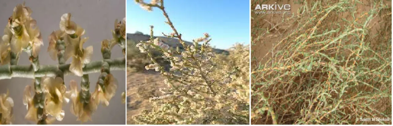
Figure 3. Arthrophytum scoparium pictures...20
Figure 4. Halogeton sativus pictures...33
Figure 5. Geografical distribution of Halogeton sativus………34
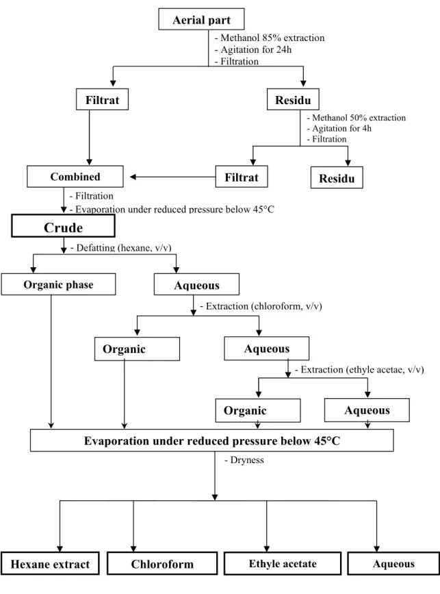
Figure 6. Diagram represents the process of extraction of phenolic compounds from A.scoparium and H.sativus aerial parts …………....………..38
Figure 7. Standard curve of gallic acid for the determination of total phenolic compounds in various plant extracts ………...…39
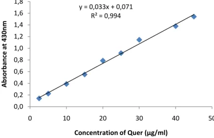
Figure 8.Standard curve of quercetin for the determination of flavonoids in various plant extracts.……….40
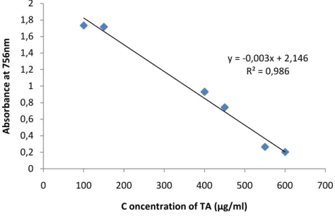
Figure 9. Standard curve of tannic acid for the determination of tannins in various plant extracts ……….……....41
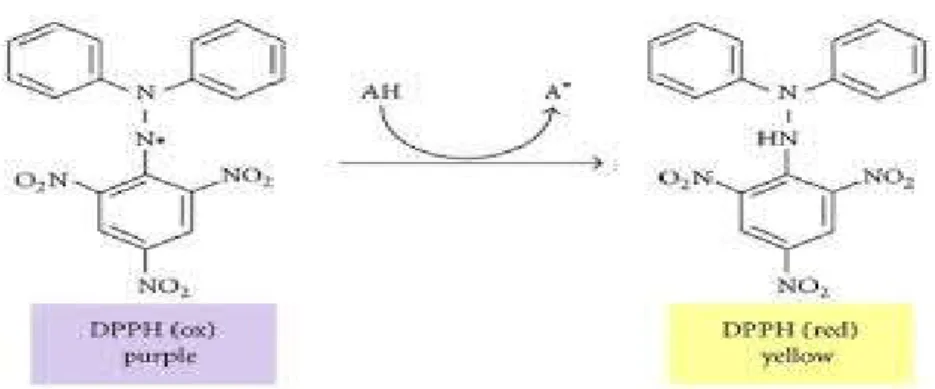
Figure10. Radical and non-radical forms of DPPH ………42
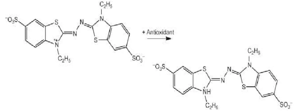
Figure 11. ABTS chemical reaction with antioxidant compound…………...43
Figure 12. IC50 values of A.scoparium and H.sativus extracts in DPPH free radical scavenging activity..……….……….…………57
Figure 13.IC50 values of A.scoparium and H.sativus extracts in ABTS radical scavenging activity……….………..58
Figure 14.The changes in the percentage of the inhibition of linoleic acid oxidation using A.scoparium and H.sativus extracts………..60
Figure 15.Antioxidant activities of A.scoparium extracts measured by β-carotene bleaching method………..60
VIII
Figure 16.Antioxidant activities of H.sativus extracts measured by β-carotene bleaching method………..61
Figure 17.EC50 values of A.scoparium and H.sativus extracts in reducing power assay……62 Figure 18.The IC50 values of ferrous iron chelating activity of A.scoparium and H.sativus ………..63
Figure 19.IC50 values of different plants extracts in hydroxyl radical scavenging activity………..65
Figure 20.The IC50 values of H2O2 scavenging activity of A.scoparium and H.sativus…….66 Figure 21.Inhibition percentage values of A.scoparium and H.sativus in antihemolytic
activity using FeSO4……….69
Figure 22.Increase in Body weight of rats treated orally with A.scoparium and H.sativus………...70 Figure 23.Biochemical parameters of control and rats treated with ASE and HSE measured during the acute toxicity………...72
Figure 24.Sections of liver and Kidney tissues of rats treated with ASE and HSE…………73
Figure 25.Effects of treatment with ASE and HSE on some biochemical parameters……...75
Figure 26.Effect of different concentrations of ASE, HSE and ViT C on DPPH radical scavenging and reducing power activities...…...76
Figure 27.Effect of treatment with ASE, HSE and vitamin C on reduced glutathione level in liver of rats………...77
Figure 28. Effect of treatment with ASE, HSE and vitamin C on catalase activity in liver of rats………78
Figure 29.Effect of treatment with ASE, HSE and vitamin C on MDA level in liver of rats………79
Figure 30.Sections of liver tissue of rats treated with ASE, HSE and ViT C compared to liver
IX
Figure 31.Effect of treatment with ASE and HSE on xylene-induced ear oedema…………81
Figure 32.Macroscopic observation of glandular regions of treated and untreated groups………..83
Figure 33.Histological Sections of stomach in normal and treated animals …….………...84
X
Table 1.Radical and non-radical ROS and RNS...04
Table 2.Extraction yields of Aerial parts of plants extracts………....………….54
Table 3.Phenolic, flavonoid and tannins content of plants extracts………...…56
Table 4.The HT50 values of anti-hemolytic activity of A. scoparium, control and aspirin………...…....………67
Table 5.The HT50 values of anti-hemolytic activity of H. sativus, control and aspirin………....………68
Table 6.Relative organs weight of rats treated with ASE and HSE……...………..71
Table 7.Effect of ASE and HSE on Ethanol Induced Gastric Ulcer in Rats……...………..82
Table 8.Percentage protection of ASE and HSE treated rats against ethanol induced gastric ulcer……….……..…83
XI
LIST OF ABBREVIATIONS
•LOO− Lipid peroxyl
•NO− Nitric oxide
•OH Hydroxyl radical
1
O2 Singlet oxygen
4-HNA 4-hydroxyl nonenal ALP Alkaline phosphatase ALT Alanine aminotransferase
AQE Aqueous extract
AST Aspartate aminotransferase CAMs Cell adhesion molecules
CAT Catalase
cGMP Cyclic guanosine monophosphate ChE Chloroform extract
COX Cyclooxygenase
CrE Crude extract
CVD Cardiovascular disease
DEC Decoction
DEN Diethylnitrosamine
EAE Ethyle acetate extract
EC50 Effective concentration of 50%
eNOS Endothelial nitric oxide synthase ER Endoplasmic reticulum
GAE Gallic acid equivalent GPx Glutathione peroxidase GSH Reduced glutathione HLA Human Leukocyte antigen HNO2
−
Nitrous acid
HT50 Half-time of 50% hemolysis
IC50 Inhibitory concentration at 50%
L• Carbon centered lipid radical
LD50 Lethal dose 50
LOOH Lipid peroxide
MDA Malondialdehyde
NAFLD Non – alcoholic Fatty liver Diseases NASH Non-alcoholic steatohepatitis
NK Natural killer
NSAIDs Nonsteroidal anti-inflammatory drugs
O2•- Superoxide
OECD Organisation for economic co-operation and development ONOO− Peroxynitrite
OS Oxidative stress
PCM Paracetamol
PUDs Peptic ulcer disease PUFA Polyunsaturated fatty acids QE Quercetin equivalent RBCs Red blood cells
RNS Reactive nitrogen species. ROS Reactive oxygen species. SOD Superoxide dismutase TAE Tannic acid equivalent XO Xanthine oxidase
XII
LISTE OF SCIENTIFIC PRODUCTIONS
ARTICLES
Kaddour Sabrina Manel, Arrar Lekhmeci, Baghiani Abderrahmane.(2020). Anti-Inflammatory Potential Evaluation (In-Vitro and In Vivo) of Arthrophytum scoparium Aerial Part, Journal of Drug Delivery and Therapeutics, 10(5):213-218.
Kaddour Sabrina Manel, Zerargui Fatima, Arrar Lekhmeci and Baghiani Abderrahmane. (2019). Acute, sub-acute and antioxidant activities of Arthrophytum scoparium aerial parts. IJPSR, 10(9): 4167-4175.
Kaddour Sabrina Manel, Guemmaz thoraya, Arrar Lekhmeci and Baghiani Abderrahmane. (2016). Hydroxyl radical scavenging and chelating activity effects of Haloxylon salicornicum extracts. Revue des Régions Arides n°43 (3/2017) – Numéro spécial – Actes du 5ème Meeting International sur l’Arido-culture et les Cultures Oasiennes : Biotechnologie végétale en zones arides et oasiennes.
COMMUNICATIONS
Kaddour Sabrina Manel, Arrar Lekhmeci et Baghiani Abderrahmane. (2016).In vitro antioxidant effect of Haloxylon salicornicum extracts. doctoriales de l’UFAS1 sur l’innovation le 19 mai 2016, école doctorales el bez sétif, Algérie.
Kaddour Sabrina Manel, Arrar Lekhmeci et Baghiani Abderrahmane. (2016).In vitro antioxidant effect of Haloxylon salicornicum extracts. doctoriales de faculté des sciences de la nature et de la vie le 31 mai 2016 sétif, Algérie.
Kaddour Sabrina Manel, Arrar Lekhmeci et Baghiani Abderrahmane. (2016).l’effet de Nigella sativa sur la production de xanthine oxydoréductase chez les rats traités par l’éthanol. 1er séminaire national de biologie, santé et stress oxydant le 9et 10 novembre 2017 Tébessa, Algérie.
Kaddour Sabrina Manel, Guemmaz thoraya, Arrar Lekhmeci and Baghiani Abderrahmane (2016).Hydroxyl radical scavenging and chelating activity effects of Haloxylon salicornicum extracts. V meeting international sur la biotechnologie végétale en zones arides et oasiennes, 19au21 décembre 2016, Zarzis, Tunisie.
Kaddour Sabrina Manel, Arrar Lekhmeci and Baghiani Abderrahmane (2017).Radical scavenging, lipid peroxidation inhibition and chelatins properties of extracts from Arthrophytum scoparium aerial parts extracts, congrès international de phytothérapie du 29 Avril au 1 mai 2017, Monastir, Tunisie.
XIII
Kaddour Sabrina Manel, Arrar Lekhmeci and Baghiani Abderrahmane (2017). Antioxidant proprieties of Haloxylon salicornicum aerial parts extracts ,8 journées scientifiques internationales sur la valorisation des bioressources 5,6 et 7 mai 2017 à Monastir, Tunisie. Kaddour Sabrina Manel and Sobhi Widad. (2017). Effet hépatoprotecteur de Nigella sativa chez les rats traités par l’éthanol. The first international congres on biotechnologies for sustainable development CIBSDD ,24-25 october , Boumerdes, Algeria.
Kaddour Sabrina Manel, Arrar Lekhmeci and Baghiani Abderrahmane. (2018).Analgesic and radical scavenging effect against hydrogen peroxyde and hydroxyl radical of Arthrophytum scoparium extracts.VI congres international de biotecghologies et valorisation des bio-ressources,organisé par l’ATBVBR du 20-23Mars 2018 à Tabarka, Tunisie.
Kaddour Sabrina Manel, Arrar Lekhmeci and Baghiani Abderrahmane. (2018). Polyphenols content, DPPH and ABTS radical scavenging activities of Halogeton sativus extracts. UFAS1, 25 Avril 2018, Setif, Algeria.
Kaddour Sabrina Manel, Arrar Lekhmeci and Baghiani Abderrahmane. (2018).Anti-inflammatoiry activity of Arthrophytum scoparium extracts.9 journées scientifiques internationales sur la valorisation des bioressources du 3-6 mai 2018, Monastir, Tunisie. Kaddour Sabrina Manel, Arrar Lekhmeci and Baghiani Abderrahmane. (2018). Antihemolytic and antioxidant effect of Arthrophytum scoparium. Congres international ; les rencontre de l’agriculture et de la biologie du 05-07 mai 2018 Constantine, Algérie.
Kaddour Sabrina Manel, Arrar Lekhmeci and Baghiani Abderrahmane. (2018). Antioxidant properties of Halogeton sativus aerial parts extracts. Auditorium M. K. Nait Belkacem, UFAS,23 juin 2018, Setif, Algeria.
Kaddour Sabrina Manel, Arrar Lekhmeci and Baghiani Abderrahmane. (2018).Total phenolic content, ion chelating and reducing power properties of Halogeton sativus extracts. The 2nd international conference on bioanalysis :food and healthHigher institute of applied sciences and technology ,December 15th, Mahdia, Tunisia.
Kaddour Sabrina Manel, Arrar Lekhmeci and Baghiani Abderrahmane. (2019). The preventive effect of arthrophyrum scoparium against gastric ulcer induced with ethanol. VII congres international de biotecghologies et valorisation des bio-ressources,organisé par l’ATBVBR du 20-23Mars 2019 à Tabarka, Tunisie.
XIV
CONTENTS
ACKNOWLEDGEMENTS...І DEDICATE...ІІ ﺺﺨﻠﻣ ... ІІІ ABSTRACT………...…….IV RESUME...VIII LISTE OF TABLES...IX LIST OF ABBREVIATIONS...XILIST OF SCIENTIFIC PRODUCTION...XII
Introduction...1
Review of literature 1. Oxidative stress/oxidant and antioxidant system...3
1 .1.Oxidative stress...3
1.2. Oxidants...3
1.2.1. Endogenous Sources of ROS………...…………...3
1.2.2. Exogenous Source of ROS………..………...5
a. Cigarette Smoke………..……...5
b. Hyperoxia...6
c. Ionizing Radiation………...6
d. Heavy Metal Ions……….……...6
1.3. Effect of oxidative stress……….…….6
a. Effects of oxidative stress on DNA………7
b. Effects of oxidative stress on lipids. ………..……...8
c. Effects of oxidative stress on proteins……….…………8
d. Effects of oxidative stress on signal transduction……….…………..9
1.4. ROS in normal physiological processes……….………….9
1.5. Antioxidants...10
1.5.1. Enzymatic Antioxidants...10
XV
b. Catalase...11
c. Glutathione system...11
1.5.2. Nonenzymatic Antioxidants...11
a. Glutathione...11
b. Vitamin C (Ascorbic Acid)……….……….…………12
c. Vitamin E (α-Tocopherol)...12
d. Melatonin...12
e. Polyphenol and flavonoid compounds……….………..………...12
2. The inflammation...15
2.1. Phases of inflammation...16
2.2. Anti-inflammatory drugs...17
2.3. Natural products in anti-inflammation...17
3. The hepatoprotective effect………...………..……..18
3.1. General description of the liver………....…………...…...…....18
3.2. Histology of the liver………....………...…...……....19
a. Hepatocytes ………....…………...…...…...20
b. Endothelial Cells………....…………...…..…....20
c. Kupffer Cells………....………..20
d. Stellate Cells………...………..…...…..21
3.3. Hepatotoxic agents used in animal models...21
a. Non-invasive method……….…...……….………...…..21 b. Invasive method………...……….……..21 3.3.1. Chemical-induced hepatotoxicity………....……….22 a. CCl4...22 b. Thioacetamide………....………23 c. Diethyl-nitrosamine………....………23 3.3.2. Drug-induced hepatotoxicity………...………23 a. NSAIDs………....………..23 b. Paracetamol………....………23 c. Anticancer drugs………...………...24
3.3.3. Metal- induced hepatotoxicity...24
a. Mercury...24
b. Cadmium...24
XVI
3. 4.Free radicals and liver diseases...25
4. Gastric ulcer...26
4.1. Etiology...26
4.2. Various ulcer models………....………..………….27
a. Ethanol-induced ulcers………27
b. Indomethacin-induced gastric ulcers………..28
c. HCl-induced ulcers……….……...28
5. Selected Plants………..….28
1. The family of chenopodiaceae………28
1.1. General description………..28
1.2. Classification of the chenopodiaceae...29
1.3. Economic importance of the chenopodiaceae…….………...30
2. Arthrophytum scoparium……….………...30 2.1. Botanical description………..……….30 2.2. Taxonomy...31 2.3. Traditional use………..………..32 2.4. Chemical composition………..………..32 3. Halogeton sativus………..………33 3.1. Botanical description………..………33 3.2. Taxonomy...34
Materials and methods 1. Materials………...………36
1.1. Plant Material………...………...36
1.2. Experimental animals………..36
1.3. Chemicals and reagents………..………36
2. Methods………..………..37
2.1. Preparation of plants extracts…..………...37
2.2. Phytochemical screening……...………...39
2.2.1. Determination of total phenolic content….………...39
2.2.2. Determination of flavonoids Content…...………...40
2.2.3. Determination of tannin content……...………...40
2.3. The evaluation of antioxidant activity in Vitro………...…41
2.3.1. DPPH radical scavenging activity………41
XVII
2.3.3.β-Carotene/linoleic acid assay………...……...43
2.3.4. Reducing power assay………..…………44
2.3.5. Ion chelating assay………..……….45
2.3.6. Hydroxyl radical scavenging assay………..………45
2.3.7. Hydrogen peroxide scavenging activity……….………47
2.3.8. Anti-hemolytic assay……….………..47
a. The AAPH-induced hemolysis……….……….47
b. ferrous ion-induced hemolysis……….……….47
2.4. In vivo……….………48
2.4.1. Acute Toxicity……….………48
2.4.1.1. Observation………..48
2.4.1.2. Plasma preparation and biochemical analysis………..48
2.4.1.3. Organs weight………49
2.4.1.4. Histological analysis………49
2.4.2. Hepatoprotective assay………...49
2.4.2.1. Histopathological studies……….49
2.4.2.2. DPPH radical-scavenging activity of plasma………..50
2.4.2.3. Reducing power assay……….50
2.4.2.4. Preparation of tissue homogenates………..50
2.4.2.5. Determination of Catalase activity………..50
2.4.2.6. Estimation of glutathione content………51
2.4.2.7. Estimation of MDA……….51
2.4.3. Xylene induced ear edema………..52
2.4.4. Ethanol- induced Gastric ulcer ………..52
2.5. Statistical analysis...52
Result and discussion 1. Extraction yield………...54
2. Total phenolics, flavonoids and tannins content in plants extracts………55
3. Antioxidant activity of A.scoparium and H.sativus extracts………57
3.1. In Vitro Antioxidant Activity……….………57
3.1.1. DPPH-scavenging assay……….………....57
3.1.2. ABTS radical cation decolorization assay……….….58
3.1.3. β-Carotene / Linoleic Acid Assay………...59
XVIII
3.1.5. Ferrous ion chelating activity……….…….62
3.1.6. Hydroxyl Radical-scavenging Assay……….…….64
3.1.7. Hydrogen peroxide scavenging activity……….…….65
3.1.8. Anti-hemolytic assay……….……..66
a. AAPH induced hemolysis………66
b. FeSO4-induced hemolysis………...68
3.2. In vivo………...70
3.2.1. Acute Toxicity………70
3.2.1.1. The effect of treatment with ASE and HSE on relative organ weights of rats……….71
3.2.1.2. Effect of plant extract on some biochemical parameters……….71
3.2.1.3. Effect of traetment with ASE and HSE on hepatic and renal tissue………73
3.2.2. Hepatoprotective assay………...74
3.2.2.1. Effect of ASE and HSE on biochemical parameters in CCl4-induced hepatotoxicity………74
3.2.2.2. Effect of ASE/HSE on plasma antioxidant capacity………...75
a. DPPH radical scavenging……….75
b. reducing power……….75
3.2.2.3. Effect of treatment with ASE /HSE on reduced Glutathione (GSH)………...77
3.2.2.4. Effect of treatment with ASE and HSE on Catalase (CAT)………...77
3.2.2.5. Effect of treatment with ASE and HSE on MDA………...78
3.2.2.6. Effect of treatment with ASE/HSE on hepatic tissue……….79
4. Xylene induced ear oedema………81
5. Ethanol-induced gastric ulcer………82
5.1. Effect of treatment with ASE/HSE on stomach tissue…...………84
Conclusion………...85
1 Introduction
Medicinal plants have been playing an essential role in the development of human culture as a source of medicines. They have always been at forefront virtually all cultures of civilizations. Medicinal plantes are regarded as rich sources of traditional medicines and from these plants many of the modern medicines are produced. For thousands of years, medicinal plants have been used to treat health disorders, to add flavor, concerve food and to prevent epidemic diseases. The present study was undertaken to valorize A.scoparium and H.sativus extracts as antioxidant and anti-inflammatory agents.
Phenolic compounds and flavonoids are present in nutrients and herbal medicines, both flavonoids and many other phenolic compounds are effective as antioxidants, antibacterial, cardioprotective agents, anti-inflammatory agents, immune system promoting, skin protecting compounds, skin protection from UV radiations, and interesting candidate for pharmaceutical and medicinal use.
WHO (World Health Organization) estimated that 80% of people worldwide rely on herbal medicines for some aspect of their primary health care needs. According to WHO, around 21.000 plant spices have the potential for being used as medicinal plants.
Reactive oxygen species (ROS), highly reactive molecules, are produced by living organisms as a result of normal cellular metabolism and environmental factors, and can damage nucleic acids and proteins, there by altering their functions. The human body has several mechanisms to counter act oxidative stress by producing antioxidants. A shift in the balance between oxidants and antioxidants in favor of oxidants is termed as “oxidative stress”.Oxidative stress results in macromolecular damage and is implicated in various disease states such as atherosclerosis, diabetes, cancer, neurodegeneration, and aging. Naturally occurring phytochemical antioxidants have occupied a prominent position as effective antioxidants for the prevention and/or treatment of several disorders and diseases. The premise for this has been the antioxidant actions of the phytochemicals as free-radical scavengers, oxidative stress relievers, and lipoperoxidation inhibitors. In recent years, plant polyphenols have been reported as effective antioxidants in the prevention and treatment of several diseases, including CVDs, cerebrovascular diseases, Alzheimer’s disease, resperatory system disease, and cancer (Liu et al., 2018).
Arthrophytum scoparium and Halogeton sativus belongs to the family of chenopodiaceae. They are worldwide distributed, especially in desert and semi desert areas. A.scoparium is a
2
local medicinal plant, which has been used extensively in the southwestern part of Algeria. It is used to treat numerous human diseases especially infectious (skin infections, urinary and genital infections), Rheumatism, diabetes, cancer, infertility, hair problems and stomach disorder (Allaoui et al., 2016, fatehi et al., 2017),however, Many studies reported the traditional use of Halogeton species in the treatment of hypertension and demonstrated the hypoglycaemic effects (TUNDIS et al., 2008).
In the present study, the following objectives are formulated:
Evaluation of the in vitro antioxidant activity of plant extracts using different methods. Estimation of the LD50 of the Arthrophytum scoparium and Halogeton sativus of crude
methanolic extracts of aerial parts extracts.
Evaluation of the acute toxicity of Arthrophytum scoparium and Halogeton sativus. Evaluation of the protective effect of crude extracts of selected plants against CCl4
-induced liver injury.
Evaluation of plasma antioxidant capacity using DPPH radical scavenging test and reducing power method.
Evaluation of the in vivo antioxiadant activity of crude extract of selected plants by assessing the MDA and GSH levels, and catalase activity in the liver of rats.
Evaluation of the antiinflammatory effect of plant extracts in mice against xylene-induced ear odema.
Evaluation of the protective effect of crude extracts of selected plants against ethanol induced gastric ulcer.
3
1. Oxidant and antioxidant system
1 .1.Oxidative stress
The term “stress” was first used in the biomedical literature as a description of hyperactivity in the hormone system, in particular concerning the corticosteroids of the adrenal cortex .The scientist summarized some 20 years later how the idea of stress, stress response, and homeostasis as a dynamic equilibrium gradually developed into a highly useful idea in general physiology and the study of diseases. According the literature, it’s reported that “stress” primarily as a factor causing disease, and even today, as exemplified by this thematic issue, modern stress research is still largely concerned with path mechanisms of human disease. (Breitenbach and Eckl, 2015). Oxidative stress (OS) is conceptually defined as the imbalance between generations and clearances of oxidants and it has increasingly become a major interested point of basic science and clinical research.
1.2. Oxidants
1.2.1. Endogenous Sources of ROS
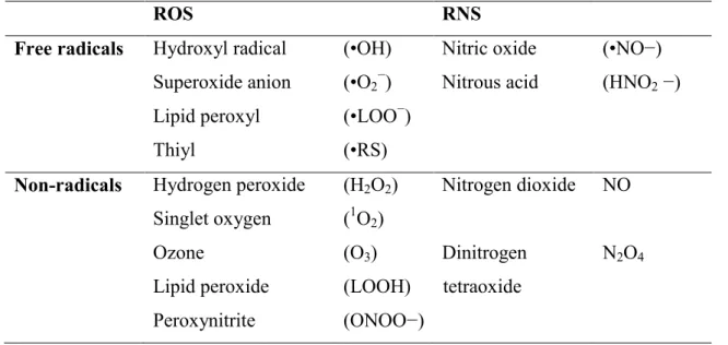
The term reactive oxygen species (ROS) includes the reduced form of oxygen and their reaction products with other molecules. ROS can be divided into two groups: free radicals and nonradical. Reactive Nitrogen species (RNS) are special forms of ROS that contain nitrogen. Similar to ROS, RNS can also include radicals and non-radicals form (Mugoni et al., 2013; Collin, 2019). Molecules containing one or more unpaired electrons and thus giving reactivity to the molecule are called free radicals. When two free radicals share their unpaired electrons, nonradical forms are created. ROS/RNS are summarized in Table 1.
4
Table 1.Radical and non-radical ROS and RNS (Phaniendra et al., 2015).
ROS RNS
Free radicals Hydroxyl radical Superoxide anion Lipid peroxyl Thiyl (•OH) (•O2−) (•LOO−) (•RS) Nitric oxide Nitrous acid (•NO−) (HNO2 −)
Non-radicals Hydrogen peroxide Singlet oxygen Ozone Lipid peroxide Peroxynitrite (H2O2) (1O2) (O3) (LOOH) (ONOO−) Nitrogen dioxide Dinitrogen tetraoxide NO N2O4
In the living organisms, ROS are generated in several cellular Systems localized on the plasma membrane, in the cytosol, in the peroxisomes, and on membranes of mitochondria and endoplasmic reticulum (Fig.1). Several soluble cell components, including thiols, hydroquinones, catecholamines, and flavins, can contribute to intracellular ROS production as they are able to undergone redox reactions (Matsubara et al., 2015). Moreover, several cytosolic enzymes produce ROS during their catalytic activity. Probably the most studied ROS producing enzyme is xanthine oxidase (XO) (Rác et al., 2015). It has now been about 50 years since mitochondrial H2O2 production in the presence of respiratory substrates was first recorded (Treberg et al., 2019), followed shortly after by the detection of mitochondrial generation of superoxide radical anion (Dudylina et al., 2019). The discovery that electron-transfer along the inner mitochondrial membrane carriers is associated with formation of ROS suggested the mitochondrial involvement in degenerative processes linked to several diseases and aging (Kausar et al., 2018). Although peroxisomes have long been known as organelles involved in cellular metabolism of H2O2, it is now clear that they are involved in several metabolic pathways (Pascual-Ahuir et al., 2017). Important functions performed by peroxisomes include fatty acid β- and α-oxidation, amino acid and glyoxylate metabolism,
5
and synthesis of lipidic compounds, and most enzymes catalyzing these processes produce ROS during their activity (Antonenkov et al., 2010; Wanders et al., 2016). The endoplasmic reticulum (ER) is involved in multiple functions, such as synthesis, folding, and transport of Golgi, lysosomal, secretory, and cell-surface proteins , calcium storage , lipid metabolism, and, in some cell types, drug detoxification (Palipoch, 2013; Schwarz and Blower, 2016). Smooth endoplasmic reticulum presents a chain of electron transport, constituted by two systems devoted to xenobiotic metabolism and introduction of double bonds in fatty acids, which are also able to produce ROS (Di Meo et al., 2016).
Figure 1. Cellular sources of ROS production. Subcellular organelles and structural and soluble cell components all contribute to production of a wide variety of reactive species (Venditti et al., 2015). 1.2.2. Exogenous Source of ROS
a. Cigarette Smoke
In the last two decades, the central role of free radical mechanisms in tobacco smoke
carcinogenesis and oxidative stress has been established by a series of studies. Cigarette smoke contains many oxidants and free radicals and organic compounds, such as
superoxide anion (O2•−) and subsequently H2O2 and the reactive hydroxyl radical (HO•), which cause oxidative damage to cellular membrane lipids, proteins, enzymes and DNA (Tian et al., 2017). ROS in the cigarette gas-phase promote the destruction of endogenous
6
antioxidants (vitamins and enzymatic antioxidants) reducing the vital role of cellular antioxidant defenses (Forouzandeh et al., 2017). Several studies show that antioxidant vitamins are lower in smokers resulting in systemic oxidative stress, whereas dietary antioxidant supplements provide only limited protection to smokers (Kim et al., 2017).
b. Hyperoxia
Hyperoxia refers to conditions of higher oxygen levels than normal partial pressure of oxygen in the lungs or other body tissues. It leads to greater production of reactive oxygen and nitrogen species (Ozougwu, 2016).
c. Ionizing Radiation
Ionizing radiation, in the presence of O2, converts hydroxyl radical, superoxide, and organic radicals to hydrogen peroxide and organic hydroperoxides. These hydroperoxide species react with redox active metal ions, such as Fe and Cu, via Fenton reactions and thus induce oxidative stress (Kashmiri et al., 2014; Collin, 2019).
d. Heavy Metal Ions
Heavy metal ions, such as iron, copper, cadmium, mercury, nickel, lead, and arsenic, can induce generation of reactive radicals and cause cellular damage via depletion of enzyme activities through lipid peroxidation and reaction with nuclear proteins and DNA (Ozougwu, 2016).
1.3. Effect of oxidative stress
Oxidative stress occurs when the balance between antioxidants and ROS are disrupted because of either depletion of antioxidants or accumulation of ROS. Higher production of ROS in body may change DNA structure, result in modification of proteins and lipids, activation of several stress-induced transcription factors, and production of proinflammatory and anti-inflammatory cytokines.
7 a. Effects of oxidative stress on DNA
Both ROS/RNS can oxidatively damage the nucleic acids. The mitochondrial DNA is more vulnerable to the ROS attack than the nuclear DNA, because it is located in close proximity to the ROS generated place. ROS, most importantly, the OH• radical directly reacts with all components of DNA such as purine and pyrimidine bases, deoxyribose sugar backbone (Phaniendra et al., 2015) and causes a number of alternations including single and double stranded breaks in DNA. A number of modified purine (e.g. 8-hydroxydeoxy guanosine, 2,6-diamino-4-hydroxy-5-formamidopyrimidine) and pyrimidine (e.g. thymine glycol, uracil glycol, 5-hydroxydeoxyuridine) base produced by attack of OH• radical and DNA- protein cross links (Fig.2). On the other hand, the RNS, most importantly, peroxynitrite (OONO-) interacts with guanine to produce nitrative and oxidative DNA lesions such as 8-nitroguanine and 8-oxodeoxyguanosine respectively (Kawanishi et al., 2016).
Figure 2. Base modifications introduced by ROS (Birben et al., 2012). b. Effects of oxidative stress on lipids
The membrane lipids, especially the polyunsaturated fatty acid residues of phospholipids are more susceptible to oxidation by free radicals. The lipid peroxidation results in the loss of membrane functioning, for example, decreased fluidity, inactivation of membrane bound enzymes and receptors (Phaniendra et al., 2015). The lipid peroxidation is initiated, when any
8
free radical attacks and abstracts hydrogen from a methylene groups (CH2) in a fatty acid (LH) which results in the formation of a carbon centered lipid radical (L•). The lipid radical can react with molecular oxygen to form a lipid peroxyl radical (LOO•). The resultant lipid peroxyl radical (LOO•) undergo rearrangement via a cyclisation reaction to form endoperoxides, which finally form malondialdehyde (MDA) and 4-hydroxyl nonenal(4-HNA),the toxic end products of lipid peroxidation that cause damage to the DNA and proteins (Hashemi, 2019). These lipid peroxyl radicals can further propagate the peroxidation process by abstracting hydrogen atoms from the other lipid molecules. Isoprostanes constitute the important product of lipid peroxidation of arachidonic acid and are considered as the makers of the oxidative lipid damage (Ayala et al., 2014; Taso et al., 2019).
c. Effects of oxidative stress on proteins
Many radical species (O2 •-, OH •, peroxyl, alkoxyl, hydroperoxyl) and non radical species (H2O2, O3, HOCl, singlet oxygen, OONO-) can be induced the protein oxidation (Ferreira et al., 2018). ROS oxidize different amino acids present in the proteins, causing formation of protein– protein cross linkages, results in the denaturing and loss of functioning of proteins, loss of enzyme activity, loss of function of receptors and transport proteins (Nita and Grzybowski, 2016). The methionine and cysteine are more susceptible to oxidation by ROS because these amino acids contained sulphur and are converted to disulphides and methionine sulphoxide, respectively. However in biological systems, only these two oxidized forms of proteins can be converted back to their native form by two different enzymes namely disulfide reductases and methionine sulfoxide reductases respectively (Phaniendra et al., 2015; dos Santos et al., 2018). The ROS induced oxidative damage of amino acid residues such as lysine, proline, threonine and arginine yields carbonyl derivatives. The presence of carbonyl groups in proteins has been considered as the marker of ROS mediated protein oxidation. The
9
other specific markers of protein oxidation are O-tyrosine (a marker for hydroxyl radical) and 3-nitrotyrosine (a marker for RNS) (Davies, 2016).
d. Effects of oxidative stress on signal transduction
ROS can induce expression of several genes involved in signal transduction.( Dudziak et al.,2019 ) Disruption of the ratio GSH/GSSG causes activation of redox sensitive transcription factors, such as NF-κB, AP-1, nuclear factor of activated T cells and hypoxia-inducible factor 1 , that are involved in the inflammatory response (Ren et al., 2017). Activation of transcription factors via ROS is achieved by signal transduction cascades that transmit the information from outside to the inside of cell (Nguyen et al., 2017). Tyrosine kinase receptors, most of the growth factor receptors (epidermal growth factor receptor, vascular endothelial growth factor receptor, receptor for platelet-derived growth factor, protein tyrosine phosphatases, and serine/threonine kinases) are targets of ROS (Alexander et al., 2017).
1.4. ROS in normal physiological processes
It has been widely documented that ROS/RNS act as intracellular signaling molecules to exert their physiological function.ROS are involved in signaling through chemical reactions with specific atoms of target proteins that lead to covalent protein modification (Griendling et al., 2016). Thus, ROS molecular recognition occurs at the atomic and not at the macromolecular level. Moreover, ROS exert their particular signaling properties by specifically targeting elements in protein. For example, O2.- preferably reacts with iron-sulfur (Fe-S) clusters. Both cysteine residues and methionine residues are the major targets of H2O2 (Mettert and Kiley, 2015). However,.OH has no preferable reactivity toward biological molecules. Furthermore, selenocysteine is more reactive with H2O2 than cysteine, and may operate in H2O2 based redox regulation (Benchoam et al., 2019). H2O2 can act as direct modifier of thiol containing
10
proteins, to produce a redox state of signaling proteins, to produce a redox dependent modification of enzyme. For the same reason not all ROS are aqually suitable for signal transduction (Song and zou, 2015; Rampon et al., 2018).
Many of low grade ROS-mediated acute responses actually protect the cell against oxidative damage and re-establish or maintain the intracellular redox homeostasis (Marengo et al., 2016). ROS are involved in important physiological responses/events, such as defense against envirommental pathogens, responce to growth factors, cellular proliferation, differentiation, apoptosis, and autophagy. Additionally, duox expression and H2O2 formation are the rate-limiting processes for the synthesis of thyroid hormones (Di Marzo et al., 2018).
1.5. Antioxidants
Just like free radicals, antioxidants can be endogenously produced (e.g. superoxide dismutase (SOD) and reduced glutathione (GSH)) and can also be introduced to the biological system exogenously, usually through diet (e,g. vitamin c, caratenoids and vitamin E) (Amber et al., 2013; Ighodaro and Akinloye, 2017). Antioxidants primarily function to balance out free radicals generated during metabolic processes including during mechanisms involved in protecting the gut from inflammation and injury (Nwosu et al., 2016). Antioxidants can be said to go about its defence system in three main ways: a: by proteins sequestering with transition metals preventing their availability for reaction with free radicals.b: inhibiting their deleterious effect, making available small molecules that have the capability of scavenging free radicals.c: specific mechanisms for the correction of ROS-induced DNA damage (Ofor et al., 2016).
1.5.1. Enzymatic Antioxidants a. Superoxide dismutase
SODs are a class of closely related enzymes that catalyse the breakdown of the superoxide anion into oxygen and H2O2 present in almost all aerobic cells and in extracellular fluids (He et al., 2017). It
11
which binds nickel. Mn-SOD has been found mostly in mitochondria and peroxisomes, Fe-SOD has been found in peroxisomes and Cu-Zn-SOD in peroxisomes and cytosol (Ighodaro and Akinloye, 2017). Superoxide dismutase’s converts superoxide to hydrogen peroxide: 2O2
+ 2H = H2O2 + O2 .
b. Catalase
This is predominant in cells exposed to oxygen and is frequently used to catalyse the decomposition of H2O2 to oxygen and water (Webb et al., 2017). Catalase has one of the highest turnover rates for all enzymes; with one molecule of catalase being able to convert approximately 6million molecules of H2O2 to water and oxygen each minute (Obeagu, 2018).
c. Glutathione system
The Glutathione system comprises of three main classes: Glutathione reductase, Glutathione peroxidise (GPx) (competes with catalase for H2O2 and is the major source of protection against low levels of oxidative stress) and Glutathione S transferase (Kivrak et al., 2017).
1.5.2. Nonenzymatic Antioxidants
Nonenzymatic antioxidants include low-molecular-weight compounds but are not limited to; Glutathione (GSH), ascorbic acid (Vitamin C), tocopherols and tocotrienols (Vitamin E) , uric acid, melatonin, alpha lipoic acid (Tan et al., 2018).
a. Glutathione
They are synthesized from constituent amino acids in cells. It is a cysteine containing peptide. In cells it is maintained as Glutathione reductase which subsequently reduces other metabolites and enzyme systems while still capable of reacting with oxidants (Lian et al., 2018).
12
Vitamin C is usually maintained in the reduced form in the body by its reaction with glutathione which can be catalysed by protein. Vitamin C is able to prevent formation of nitrosamines, neutralise ROS such as H2O2, thus boosting immunological response (Pehlivan, 2017). c. Vitamin E (α-Tocopherol)
They are fat soluble vitamins that exist in eight different forms and possess antioxidative properties. The main function of vitamin E is protecting the cell from lipid peroxidation. In the human biological system, α-tocopherol is the most studied and is said to have the highest bioavailability with the body preferentially absorbing and metabolizing this most active form (Obeagu, 2018).
d. Melatonin
This is a powerful antioxidant that has the ability to easily cross cell membranes and blood-brain barrier. Unlike other antioxidants melatonin does not undergo redox recycling (repeated reduction and oxidation), once oxidized cannot be reduced to previous state because it forms several stable end products when it reacts with free radicals. Thus has been termed terminal (or suicidal) antioxidant (Galano et al., 2018; Debnath et al., 2019).
e. Polyphenol and flavonoid compounds
Plant metabolism is mainly classified as primary or secondary. Compounds produced through primary metabolism, which are generally referred to as primary metabolites; include sugars, fatty acids, amino acids and nucleic acids (Singh et al., 2017). Primary metabolites are required for maintenance of plant cells while secondary metabolites are essential to the normal growth, development and defense of plants (Wang et al., 2019). To date, thousands of different types of secondary metabolites have been identified in plants chemically, these compounds are either nitrogen-containing (alkaloids) or nitrogen-deficient (terpenoids and
13
phenolics) (Patra et al., 2013; Deepak et al., 2015).).Plant phenolics are mainly classified into five major groups, phenolic acids, flavonoids, lignans, stilbenes and tannins (Myburgh, 2014). Phenolic compounds generally possess one or more aromatic rings with one or more hydroxyl groups (Tanase et al., 2019). It has commonly been assumed that the antioxidant capacity of phenolics will increase with the number of free hydroxyls and conjugation of side chains to the aromatic rings Flavonoids and phenylopropanoids are also oxidized by peroxidase, and act as H2O2 scavengers (Lee et al., 2017).
The basic structure of a phenolic compound comprises of an aromatic ring with one or more – OH groups. However, phenolic compound found in Nature are structurally diverse from simple phenolic molecules to complex polymerized compound (Bhuyan and A Basu, 2017). Phenolics found in food material can be divided into three major groups: simple phenol and phenolic acids, hydroxycinnamic acid derivatives and flavonoids. Phenolic acids, flavonoids and tannins are considered as the main dietary phenolics (Ștefănescu et al. 2019). Flavonoids constitute the largest group of low-molecular-weight plant phenolics and have been studied most extensively. They are also the most important plant pigments. Over 4,000 different types of flavonoids are found in Nature (Panche et al., 2016). Flavonoids usually occur bound to sugar molecules and consist mainly of catechins, proanthocyanins, anthocyanidins, flavons and flavonols and their glycosides (Talapatra and Talapatra, 2015). Phenolic acids are divided into two subgroups: hydroxybenzoic and hydroxycinnamic acids (Bhuyan et al. 2015). Phenolic acids are significant components of fruit and vegetables. These compounds play an important role in color stability, aroma profile and antioxidant activity. They act as acids because of their carboxylic group (Baião et al. 2017). Tannins are the third important group of polyphenolics which can further be divided into two subcategories: condensed and hydrolysable tannins (Bhuyan and Basu, 2017). These are high-molecular-weight polymers. Fruits, grains and legumes consist of condensed tannins which are mainly polymers of
14
catechins or epicatechins, whereas hydrolysable tannins are polymers of gallic or ellagic acid and found in berries and nuts (Smeriglio et al. 2017).
Numerous studies have reported the potential health benefits of plant polyphenolics in particular. Due to their potent antioxidant properties, plant phenolics have scientifically proven to prevent various oxidative stress-related as well as chronic diseases, such as cancer, cardiovascular and neurodegenerative diseases (Koch, 2019).
Oxidative stress plays an important role in carcinogenesis. Several mechanisms contribute to the overall formation of tumours from oxidative damage. (Lee et al. 2014). Therefore, phenolics with antioxidant properties have been found to be beneficial in preventing or treating the oxidative damage that can induce cancer (Liu et al. 2018). Polyphenols may affect the molecular events in the initiation, promotion and progression stages of carcinogenesis and isoflavones and lignans may affect the estrogen-related activities related to tumour formation (Hsu et al. 2015 ; Moein, 2015). Flavonoids have been reported to modulate key enzymes and receptors involved in signal transduction pathways of cellular proliferation, differentiation, apoptosis, inflammation, angiogenesis, metastasis and reversal of multidrug resistance (Baião et al. 2017).
Cardiovascular disease (CVD) is one the major killers in all developed countries with a rise in prevalence (Tome-Carneiro and Visioli 2016). Several epidemiologic studies and intervention trials suggest that polyphenols present in fruits and vegetables are associated with decreased risk of cardiovascular diseases (Rangel-Huerta et al. 2015). As polyphenols are known for their antioxidant activities, increased intake of dietary antioxidants may protect against the development of CVD. In addition, recent evidence suggests that polyphenols have immunomodulatory and vasodilatory properties which may also contribute in reducing the risk of CVD (Mahmoud et al., 2019).
15
2. The inflammation
Inflammation is biological response of the immune system’s that can be triggered by a variety of
factors, such as pathogens, damaged cells, toxic compounds, or irradiation. These factors may
induce acute and/or chronic inflammatory responses in the heart, pancreas, liver, kidney, lung, brain, intestinal tract and reproductive system, potentially leading to tissue damage or disease. (Chen et al., 2018; Liu et al., 2019), and acts by removing injurious stimuli and initiating the healing process. Inflammation is therefore a defense mechanism that is vital to health (Zhou et al., 2016). Usually, during acute inflammatory responses, cellular and molecular events and interactions efficiently minimize impending injury or infection. This mitigation process contributes to restoration of tissue homeostasis and resolution of the acute inflammation. However, uncontrolled acute inflammation may become chronic, contributing to a variety of chronic inflammatory diseases (Chen et al., 2018; Donald et al., 2018). At the tissue level, inflammation is characterized by redness, swelling, heat, pain, and loss of tissue function, which result from local immune, vascular and inflammatory cell responses to infection or injury (Gallo et al., 2017). Important microcirculatory events that occur during the inflammatory process include vascular permeability changes, leukocyte recruitment and accumulation, and inflammatory mediator release (Chertov et al., 2000; Chen et al., 2018).
Mechanism of inflammation represents a chain of organized, dynamic responses including both cellular and vascular events with specific humoral secretions. These pathways involve changing physical location of white blood cells (monocytes, basophils, eosinophils, and neutrophils), plasma, and fluids at inflamed site (Huether and McCance, 2015). A group of secreted mediators and other signaling molecules (e.g., histamine, prostaglandins, leukotrienes, oxygen- and nitrogen-derived free radicals, and serotonin) are released by immune defense cells principally in the mechanism which can contribute in the event of inflammation (Abdulkhaleq et al., 2018). Whatever, the inflammatory response is triggered
16
through two phases: (a) acute and (b) chronic, and each is apparently mediated by a different mechanism (Serhan et al., 2015). These immune responses which involved in acute inflammation can be divided into vascular and cellular (Nguyen, 2012).
2.1. Phases of inflammation
The responses which occur in microvasculature normally appear in few minutes following tissue injury or microbial infection in the presence of other inflammatory stimuli named vascular events ( Boffet al., 2018). The occurrence of these processes is rapid and eventually will lead to vasodilatation and subsequently makes the vessels become more permeable. This process will result in entry of inflammatory mediators and produces interstitial oedema (Porter, 2013; Simon et al., 2018).Infiltration of white blood cells from circulatory system is essential during inflammatory responses (Goljan, 2014; Kumar et al., 2013). A group of chemotactic agents such as microbial endotoxins holding amino terminal N-formyl methionyl groups, C5a complement fragment, and interleukins along with the secretions of basophils such as platelets activating factor, histamine, and leukotriene B can stimulate intense leukocytes infiltration within few minutes (Bitencourt et al., 2013; Abdulkhaleq et al., 2018). Among the leukocytes, neutrophils are the first inflammatory cells that are recruited at the acute inflammation site (Curcic et al., 2015). Infiltration of immune cells triggered via a complicated mechanism in which white blood cells work together with endothelium in postcapillary venules (Filippi, 2016). Cellular events encompass the successive capture, trundling, and firming an adhesion to the microvascular endothelium (Leick et al., 2014). These events in the mobilization pathway are arranged by cell adhesion molecules (CAMs). Following a period of stationary adhesion, the white blood cells may leave the postcapillary venules extending pseudopodia between endothelial cells and reach into the subendothelial space. This complex event is often referred as white blood cell extravasations and transendothelial migration (Abdulkhaleq et al., 2018; Muro, 2018). The inflammations of
17
chronic events are distinguished by mononuclear cell infiltration (e.g., monocyte and lymphocytes), fibroblasts proliferation, collagen fibers, and connective tissue formation (Shahid et al., 2018). With chronic inflammation, the tissue degeneration is normally mediated by nitrogen species, proteases, and other reactive oxygen species released from infiltrated inflammatory cells (Murakami, 2012).
2.2.Anti-inflammatory drugs
The main anti-inflammatory drugs are either steroidal (e.g., betamethasone, prednisolone, and dexamethasone) or nonsteroidal (Juthani et al., 2017) (e.g. aspirin, diclofenac, ibuprofen and indomethacin.) used to treat both acute inflammatory condition and chronic inflammatory diseases such as osteoarthritis and rheumatoid arthritis (Abdulkhaleq et al., 2018).However, their prolonged use is associated with various side effects; for example, steroidal drug causes adrenal atrophy osteoporosis, suppression of response to infection or injury, euphoria. Cataracts, glaucoma (Phalitakul et al., 2011; Juthani et al., 2017), and non-steroidal drug cause peptic ulcers and bronchospasm due to blockade of both the physiological and inflammatory prostaglandins and concurrent production of leukotrienes (Gunaydin and Bilge, 2018). Thus taking into account the adverse effects and high cost of synthetic conventionally available steroidal or non-steroidal drugs, the search for new anti-inflammatory agents from herbal sources is getting popular with the objective to obtain greater safety, better efficacy, and a more economical way to treat inflammation (Arulselvan et al., 2016).
2.3.Natural products in anti-inflammation
Medicinal plants continues to be an accepted form of treatment in the Orient, and plant drugs based on traditional practice represent a huge portion of the pharmaceutical products in modern western countries (Levin and Laufer, 2012; Yuan et al., 2016).First, concerns have been raised that modern pharmaceutical practice too often involves costlydrugs that produce
18
unacceptable side effects (Lemonnier et al., 2017); second, the experience shows that natural substances can apparently address several modern health concerns with fewer side effects (Shao et al, 2017); and third, experience shows that modern medicine and traditional herbal medicine can be combined (Yuan et al., 2016).
3. The hepatoprotective effect
The liver is the largest solid organ, the largest gland and one of the most vital organs that functions as a centre for metabolism of nutrients and excretion of waste metabolites (Ozougwu and Eyo, 2014). Its primary function is to control the flow and safety of substances absorbed from the digestive system before distribution of these substances to the systemic circulatory system (Ozougwu, 2017).
3.1. General description of the liver
The liver weighs approximately 1500g and accounts for approximately 2.5% of adult body weight. The liver lies mainly in the right upper quadrant of the abdomen where it is hidden and protected by the thoracic cage and diaphragm (Mubbunu et al., 2018). The liver is divided into 4 lobes: right, left, caudate, and quadrate. The right and left lobes are the largest, while the caudate and quadrate are smaller and located posteriorly (Kumon, 2017). Adjacent to the caudate lobe is the sulcus for the inferior vena cava. Just inferior to the caudate lobe is the porta hepatis, where the hepatic artery and hepatic portal vein enter the liver (Lowe and D'Angelica, 2016). The portal vein carries nutrient laden blood from the digestive system. Inferior to the porta hepatis is the bile duct which leads back to the gallbladder, also the hepatic vein, where post-processed blood leaves the liver, is found inferior and adjacent to the sulcus for the inferior vena cava (Singh and Rabi, 2019).