Cotutelle avec l’Université Sétif 1
ÉCOLE DOCTORALE DES SCIENCES DE LAVIE ET DE DA SANTE
Laboratoire de Biophotonique et Pharmacologie – UMR 7213
THÈSE
présentée par :Redouane BOUCHAALA
Soutenue le : 18 Septembre 2017
Pour obtenir le grade de : Docteur de l’université de Strasbourg
En cotutelle avec l’Université Sétif 1
Discipline/ Spécialité
: Nanophysique-Biophysique
Nanoparticules organiques fluorescentes à
base de lipides : intégrité et relargage de
principes actifs in vitro et in vivo
THÈSE dirigée par :
M. KLYMCHENKO Andrey Directeur de recherche CNRS, Université de Strasbourg
M. DJABI Smail Professeur, Université de Sétif 1-Algerie
RAPPORTEURS :
Mme. TEXIER Isabelle Directeur de recherche CEA, Grenoble
M. GUERIOUNE Mohamed Professeur, Université Badji Mokhtar -Annaba -Algérie
AUTRES MEMBRES DU JURY :
M. ZUBER Guy Directeur de recherche CNRS, Université de Strasbourg M. BOUAMAMA Larbi Professeur, Université de Sétif
1
Acknowledgements
This thesis work is an important part in my life, a real adventure that will leave many memories. My strongest gratitude goes to my supervisor Dr. Andrey Klymchenko, the man who taught me everything that I know in scientific research. I appreciate his continuous support during my PhD, motivation, advises, patience, immense source of knowledge and inspiration. Thanks for his guidance during my research and thesis writing, thanks for being a boss and a friend. We say that in life there are encounters that mark you forever, Andrey was one of them, with his unique personality, interesting discussions, passion, generosity, great knowledge, positive attitude and laughters. I could not imagine a better supervisor than him.
This PhD would never be possible without some helpful people. I would thank Pr. Yves Mély for welcoming me in his laboratory, Pr. Djabi Smail my co-supervisor for his continuous support, guidance and help, Pr. Abdelkrim Beniaiche without whom this adventure would not be made.
I’d like to thank the jury members Dr. Isabelle Texier, Pr. Mohamed Guerioune, Pr. El Arbi Bouamama and Dr. Guy Zuber for accepting our request to evaluate this work.
Many thanks to our numerous collaborators, namely Dr. Nicolas Anton, Pr. Thierry Vandamme, Dr. Jacky G Goetz, Luc Mercier, Dr. Olivier Lefebre. It was a pleasure to work together on different projects and to learn a lot from them.
I’m thankful to Dr. Youri Arntz, Dr. Julien Godet, Dr. Nicolas Humbert, Dr. Halina Anton, Dr. Jurga Valanciunaite, Dr. Andreas Reisch for discussions. Dr. Ludovic Richert, Dr. Frederic Pryzbilla and Pr. Pascal Didier for their help with the microscope. Moreover, I’d like to thank Ingrid Barthel and Romain Vauchelles for their assistance, and especially Marlyse Wernert for her availability, help and smile. To Dr. Guy Duportail for his help with French papers, his kindness and for providing me newspaper.
I thank all my colleagues from Laboratory of Biophotonics and pharmacology for the friendly atmosphere in the lab, useful discussions and happy moments. Specially Rajhans, Bogdan, Marianna, Lesia, Katya, Sasha, Oleksii, Evgen, Hassan, Waseem, Tanveer, Doriane, Iryna,
2 Nina, Ashokkumar, Anne, Taras, Kyong, Luca. Thank you guys for creating pleasant atmosphere and good memories.
I would like to express my thanks to Dr. Mayeul Collot for our scientific, political and friendly discussions, I highly appreciate these moments. To my friends, Avisek Ghose with whom I had a good time, discovering the city and Indian cuisine. Salah El Meshri, Yusuke Niko and Kamal Sharma with whom I laughed a lot. Special thanks to my good friend Manuel Pires, with whom I shared great moments, the same passions, unfished discussions, hoping that this friendship will continue. Big thanks to Liliana Zaiter for her help, kindness, our discussion, debate and all the moments. To Hicham Tasfaout, Redouane Thabti and my brother Mohamed Attia with whom I created a real friendship forever.
I want to acknowledge the university of Setif, and ERC for providing Financial support to this work. To Doctoral School of Life and Health Sciences of Strasbourg and Institute of optics and precision mechanics of Setif.
I’m deeply thankful to the most important people in my life my family, my mother Abla and my father Hocine I own them what I am, what I did and what I will do. They will be always near my heart. To all my Friends in Algeria for their support.
Finally, I’m Grateful to my brother Khalil that without him I could not accomplish that, thanks for your patience, enormous support, I will never forget what you did.
3
Contents
Acknowledgements ... 1
Contents ... 3
List of Abbreviations ... 6
Aim of PhD thesis ... 7
PART 1-Bibliographical overview ... 8
Chapter 1: Fluorescence ... 9
1- Principles of fluorescence ... 9
2- Characteristics of fluorescence ... 10
2.1- Emission and excitation spectra / absorbance ... 10
2.2- Stokes Shift and Solvatochromism ... 11
2.3- Quantum yield and fluorescence brightness, Fluorescence lifetime ... 12
2.4- Quenching ... 13 2.5- Anisotropy ... 13 3- Fluorescent Probes ... 15 3.1- Natural fluorophores ... 15 3.2- Synthetic dyes ... 15 3.3- Fluorescent nanoparticles ... 18
4- General requirements for fluorescent probes ... 18
5- Near-IR Dyes ... 19
Chapter 2: Nanocarriers ... 21
1- Terminology clarification ... 21
2- Nanoparticles in biomedical applications: ... 22
2.1- Inorganic nanoparticles ... 22
2.1.1- Quantum Dots (QDs) ... 22
2.1.2- Upconversion Nanoparticles (UCNPs) ... 23
2.1.3- Carbon Nanotubes ... 24
2.1.4- Gold Nanoparticles ... 25
2.1.5- Mesoporous Silica Nanoparticles (MSNs) ... 27
2.2- Organic Nanocarriers ... 28
4
2.2.2- Micelles ... 28
2.2.3- Polymeric Nanocarriers ... 30
2.2.4- Lipid Nanocarriers ... 33
2.2.4.1- Liposomes ... 34
2.2.4.2- Solid Lipid Nanocarriers (SLNs) ... 36
2.2.4.3- Nanostructured Lipid Carriers (NLCs) ... 38
2.2.4.4- Lipid Nanocapsules ... 40 2.2.4.5- Lipid Nanoemulsions ... 41 a) Introduction: ... 41 b) Components ... 42 c) Formulation methods ... 43 d) Nanoemulsion stability ... 47
e) Application of nanoemulsions in biomedical field ... 49
Chapter 3: Optical instrumental techniques for nanocarriers
characterization ... 53
1- Fluorescence correlation spectroscopy (FCS) ... 54
1.1- Theoretical view ... 54
1.1.1- Autocorrelation: principle and modelling of data ... 55
1.1.2- Instrumentation ... 56
1.2- Application of FCS for nanoparticles characterizations ... 58
2- Förster resonance energy transfer (FRET) ... 61
2.1- Theoretical view ... 61
2.1.1- Mathematical formalism of FRET ... 61
2.1.2- Distinction in energy transfer process ... 64
2.1.3- FRET applications: ... 65
2.2- Application of FRET for nanoparticles characterizations ... 65
3- Fluorescence-lifetime imaging microscopy FLIM ... 70
4- Intravital microscopy IVM ... 72
5- Dynamic light scattering DLS ... 76
6- Other different techniques ... 77
PART II
-Results and discussions ... 80
1- Integrity of lipid nanocarriers in bloodstream and tumor quantified by near-infrared ratiometric FRET imaging in living mice (Publication-1) ... 81
2- Fluorescence correlation spectroscopy to study release from lipid nanocarriers (Publication-2) ... 110
5 3- Encapsulation and release of molecules in lipid and polymer nanocarriers evaluated by
chemical bleaching ... 126
4- Light-triggered release from dye-loaded fluorescent lipid nanocarriers in vitro and in vivo (Publication-3) ... 134
PART III-
Conclusion and perspectives ... 148PART IV-Materials and Methods ... 152
1- Materials ... 153
2- Methods ... 153
2.1- Synthesis of NR668 and F888 ... 153
2.2- Synthesis of Cy5.5LP and Cy7.5LP ... 154
2.3- Preparation of lipid nanocarriers ... 156
2.4- Preparation of polymer nanoparticles ... 157
2.5- Nanocarriers size characterization ... 157
2.6- Fluorescence spectroscopy ... 157
2.7- FRET-based stability test ... 158
2.8- Fluorescence correlation spectroscopy (FCS) and dyes release ... 158
2.9- Microscopy ... 159
2.10- Cellular studies ... 160
2.11- In vivo studies in zebrafish ... 160
2.12- Subcutaneous tumor grafting and administration of FRET nanocarriers ... 161
2.13- In vivo whole animal FRET imaging ... 162
2.14- Calibration of the ratiometric response of NCs to disintegration ... 162
2.15- Image analysis of the FRET signal in living mice ... 162
2.16- Statistical analysis ... 163
2.17- Bleaching of Nile Red and NR668 with Sodium Dithionite ... 163
References ... 164
List of publications... 189
List of Conferences ... 190
6
List of Abbreviations
CMC Exceeds critical micelle concentration
Cy Cyanine dyes
DLS Dynamic light scattering
DOPC 1,2-Dioleoyl-sn-glycero-3-phosphocholine
DOX Doxorubicin
EPR Enhanced permeation and retention
FCS Fluorescence correlation spectroscopy
FRET Förster resonance energy transfer
FLIM Fluorescence lifetime imaging microscopy
FBS Foetal bovine serum
F888 49-Dioctylamino-3-octyloxyflavone
HLB Hydrophilic-lipophilic balance
IVM Intravital microscopy
ICG Indocyanine Green dyes
MSNs Mesoporous Nanoparticles
MCT Medium-chain triglycerides
MRI Magnetic resonance imaging
NR668 Lipophilic Nile red
NIR Near-infrared
NR Nile red dyes
NPs Nanoparticles
NLC Nanostructured Lipid Carriers
NCs Nanocarriers
OCT Optical coherence tomography
PEG Poly(ethylene glycol)
PTT Photothermal therapy
PAT Photoacoustic tomography
PLGA Poly (D, L-lactide-co-glycolide)
PTX Paclitaxel
PDT Photodynamic therapy
PIT Phase inversion temperature
PBS Phosphate-buffered saline
PK Pharmacokinetics
QY Quantum yield
QDs Quantum Dots
ROS Reactive oxygen species
SLN Solid Lipid Nanocarriers
TIRF Total internal reflection fluorescence microscopy
TPB Tetraphenyl borate
7
Aim of PhD thesis
Nowadays, nanocarriers present an attractive field for researchers and pharmaceutical industry, on account of their possible application as a drug delivery system in biomedical applications. They have the ability to improve the pharmacokinetics, solubility and toxicity profile of certain drugs, and potentially augmenting their therapeutic index. However, disappointing results in recent clinical trials have compelled some to question nanocarriers potential as drug delivery and create debate about delivery problems. The poor delivery may be the result of stability, design, pharmacokinetics or bioavailability, or simply due to intrinsic biological variability. Nanocarriers complexity requires in-depth in vitro and in vivo quantitative evaluation of their behavior, with a focus on encapsulation, stability, blood interactions and release properties.
The aim of my PhD project is the development of fluorescence based techniques for
characterization of lipid nanocarriers integrity and the release of active molecules in vitro and in vivo.
Using a specially designed dyes as models of drugs, the following points have been addressed: The integrity of the lipid nanocarriers and the release followed by Förster resonance
energy transfer (FRET) technique in vitro and in vivo.
The integrity of the lipid nanocarriers and the release followed by fluorescence correlation spectroscopy (FCS).
Encapsulation of organic nanocarriers and the release characterized by chemical bleaching.
Photo-triggered controlled release of active molecules from lipid nanocarriers in vitro and in vivo.
Adopting and developing these techniques, could help overcoming the challenges and the difficulties in obtaining rapid, quantitative characterization of nanocarriers stability and cargo release directly in biological media and living organisms.
8
PART 1
9
Chapter 1: Fluorescence
During the past 20 years there has been a remarkable growth in the use of fluorescence in many disciplines, especially in biology and imaging. This use is going more and more at the level of single-molecule detection and in vivo imaging, because fluorescence is a highly sensitive in situ technique. This chapter first introduces the basic concepts of fluorescence, its main characteristics and describes some essential phenomena that were used in this thesis, such as fluorescence correlation spectroscopy and energy transfer.
1- Principles of fluorescence
Fluorescence is a part of a more general phenomenon called photoluminescence, which correspond to the emission of light from an electronically excited state of a given molecule. Depending on the nature of exited state either a singlet or a triplet state, photoluminescence includes fluorescence and phosphorescence. A singlet or a triplet state indicates the decay of the excited energy level and depends on the spin orientation of two paired electrons, singlet is formed if the spins are not parallel, and consequently, if the spins are parallel, a triplet is formed. The fluorescence phenomena occur if the emission takes place from the singlet excited state that has been populated after light absorption. The emission rates of fluorescence are typically 108 s–1, so that a typical fluorescence lifetime is around a nanosecond. When the emission
occurs from the triplet excited state, the phenomenon is described as phosphorescence; transitions to the ground state are forbidden and the emission rates are slow (>10-3 s–1), so that
phosphorescence lifetimes are typically on the time scale of milliseconds.
The fluorescence process is usually described by the Jablonski diagram [1] (Figure 1.1). For any molecules, different electronic states exist (S0, S1, S2 and T1), each electronic states are
subdivided into vibrational and rotational energy levels. Fluorescence includes several events; first, the molecule is excited by absorption of an exciting photon (represented by the energy hν1) leading to a transition to a higher electronic energy state S2, then rapidly relax to the lowest
excited state S1 by dissipating a part of its energy in the surrounding environment, this phenomenon is usually called internal conversion and occurs within 10-12 s.
10
Figure 1.1. Jablonski diagram illustrating the transition between electronic states and major phenomena resulting from this transition. Adopted from ref [2].
From the excited state S1, the molecules will reach the ground state S0 via different competitive
processes. Molecules relax (S1 → S0) directly by emitting another photon (hν2) and it is known
as fluorescence, or can undergo a spin conversion (S1 → T1) called intersystem crossing. As
transition T1 to the singlet ground state is forbidden (but it can be observed because of spin – orbit coupling), the rate constants for triplet emission are several orders of magnitude smaller than those for fluorescence. Emission from T1 is termed phosphorescence, and is generally shifted to longer wavelengths (lower energy) relative to the fluorescence. Other important non-radiative process is Forster Resonance Energy Transfer, where a molecule in S1 state (donor)
gives the energy to another molecule (acceptor). This phenomenon will be described in the chapter 3. Molecules which are able to show electronic transitions resulting in fluorescence are known as fluorophores or fluorescent dyes. A fluorophore can be excited either by one photon or multiphoton excitation (two-photon generally), the latest one is more and more used in bio-imaging because it provides higher resolution of focal volume and deep tissue bio-imaging through the optical windows of living tissue (700- 1400 nm).
2- Characteristics of fluorescence
2.1- Emission and excitation spectra / absorbance
Emission spectrum is the most common measured fluorescence parameter. Basically, it is the intensity of the emitted light recorded as a function of wavelength at a fixed excitation. This spectrum allows us to deduce interesting behaviours of the system under investigation,
11 regarding the shift in emission maximum or / and the variation of emission intensity. Some biological and chemical assays are based on the variation in these parameters. Generally, the same fluorescence emission spectrum is observed independently of the excitation wavelength, this is known as Kasha's rule [3]. Excess energy is rapidly dissipated upon excitation into higher electronic and vibrational levels, leaving the fluorophore in the lowest vibrational level of S1. This relaxation occurs in about 10–12 s and because of it, emission spectra are mostly independent of the excitation wavelength.
By contrast, excitation spectrum is obtained by fixing the emission wavelength and the fluorescence intensity variation is recorded as the exciting wavelength is changed. Typically, the excitation spectrum covers a large wavelength range, which usually corresponds to the fluorophore absorption spectrum, unless the existence of some species in the ground state in different forms (aggregates, complexes, tautomeric forms, etc.), the excitation and absorption spectra are no longer super-imposable. The measurements of emission and excitation spectra should take into account the characteristics of the detector.
The ability of a molecule to absorb light at particular wavelengths can be considered in the context of the Beer–Lambert law, which is a mathematical equation expressing how matter is absorbing light 𝐶
=
ɛ×LA . This law illustrates that the light emerging from molecules is regulated by three phenomena: (i) the concentration (C) of the molecules, (ii) the optical path length (L) that the light must travel through the sample, (iii) the photon probability to be absorbed by the sample (the extinction coefficient ɛ of the substance). One of the absorption spectroscopy applications is the determination (A) of the concentration of molecules in solution, by the knowledge of the extinction coefficient.2.2- Stokes Shift and Solvatochromism
The Stokes shift is the gap between the two maxima of absorption and emission spectra (expressed in wavenumbers). This parameter gives information on the exited states and it is due to the rapid relaxation to the lowest vibrational level of S1. Moreover, fluorophores further relax
to the higher vibrational levels of S0, resulting in extra loss of excitation energy. In addition to
these effects, fluorophores can display additional Stokes shifts due to complex formation, solvent effects, and/or energy transfer, charge transfer, proton transfer and other excited-state reactions.
12 Solvent polarity can affect strongly the fluorescence of a particular fluorophore possessing high change in the dipole moment on electronic excitation. This solvent effect becomes stronger with the increase in the solvent polarity, leading to a shift of the emission to lower energies. This shifting is called fluorescence solvatochromism.
2.3- Quantum yield and fluorescence brightness, Fluorescence lifetime
Fluorescence quantum yield and fluorescence lifetime are among the most important characteristics of a fluorophore. The quantum yield QY of a fluorophore is the number of emitted photons relative to the number of absorbed photons, according to the following equation:
𝑄𝑌 = 𝑘𝑓
𝑘𝑓+ 𝑘𝑛𝑟 (1.1)
Where the two rate constants 𝑘𝑓 and 𝑘𝑛𝑟 are, respectively, the radiative rate constant, and the
non-radiative rate constant. These two cover all possible competing deactivation pathways, such as intersystem crossing, internal conversion or other intra- and intermolecular quenching mechanisms. Practically, the simplest method to determine the quantum yield is the measurement of fluorescence efficiency relative to that of a standard fluorophore with known QY value. For that, absorption and emission spectra for our sample and the reference, are measured at the same excitation wavelength with the same condition. the QY is calculated as:
𝑄𝑌 = 𝑄𝑌𝑟𝑒𝑓 ɳ 2 ɳ 𝑟𝑒𝑓2 𝐼 𝐴 𝐴𝑟𝑒𝑓 𝐼𝑟𝑒𝑓 (1.2)
Where 𝑄𝑌𝑟𝑒𝑓 is the quantum yield of our reference fluorophore, ɳ is the solvent refractive index, 𝐼 is the integrated fluorescence intensity and 𝐴 is the absorbance at excitation wavelength. In this setup experiment to avoid inner filter effect and to assure intensity linearity response, the absorbance A should be kept ≤ 0.1. On the other hand, the brightness of a given fluorophore is determined by the molar extinction coefficient and quantum yield, both of which are specific for each fluorophore : 𝐵 = 𝑄𝑌 × 𝜀.
The fluorescence lifetime 𝜏𝑓 is defined by the average time that given molecules stay at the excited state before emitting a photon:
𝜏𝑓 =
1 𝑘𝑓+ 𝑘𝑛𝑟
13 Two popular methods for the determination of fluorescence lifetime exist, time-correlated single-photon counting TCSPC (time domain) and the phase modulation method (frequency domain). Based on lifetime a very popular and powerful microscopy technique emerged: the fluorescence lifetime imaging microscopy (FLIM) [3].
2.4- Quenching
The decrease in the fluorescence intensity by different processes is called quenching. As example, we can cite the collisional quenching upon contact with some other molecule (the quencher) in solution when the excited-state fluorophore is deactivated, the molecules are not chemically altered in the process. Different molecules can act as collisional quenchers, such as halogen anions, amines, oxygen and electron-deficient molecules like acrylamide [4]. The mechanism of quenching varies with the fluorophore-quencher pair. Other type of process leading to quenching is where the fluorophore can form non-fluorescent complexes with quenchers. This process called static quenching, occurs in the ground state without any diffusion. Quenching can also occur by a non-molecular mechanism, such as attenuation of the incident light by the fluorophore itself or other absorbing species. Also we can cite auto-quenching where many fluorophores show different spectroscopic properties in concentrated solutions: fluorescence often becomes quenched while increasing the concentration of dyes. This phenomenon is termed “aggregation-caused quenching”.
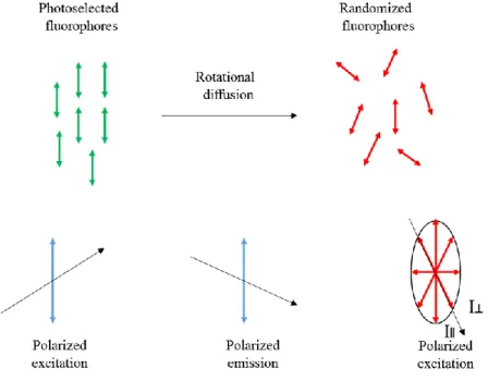
2.5- Anisotropy
Measurements of anisotropy are based on the photoselective excitation of fluorophores by polarized light (Figure 2.1). Dyes absorb preferentially photons whose electric vectors are aligned parallel to the dye transition moment. This transition has a defined orientation with respect to the molecular axis. In isotropic solution the fluorophores are randomly oriented, any molecules whose transition dipole moments are oriented parallel to the electric field vector E will be excited preferentially [1]. As a result, this selective excitation drives to partially oriented fluorophores population and in partially polarized fluorescence emission. Emission also occurs with the light polarized along a fixed axis in the fluorophore. The relative angle between these moments determines the maximum measured anisotropy (r0). The fluorescence anisotropy (r)
and polarization (P) are defined by:
𝑟 = 𝐼∥− 𝐼⊥
14 𝑝 = 𝐼∥− 𝐼⊥
𝐼∥+ 𝐼⊥ (1.5)
where I∥ and I⊥ are the fluorescence intensities of the vertically and horizontally polarized
emission, when the sample is excited with vertically polarized light. Anisotropy and polarization are both expressions for the same phenomenon.
Figure 1.2. Effect of polarized excitation and rotational diffusion on the polarization or
anisotropy of the emission. Adopted from ref [1].
The measured anisotropy can be decreased to lower values than the maximum theoretical values by several phenomena. One of them is rotational diffusion, which occurs during the excited state lifetime and displaces the emission dipole of the fluorophore. Measurement of this parameter provides information about the relative angular displacement of the dyes between the times of emission and absorption. In liquid solution most fluorophores rotate in 50 to 100 ps. Thus, during excited-state lifetime the molecules can rotate many times, polarized emission orientation is randomized. For this reason, fluorophores in non-viscous solution have anisotropies close to zero. Transfer of excitation between fluorophores also results in decreased anisotropies. Measurements of anisotropy of fluorophore dispersed in nanoparticles can inform us about the environment around this fluorophore (liquid or solid).
Anisotropy is commonly used in the biochemical fluorescence sensing applications [5]. It provides information’s on the size and the rigidity by giving response in the change of rotational mobility for fluorescent reporter in various molecular environments. For example, if the free
15 fluorescent molecule has a small size, it rotates rapidly and exhibits a low value of anisotropy. On target binding the size of this rotating unit increases producing a sharp rise in anisotropy. When the molecules are in solid environment, the rotation is very low, which also results in high values of fluorescence anisotropy.
3- Fluorescent Probe
Fluorescent probes are very important tools in biological research. The choice of the appropriate probe depends on the application. We can distinguish:
i) Natural fluorophores such: tryptophan, fluorescent proteins, plant pigments.
ii) Synthetic dyes.
iii) Fluorescent nanoparticles.
3.1- Natural fluorophores
Known as “intrinsic fluorophores” they present conjugated double bonds. Most proteins exhibit absorption and emission in the ultraviolet (UV) region due to aromatic amino acids. Other natural fluorophores in tissue include the reduced form of nicotinamide adenine dinucleotide (NADH) and flavin adenine dinucleotide (FAD), porphyrines, etc. Fluorescent proteins, such as green fluorescent protein, are special family of proteins, containing a special fluorophore formed from several amino acids [6]. These proteins are used as tags to label a protein of interest inside live cells.
3.2- Synthetic dyes
Organic fluorophores became an indispensable tool for fluorescent imaging and sensing. They are used for labelling biomolecules or fluorescence sensing [7,8]. Typically, they contain several combined aromatic rings and are characterized by a strong absorption and emission bands in visible range extending to near-IR of the electromagnetic spectrum, due to presence of delocalized π-electrons forming discrete energy states. Their size is commonly less than 1 nm and their quantum yields can vary from zero to nearly 100 %. The small size allows their incorporation into biological structures such as double-stranded DNA and bio-membranes with minimal perturbation [9].
Organic dyes offer tremendous possibilities in applications due to immense variations of their structures and diversity of their spectroscopic properties.
16
Figure 1.3. Fluorophore brightness versus the wavelength of maximum absorption (λmax) for
different classes of fluorophores. The colors of depicted structure illustrate their wavelengths of emission (λem). Adopted from ref [10].
With regard to applications, fluorescent dyes could be classified into two categories: non-responsive (labels) and non-responsive (sensors/probes).
For the “label” activity the dyes must show highest brightness and contrast, also optimal protection against various quenching and bleaching effects or perturbations by the interactions with the medium [11]. There are several classes of dyes that conform to optimal labels criteria like derivatives of fluorescein and rhodamine [12], BODIPY [13] and cyanine dyes [14]. These dyes families having rigid skeleton, exhibit minimal vibrations-related energy losses. Their electronic density is delocalized symmetrically over the whole structure, which provides high QY and low spectral sensitivity to the environment. As example of this kind of dyes we present below the cyanine dyes which were used in one of our studies.
Cyanine dyes are composed of a polymethine chain between two amino/imino groups, which
can be illustrated by the simple representation (i.e., R2N-(CH=CH) n - CH=N+R2), where n is a
17 the conjugated chain usually form part of a heterocyclic system [15]. This family of dyes is widely used in ultrasensitive imaging and spectroscopy, especially for biological applications. By variation of the polymethine chain length, the absorption can be tuned from the visible and near-infrared region. This family of dyes is characterized by high extinction coefficients (1.2 and 2.5×10⁵M-1 cm-1) and moderate to high quantum yields between 0.04 and 0.4 as well as good photostability, with a lifetime in the ranging from 0.2 to 2.0 ns.
Figure 1.4. Examples of different cyanine dyes. Adopted from ref [16].
Alternatively, for the “sensors” activity, dyes must show high brightness to reach the highest dynamic range of response in terms of variations of lifetime, intensity or of the wavelength of emission. To achieve that, some photophysical mechanisms of response need to be exploited, like ESIPT (excited-state intramolecular proton transfer) [17], ICT (intramolecular charge transfer), FRET (Forster resonant energy transfer) [18], quenching, isomerization, formation of exciplexes, etc. Based on organic dyes family as hydroxychromone derivatives [19], Pyrene and Nile red [9], probes for sensing biomolecular interaction and monitoring biophysical properties of biomembranes were developed [20]. Other types of molecular probes were also developed for sensing pH, temperature, oxygen, metal ions in living systems [21]. Here, a particular attention will be paid to Nile red [20,22].
Nile red is a solvatochromic dye that shows a poor solubility in water, but in solvent it
demonstrates strong fluorescence, with intensity maxima as indicators of the fluorophore environment properties. Nile red shows interesting spectroscopical properties, such as large stokes shift, good quantum yields (up to 50%) and extinction coefficient (45,000 M-1 cm-1)
18 which is suitable for intracellular imaging. With increase in solvent polarity, its emission maximum shifts to the red and the QY decreases.
Figure 1.5. Nile red Fluorophore
3.3- Fluorescent nanoparticles
Fluorescent nanoparticles have attracted a lot of attention as a new class of probes for bioimaging [23]. They demonstrate unique electronic structures and optical properties, unusual chemical and physical characteristics. As example we can cite: quantum dots and clusters, upconversion nanoparticles, carbon dots, fluorescent organic dye nanoparticles, dye-doped silica nanoparticles, dye-loaded polymer or lipid nanoparticles. More description of these systems will be developed further in this manuscript.
4- General requirements for fluorescent probes
For high-quality imaging and sensing in biology, fluorescent probes should fit some criteria [24,25]:
High quantum yield (QY); it means that some photochemical processes such as bleaching or radical formation will be reduced leading to higher fluorescence intensity. High extinction coefficient (ε); in this case higher brightness will be achieved, driving to the use of low excitation intensity. The latest one could be useful in imaging for living tissue without damage or imaging of very low quantity of fluorophores [26].
Optimal excitation wavelength (λex); to avoid autofluorescence, the excitation
wavelengths longer than 400-460 nm are preferable. For deeper penetration in tissue imaging, NIR sources of excitation are optimal [27].
Optimal emission wavelength (λem); it should be selected with the account of
wavelength dependence of the detector sensitivity and sample transparency.
Large Stokes shift; for reducing light-scattering effects, a strong separation between absorption and emission bands is needed. Also for more efficient collection of emitted
19 light using broad-band filters or larger monochromator slits. Moreover, it can reduce homo-FRET (excitation energy transfer between the same dye molecules).
Optimal fluorescence lifetime τ; depending on the application it can be selected long or short by choosing the dye and its environment, because the lifetime depends on the temperature and the dye immediate environment [28].
High photostability; higher dye chemical reactivity in the excited states leads to the degradation of the fluorophore (photobleaching). This photobleaching is due to some photochemical reaction that often involves molecular oxygen and is coupled with the production of singlet oxygen. In sensing applications, photostability is not a big problem because it does not require exposure to intensive light. But in single molecular studies and super-resolution microscopy, where the dye molecules are subjected to high light intensities, photostability is an important issue. Regarding this, in many of these experiments oxygen scavenging systems are used for improving the dye photostability [29].
Solubility and environmental stability. The physical and chemical properties of the dyes (polarity, charge distribution, reactivity to form covalent bonds, ability to participate in π–π stacking, hydrogen bonding and other noncovalent interactions) determine its distribution in a heterogeneous system, moreover, the dye has to be chemically stable and less sensitive to external factor like pH or temperature if this sensitivity is not wanted.
5- Near-IR Dyes
Fluorescence-based imaging techniques offer high temporal and spatial resolution compared to ultrasound imaging, CT, MRI, PET [30,31]. For in vitro applications, super-resolution microscopy offers optical resolution well below 100 nm and fast image acquisition on the millisecond timescale allowing real-time imaging for many cellular processes [30].
For in vivo experiments, fluorescent techniques are limited by the scattering and strong attenuation of visible light (400 nm–650 nm) by biological matter (collagens, hemoglobin, lipid membranes, etc.), in addition of strong auto-fluorescence throughout the visible spectrum [32,33], rendering deep tissue imaging practically impossible in this spectral region. To overcome these problems, attention has shifted to the two so-called near-infrared biological windows from 650 nm to 950 nm (NIR1) and 1000 nm to 1350 nm (NIR2) [34,35]. In this spectral regions, light can penetrate much deeper into biological samples with minimum
20 absorbance and auto-fluorescence with reduced scattering, affording high signal-to-background ratio SBR (Figure 1.6). Rendering fluorescence-based imaging a viable alternative with a high spatial and temporal resolution allowing precise quantifications.
Figure 1.6. Near-infrared optical windows in biological tissues. The effective attenuation
coefficient as a function of wavelength show that absorption and scattering from oxygenated blood, deoxygenated blood, skin and fatty tissue is lowest in the NIR I and NIR II spectral region. Adopted from ref [36].
Last decade with advances in imaging instrument allowed efficient detection of long-wavelength NIR photon. However, few fluorophores with sufficient fluorescence quantum yields in the NIR region were developed, especially in aqueous surroundings. One should mention cyanines 5.5, 7 and 7.5, some squaraine dyes [37], methylene blue (MB) and the FDA approved Indocyanine green (ICG) [38]. In addition to dyes, we can also cite quantum dots, carbon nanotubes, UCNPs, and gold nanorods.
These recently developed NIR emitters have emerged in biomedical imaging from contrast-enhanced imaging and molecular imaging of specific biomarkers, both for preclinical animal studies and clinical diagnostics and interventions.
21
Chapter 2: Nanocarriers
Nanotechnology emerged as new field involving manipulation of the matter at nanometer scale, which outcomes in a novel class of materials with innovative properties for a wide range of applications. Nanoparticles are a part of these new materials, with a size between 1–100 nm. Their use in diverse areas has been vastly explored in recent years, particularly in the biomedical field. Nanoparticles were used in diverse range of applications, such as biosensors, drug delivery, molecular imaging, and novel theranostic systems. In this chapter, we will introduce different kind of nanoparticles and their applications, with stress on lipid nanocarriers, their definition, methods of synthesis and characterisation. Additionally, their use in research as fluorescent nanocarriers are summarized.
1- Terminology clarification
Before beginning this chapter, some term definitions should be clarified. The first one is between nanoparticles (NPs) and nanocarriers (NCs). The term nanoparticle is the most general term for nanosystems with the size under 100 nm, with more than 600,000 times used in published article titles according to Web of Science database. By contrast, nanocarriers was used 8,000 times. Also NPs are used as term in all kind of application starting from manufacturing and materials, environment, energy and electronics, food applications, finishing by biomedical applications as sensor, drug delivery and imaging agent. NCs is used like a sub-class of nanoparticles, as a transport module for another substance (Protein, DNA, RNA, Drug and Contrast agent); it is more related to the use in biomedical field. Most of research on nanocarriers is being applied to their potential use in nanomedicine and drug delivery, especially in chemotherapy. Term nanocarriers generally refers to organic nanoparticles, composed of lipids or polymers. Among lipid nanocarriers one should mention liposomes, nanoemulsions, solid lipid nanoparticles, lipid nanocapsules and nanostructed lipid carriers. Sometimes different terms are used for relatively similar lipid materials. According to Benoit and co-workers, nanoparticles containing oil core and shell composed of PEGylated phospholipids are called nanocapsules [39,40]. On the other hand, Muller et al as well as McClements et al used term nanoemulsion for different lipid based nanocarriers.
22 To simplify the situation in the terminology, I will call all these systems lipid nanocarriers. Moreover, all lipid systems containing liquid core will be termed nanoemulsions.
2- Nanoparticles in biomedical applications :
2.1- Inorganics nanoparticles
2.1.1- Quantum Dots (QDs)
QDs have semiconductor nanocrystal core, generally from the group II–VI or group III–V elements in periodic table (CdS, CdSe, CdTe, ZnS, PbS), coated by a shell to modify their physicochemical properties and promote solubility (Figure 2.1). With size ranging from 2 to 30 nm, they possess unique luminescent properties such a high photostability, high quantum yields and absorption coefficient. Their photoluminescence emission band can easily be tuned from the UV to the IR regions by varying the particle size or composition [41].
QDs particular characteristics make them attractive for the biomedical field such as imaging, diagnosis and therapeutic applications [42]. Thus, QDs were used for multicolor imaging and single-cell molecular profiling [3], also for simultaneous multi-species tracking in live cells [44]. In addition, QDs were proved promising for multiplexing, as example, for in situ molecular profiling of breast cancer biomarkers [45,46]. Recently, to overcome the poor transmission of visible light through biological tissues, NIR emitting QDs were proposed. The examples of their applications include in vivo imaging of tumor markers, such as EGFR [47,48]. Moreover, QDs operating in the second near-infrared region (1100 – 1400 nm) emerged as probes for deep-tissue dynamic imaging of the circulatory system [49], including lymphatic drainage, vascular networks, and angiogenesis [50]. In these cases, exceptional image resolution was achieved, because of deeper penetration of light in this region. Additionally, the use of QDs in targeted delivery have increased with the possibility to graft on their surface chemically reactive ligands as well as active biomolecules, such as proteins and nucleic acids. Despite the research of different groups to use QD nanoparticles as a drug delivery nanocarriers [51], the application of QDs nanocarriers for drugs present a lot of challenges, such as limited encapsulation capacity, presence of toxic elements and incomplete excretion pathway [52,53].
23
Figure 2.1. Schematic representation of QD and its energy levels. A) Schematic structure of
QDs. Image adopted from physicsworld.com. B) Energy splitting diagrams for QDs due to the quantum confinement effect: semiconductor band gap increases with decrease in size of the nanocrystal. C) Example of QDs with a size-dependent luminescence. Adopted from ref [54].
2.1.2- Upconversion Nanoparticles (UCNPs)
UCNPs have emerged as a new class of theranostic agents [55]. In general, it consists of the host matrix such as NaYF4 or CaF2 doped with the rare earth ion like Yb3+, Tm3+, Er3+, or Ho3+
in which electronic transitions occur (Figure 2.2). UCNPs exhibit unique optical properties; they absorb NIR light and emit shorter wavelength UV/visible photons [56]. In addition to these properties, which allow elimination of background signal and deeper tissue penetration of light, UCNPs are highly photostable [57], without blinking and have narrow emission bands, controlled shapes and sizes. These make them promising candidates for biomedical applications such as imaging, photodynamic therapy (PDT) and drug delivery.
In photodynamic therapy application, a photosensitizer is in proximity with UCNPs. At NIR light illumination, UCNPs emit visible light allowing FRET to the photosensitizer and the production of ROS (reactive oxygen species), which affect cell viability. The photosensitizer can be loaded either by chemical linkage to the carbon chains at the surface of UCNPs [58], or physically loaded in nanoparticles. As example, two photosensitizers MC540 and ZnPc were loaded into UCNPs mesoporous silica shell, where the emission of the upconversion nanoparticles matched with the absorbance of these two photosensitizers. These photosensitizers activation at single 980-nm wavelength enhanced the therapeutic efficacy of PDT in mice xenograft melanoma tumor [59].
In the same way, UCNPs were tested for drug delivery systems using a photorelease mechanism. Doxorubicin anti-cancer drug was loaded into mesoporous silica-coated UCNPs,
24 modified by azobenzene molecules [60]. By irradiation with NIR laser light the trans–cis photo-isomerization of the azobenzene molecules lead to the release of the drug in biological samples. Alternatively, photo-cleavable moieties were implemented for release of siRNA [61], and dyes molecules from UCNPs [62].
UCNPs can also be suitable material for bio-imaging due to their NIR excitation and can provide high image resolution. In multiplexed in vivo imaging research, UCNPc were used for lymph node mapping and in vivo tumor cell tracking, where PEGylated particles with three emission colors were synthesized by varying the molar ratio between Yb and Er. The resulting image exhibited higher in vivo detection and sensitivity compared to quantum dots [63]. In spite of UCNPs different advantages, it remains to overcome some major limitations, like low quantum yield, need of strong laser powers, low solubility and biocompatibility.
Figure 2.2. Schematic representation of a core/shell upconversion nanoparticle showing
absorption of NIR light at 980 nm upconverted to visible light. Adopted from ref [64].
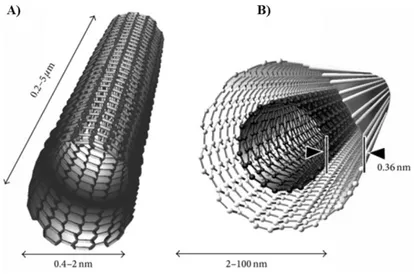
2.1.3- Carbon Nanotubes
Carbon nanotubes are tubular hydrophobic networks of carbon atoms prepared by rolling up of graphene, with unique structural, electronic, optical and mechanical properties. They are divided into single-walled carbon nanotubes (SWNTs) and multiwalled nanotubes (MWNTs) [65] (Figure 2.3).
SWNTs present remarkable optical properties, as NIR region absorbance, high photothermal effect, in addition of large endocytosis. Hence, they present a large interest of the scientific community and have been considered for biomedical imaging [66], drug delivery [67], photothermal therapy [68], as well as PDT. Carbon nanotubes are insoluble in most aqueous media, creating toxicity problems. Various surfactants and polymers were used to overcome
25 their poor solubility, however, their toxicity remains a controversial issue. Specifically, the problem of delayed clearance in the lungs was reported by some researchers [69,70].
Figure 2.3. Schematic representation of carbon nanotubes showing typical dimensions of
length, width. A) Single-walled carbon nanotube (SWCNT). B) Multiwalled carbon nanotube (MWCNT). Adopted from ref [71].
2.1.4- Gold Nanoparticles
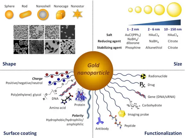
Gold nanoparticles (AuNPs) have recently been widely used in many therapeutic and diagnostic procedures in the biomedical field [72], mainly due to their unique structural and optical characteristics.
AuNPs are particles composed of an Au core and a surface coating. They can be colloidal or clustered and can be synthetized through different methods [73]. The most used is the chemical reduction method, namely reduction of a gold salt solution with reducing agent such as borohydrides or citrate generating Au (0). Then, they are stabilised to avoid agglomeration by stabilizing/capping agents such as citrate or alkane thiols. The synthetic versatility of AuNPs facilitates manipulation of particle size, shape and surface like shown in Figure 2.4, offering unique chemical and optical properties.
AuNPs absorption depends on size and shape. Sphere nanoparticles absorb well in the visible region, whereas gold nanorods and nanoshells show strong light absorption in the NIR region. Under external light excitation collective oscillation of electrons occurs on their surface. This phenomenon is called plasmon resonance (LSPR), and is dependent on the size and shape of AuNPs [74]. With size increasing, the surface plasmon absorption shifts to red or NIR region.
26 With shape changing from sphere to rode, both longitudinal and transverse oscillation of electrons can be induced.
Figure 2.4. Versatility of AuNPs offer a unique platform for tailoring particle size, shape,
surface coating and functionalization. Adopted from ref [75].
Due to LSPR proprieties and NIR absorption, gold nanoparticles are applied in different imaging techniques like optical coherence tomography (OCT) imaging [76], photoacoustic tomography (PAT), as well as fluorescence imaging. In early study, Chulhong Kim et al explored gold nanocages conjugated with melanocyte stimulating hormone, as a contrast agent for quantitative molecular PAT of melanomas in vivo, achieving contrast 300% higher than the control [77]. Moreover, gold nanoparticles have a high electron density, leading to efficient absorption of X-rays, which brings them in the area of X-ray-based imaging as contrast agent for computed tomography (CT) [78].
AuNPs efficiently absorb light and convert it to heat; therefore they can be used for ablation of tumor cells in photothermal therapy [79,80], and thermoresponsive controlled drug release. In drug delivery application, AuNPs are associated with organic nanocarriers such polymers or liposomes to deliver the cargo drugs or peptide upon external laser irradiation. As example, liposomes and gold nanoparticles were used to construct a cluster “bomb” structure for and
27 multi-order Paclitaxel release for liver tumor treatment upon laser illumination of the gold nanoparticles [81]. In addition to all these multiple applications of AuNPs, they are largely used as a sensor for in vitro detection of biomolecules [82].
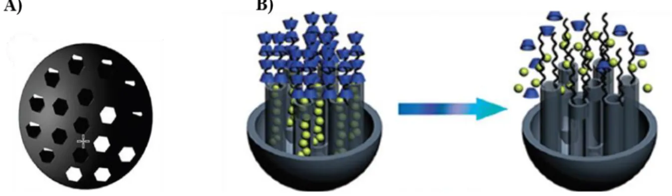
2.1.5- Mesoporous Silica Nanoparticles (MSNs)
Mesoporous silica nanoparticles (MSNs) are class of silica nanoparticles with periodic mesoporous of hexagonal, cubic or lamellar structures in the range of 2 to 50 nm [83]. Diverse mesoporous silica nanoparticles were developed using different synthetic methods, such a modified Stöber method, dissolving reconstruction and self-assembly, soft and hard templating [84].
MSNs have been widely applied in biomedical research, especially as delivery nanocarriers for different drugs, dyes and active molecules. This interest is due to the intrinsic propriety of mesoporous nanoparticles, including pore size and volume, favourable chemical properties allowing surface modifications (Figure 2.5).
Even though their inorganic nature, the mesoporous MSNs are promising solution for responsive drug release under triggers. As an example, Huang et, al developed 50 nm nanoparticles with bio-cleavable disulfide bonds (-S-S-) directly incorporated, which break within tumor microenvironment [85]. As a result, high chemotherapeutic outcome was achieved in vitro and in vivo.
Figure 2.5. Representation of mesoporous nanoparticles system. A) Structural illustration of MSNs. B) responsive release switches on MSNs. Adopted from ref [86].
28
2.2- Organics Nanocarriers
2.2.1-Dendrimers
Dendrimers are a family of three-dimensional, nanoscale hyperbranched polymers, characterized by a symmetrical branched architecture and ultra-small size (typically <5 nm) with high monodispersity (Figure 2.6). They can be functionalized and conjugated with drugs or dyes, and their synthesis is based on the principles of robust high-yield chemistry. Nowadays, they are conjugated either with drug [87], or dyes for using as molecular probes [88]. Fluorescent dendrimers have relatively low quantum yield when used as fluorescent contrast agent because of dye self-quenching and their brightness is controlled by their size. However, these organic nanoparticles are costly to fabricate, and majority of them are toxic producing cell membrane damage [89].
Figure 2.6. Architectural components of dendrimers. Adopted from ref [90].
2.2.2- Micelles
Micelles are amphiphilic macromolecules that self-assemble into core−shell structured nanocarriers, in which the hydrophilic parts are in contact with surrounding solvent and hydrophobic parts are in the center; their diameter is between 5 and 100 nm (Figure 2.7). They can be classified according to their amphiphilic core: polymeric or lipid micelles.
Lipid micelle are prepared with water soluble surfactant bearing hydrophobic chain linked to a polar head group. The latter can be small charged group or hydrophilic polymer, such as Polyethylene Glycol (PEG) [91], Cremophor ELP being an important example [92]. Polymeric micelles are made from block copolymers composed of hydrophobic and hydrophilic blocks
29 [93]. Due to their lower CMC (critical micelle concentration), polymeric micelles display better stability against dilution compared to lipid one, allowing retention of active molecule for a longer period of time in blood pool [94].
Figure 2.7. Figure illustrating micelle architecture.
Micelles are used as drug delivery systems to carry hydrophobic drugs entrapped in or covalently bound to the hydrophobic core leading to dramatic improvement in their aqueous solubility. As example, the solubility of anticancer drug paclitaxel (PTX) was increased 5,000 fold when it was formulated in poly(d,l-lactide)-MePEG diblock copolymer [95], and this formulation is currently in clinical trials. Other common utilization of micelles is in stimuli-responsive delivery mode, where drug can be released either by internal stimulus such as pH [96], enzymatic reactions [97], reactive oxygen species (ROS) [98], and temperature. Alternatively, external signal such as ultrasound, heat and light, can be used, where these last stimuli are widely associated with photothermal and photodynamic therapy (PDT) issues. In a recent study, Gao et al reported that photosensitizer (Ce6)-loaded micelles integrating cyanine dye have huge potential as theranostic tool for tumor localization via NIR / photoacoustic imaging modalities with superior cancer effect via subsequent combining photothermal therapy (PTT)/photodynamic therapy (PDT) [99].
For other applications, several micellar forms of contrast agent have been established for different imaging modes like magnetic resonance imaging (MRI) and X-ray computed tomography (CT). Recently, bimodal fluorescent/photoacoustic imaging agent, based on NIR Squaraine dye encapsulated into Pluoronic F-127 micelles was proposed, exhibiting high photostability and low cytotoxicity in biological conditions, with good contrast in animal experiment using fluorescence imaging and photoacoustic tomography modalities [100].
30 Recently, Shulov et al presented an interesting concept of new shell-cross-linked fluorescent micelles, with PEGylated cyanine 3 and 5 bis-azides forming a covalently attached shell on the surface of micelles of amphiphilic calixarene bearing four alkyne groups. The final micelles displayed small size of 7 nm, good quantum yield and stability in biological media. They were 2-fold brighter than quantum dots (QD-585) under microscopy, which make them suitable new platform for developing bright protein-sized responsive nanoparticles for bioimaging [101].
2.2.3- Polymeric Nanocarriers
Polymeric nanoparticles NPs are colloid systems with an average size of 10-300 nm prepared from natural or synthetic polymers. Recently, they have gathered a lot of interest in nanomedicine as drug or protein nanocarriers, also in imaging as fluorescent nanoparticles. Within in vivo applications, they have the capacity to protect their active content, with long circulation time and controlled release. In addition, they offer great versatility in modifying their size, chemical composition, morphology, biodegradability and surface functionality [102]. Nowadays, the most used polymers for nanoparticles include poly (lactic acid) (PLA), poly (glutamic acid) (PGA) and poly (D, L-lactide-co-glycolide) (PLGA), due to their biocompatibility and biodegradability properties [103]. Beside, poly (methyl methacrylate) (PMMA), poly(caprolactone) (PCL), N-(2-hydroxypropyl)-methacrylate copolymers (HPMA), poly (ortho ester) (POE) and Chitosan are aslo widely used [104].
Various methods for preparation of polymer NPs were proposed. One of them is direct polymerization of monomers (Fig 2.8). In different types of emulsions, such as (conventional) emulsion [105], mini-emulsion[106], and micro-emulsion [107]. The type of emulsion depends on the surfactant concentration (> cmc in emulsion, < cmc in mini-emulsion, >> cmc in micro-emulsion) and the method of homogenization (shear in emulsion, high shear, ultrasound in mini-emulsion and low shear in micro-emulsion) [108].
31
Figure 2.8. Preparation of polymer NPs though polymerization of a monomer. Adopted and
modified from ref [108].
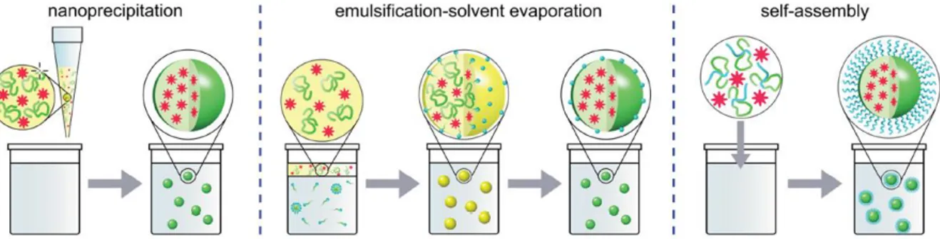
The other strategies for polymeric NPs synthesis is based on preformed polymers, following three main approaches: emulsification solvent evaporation, nanoprecipitation, and self-assembly (Figure 2.9). In emulsification solvent evaporation method, the polymer is dissolved in water-immiscible solvent and dispersed in an aqueous phase containing stabilizer. Then nanoparticles are generated under sonication or high-speed homogenization, followed by evaporation of the solvent. NPs of size 100-200 nm could be obtained [109].
Nanoprecipitation (also called solvent displacement technique) is based on the addition of polymer, dissolved in a solvent, to aqueous phase [110]. Formation of NPs occurs due to rapid diffusion of the solvent into the aqueous phase (and vice versa) and polymer supersaturation. Concentration of polymer, amount of organic and aqueous phase and mixing procedure influence the size. NPs from <10 nm up to hundreds nm could be obtained. Currently, nanoprecipitation method is probably the most used method to obtain fluorescent polymer NPs and therapeutic polymeric nanocarriers. Reisch et al based on this technique obtained very small fluorescent polymeric nanoparticles with size nearly 15 nm using different charged polymer [111]. In other study, Zhu et al used it as single self-assembly step to generate polymeric nanoparticles with simultaneous installation of targeting proteins on the exterior and loading of therapeutic proteins in the interior [112].
Finally, the self-assembly method is used for some amphiphilic polymer, who can self-assembly into NPs in form of micelles under thermodynamic conditions. In this case, a solution of
32 polymer dissolved in organic solvent is mixed with an aqueous phase, then the aggregation of the hydrophobic part of polymer takes place when the concentration exceeds critical micelle concentration (CMC) [113]. In fact, this approach lead to polymeric micelles, which were already mentioned above.
Figure 2.9. Techniques used for the preparation of polymeric NPs from preformed polymers.
Hydrophobic polymer segments are shown in green, hydrophilic ones in blue, organic solvent in yellow. Adopted and from ref [108].
Polymeric nanoparticles serve as excellent drug or protein therapeutic carriers; the release can be tailored via controlled polymer biodegradation or appropriate stimulus effect. One of the most successful translation from bench to clinical trial was based on work led by Farokhzad and co-workers. They developed docetaxel encapsulated in to PLGA-b-PEG polymeric nanoparticles and surface functionalized with RNA aptamers, that target prostate cancer cells over-expressing the prostate-specific membrane antigen (PSMA) receptor, with up to 77-fold increase in binding compared to non-targeted controls [114]. Therefore, administration of this aptamer-NPs containing docetaxel was highly effective at tumor size reduction, showing almost complete tumor reduction and 100% survival.
Next, this concept was taken by BIND Therapeutics to clinical development translation with the most optimal hit; BIND-014- as the first targeted and controlled release polymeric NPs, for cancer chemotherapy. However, until now in clinical trial, polymeric nanocarriers failed to demonstrate higher activity against tumors from their parent drug. Highlighting the need for better patient selection [115,116], and better understanding of the release mechanism undergoing with these nanoparticles in vitro and in vivo [117]. Moreover, the engineering of polymeric nanocarriers that can avoid the immune system with prolonged circulation is needed. Among the lines of reflection to this problem, researchers turned to nature by designing a biomimetic nanoparticles. In one report, the group of Zhang report that PLGA nanoparticles
33 coated with the membrane of blood platelets can hide from the body’s immune responses, and possess higher binding properties that allow them to target desired cells and tissues [118]. In other approaches, polymeric nanoparticles are used for fluorescence bioimaging and sensing in order to achieve extreme brightness and contrast using biodegradable materials. For that purpose, dyes with high concentration are encapsulated inside the nanoparticles. However, because of dyes aromatic flat structure, this encapsulation induces quenching caused by aggregation of these fluorophores in tight confinement, besides the excitation energy transfer phenomena. These processes affect the fluorescence quantum yield and reduce the brightness of dye-loaded polymer NPs. Recently, an original approach was employed by Klymchenko group, where they encapsulated inside PLGA NPs, an association of bulky hydrophobic counterions (tetrakis (pentafluorophenyl) borate) with ionic dyes (Rhodamine C18) in the form of salts, achieving small nanoparticles of 40 nm diameter and high dyes loading up to 5 wt% with minimized quenching [119]. These PLGA fluorescent nanoparticles express a QY of 23% being ≈6-fold brighter than QDs-605. What is more, these NPs showed reversible and nearly complete ON/OFF switching (blinking), opening the way for further application of dye-doped NPs for super-resolution imaging by direct stochastic optical reconstruction microscopy (dSTORM) [120]. Also, this systems was applied successfully to cyanine dyes leading to bare coded fluorescent nanoparticles for single cells tracking experiment [121].
2.2.4- Lipid Nanocarriers
Over the past decade, there have been major advances in the development of nanocarriers as delivery systems for drugs, peptides, proteins and contrast agents. Among them, lipid-based nanocarriers have great potential to solubilize, protect, encapsulate and deliver lipophilic bioactive components to desired target, achieving bioavailability and avoiding side-effects. These nanocarriers are made from biocompatible lipids, such as phospholipids, fatty acids, cholesterol and triglycerides, in addition of surfactants and aqueous phase. Numerous advantages of the lipid matrix make the lipid-based nanocarriers potentially ideal drug delivery systems. Due to bio-compatibility and bio-degradability characteristics, these systems are prone to be less toxic as compared to other delivery systems, such as polymeric nanoparticles. Before discussing nanoemulsions, which was the main subject of my research work, it is useful to begin with a brief overview of the other major lipid nanocarriers, focusing on their advantages and limitations.
34
2.2.4.1- Liposomes
First described in the 1960s by Bangham [122], liposomes are spherical vesicles formed from lipid bilayer shells enclosing aqueous interior compartments. PEGylated or not PEGylated, they can encapsulate hydrophilic agents inside the central aqueous compartment and entrap hydrophobic agents within the lipid bilayer. Liposomes are prepared mainly with lipid and/or phospholipid molecules, dissolved in an organic solvent (chloroform generally), followed by an evaporation step, rehydration of the film in an aqueous solvent, and finally processing with different techniques such as [123]:
- Extrusion: with high pressure extruder, liposomes are structurally modified to large unilamellar vesicles (LUV) or nanoliposomes, depending on the pore-size of the filters employed [124].
- Sonication: which is the most widely applied method because of its simplicity [125]. It can be realized by bath or probe sonication. However, this method presents some disadvantages, like low internal volume/encapsulation efficiency and metal pollution by the probe.
- Microfluidization: employing a microfluidizer without using solvent, based on divided pressure stream into passing each part across a tiny aperture and leading the flows to each other inside the chamber of microfluidizer [126]. This method has been applied in pharmaceutical field to produce liposomes due to the large volume production capacity, with adjustable average size and high capture efficiency.
Figure 2.10. Schematic diagram of a liposome. Adopted from ref [127].
Liposome technology used as nanocarriers is extensively developed, a number of liposome-based drug formulations are available for human use and many products are under different clinical trials [128]. Doxil® was the first successful liposome-based product, introduced in US market in 1995, for the treatment of patient with ovarian cancer. Barenholz and co-workers were behind the development of this approach [129]. It consists of a Doxorubicin-loaded
35 PEGylated liposomal bilayer with a size of 80–90 nm, comprising hydrogenated soy phosphatidylcholine (HSPC), cholesterol (CHOL) and methyl-distearoyl phospho-ethanolamine PEG 2000 (DSPE–PEG) sodium salt. Despite its large use in the cancer therapy due to its long circulation time, good pharmacokinetics and pharmacodynamics, the Doxil® does not improve therapy efficiency comparing to normal doxorubicin, but offers better life for the patient and less side effects like hair loss and reduced cardiotoxicity. Subsequently, other formulations based on liposomes came to market, including Myocet®, Depocyt®, and Onivyde® [130].
Supplementary, huge effort is made in order to confer stimuli-responsive properties to liposomes for delivery of active molecules (drug, siRNA, protein, etc.), with different chemical and physical activation methods (pH, enzyme, redox, and light) [131]. Light triggered release from liposomes is one of the most popular method, where a photosensitizer, NIR dyes or gold nanoparticles are encapsulated inside the liposome and activated by photons to initiate the release process. Recently, Kohane group developed, based on this technique, an ultrasensitive phototriggered local anesthesia in superficial or deep tissues, where gold nanorods were attached to low temperature sensitive liposomes (LTSL), encapsulating tetrodotoxin and dexmedetomidine. Near-infrared light (808 nm) engender heat gold nanorods leading to rapid release of the anesthesia agent. They demonstrate that for in vivo situation, only 1-2 min irradiation at ≤ 272 mW/ cm2 is needed to produce repeatable and adjustable on-demand
infiltration anesthesia or sciatic nerve blockade with minimal toxicity. This effect is correlated with the power and the time of irradiation [132].
Furthermore, dyes can be also associated with liposome for photothermal and photodynamic therapy, photosensitizer dyes are encapsulated inside in order to generate singlet oxygen [133]. Also NIR dyes are used, due their deep penetration to generate the photothermal effect [134]. These last years, Indocyanine Green (ICG) was a subject of multiple studies for photothermic and photodynamic therapy [135]. Yoon et al engineered liposomal Indocyanine Green (ICG) for photothermal purpose. The photothermal effect of different lipid compositions and ICG ratio was evaluated. They showed that optimized formulation has greater anticancer effects in a mouse tumor model compared with other liposomal formulations and the free form of ICG. Then it was served to visualize the metastatic lymph node around the primary tumor under fluorescence imaging guidance, and ablate the lymph node with the enhanced photothermal effect, indicating the potential for selective treatment of metastatic lymph node [136].
36
2.2.4.2- Solid Lipid Nanocarriers (SLNs)
Solid lipid nanocarriers were introduced at the beginning of the 1990s by Müller research group [137,138], in order to develop an alternative carrier system to liposomes, emulsions and polymeric nanoparticles. SLN are composed of lipid being solid at physiological temperatures such as mono-, di- and triglycerides, fatty acids, waxes and steroids, stabilized by surfactants [139]. Having a highly-ordered crystalline structure, they are very stable. This type of nanoparticles designed with the idea that the usage of solid lipids instead of liquid oils may provide prolonged drug release, as the bioactive substance has much lower diffusion rate and it can be controlled by controlling the physical state of the lipid matrix of SLN [140]. The average diameter of SLNs is from 100 to 1000 nm. Solid lipid nanocarriers appear to be promising drug delivery systems, especially for oral administration . The SLN formulations are generated by both high- and low-energy methods [141].
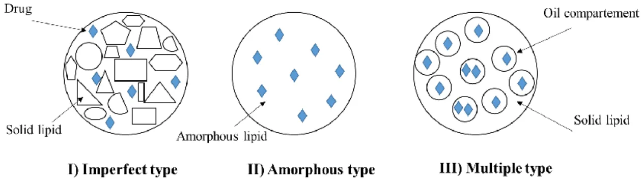
Three models for the location of the drug within the SLN were described [142] (Figure 2.10): The homogeneous matrix model.
The drug enriched shell model. The drug-enriched core model.
Figure 2.11. Models for drug incorporation into SLNs.
Among these models, the difference is mainly due to the chemical nature of the active ingredient, lipid and surfactant, as well as the process parameters [143]. For the matrix model, there is a homogeneous dispersion of the drug in the lipid matrix. It occurs when the nanocarriers are produced by cold homogenization technique without the use of a drug solubilizing- surfactant, or when lipophilic drugs are incorporated using hot homogenization technique into SLNs. In the drug-enriched shell model, the lipid core is surrounded by lipid corona, due to the lipid precipitation mechanism that occurs at cooling stage when the
![Figure 2.15. Setup and functioning of a microfluidizer for nanoemulsion formulation. Adopted from ref [199]](https://thumb-eu.123doks.com/thumbv2/123doknet/3447727.100697/44.892.273.627.795.1062/figure-setup-functioning-microfluidizer-nanoemulsion-formulation-adopted-ref.webp)
![Figure 3.2. Scheme of a confocal set up for Fluorescence Correlation Spectroscopy. Adopted from ref [242]](https://thumb-eu.123doks.com/thumbv2/123doknet/3447727.100697/57.892.168.716.256.693/figure-scheme-confocal-set-fluorescence-correlation-spectroscopy-adopted.webp)
![Figure 3.3. Timescales of various processes monitored by autocorrelation analysis. Adopted from ref [243]](https://thumb-eu.123doks.com/thumbv2/123doknet/3447727.100697/59.892.165.655.181.560/figure-timescales-various-processes-monitored-autocorrelation-analysis-adopted.webp)
