The inhibition of Bid expression by Akt leads to resistance to TRAIL-induced apoptosis in ovarian cancer cells
By
Nadzeya Goncharenko Khaider
Département de Microbiologie et Infectiologie
Thesis is presented to the Faculté de Médecine et des Sciences de la Santé in order to obtain the Diploma of Master of Sciences (M.Sc.) in Microbiology
Sherbrooke, Québec, Canada May,2011
Members of the evaluationjury: from département de microbiologie et infectiologie, Dr Alain Piché (Director of the research) and Dr Brendan Bell and from départment
d'anatomie et biologie cellulaire, Dr Nathalie Perreault
NOTICE:
Branch
395 Wellington Street Ottawa ON K1A ON4 Canada
The author has granted a
non-exclusive license allowing Library and Archives Canada to reproduce, publish, archive, preserve, conserve, communicate to the public by
telecommunication or on the Internet, loan, distrbute and sell theses
worldwide, for commercial or non-commercial purposes, in microform, paper, electronic and/or any other formats.
The author retains copyright ownership and moral rights in this thesis. Neither the thesis nor substantial extracts from it may be printed or otherwise reproduced without the author's permission. ln compliance with the Canadian Privacy Act some supporting forms may have been removed from this thesis.
While these forms may be included in the document page count, their removal does not represent any loss of content from the thesis.
Canacfâ
Patrimoine de l'édition 395, rue Wellington Ottawa ON K1A ON4 Canada
AVIS:
Your file Votre référence
ISBN: 978-0-494-83764-1 Our file Notre référence
ISBN: 978-0-494-83764-1
L'auteur a accordé une licence non exclusive permettant à la Bibliothèque et Archives Canada de reproduire, publier, archiver, sauvegarder, conserver, transmettre au public par télécommunication ou par l'Internet, prêter, distribuer et vendre des thèses partout dans le monde, à des fins commerciales ou autres, sur support microforme, papier, électronique et/ou autres formats.
L'auteur conserve la propriété du droit d'auteur et des droits moraux qui protege cette thèse. Ni la thèse ni des extraits substantiels de celle-ci ne doivent être imprimés ou autrement
reproduits sans son autorisation.
Conformément à la loi canadienne sur la protection de la vie privée, quelques formulaires secondaires ont été enlevés de cette thèse.
Bien que ces formulaires aient inclus dans la pagination, il n'y aura aucun contenu manquant.
cellules de cancer ovarien par
Nadzeya Goncharenko Khaider
Département de Microbiologie et Infectiologie
Mémoire présenté à la Faculté de Médecine et des Sciences de la Santé en vue de l'obtention du diplôme de maître ès sciences (M.Sc.) en Microbiologie, Faculté de
Médecine et des Sciences de la Santé, Université de Sherbrooke, Sherbrooke, Québec, Canada, JlH 5N4
Le cancer épithélial ovarien (CEO) est la principale cause de décès parmi les cancers gynécologiques. La majorité des patientes sont diagnostiquées à un stade tardif de la maladie. La survie de ces patientes est limitée à cause de la récurrence de la maladie devenue résistante à la chimiothérapie. Le développement de nouveaux agents thérapeutiques pour le traitement de ce cancer est ainsi une priorité importante dans le domaine afin d'amél.iorer le taux de survie. La cytokine TRAIL ("TNF-related apoptosis-inducing ligand") est un nouvel agent prometteur pour le traitement du cancer, incluant le cancer ovarien, car il a la capacité unique de déclencher l'apoptose dans les cellules cancéreuses et d'épargner les cellules normales.
Précédemment, notre laboratoire a montré qu'un nombre significatif de lignées cellulaires dérivées de cancer ovarien et de cellules provenant de cultures primaires de cancer ovarien sont intrinsèquement résistantes à l'apoptose induite par le TRAIL. Les mécanismes menant à la résistance intrinsèque sont largement inconnus.
Les cellules résistantes au TRAIL montrent souvent une activation accrue de la voie de survie PBK/ Akt. En se basant sur les observations que les ascites provenant de cancer ovarien induisent une activation de la voie PBK/ Akt chez les cellules de cancer ovarien sensibles au TRAIL, résultant en une inhibition de l'apoptose produite par le TRAIL, nous proposons l'hypothèse que l'activation de la voie PBK/Akt dans les cellules CEO joue un rôle important dans la résistance intrinsèque à l'apoptose induite par le TRAIL.
Les objectifs de mon projet sont de démontrer qu' Akt est impliqué dans la régulation de l'apoptose induite par le TRAIL dans les cellules CEO et de déterminer les mécanismes par lesquels Akt contribue à la résistance au TRAIL.
Nous avons démontré que l'activation d'Akt réduit la sensibilité des cellules CEO au TRAIL. Les cellules résistantes au TRAIL furent sensibilisées à l'apoptose induite par le TRAIL via traitement avec des inhibiteurs de la voie PBK ou Akt. L'inhibition de la voie PBK/Akt n'a pas interférée avec le recrutement et l'activation du pro-caspase-8 au DISC (death-inducing signaling complex). Parallèlement, une surexpression stable d' Aktl dans les cellules sensibles au TRAIL a résulté en une certaine résistance au TRAIL. Malgré le fait que la pro-caspase-8 était recrutée et activée au DISC dans les lignées cellulaires
l'activation d' Akt. La déplétion de Bid par siRNA dans les cellules de CEO sensibles au TRAIL fût associée à une baisse de l'apoptose médiée par TRAIL et une surexpression de Bid dans les cellules résistantes au TRAIL résultât en une hausse de l'apoptose médiée par TRAIL.
Dans leur ensemble, ces résultats suggèrent qu' Akt est un facteur critique pour la résistance intrinsèque chez les cellules CEO et qu'un mécanisme important par lequel l'activation d' Akt contribue à la résistance au TRAIL, est la régulation de l'expression de la protéine pro-apoptotique Bid.
Suite à ces données, nous spéculons que l'activation d' Akt pourrait être un biomarqueur potentiel pour prédire la réponse des patientes à une thérapie basée sur TRAIL et que l'inhibition de la voie PI3K/Akt pourrait devenir une des stratégies pour surmonter la résistance à TRAIL dans le cancer ovarien.
cancer cells By
Nadzeya Goncharenko Khaider
Département de Microbiologie et Infectiologie
Mémoire is presented to the Faculté de Médecine et des Sciences de la Santé in order to obtain the Diploma ofMaster of Sciences (M.Sc.) in Microbiology, Faculté de Médecine et
des Sciences de la Santé, Université de Sherbrooke, Sherbrooke, Quebec, Canada, JlH 5N4
EOC (epithelial ovarian cancer) is a leading cause of death from gynecological cancers. The majority of the patients with EOC are diagnosed at a late stage. The survival of these patients is limited due to recurrence of chemotherapy resistant disease. Therefore, the development of navel therapeutic agents for the treatment of EOC is urgently needed and it is a high priority in the field. TNF-related apoptosis-inducing ligand (TRAIL) is a promising navel agent for the treatment of cancer, including EOC, because of its unique ability to trigger apoptosis in cancer cells and spare normal cells.
Our laboratory has previously shown that a significant number of EOC cell lines and primary EOC samples are intrinsically resistant to TRAIL-induced apoptosis. The mechanisms leading to intrinsic resistance are largely unknown. TRAIL-resistant cells often display increased activation of the pro-survival PBK/Akt pathway. Based on our observations that EOC ascites induced activation of the PBK/ Akt pathway in TRAIL-sensitive EOC cells which resulted in inhibition of the TRAIL-mediated apoptosis, we hypothesized that activation of the pro-survival PBK/Akt pathway in EOC cells plays an important role in the resistance to TRAIL-induced apoptosis.
The objectives of my project were to demonstrate that Akt is implicated in the regulation of TRAIL-induced apoptosis in EOC cells and to investigate the mechanisms by which Akt contributes to TRAIL resistance.
We report that Akt activation reduces the sensitivity of ovarian cancer cells to TRAIL. TRAIL-resistant cells were sensitized to TRAIL-induced apoptosis by treatment with PBK or Akt inhibitors but inhibition of PBK/ Akt signaling pathway did not interfere with the recruitment and processing of the pro-caspase-8 to the death-inducing signaling complex (DISC). Conversely, overexpression of Aktl in TRAIL-sensitive cells promoted resistance to TRAIL. Despite the fact that TRAIL-induced caspase-8 activation was observed in both TRAIL-sensitive and -resistant cell lines, Bid cleavage occurred only in TRAIL-sensitive cells. Akt activation in TRAIL-sensitive cells inhibited TRAIL-induced Bid cleavage. Furthermore, Bid expression was downregulated by Akt activation. Depletion of Bid by siRNA in TRAIL-sensitive EOC cells was associated with a decrease in TRAIL-mediated apoptosis and Bid overexpression in resistant cells resulted in increased TRAIL-mediated apoptosis.
Bid.
Given these data, we speculate that Akt activation may be a potential biomarker to predict patient's response to TRAIL therapy and that the inhibition of the PBK/Akt pathway can become one of the strategies to overcome resistance to TRAIT, therapy in ovarian cancer. Key words: epithelial ovarian cancer; TRAIL; apoptosis; resistance; Bid.
Table of contents ... , ... VI List of tables ... IX -List of figures ...... X List of abbreviations ...... XII
INTRODUCTION ... 1
1 The cancer ... 1
1.1 General remarks ... 1
1.2 The hallmarks of cancer ... 1
1.2.1 Self-sufficiency in growth signals ... 3
1.2.2 Insensitivity to antigrowth signals ... 4
1.2.3 Resistance to apoptosis ... 5
1.2.4 Limitless replicative potential ... 5
1.2.5 Sustained angiogenesis ... 6
1.2.6 Tissue invasion and metastasis ... 6
1.2. 7 Additional hallmarks: the stress phenotype of cancer ... 7
2 Ovarian cancer ... 8
2.1 General remarks ... 8
2.2 Risk factors ... 9
2.3 Diagnostic eues ... 10
2.3.1 Serum biomarkers ... 10
2.4 Grading of ovarian cancer ... 11
2.5 Role of EOC ascites in promotion of tumor cell survival ... 13
2.6 Origin of ovarian cancer ... 16
2.6.1 Ovarian surface epithelium and cortical inclusion cyst ... 16
2.6.2 Two pathway model of ovarian carcinogenesis ... 18
2.6.3 Fallopian tube as a site of origin ... 20
2.7.1 Mechanisms ofresistance to chemotherapy ... .' ... 23
2.7.2 Targeted therapies for ovarian cancer ... 25
3. TRAIL as a promising agent for cancer treatment ... 27
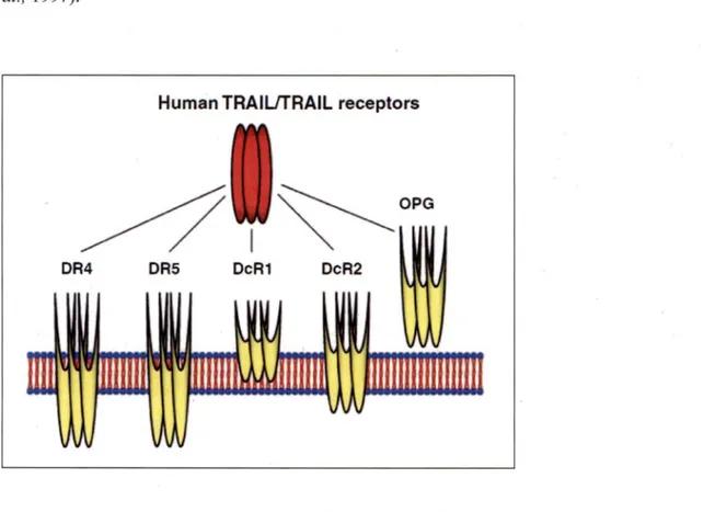
3.1 TRAIL and its receptors ... 27
3.2 The physiological role of TRAIL and its receptors ... 29
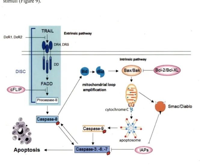
3 .3 Apoptotic TRAIL signaling ... 30
3.3.1 Members ofBcl-2 family ... 32
3.4 Additional signal transduction pathways activated by TRAIL ... 37
3 .5 TRAIL receptor targeting agents ... 39
3 .6 TRAIL resistance ... 40
3. 7 Resistance to TRAIL-induced apoptosis among ovarian cancer cells ... 43
4. PI3K/Akt pathway ... 45
4.1 PI3K/ Akt pathway components and activation mechanisms ... 45
4.2 Akt/PKB ... 46
4.3 Cellular processes regulated by A.kt ... 48
4.3.1 Direct modulation of apoptosis by A.kt ... 48
4.3.2 Indirect modulation of apoptosis by A.kt ... 51
4.3.3 Regulation of cell growth and cell cycle progression by Akt ... 53
4.4 Inhibitors targeting PI3K/ Akt pathway ... 55
5. Hypothesis and objectives of the project ... 56
AVANT-PROPOS DE L'ARTICLE ... 58 RÉSUMÉ DE L'ARTICLE ... 59 ORIGINAL ARTICLE ... 61 Abstract ... 61 Introduction ... 62 Results ... 64
Overexpression of Aktl inhibits TRAIL-induced apoptosis in CaOV3 cells ... 66
PI3K and Akt inhibitors enhance TRAIL-induced apoptosis in resistant cells ... 68
Akt activation in human primary samples of EOC correlated with decreased TRAIL sensitivity ... 68
TRAIL resistance is downstream of caspase-8 but upstream of mitochondria ... 70
tBid is not detected in resistant cells ... 74
Akt activation decreases Bid protein levels ... 75
Depletion ofBid inhibits TRAIL-induced apoptosis ... 77
Bid overexpression promotes TRAIL-induced apoptosis in resistant cells ... 79
TRAIL resistance is not affected by XIAP downregulation ... 80
Discussion ... 82
Materials and methods ... 85
Ovarian cancer tumor samples and cell culture ... 85
Reagents ... 86
Retro viral preparation and transduction efficiency ... 86
Quantitative RT-PCR ... 86
Cell viability assays ... 87
Apoptosis and caspase assays ... 87
Western blot analysis ... 88
Knockdown of Bid and XIAP ... 88
Statistical analysis ... : ... 89
Conflict of interest ... 89
Acknowledgements ... 89
Ref erences ... 90
Discussions and conclusions ... 94
Conclusions ... 100
Perspectives ... · ... 102
Acknowledgments ... 103
The major histologie subtypes of ovarian carcinoma ..... 12
The mechanisms of PI3K/Akt pathway activation by ascite ... : ... 15
Model of the development of an EOC tumor ...... 17
Two pathway model of ovarian carcinogenesis .... 19
Fallopian tube as a site of origin ... 21
Acquired drug-resistant models possess multiple drug-resistance mechanisms ... 14
TRAIL and its receptors ... 28
Apoptotic TRAIL signaling ... 30
Members ofBcl-2 family ... 33
Models for Bax/Bak activation by BH3-only proteins ... 36
Alternative TRAIL signaling ... 37
Mechanisms of resistance to TRAIL ... 42
Resistance to TRAIL ... 44
Mechanisms of Akt activation by PI3K ... 46
Three human Akt/PKB isoforms ... 4 7 Direct modulation of apoptosis by Akt ... 50
Indirect modulation of apoptosis by Akt ... 51
Regulation of cell cycle progression by Akt ... 54
Effect of TRAIL on EOC cells ... 57
Figure 1 of the article: Tumor necrosis factor-related apoptosis-inducing ligand (TRAIL) resistance correlates with Akt activation in ovarian cancer cells ... 65
Figure 2 of the article: Overexpression/phosphorylation of Akt inhibits TRAIL-induced apoptosis in CaOV3 cells ... 67
Figure 3 of the article: Inhibition of the PI3K/Akt pathway sensitizes ovarian cancer cells to TRAIL-induced apoptosis ... 69
Figure 4 of the article: Blockade of the TRAIL signaling cascade is located downstream of caspase-8 in resistant cells ... 71
Figure 5 of the article: Lack of mitochondrial activation in TRAIL-resistant cells ... 72
Figure 6 of the article: Effect of Akt on Bid cleavage ... 74
Figure 7 of the article: Akt downregulates Bid expression ... 75
Supplementary figure Sl of the article ... 76
Figure 9 of the article: Overexpression of Bid in resistant cells increases TRAIL-induced apoptosis ... 79 Figure 10 of the article: Effect ofX-linked inhibitor of apoptosis (XIAP) depletion on TRAIL-induced apoptosis ...
81
Apoptotic cascade activation in EOC cells ... 97 Proposed model of apoptotic cascade activation in TRAIL-resistant EOC cells ...
101
Al/Bfl-1 ABX-EGF APAFl APST ASK 1 ATCC ATP Bad Bak Bax Bcl-2 Bel-XL Bid Bik Bim Bmf BNIP3L Bok Boo/Diva bp BRAF BRCAl BRCA2 BSO CA 125 CAMs c-FLIP1 c-FLIPs
crc
coxrv
CREB CTL DcRl DcR2 DD DED DISC DMEM DNA DR4 DR5 ECM LIST OF ABBREVIATIONS Bcl-2 related protein AlHuman monoclonal antibody specific to the epidermal growth factor receptor
Apoptotic protease activating factor-1 Atypical proliferative serous tumor Apoptosis signal-regulating kinase 1 American Type Culture Collection Adenosine-5'-triphosphate
Bcl-2-associated death promoter Bcl-2 homologous antagonist/killer Bcl-2-associated X protein
B-cell lymphoma 2
B-cell lymphoma-extra large
BH3 interacting domain death agonist Bcl-2-interacting killer
· Bcl-2 interacting protein Bcl-2-modifying factor
Bcl-2/adenovirus ElB 19 kDa protein-interacting protein 3-like Bcl-2-related ovarian killer protein
Divergent Bcl-2 protein Base pair( s)
B-Raf proto-oncogene serine/threonine-protein kinase Breast cancer type 1 susceptibility protein
Breast cancer type 2 susceptibility protein Bilateral salpingo-oophorectomy
Cancer antigen 125 or carbohydrate antigen 125 Cell adhesion molecules
FLICE-inhibitory protein long form FLICE-inhibitory protein short form Cortical inclusion cysts
Enzyme cytochrome c oxidase or Complex IV Cyclic AMP-response element-binding protein Cytotoxic T lymphocytes
Decoy receptor 1 Decoy receptor 2 Death domain
Death effector domain
Death inducing signaling complex
DulbeccoN ogt modified Eagle's minimal essential medium Deoxyribonucleic acid
Death receptor 4 Death receptor 5 Extracellular matrix
EDTA EGFR EOC ER
ERK
EV FADD FAK FBS FGF FOXO FSH FTE Gl phase GF GFP GPCR GPI GSK-3 HE4 He La Her2/neu Hrk IAP ICso IkB IKK IL JNK Kb kDa KRAS LH LPA mAbs MAPK Mcl-1 Mdm2 MKK4 MLK3 MMP MMP MPST mRNA mTOR NCCN NFkB Ethylenediaminetetraacetic acid Epidermal growth factor receptor Epithelial ovarian cancerEndoplasmic reticulum
Extracellular-signal-regulated kinase Empty vector
Fas-associated protein with death domain Focal adhesion kinase
Fetal bovine serum Fibroblast growth factor F orkhead box proteins F o llicle-stimulating hormone Fallopian tube epithelium Growth phase of cell cycle Growth factor
Green fluorescent protein G-protein-coupled receptor Glycosylphosphatidylinositol Glycogen synthase kinase 3 Human epididymis protein 4 Immortal cell line
Human Epidermal growth factor Receptor 2 Activator of apoptosis harakiri
Inhibitor of apoptosis proteins
Half maximal inhibitory concentration Inhibitor ofNF-kappaB kinase
IkappaB kinase Interleukin
C-Jun N-terminal kinase Kilo base pairs
kilodaltons
V-Ki-ras2 Kirsten rat sarcoma viral oncogene homolog Luteinizing hormone
Lysophosphatidic acid Monoclonal antibodies
Mitogen-activated protein (MAP) kinases Myeloid Cell Leukemia 1
Murine double minute 2
Mitogen-activated protein kinase kinase 4 Mixed lineage kinase 3
Matrix metalloproteinase
Mitochondrial membrane permeabilization Micropapillary serous carcinoma
Messenger RNA
Mammalian target of rapamycin
National Comprehensive Cancer Network
NK NOXA OCP OPG OSE P21/WAF P27/Cip/Kip P38 pAkt PCOS PDG FR PDK-1 PH PBK PIP2 PIP3 PKA
PKB
PKC pRb PTEN PUMA qRT-PCR Raf Ras RIP RNA ROS RPTK S phase SAPK SDS-PAGE Ser siRNA Smac/Diablo TAMs tBid Thr TIC TM TNF TP53 TRAF-2 TRA IL TRAIL-R TSC Natural killer Phorbol-12-myristate-13-acetate-induced protein 1 Oral contraceptive pillOsteoprotegerin
Ovarian surface epithelium
Cyclin-dependent kinase inhibitor 1 Cyclin-dependent kinase inhibitor lB P38 mitogen-activated protein kinases Phosphorylated Akt
Polycystic ovary syndrome
Platelet-derived growth factor receptor
Protein serine/threonin kinase 3-phosphoinositide-dependent kinase-1 Pleckstrin-homology domain
Phospatidylinositol-3 kinase
Phospatidylinositol ( 4, 5)-biphosphate Phospatidylinositol (3, 4, 5)-trisphosphate Protein kinase A
Protein kinase B (Akt) Protein kinase C Retinoblastoma protein
Phosphatase and tensin homologue deleted on chromosome 10 P53 upregulated modulator of apoptosis
Quantitative real time polymerase chain reaction Proto-oncogene serine/threonine-protein kinase Protein superfamily of small GTPases
Receptor interacting protein 1 Ribonucleic acid
Reactive oxygen species
Receptors with protein tyrosine kinase activity Synthesis phase
Stress-activated protein kinase
Sodium dodecyl sulfate polyacrylamide gel electrophoresis Serine
Small interfering RNA
Second mitochondria-derived activator of caspase/direct IAP binding protein with low pl
Tumor-associated macrophages
Truncated BH3 interacting domain death agonist Threonine
Tubai intraepithelial carcinoma Transmembrane
Tumor necrosis factor Tumor protein 53 gene
TNF receptor associated protein
Tumor necrosis factor (TNF)-related apoptosis inducing ligand
Tumor necrosis factor (TNF)-related apoptosis inducing ligand receptor Tuberous sclerosis complex
TVS VEGF WHO WT XIAP Transvaginal sonography
Vascular endothelial growth factor W orld health organization
Wild type
1. Cancer
1.1 General remarks
Cancer is a genetic disease that is responsible for one in eight deaths worldwide (GARCIA et al., 2007). The two main characteristics of the cancer are the uncontrolled growth of the cells in the human body and the ability of these cells to migrate from the original site and spread to distant sites. Cancer is a leading cause of death in Canada. Based on the 2009 incidence rates, 40% of Canadian women and 45% of men will eventually develop cancer. An estimated 1 out of every 4 Canadians are expected to die from cancer. The most common types of cancer are lung, breast and prostate cancer (CANADIAN CANCER STATISTICS, 2009).
1.2 The hallmarks of cancer
Development of cancer involves dynamic changes in the genome. In order to be able to transform from normal to malignant, cells can accumulate many genetic alterations during lifetime, which explains the dramatic age-dependent escalation in cancer risk. Cancer cells acquire a common set of properties such as self-sufficiency in growth signais, evasion of programmed cell death (apoptosis), insensitivity to growth-inhibitory signais, limitless replicative potential, sustained angiogenesis, and tissue invasion and metastasis. These new properties are shared by most types of human tumors (HANAHAN and WEINBERG, 2000).
Multiple systems in the cells ensure that the mutations are rare events. So rare that the multiple mutations known to be present in tumor cell genomes are highly unlikely to occur during human lifetime. Y et cancers do appear because genomes of tumor cells have increased mutability due to malfunction of specific components of genomic caretaker
systems. Not surprisingly, in addition to six classical hallmarks of cancer mentioned above, a set of additional hallmarks were proposed: DNA damage stress, proteotoxic stress, mitotic stress, metabolic stress, oxidative stress and evading immune surveillance (LUO et al. , 2009) (Figure 1 ). Tissue Invasion & metastasis Evading apoptosis Proteotoxic stress Self-sufficiency in growth signal Mitotic stress Lm tles replicative potentlal
Figure 1. The hallmarks of cancer (LUO et al., 2009). In addition to the six hallmarks of cancer originally proposed by Hanahan and Weinberg (top half, white symbols) and evasion of immune surveillance proposed by Kroemer and Pouyssegur, a set of additional hallmarks were highlighted (lower half, colored symbols).
1.2.1 Self-sufficiency in growth signais
Normal cells require mitogenic growth signais to proliferate. These signais are transmitted into the cells by transmembrane receptors that bind number of signaling molecules: GFs (growth factors), components of ECM (extracellular matrix), and cell-to-cell adhesion/interaction molecules. Normal cells can not proliferate in absence of such stimulatory signais. Tumor cells generate many of their own GFs, thereby reducing their dependence on stimulation from their normal tissue microenvironment (FEDI et al., 1997). Cancer cells can achieve independence from exogenously derived GFs by three common molecular strategies, involving alterations of extracellular growth signais, or transcellular transducers of those signais, or intracellular molecules that translate those signais into action.
Successful tumor cells can influence their normal neighbours by inducing them to release groWt:h-stimulating factors (SKOBE and FUSENIG, 1998). In addition, the inflammatory cells attracted to sites of neoplasia may promote (rather than eliminate) cancer cells (HUDSON et al., 1999; COUSSENS et al., 1999). Tumor cells produce various cytokines and chemokines that attract the diverse leukocyte population - for example, neutrophils, dendritic cells, macrophages, eosinophils and mast cells, as well as lymphocytes - all of which are capable of producing an assorted array of cytokines, cytotoxic mediators including reactive oxygen species, serine and cysteine proteases, MMPs and membrane-perforating agents, and soluble mediators of cell killing, such as TNF-a, interleukins and interferons (KUPER et al., 2000). But the metabolic microenvironment of tumor cells may inhibit the function of antitumor immune effectors such as cytotoxic T lymphocytes (CTLs) and natural killer (NK) cells while attracting inflammatory cells that participate in tumor progression such as tumor-associated macrophages (T AMs) (WAHL and KLEINMAN, 1998). TAMs and tumor cells produce IL-10, which effectively blunts the anti-tumour response by cytotoxic T cells (KROEMER and POUYSSEGUR, 2008). TAMs facilitate angiogenesis, promote tumor cell migration, and exert local immunosuppressive effects
(CONDEELIS and POLLARD, 2006). In addition, acidification of tumor beds can inhibit the activity ofNK cells (LARDNER et al., 2001).
· The cell surface receptors that transduce growth-stimulatory signais into the cell are often overexpressed in cancers. Cancer cells can also switch the types of the extracellular receptors (integrins) they express, favouring the ones that transmit pro-growth signais (GIANCOTTI and RUOSLAHTI, 1999).
A number of intracellular growth signaling pathways are deregulated in cancer cells. One of them is SOS-Ras-Raf-MAP kinase mitogenic cascade (HUNTER, 1997) which is linked to a variety of cross-talking pathways. For example, the direct interaction of Ras protein with the survival-promoting PI3K/ Akt pathway enables GFs to stimulate survival signais within the cell (DOWNWARD, 1998).
1.2.2 Insensitivity to antigrowth signais
Within a normal tissue, multiple antiproliferative signais can block proliferation by two distinct mechanisms. Cells may be forced out of proliferative cycle into the quiescent (GO) state from which they can recover upon GFs stimulation. Alternatively, cells may be induced to permanently exit their proliferative state by entering into a postmitotic state. For example, when they are terminally differentiated, cells permanently enter GO phase but continue to perform their main functions for the rest of the organism's life (HANAHAN and WEINBERG, 2000).
Tumor cells use various strategies to avoid terminal differentiation. The classical example is negative regulation of pRb tumor suppressor gene which normally blocks proliferation by altering function of E2F transcription factor that is essential for progression from G 1 to S phase (WEINBERG, 1995). Another strategy is overexpression of the c-Myc oncoprotein that drives cell growth and proliferation (FOLEY and EISENMAN, 1999).
1.2.3 Resistance to apoptosis
The apoptotic program is present in latent form in almost all cell types. Intracellular sensors monitor the cell's well being and activate the death pathway in response to detected diverse abnormalities such as DNA damage, signaling imbalance provoked by oncogene action, survival factors insufficiency, or hypoxia (EV AN and LITTLEWOOD, 1998).
Resistance to apoptosis is acquired by cancer cells through occurring loss of pro-apoptotic regulators such as p53 tumor suppressor gene that is seen in more than 50% of human cancers (HARRIS, 1996). Additionally, constitutive activation of the PBK/ Akt pro-survival pathway that counteracts apoptosis is often observed in cancer cells (EV AN and LITTLEWOOD, 1998; DOWNWARD, 1998; CANTLEY and NEEL, 1999). Possible mechanisms of action are described in section 4.3.l and 4.3.2.
1.2.4 Limitless replicative potential
Once normal cell populations have progressed through a certain number of doublings, they stop growing (a process termed senescence). This process in independent of cell-to-cell signaling pathways described above. The number of cell divisions is controlled by the end of the chromosomes (telomeres). Telomeres keep chromosomes protected and prevent them from fusing into rings or binding with other DNA. After each·cell replication 50 to 100 bp of telomeric DNA are lost. As human telomeres grow shorter, eventually cells reach the limit of their replicative capacity and progress into senescence. However, further cell proliferation can be achieved by inactivation of p53 and pRb pathways. Cells entering proliferation after inactivation of p53 and pRb pathways undergo crisis. Crisis is characterized by gross chromosomal rearrangements and genome instability, and almost all cells die (COUNTER et al., 1992). But 85-90% oftumor cells are able to maintain telomere length by upregulating expression of the enzyme telomerase that adds hexanucleotide repeats at the end oftelomeric DNA, which in turn permits unlimited multiplication of cells (BRYAN and CECH, 1999).
1.2.5 Sustained angiogenesis
The nutrients and oxygen supply by the vasculature are crucial for cell function and survival. The process of new blood vessels formation (angiogenesis) is carefully regulated in normal tissues. Counterbalancing positive and negative signais encourage or block angiogenesis. Sorne examples of angiogenesis-initiating factors are VEGF (vascular endothelial growth factor) and FGFl/2 (basic fibroblast growth factors) (VEIKKOLA and ALITALO, 1999) and one of the inhibitors is thrombospodin-1 (BULL et al., 1994). Tumors appear to activate the angiogenic switch by changing the balance of angiogenesis inducers and countervailing inhibitors (HANAHAN and FOLKMAN, 1996).
1.2.6 Tissue invasion and metastasis
The distant settlements of tumor cells (metastases) are the cause of 90% of human cancer deaths (SPORN, 1996). The capability for invasion and metastasis enables cancer cells to escape the primary tumor mass and form new tumors in other parts of the body where, at least initially, nutrients and space are not limiting. Severa! classes of proteins are involved in this process: the cell-cell adhesion molecules (CAMs), the integrins, which link cells to extracellular matrix, the cadherins and extracellular proteases (WOODHOUSE et al., 1997).
The most widely observed alteration in cell-to-environment interactions in cancer involves E-cadherin, a homotypic cell-to-cell interaction molecule expressed on cell surface of epithelial cells. E-cadherin function is apparently lost in a majority of epithelial cancers (CHRISTOFORI and SEMB, 1999) with the exception of epithelial ovarian cancer. Up-regulation of E-cadherin is an early defining event in ovarian cancer and may play a significant role in the initial development of the primary ovarian tumor (ANSENBERGER et al., 2009).
Another general parameter of the invasive metastatic capability involves extracellular proteases (COUSSENS and WERB, 1996). Protease genes are upregulated, protease inhibitor genes are downregulated, and inactive zymogen forms of proteases are converted into active enzymes in cancer cells (STETLER-STEVENSON, 1999).
1.2. 7 Additional hallmarks: the stress phenotypes of cancer
DNA damage stress: Tumors pass through stages of extreme genomic instability that result
in accumulation of point mutations, deletions, complex chromosomal rearrangements, and extensive aneuploidy (HARTWELL and KASTAN, 1994). This is due to a constitutive level of endogenous DNA damage, which leads to activation of the DNA damage checkpoint and thereby to apoptosis or cell cycle arrest. Cancer cells are able to overcome the antiproliferative effects of DNA damage, continuing to replicate in the presence of damage.
Proteotoxic stress: The long-term health of the cell has been linked to protein quality
control. Under optimal conditions this is accomplished by protein homeostasis, a highly complex network of molecular interactions that balances protein biosynthesis, folding, translocation, assembly/disassembly, and clearance. If unattended, misfolded proteins can result in severe molecular damage to the cell. Adaptation and survival requires the ability to sense damaged proteins and to coordinate the activities of proteotoxic stress response pathways and chaperone networks. Y et, despite the abundance and apparent capacity of chaperones and other components of homeostasis to restore folding equilibrium, cancer cells have been shown to contain increased amount of toxic, unfolded protein aggregates (DENOYELLE et al., 2006).
Mitotic stress: The completion of spindle formation is a crucial transition point in the cell
cycle called the spindle assembly checkpoint. If some chromosomes are not properly attached to the mitotic spindle by the time of this checkpoint, the onset of anaphase will be delayed. A subset of tumors have increased defects in mitotic proteins that execute chromosome segregation and defects in the spindle assembly checkpoint, which
coordinates anaphase entry with the proper alignment of chromosomes on the mitotic spindle (CAHILL et al., 1998).
Metabolic stress: Normal cells carry out mitochondrial oxidative phosphorylation to produce A TP. Most cancer cells produce energy by less efficient method of glycolysis and secrete a large amount of lactic acid, even under high oxygen conditions (DEBERARDINIS et al., 2007) which provides several advantages to the tumor including adaptation to a low oxygen environment and acidification of the surrounding microenvironment, which promotes tumor invasion and suppresses immune surveillance (GATENBY and GILLIES, 2004).
Evading immunosurveillance: The metabolic microenvironment of tumor cells may inhibit the function of antitumor immune effectors such as cytotoxic T lymphocytes (CTLs) and natural killer (NK) cells while attracting inflammatory cells that participate in tumor progression. Tumor-associated macrophages (TAMs) are often enriched in hypoxie and tumor perinecrotic areas and constitute a negative prognostic marker. T AMs facilitate angiogenesis, promote tumor cell migration, and exert local immunosuppressive effects (CONDEELIS and POLLARD, 2006).
2. Ovarian cancer
2.1 General remarks
Ovarian cancer occurs in approximately 204 000 women worldwide each year (JEMAL et al., 2008). Globally it claims 125 000 lives per year, making it the seventh leading cause of cancer-related deaths among women. Over 2500 Canadian women are· diagnosed every year; and every year 1700 women succumb to this disease (CANADIAN CANCER STA TISTICS, 2009). Despite its relatively low incidence rate, ovarian cancer is an extremely lethal disease. Its high mortality is mostly due to the difficulty in diagnosing the disease at early stage. Although the five years survival rate for stage I ovarian cancer is
>90%, stage I diagnoses are more often the exception than the rule. Most patients (75%) present with advanced stage (Ill/IV) tumors, for which the five year survival rate is at best 30% (BOYLE and LEVIN, 2008). Consequently, ovarian cancer is frequently nicknamed the "silent killer".
2.2 Risk factors
Age: Epithelial ovarian cancer (EOC) is a disease of older age. With the exception of the
hereditary forms of the disease, ovarian cancer is uncommon before the age of 40 years (COLOMBO et al., 2006).
Environmental and hormonal factors: Significant geographic and ethnie variations in
ovarian cancer incidence have been observed. Rates are highest for Caucasian women in industrialized countries of North America and Europe which could be explained by differences in reproductive pattern and diet. Epidemiological research has shown that multiple pregnancies, lactation, and oral contraceptive use are associated with a reduced risk of developing ovarian cancer (TORTOLERO-LUNA and MITCHEL, 1995). Conversely, nulliparity, early menarche, and late menopause are associated with an increased risk (OZOLS et al., 2004). Studies that analyzed dietary factors found that diet, which is high in meats and low in vegetables, may be positively associated with ovarian cancer incidence. Vegetable, but not fruit, consumption may be associated with beneficial effects (SCHULZ et al., 2004). Exposure to talc or asbestos may represent factors that m_ay initiate ovarian cancerogenesis, inducing a chronic inflammation of ovarian epithelium (COLOMBO et al., 2006).
Hereditary predisposition: A strong family history of either breast or ovarian cancer is the
most important risk factor for the development of EOC. Approximately 10% to 15% of all EOC have a hereditary predisposition (PAL et al., 2005). Hereditary ovarian cancer is seen commonly within the breast-ovarian cancer family syndrome because of mutations in BRCAlor BRCA2 (GARBER and OFFIT, 2005). BRCAl and BRCA2 genes are located on chromosomes 17q and 13q respectively. The proteins encoded by these genes are
implicated in DNA repair (SCULL Y et al., 1996). Ovarian cancer has also been seen in families with hereditary non-polyposis colon cancer (Lynch syndrome II), along with an excess of colorectal and endometrial cancers. Lynch syndrome II results from inherited mutation in DNA mismatch repair genes (LINDOR et al., 2006).
2.3 Diagnostic eues
Symptoms:
Ovarian carcinoma does not produce specific symptoms. Abdominal discomfortor vague pain, abdominal fullness, bowel habit changes, constipation, early satiety, dyspepsia, and bloating, urinary frequency are frequent presenting symptoms. Often these symptoms occur when the disease is already spread throughout the abdominal cavity (ALETTI et al., 2007).
Signs: The most important sign of ovarian carcinoma is the presence of a pelvic mass on physical examination. The avaries are a pair of tiny organs, only 2-4 cm in diameter, suspended on either side of uterus and not readily accessible by pelvic examination unless significantly enlarged (Figure 2). By definition, a stage I tumor is confined to the ovary and is unlikely to be noticed without the aid of a sensitive screening test. The best tools currently available are transvaginal sonography (TVS) and serum biomarker testing (KARST and DRAPKIN, 2010).
2.3.1 Serum biomarkers
The most well-studied ovarian cancer biomarker is CA-125, à high molecular weight
transmembrane glycoprotein expressed by coelomic- and Müllerian-derived epithelia. It is not expressed by normal ovarian epithelium (KABAWAT et al., 1983). CA-125 is detected at low levels ( <35 U/ml) in the serum of healthy individuals but is elevated in 50% of stage I ovarian cancer patients and 9.0% of advanced-stage patients (BAST et al., 2002). However, it was later discovered that serum CA-125 can also be increased in different benign conditions (such as pelvic inflammatory disease, endometriosis, uterine fibroids, and ovarian cysts) making false positivity a problem (MEDEN and FATfANI-MEIODI,
1998). In addition, 25% of ovarian carcinomas are CA-125 negative (MANN et al., 1988). Although, CA-125 has not demonstrated adequate sensitivity to support its use in screening for early-stage ovarian cancer, it remains a valuable tool for monitoring response to chemotherapy and for detecting disease relapse following treatment (GADDUCCI et al.,
1995). Recent studies identified several new candidates for markers such as LPA (lysophosphatidic acid) that is found to be elevated in serum and ascites fluid (XU et al.,
1998), mesothelin (SCHOLLER et al., 1999), HE4 (SCHUMMER et al., 1999), osteopontin (MCINTOSH et al., 2004), VEGF and IL8 (LU et al., 2004) and macrophage colony-stimulating factor (XU et al., 1991 ).
2.4 Grading of ovarian cancer
90% of ovarian cancers arise from cells that make up the epithelial layer that covers the surface of the avaries. Other two types are germ cell tumors and stromal tumors. Stromal tumors arise in hormonally active elements within the connective tissue stroma of the ovary. Germ cell tumors and stromal tumors each account for approximately 5% of ovarian cancers (GERSHENSON et al., 2005). Epithelial ovarian cancer (EOC) represents a heterogeneous group of neoplasm that exhibit a wide range of tumor morphologies, clinical manifestations, and underlying genetic alterations.
Ovarian tumors are surgically staged to determine how far they have extended beyond ovary. Stage 1 indicates confinement to ovary. Stage II tumors extended beyond ovary to adjacent pelvic structures such as fallopian tube or uterus. Stage III indicates metastasis to the peritoneum and/or regional lymph nodes. Stage IV tumors have metastasized beyond the peritoneum to distant sites.
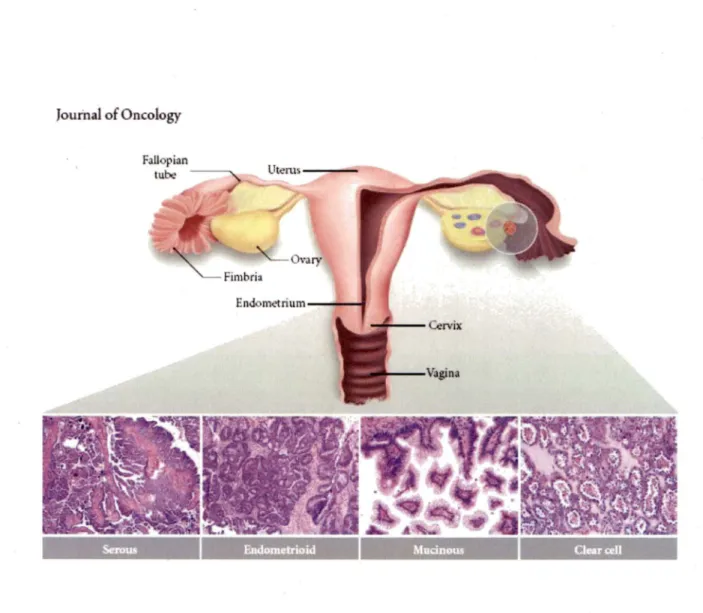
World health organization (WHO) recognizes eight histologie tumor subtypes of EOC: serous, endometrioid, mucinous, clear cell, transitional cell, squamous cell, mixed epithelial, and undifferentiated (TAVASSOLI and DEVILEE, 2003). The three most common (serous, endometrioid, and mucinous) are characterized by their morphological resemblance to various mucosal tissues of female reproductive tract, all of them exhibit
Müllerian differentiation (Figure 2). lt has been estimated that 50% of malignant ovarian tumors are serous carcinomas, while 25% are endometrioid, 10% are mucinous and only 5% are clear cell carcinomas (CHE et al., 2003).
Journal of Oncology
Ovary
Fimbria
Mucinous Clearœll
Figure 2. The major histologie subtypes of ovarian carcinoma (KARST and DRAPKIN, 2010). Serous carcinomas resemble fallopian tube epithelium, endometrioid carcinomas resemble endometrial glands, and mucinous carcinomas resemble endocervical epithelium. Photographs show representative tumor sections stained with hematoxylin and eosin. The shaded circle represents the general anatomical location from which ovarian carcinomas are thought to arise. The pink and blue entities within the cross-sected ovary represent maturing ovarian follicles.
Within each subtype, tumors are further described as benign (localized tumors that has not spread to other parts of the body), malignant (tumors that invade and damage other tissues and organs), or borderline (semimalignant tumors which are considered to have low malignant potential) and, depending upon tumor subtype, classified as low-or high-grade.
Low grade tumors are composed of uniform cells that look very like normal cells of ovary
with mild or moderate nuclear atypia and low mitotic index. They are associated with a serous neoplasm of low malignant potential. High grade tumors are composed of pleomorphic cells that look very abnormal with marked nuclear atypia and high mitotic index and associated with high malignant potential (MALPICA, 2008).
2.5 Role of EOC ascites in promotion of tumor cell survival
Epithelial ovarian cancer (EOC) is highly metastatic cancer characterized by widespread intraperitoneal dissemination and ascites formation (an abnormal accumulation of fluid in the abdomen) (SHEN-GUNTHER and MANNEL, 2002). EOC ascites may contain a variety of cytokines, growth factors, bioactive lipids, hormones, and components of ECM which contribute to cancer cell migration and invasion (Y AMADA et al., 2004; GRAVES
et al., 2004). Our laboratory has demonstrated that EOC ascites contrib~te to tumor cell
survival by activating the pro-survival PI3K/Akt pathway (LANE et al., 2007; LANE et al.,
2010). Activation of a number of cell surface receptors may contribute to the activation of PI3K/ Akt pathway (Figure 3).
Ovarian cancer ascites contains growth factors that could potentially activate tyrosine kinase receptors (ABENDSTEIN et al., 2000; MIY AMOTO et al., 2004). The binding of growth factors to tyrosine kinase receptors stimulates the phosphorylation of phosphatidylinositol 3-kinase (PI3K), which in tum leads to Akt activation_ (Figure 3). Epidermal growth factor receptor (EGFR), platelet-derived growth factor receptor (PDGFR), and Her2/neu are commonly overexpressed in EOC and are involved in inducing the tumor cell proliferation (ALPER et al., 2001; MA TEI et al., 2006).
PBK may also become activated through G-protein-coupled receptors (GPCR) (Figure 3). G-protein-coupled receptors regulate a variety of cell functions including cell proliferation, survival, and motility, and have recently emerged as important players in tumor growth and survival (MILLS and MOOLENAAR, 2003). One of the ligands of G-protein coupled receptors is the lysophosphatidic acid (LPA) that has been shown to induce cell survival signaling through the PBK/Akt pathway in ovarian cancer cells (KANG et al., 2004).
Ascites fluid contains significant levels of LP A, which exceed levels required to activate
LPAreceptors (YAMADAetal., 2004).
PBK/Akt pathway may also become activated by the interactions between extracellular matrix proteins (ECM) with cell surface integrins (HYNES, 1992). Integrins transmit signais directly through ligation-dependent recruitment of the focal adhesion kinase (F AK) leading to the activation of several cell-signaling pathways including the PBK/Akt pathway (STUP ACK and CHERESH, 2002).
Our laboratory investigated the mechanisms by which EOC ascites activates Akt in EOC cells and we demonstrated that survival promoting activity of ascites was not affected by inhibitors of GF receptors including EGFR, VEGFR, FGFR, Her2/neu, and IGF-Rl. We also concluded that LP A does not contribute to the pro-survival activity of EOC ascites. However, the integrin pathway was involved in ascites-mediated protection from TRAIL-induced apoptosis. We demonstrated that ovarian cancer ascites induces F AK and Akt activation in an av~5 integrin-dependent pathway (LANE et al., 2010).
Growth
factors
l
EOC
ascites
Bioactive
Lipids
(LPA)
!
activation
Interaction
between ECM
proteins and
integrins
!
Cell survival and proliferation
Figure 3. The mechanisms of PBK/Akt pathway activation by ascites. Ovarian cancer ascites contains a number of growth factors that could potentially activate tyrosine kinase receptors such as EGFR, PDGFR, Her2/neu, etc. Ascites fluid also contains bioactive lipids, such as LPA, which is one of the ligands of G-protein coupled receptors (GPCR). Activation of these receptors can result in downstream activation of PI3K/Akt. Akt pathway can also become activated due to the interactions between extracellular matrix (ECM) proteins with cell surface integrins. Such interactions activate the focal adhesion kinase (F AK) and subsequently the PI3K/ Akt pathway.
2.6 Origin of ovarian cancer
Severa! hypotheses have been proposed about the underlying physiological processes that increase risk of the ovarian cancer (Table 1) (LA DEN et al., 2008).
Table 1. Hypotheses on physiologie susceptibilities to epithelial ovarian cancer
othes1s Pro osed mecharnsms Best evidence
Incessant owlabon OSE damaged dunng owlabon R1sk of EOC decreases with decreased with repair making ceHs succeptible number of cycles. such as pregnancy,
_ _ _ _
1,,.,.,.... ... . utabons lactati
~l!&l.)--....-..iOM..-Gonadotropm s mula- snmulatory effec of FSH and LH lncreased EOC risk with infe ility. PCOS: tion promote growth, increased cell Decreased nskwith progesteron.only Hormonal sbmulation
ln lamma on
dMs1ons: and mu a ons OCPs: FSH upregula es many oncogenes H1gh concentrations of androgenes
m the tumor m1croe™ronment promote cancerogenesis. whereas progesbns decrease nsk
and romo es growth in reclimcal mo els Cond1bons of h1gh c1rculatJng androgenes (PCOS. hlrsUbsm. acne) assoc1ated with increased risk. androgenes are the domi-nant hormones m the inclusion cyst, pr0-gesbn use decreases nsk of EOC and mduces OSE apoptosis
Oamaged OSE with ovulation Possible reduced nsk with NSAID use~ induces in ammanon. wh1ch pro • increased risk with talc or asbestos. motes reconstrucnon and muta on abundanœof in lammatory mediators
susc p b1litv in mors
Abbre'Vla ons: OSE. ovanan surface ep1theltum. EOC. ep1thellal ovanan cancer. OCP. oral contracep. ive ill FSH. follicle-s ·mulatin hormone, LH lutem1zm ·hormone. PCOS. o c ticovanan? drome
2.6.1
Ovarian surface epithelium and cortical inclusion cyst
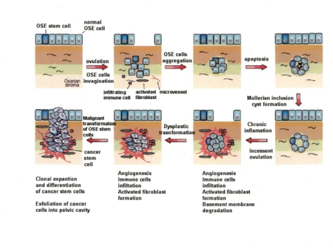
lt is widely thought that most ovarian cancers develop from the surface epithelium (OSE). OSE is a flat-to-cuboidal layer of uncommitted mesothelial cells covering the. exterior surface of the ovary. During ovulation, follicular rupture and oocyte release causes physical trauma upon the ovarian surface, creating a breach in OSE that must be repaired. Over the course of a woman' s life, this process of damage and repair is repeated multiple times. Accordingly OSE exhibit a high degree of plasticity that facilitates tissue remodeling (KRUK et al., 1994). ln addition to physical trauma, OSE cells are subjected to ovulation-associated inflammatory cytokines and oxygen species that are capable damaging D A
(MURDOCH and MARTINCHICK, 2004). Furthermore, as women age, the ovarian surface develops numerous invaginations into the cortical stroma. These invaginations frequently pinch off and become entrapped within the stroma, forming circular OSE-lined structure termed "cortical inclusion cysts" (CICs) (Figure 4). Once inside the ovary, the epithelial cells lining CICs are exposed to a new hormone-rich milieu that is thought to induce a differentiation into more complex epithelium resembling that of Müllerian-derived organs (DRAPKIN and HECHT, 2006). If the epithelial cells also happen to harbour unresolved DNA damage, they may be prime targets for neoplastic transformation, eventually giving rise to ovarian carcinomas.
normal
OSE stem cell OSE cell
,_.,,.1 ...--.--.--.--..,,.-;"
-
-
,....,ovulation
.-.;•qli!J;j
aggregationOSE cells ;
dtJ'.J
Ovarian invagination . - -~\--
stroma I \ ~~iiii,,;;;---cancer stem cell Clonai expantion and differentiation of cancer stem cells Exfoliation of cancer cells into pelvic cavityinfittrating actiwted microvessel
immune cell fibroblast
Angiogenesis Immune œlls infiltation Activated fibroblast formation Angiogenesis Immune œlls infiltation apoptosis Chronic inflamation ln cessent ovulation Activated fibroblast formation Basement membrane degradation
-Figure 4. Model of the development of an EOC tumor (FARLEY et al., 2008). Incessant ovulation and wound repair increases the risk of genetic abnormalities, Jeading to changes in epithelial cells lining the Müllerian inclusion cyst. Stroma! microenvironment in the forms of activated fibroblast formation, microvessel proliferation, and growth factors contributes to dynamic formation and eventual malignant transformation.
2.6.2 Two pathway model of ovarian carcinogenesis
The two pathway model of ovarian carcinogenesis proposed by KURMAN and SHIH (Figure 5) takes into account the diverse nature of ovarian cancer and correlates the clinical, pathological, and molecular features of the disease. In this model, ovarian tumors are divided into type I and II. Type I tumors are slow growing, generally confined to the ovary at the diagnosis, and develop from well-established precursor lesions. Type I tumors include low-grade micropapillary serous carcinoma, mucinous, endometrioid, and clear cell carcinomas. They are genetically stable and· characterized by mutations in number of different genes including KRAS, BRAF, PTEN, and ~-catenin. Mutations of KRAS and
BRAF lead to the activation of MAPK signaling pathway (PEYSSONNAUX and EYCHENE, 1996). PTEN mutations result in constitutively PI3K/ Akt signaling (OBATA
et al., 1998).
Type II tumors are rapidly growing highly aggressive neoplasms for which well-defined precursor lesions have not been described. Type II tumors include high-grade serous carcinoma, malignant mixed mesodermal tumors, and undifferentiated carcinomas. This group has a high level of genetic instability and is characterized by mutation of TP53. These tumors may also exhibit gene amplification and overexpression of HER2/neu oncogene (10-20%) (ROSS et al., 1999) and Akt2 oncogene (12-18%) (CHENG et al.,
Molecular genetic changes KRASIBRAF
---TP53 1 Serous cystadenoma 1KR~~~~
BRA7
APSTl
MPSC!~
Low-grade High-grade serous CA serous CA (grade 1) (grade 3) Mutations Mutations APSTl
MPSC High-grade serous CA (grade 2) Wild-typeOvarian surface inclusions Peritoneum Fallopian tube High-grade serous CA (grade 3) Wild-type ---
--- ---
---Wild-type Wild-type Mutations Mutations
L_
__j. " ' - - - - _ _ _ _ _/~
~
Type 1 Type Il
Figure 5. Two pathway model of ovarian carcinogenesis (KURMAN and SHIH, 2008). Proposed pathways for the development of different types of serous carcinoma. APST indicates atypical proliferative serous tumor; MPSC, micropapillary serous carcinoma.
2.6.3 Fallopian tube as a site of origin
Severa! recent studies have demonstrated that many high-grade serous ovarian tumors may actually be of tubai origin, arising from the distal region of the fallopian tube, but then quickly spreading to nearby ovary (MEDEIROS et al., 2006; KINDELBERGER et al.,
2007; LEE et al., 2007). The fimbria (the end of the Fallopian tube (Figure 2)) lies in extremely close proximity to ovarian surface epithelium and is therefore exposed to the same inflammatory and potentially carcinogenic microenvironment. Lee et al. formulated a model of ovarian cancer which incorporates the fimbria as a major site of origin for serous carcinomas. This model states that there are two distinct pathways leading to ovarian tumorigenesis. The first route is the traditional OSE-CIC pathway which leads to formation of Kurman's and Shin's "Type I" tumors. The second pathway involves the fallopian tube fimbria, where a combination of TP53 mutation and genotoxic stress leads to the clonai expansion of secretory epithelial cells, forming a pre-neoplastic precursor lesion or "p53 signature". Additional genetic "bits" in the absence of functional TP53 enable these cells to acquire a proliferative capacity, facilitating the progression of tubai intraepithelial carcinoma (TIC) (Figure 6). This second pathway leads to formation of Kurman's and Shin's "Type II" tumors.
0 D Ovula tory cytokines +ROS 0 0 D 0 D 0 • 0 • 0 0 0 0 0 of pcritoncum or ovary
Figure 6. Fallopian tube as a site of origin (KARST and DRAPKIN, 2010). The fallopian tube epithelium (FTE) is composed of a single layer of ciliated and secretory cells that are exposed to ovulation-associated inflammatory cytokines and reactive oxygen species (ROS). Repetitive genotoxic stress causes DNA damage and induces p53 mutation, leading to the clonai expansion of normal looking FTE cells of secretory phenotype. These damaged cells stain strongly for p53 ("p53 signature"). Further genetic "hits" enable cells to acquire a proliferative capacity, giving rise to tubai intraepithelial carcinoma (TIC). As a TIC progresses to invasive serous carcinoma, malignant cells are exfoliated from the fimbria and they may spread rapidly to the surface of the peritoneum and/or ovary. Exfoliation may also occur from TICs prior to fimbrial invasion.
2.7 Current treatment of EOC and resistance
The bilateral salpingo-oophorectomy (BSO) is generally recommended for women with high risk of developing ovarian cancer (with a known germline mutations of BRCAl and BRCA2) at approximately 35 to 40 years of age as apart of the prophylactic treatment (NCCN, 2005). Although prophylactic BSO reduces the risk of ovarian cancer by more than 90%, high-risk patients with BRCAl and BRCA2 mutations can still develop primary peritoneal cancer, which develops in 4% to 5% of women at 20 years after BSO (FINCH et al., 2006).
The standard treatment for early-stage ovarian cancer patients is typically cytoreductive surgery. Stage I patients with favourable prognosis factors have more than 90% cure rate with surgery alone. Early-staged patients with unfavourable prognostic features (such as poorly differ,entiated tumors or evidence of spread) have 70 to 80% cure rate (OZOLS et al., 2004).
After completion of surgery, patients with advanced disease reqmre chemotherapy or radiotherapy. EOC is considered to be a chemosensitive neoplas.m, with initial overall response rates to systemic therapy exceeding 80% when integrated with surgery. However, among women with advanced stage disease at diagnosis, long term survival remains poor due to eventual tumor recurrence and emergence of drug-resistant disease. Combination chemotherapy using paclitaxel with a platinum-based regimen is currently the standard first-line treatment for ovarian cancer (COLOMBO et al., 2006). Whereas cisplatin-paclitaxel combination chemotherapy has shown significant efficacy over previous drug combinations, 20-30% of patients fail to respond to this combination and 90% of patients that initially responded to therapy will eventually develop chemotherapy-resistant disease (MCGUIRE et al., 1996). Patients that do not respond to the first-line chemotherapy are given second-line and third-line regimens of chemotherapy in an attempt to pro long life and palliate symptoms. Second-line chemotherapeutic agents are rarely curative, with initial response ranging from 10-33% (JUDSON et al., 1999). The discovery of navel and effective therapy against chemotherapy-resistant ovarian cancer remains a high priority.
2. 7.1 Mechanisms of resistance to chemotherapy
A wide range of metabolic or structural properties within tumour cells may lead to drug resistance. The identified mechanisms include: decreased drug uptake, increased drug efflux, increased repair of DNA damage induced by chemotherapy and reduced ability to undergo apoptosis (GOLDENBERG et al., 1998). In addition, because cancer cells are heterogeneous, more than one mechanism of drug resistance may be present in any particular case.
Initially sensitive tumor cells may become resistant to drugs during repetitive chemotherapy, which is so called acquired (induced) resistance, or they may be resistant without previous exposure to drugs- intrinsic resistance. Cells with acquired drug resistance can be produced experimentally by the successive exposure of parent cells to a particular drug in vitro (BOSCH and CROOP, 1996; Lane et al., 2006). Severa! mechanisms of acquired drug resistance have been demonstrated, including reduced accumulation of drug, changes in the expression of enzymes involved in the glutathione detoxification pathway, alterations in DNA repair, c'ell cycle checkpoints and ratio of pro and anti-apoptotic Bcl-2 family members (Figure 7). In contrast to acquired drug resistance, few studies have examined the mechanisms responsible for intrinsic drug resistance. The reason why some tumor cells are inherently resistant without previous drug treatment is not known. In addition, importance of the de-nova (environment-mediated) drug resistance has been acknowledged. This type of resistance is mediated by tumor microenvironment which represents a rich source of both soluble factors and components of ECM, both ofwhich can favour cell survival following drug exposure. Mechanisms associated with de-novo drug resistance may contribute to the failure to eliminate minimal residual disease and facilitate the emergence of acquired drug resistance (MEADS et al., 2009; LANE et al., 2010; KERBEL et al., 1994).
Altcred Drug Target
Repair D A damage Altered cell cycle checkpoint Decreased DNA damage re ·pon e
Apoptosis
'
' APAF-1c
Caspa e-9
*
Drug metabolism*
*
Drug Bax.BAD. BCL-2. MCL-1 , BCL-XLFigure 7. Acquired drug-resistant models possess multiple drug-resistance mechanisms (HAZLEHURST et al. , 2003). These mechanisms include: (a) decreased intracellular concentration of the drug characteristic of overexpression of drug transporters; (b) alterations in the drug target su ch as point mutations or overexpression of the target; ( c) increased detoxification of the drug such as glutathione conjugation; (d) changes in the repair of DNA damage induced by the drug (e) alterations in the cell cycle checkpoint such as p27Kipl or p21 ; (e) changes in the ratio of pro and anti-apoptotic Bcl-2 family members.
The ability of most types of cancer to metastasize was also linked to their capacity to express resistance to anti-cancer drugs, including all major classes of drugs used in chemotherapy. Expression of certain dominantly acting oncogenes or altered expression of tumor suppressor genes can enhance not only tumor cell growth and malignant
aggressiveness, but also the relative expression of drug resistance (HENNEQUIN et al.,
2003). Sorne of these genetic alterations, mutations of suppressor gene p53 being a good example, often occur in more advanced stages of the disease (LASSAM et al., 1993) and are relevant to acquisition of malignant properties and also drug resistance.
Interestingly, a number of studies demonstrated connection between increased activation of the pro-survival PI3K/Akt pathway in ovarian cancer cells and resistance to chemotherapy.
ln vivo and in vitro studies demonstrated that inhibition of PI3K enhanced
paclitaxel-induced apoptosis in the human ovarian cancer cells (HU et al., 2002). XIAP (X-linked inhibitor of apoptosis protein), Akt2 and p53 are important mediators of chemoresistance in EOC. Inhibition of XIAP and/or Akt expression/function is considered to be effective of overcoming chemoresistance in EOC cells (FRASER et al., 2003). Targeting PI3K/Akt pathway has been shown to enhance sensitivity to docetaxel in breast and ovarian cancer (XING et al., 2008). Recent study demonstrated that Akt confers resistance to cisplatin, by modulating cisplatin-induced, p53-dependent c-FLIP (FLICE-inhibitory protein) ubiquitination (ABEDINI et al., 2009). Combined treatment of carboplatin and PI3K inhibitor L Y294002 has been demonstrated to effectively decrease ovarian tumor progression (WESTF ALL and SKINNER, 2005). However, another study indicates that efficacy of L Y294002 may be greatly affected by the tumor p53 status (BAR et al., 2005).
2. 7.2 Targeted therapies for ovarian cancer
Despite evidence of considerable heterogeneity in their histological phenotypes and molecular profiling, most cases of ovarian cancer are treated in a similar fashion. It became apparent that the focus should be towards the development of new targeted therapies capable of exploiting molecular and genetic characteristics of individual tumor subtypes. At the moment, there is rapid development of navel compounds, including antiangiogenic reagents, humanized monoclonal antibodies, selective hormonal agents, and small molecules that target key components in signal transduction pathways associated with cell growth, tumor vascularity, and invasive potential.
A number of studies have focused on the epidermal growth factor receptor (EGFR), which is overexpressed in 30 to 70% of EOC (BASELGA, 2002). EGFR inhibitors such as cetuximab, ABX-EGF, erlotinib have been evaluated in clinical trials (FINKLER et al., 2001 ). In spite of encouraging preclinical models, early results from large trials have not shown a benefit (OZOLS et al., 2004).
Vascular endothelial growth factor (VEGF) has a key role in ascites formation. Antibody against VEGF has been shown to prevent and even reverse ascites formation in preclinical studies on mice (HU et al., 2002). Bevacizumab (Avastin) is a recombinant humanized monoclonal antibody against VEGF, which has shown activity in ovarian cancer. Bevacizumab was given as a single agent to the women with rec.urrent ovarian or primary peritoneal cancer. 21 % of women responded to therapy and 40% of patients had progression-free survival for 6 months and more (BURGER et al., 2005). However, a serious adverse effect (bowel perforation) has been reported due to use of Bevacizumab (HEINZERLING and HUERTA, 2006).
Replacing defective genes that cause malignant behaviour of cancer cell is another approach. One such target is TP53. Promising preclinical data from in vitro systems led to phase I trials, but this trial was closed after first analysis due to the lack of any signs of efficacy (ZEIMET and MARTH, 2003). One of the possible problems is the presence of adenovirus-neutralizing antibodies in EOC ascites. Another study demonstrated that adenoviral transduction of primary ovarian cancer samples was abolished by autologous ascitic fluids (INGRAM et al., 2010).
It is now recognized that in addition to genetic alterations, epigenetic mechanisms, such as DNA methylation, histone modifications and nucleosome remodeling, play an important role in the development and progression of ovarian cancer by modulating chromatin structure, and gene and miRNA expression. Furthermore, epigenetic alterations have been recognized as useful tools for the development of novel biomarkers for diagnosis, prognosis, therapeutic prediction and monitoring of diseases. Moreover, new epigenetic therapies, such as DNA methyltransferase inhibitors and histone deacetylase inhibitors,