D
EPARTMENT OFM
ICROBIOLOGYThesis
Presented by
NOUMEUR Sara Raouia
For the fulfillment of the requirements for the degree of
Doctorate of Sciences
In Biology
Option: MiCrobiology
Topic
Identification of bioactive natural products from
endophytic fungi isolated from Globularia alypum
Presented publically in 02/07/2018 Jury
President Mohamed Mihoub Zerroug Pr. UFA Sétif 1
Supervisor Daoud Harzallah Pr. UFA Sétif 1
Examiners Ammar Ayachi
Tayeb Idoui
Pr. Univ. Batna 1 Pr. Univ. Jijel
Marc Stadler Pr. HZI (Germany)
Samia Mezaache-Aichour MCA. UFA Sétif 1
Invited guest Soleiman Elsayed Helaly
Laboratory of Applied Microbiology
Dr. Univ. Aswan (Egypt)
Université Ferhat Abbas Sétif 1 Faculté des Sciences de la
Nature et de la Vie
ةيبعشلا ةيطارقميدلا ةيرئازجلا ةيروهمجلا
ملعلا ثحبلا و يلاعلا ميلعتلا ةرازو
ي
فيطس ،سابع تاحرف ةعماج
1ةيلك
مولع
ةايحلا و ةعيبطلا
N°……….…………..…….……/SNV/2018
خـلم
ـــــــــــــــ
ص
ردصم ةيلخادلا تايرطفلا ربتعت ا ينغ ا ةيعيبطلا تابكرملاب نم اهتبستكا نيب ةيئفاكتلا ةقلاعلا للاخ يب و اه لئاعلا تابنلا ن مت ، للاخ لا هذه ةسارد ةفرعم ةيلخادلا تايرطفلل يجولويبلا عونتلا نم ةلوزعملا ةتبن روذج Globularia alypum Plantaginaceae) ، (Scrophulariales يتلا نم اهينج مت ةنتاب ةيلاو ) رئازجلا ( . ىلا ةساردلا تدأ لزع 71 ةيرطف ةللاس و ىوتسم ىلإ تفرع يتلا مادختساب عونلا صئاصخ ثيح روطتلاو ءوشنلا ليلاحتو ةيجولوفروم تنكم ةريخلأا هذه ىلإ تنا تايرطفلا ءام ةبعش يف تائف ثلاث ىلإ ةلوزعملا Ascomycota : Eurotiomycetes ، Dothideomycetes و dariomycetes Sor و سانجأ ةعست يف ةلثمتملا : Alternaria ، Aspergillus ، Chaetomium ، Dendrothyrium ، Diaporthe ، Fusarium ، Macrophomina ، Penicillium و Preussia قاطن ىلع ريمختلل ةينامث ةلوزعملا تايرطفلا نيب نم رايتخا مت . صلختسملا تتبثأ .ةفلتخم ةيئاذغ طاسوا ةثلاث يف ريغص تا لصحتملا يبوركيم دض اطاشن اهيلع ا دعب . اه سمخ ريمخت مت تايرطف ة : نم تلالاس ثلاث Preussia similis ، variisporum Dendrothyrium ، Chaetomium madrasense قاطن ىلع .ربكا لزع ىلإ ريمختلا اذه ىدأ 82 هنيب نم يوناث بلقتسم ا 78 ديدج ابكرم ا فرعتلا مت . ةغيص ىلع بكرملا تا ةيفيط قرط ةطساوب ةلوزعملا ةيئايميكلا 1D و 2D NMR spectroscopy ، high-resolution mass spectrometry ، CD spectroscopy سرد . ت ةفورعملا وأ اهنم ةديدجلا ءاوس ةلوزعملا تابكرملل ةيجولويبلا تاطاشنلا لزع مت .عساو لكشب 71 دقعملا نم بكرم Preussia similis نيب نم اهتيمست مت ةقلحلا ةيئانث دياتيكيلوب :ةديدج تابكرم ةتس اه preussilides A-F ديدج بكرموdimer of 2-amino benzoid acid moieties لثم ةفورعم ىرخأ تابكرم ىلإ ةفاضلإاب .نوتنازغلا تاقتشمو نيزلااكوتياسلا ارهظأ preussilide A و preussilide C يف رابتخإ preussilides A-F طاشن ا طبثم ا يئاقتنا ا ناطرس ايلاخ ايجولوفروم ىلع اريثأت ،ةاونلا تايقيقح دض امك .ماظعلا ي ذه فدهتس ا ىلع لوؤسم ميزنإ دياتيكيلوبلا قيسنت .ايلاخلا ماسقنإ ةرود نم ةيوناثلا تابلقتسملا جاتنا ىلع تيرجأ يتلا تاساردلا ترفسأ رطف Dendrothyrium variisporum ع ن لزع 78 م ،بكر اهنم H و Massarilactones D و نيسسيئر نيبكرمك 8 نوناريفلل ةديدجلا تاقتشملا نم اهتيمست مت يتلا (5S)-cis-gregatin B و graminin D ىلإ ةفاضلإاب 1 كيلينارتنلأا ضمح رئاظن نم نينثا ،كيلارتنولأا ضمح نم ةديدج تاقتشم ا براجت ترهظأ .سديتببولكيس ةثلاثو ةفورعملا نأ يبوركيم دض طاشنل لل نيبكرم 2-phenylethyl-3-hydroxyanthranilate و 2-phenylethylanthranilate ةعساو ةيبوركيم دض ةيطيبثت ةيطاشن ، ةيناطرسلا ايلاخلا رثاكتل هطيبثت بكرملا رهظا نيح يف . HeLa لزع مت اريخأ 1 نم ةفورعم تابكرم :Chaetomium madrasense A1 ، A3 xanthoquinodins و N-acetytryptamin . ةيحاتفم تاملك ،ةيوناثلا تابلقتسملا ،يجولويبلا عونتلا ،ةيلخادلا تايرطفلا : فيرعت لا ،ةيئايمكلا ةغيص Dendrothyrium variisporum ، Chaetomium madrasense ، Preussia similis
Abstract
The endophytic mycobiota constitute a prolific source of bioactive natural products that has evolved during the symbiotic endophyte−plant relationship. The diversity of fungal endophytes isolated from the roots of the plant Globularia alypum (Plantaginaceae, Scrophulariales) collected in Batna (Algeria) were studied in the present thesis. In total, seventeen fungal strains were isolated and identified to species level by means of morphological and molecular phylogenetic methods. The phylogenetic analysis of these fungal endophytes revealed their affinities to three classes of the phylum of Ascomycota: Eurotiomycetes, Dothideomycetes and Sordariomycetes representing nine genera: Alternaria, Aspergillus, Chaetomium, Dendrothyrium, Diaporthe, Fusarium, Macrophomina,
Penicillium and Preussia. Among the isolated fungal endophytes, eight were selected for small scale
fermentation in three different liquid media. Their extracts showed prominent antimicrobial activity in a screening for novel antibiotics by the serial dilution assay. A scale-up of fermentation of selected five fungi including three strains of Chaetomium madrasense, Dendrothyrium variisporum and
Preussia similis, yielded twenty eight secondary metabolites, twelve of which turned out to be novel
natural products. The structures of the isolated metabolites were elucidated using a combination of spectral methods, including 1D and 2D NMR spectroscopy, high-resolution mass spectrometry, and CD spectroscopy. The biological activities of both, the known and new compounds were extensively studied. Thirteen metabolites were isolated from the Preussia similis complex including six new bicyclic polyketides (for which the trivial names preussilides A−F are proposed) and one new dimer of 2 amino benzoid acid moieties, along with several known cytochalasins and xanthones derivatives. The preussilides were tested for antimicrobial and antiproliferative effects, and, in particular, preussilides A and C showed selective activities against eukaryotes. Subsequent studies on their influence on the morphology of human osteosarcoma cells (U2OS) suggest that these polyketides might target an enzyme involved in coordination of the cell division cycle. Hence, they might, for instance, affect timing or spindle assembly mechanisms, leading to defects in chromosome segregation and/or spindle geometry. Studies on the secondary metabolite production of
Dendrothyrium variisporum in various culture media led to the isolation of twelve compounds.
Massarilactones D and H were isolated as the major components as well as two new furanone derivatives for which we propose the trivial names (5S)-cis-gregatin B and graminin D, three new anthranilic acid derivatives, two known anthranilic acid analogues and three cyclopeptides. The new anthranilic acid derivatives, 2-phenylethyl-3-hydroxyanthranilate and 2-phenylethyl anthranilate exhibited antimicrobial activity while 2-phenylethyl anthranilate showed cytotoxicity against HeLa cells. Finally, from cultures of Chaetomium madrasense, three known compounds, xanthoquinodins A1, A3 and N-acetytryptamin were isolated.
Keywords: Fungal endophytes, biodiversity, Globularia alypum, secondary metabolites, Preussia
Résumé
Le mycobiote endophyte constitue une source riche de produits naturels bioactifs qui ont été développé au cours de la relation symbiotique endophyte-plante. La diversité des endophytes fongiques isolés à partir des racines de la plante Globularia alypum (Plantaginaceae, Scrophulariales), collectée à Batna, (Algérie), a été étudiée dans le présent travail. Au total, dix-sept souches fongiques ont été identifiées jusqu’au rang de l'espèce à l'aide de méthodes morphologiques et phylogénétiques. L'analyse phylogénétique de ces endophytes fongiques a révélé leurs affinités à trois classes du phylum d'Ascomycota: Eurotiomycètes, Dothideomycètes et, Sordariomycètes représentées par neuf genres: Alternaria, Aspergillus, Chaetomium, Dendrothyrium, Diaporthe, Fusarium, Macrophomina,
Penicillium et Preussia. Parmi les endophytes fongiques isolés, huit ont été sélectionnés pour une
fermentation à petite échelle dans trois milieux liquides différents. Leurs extraits ont montré une activité antimicrobienne importante au cours du screening pour de nouveaux antibiotiques par le test de dilution en série. La fermentation à grande échelle de cinq champignons sélectionnés, y compris trois souches de Preussia similis, Dendrothyrium variisporum et Chaetomium madrasense, a donné vingt-huit métabolites secondaires, dont douze ont été identifiés comme nouveaux produits naturels. Les structures des métabolites isolés ont été élucidées en utilisant une combinaison de méthodes spectrales, y compris la spectroscopie RMN 1D et 2D, la spectrométrie de masse à haute résolution et la spectroscopie CD. Les activités biologiques des composés connus et nouveaux ont été largement étudiées. Treize métabolites ont été isolés du complexe Preussia similis, y compris six nouveaux polycétides bicycliques (pour lesquels les noms triviaux preussilides A-F ont été proposés) et un nouveau dimère de l’acide 2-amino benzoïque, ainsi que plusieurs autres métabolites connus comme les cytochalasines et des dérivés de xanthone. Les preussilides ont été testés pour leurs effets antimicrobiens et antiprolifératifs et, en particulier, les preussilides A et C qui ont montré des activités sélectives contre les eucaryotes. Des études ultérieures sur leur influence sur la morphologie des cellules d'ostéosarcome humain (U2OS) suggèrent que ces polycétides pourraient cibler une enzyme impliquée dans la coordination du cycle de la division cellulaire. Par conséquent, ils pourraient, par exemple, affecter les mécanismes de la synchronisation ou d'assemblage des fuseaux mitotiques, conduisant à des défauts de ségrégation des chromosomes et / ou de la géométrie du fuseau mitotique. Des études portées sur la production de métabolites secondaires de Dendrothyrium variisporum ont conduit à l'isolement de douze composés. Les massarilactones D et H ont été isolés comme principaux composants ainsi que deux nouveaux dérivés de furanone pour lesquels les noms triviaux (5S) -cis-grégatine B et graminine D ont été proposés, trois nouveaux dérivés d'acide anthranilique, deux analogues connus de l'acide anthranilique et trois cyclopeptides. Les nouveaux dérivés de l'acide anthranilique, le 2-phényléthyl-3-hydroxyanthranilate et le 2-phényléthyl anthranilate, ont présenté une activité antimicrobienne tandis que le 2-phényléthyl anthranilate présentait une cytotoxicité contre les cellules HeLa. Enfin, à partir des cultures de Chaetomium madrasense, trois composés connus à savoir xanthoquinodines A1, A3 et N-acétyltryptamine ont été isolés.
Mots-clés: Endophytes fongiques, biodiversité, Globularia alypum, métabolites secondaires,
Dedications
My dedications go with love and affection to my mother. Thank you for being the embodiment of sacrifice to ensure a better life for me. Without your enormous effort and unconditional love and care, I would not have never become the individual that I am today. You have been a source of motivation and strength during moments of despair and discouragement. There are not enough words I can describe how important my mother was to me and what a powerful influence she continues to be.
I also dedicate this work to my husband Bilal, whose patience, constant encouragement, limitless giving and understanding, helped me to accomplish my degree. You were always beside me during the challenges of my PhD research and life. I truly thank God for having you in my life.
I dedicate this work to my lovely daughter Olina, the major source of my joy and strength.
My dedications go to my beloved sister Rima and brother Abdelmoutaleb. Thank you for the love, encouragement and support you have given me in numerous ways.
To the memory of my beloved father
I love you all
Acknowledgments
First and foremost, I would like to thank Allah Almighty for giving me the strength, knowledge, ability and opportunity to undertake this research study. Without his blessings and guidance, this achievement would have never become true.
I am deeply grateful to my supervisor Prof. Daoud Harzallah (University of Ferhat Abbas, Sétif 1) for his patience and for the many times he supplied me with advice and suggestions. Thank you for being always generous during all phases of the research.
I am greatly indebted to my co-supervisor Prof. Dr. Marc Stadler, head of Department of Microbial Drugs at Helmholtz Center for Infection Research, Braunschweig (Germany) for giving me the opportunity to work in his department and let me dive into the world of fungi and natural products. I would like to thank you explicitly for your expert advice, your excellent work facilities, steadfast support and encouragements that have been great contributors in the completion of the thesis. Thank you Marc not only for your tremendous academic support, but also for giving me so many wonderful opportunities which have a positive influence in growing my career.
My acknowledgements are also expressed to Prof. Mohamed Mihoub Zerroug (University of Ferhat Abbas, Sétif 1) for having honored me by being the chair of the jury, to Prof. Ammar Ayachi (University of Batna 1), Prof. Idoui Tayeb (University of Jijel) and Dr. Samia Mezaache-Aichour (University of Ferhat Abbas, Sétif 1) for showing their willingness to examine this work and to be members of the jury.
I highly appreciate the valuable time and the efforts expended by Dr. Soleiman Essayed Helaly, without his tireless devotion and chemical skills, this work would not have seen the light.
Thank you for being kind and patient for teaching me chemistry. Your assistance proved to be a
I also would like to express my wholehearted thanks to Prof. Dr. Theresia Stradal, head of Department of Cell Biology at Helmholtz Center for Infection Research, Braunschweig (Germany) for allowing me to use her laboratory facilities and for the generous support she provided me during time pressure and challenges throughout my cell biology experiments. I also extend my thanks to her lab team: Anika, Annette, Markus, Jana, Stephanie, Marco and Jessica for the excellent technical assistance in cell biology experiments. You were all wonderful.
I offer my thanks to Dr. Rémy Bertrand Teponno for our pleasant and fruitful collaboration. I want to express my deep thanks to Simone Heitkämper for her kind and excellent assistance. Further, I would like to thank Wera Collisi for her patience and her excellent technical assistance in conducting the bioassays. I am very grateful to Cäcilia Schwager for conducting HPLC-MS data, you were really kind and generous. Also, many thanks go to Silke Reinecke, for her generous help.
I would like to take this opportunity to say warm thanks to all my beloved friends Wilawan, Sandra, Sara, Lucile.
Many thanks to the amazing team of microbial drugs: Christiane, Rolf, Kathrin, Frank, Christian, Clara, Zeldjka, Stephan, Birthe, Lucky, Eric, Enge, Kerstin, Anke and to all the international students I have met during my internship, I have learnt a lot from you.
I owe my profound gratitude to Dr. Manfred Rohde, head of the Research Group of Molecular mechanisms of streptococci at Helmholtz Center for Infection Research, Braunschweig (Germany) for his support with electron microscopy.
I gratefully thank Dr. Carlos Plaza for kindly providing fluorescent substrate Cell Event Caspase 3/7 and staurosporine and also for his support with flow cytometry.
I acknowledge the Ministry of Higher Education and Scientific Research of Algeria for the financial support.
Last but not least, deepest thanks go to all people who took part in making this thesis real. To all of you, thank you very much
List of abbreviations
BLAST CBS CD COSY DAD DAPI DCM DMEM DSMZ EDTA EF1-α EtOAc FACS GC –MS GMAK GTR HA HPLC HRESIMS ITS LSU MAFFT MEA MeOH MIC MS MTT MUCL NA NHA NMR OMA PBS PCR PDA PFA PhyML PI RAxML ROESY RP SEM TAE TFA tR STMA YEA YMGBasic Local Alignment Search Tool
Centraalbureau Voor Schimmelcultures, Utrecht, The Netherlands Circular Dichroism
Correlation Spectroscopy (NMR) Diode Array Detection
4',6-Dia midino-2-P henylindole Dichloromethane
Dulbecco’s modified eagle medium
German Collection of Microorganisms and Cell Cultures Ethylene Diamine Tetraacetic acid
Elongation Factor 1- α Ethyl Acetate
Fluorescence-activated cell sorting. Gas Chromatography - Mass Spectrometry Genomanalytik (Genome Analytics Group) Generalised time reversible
Habitat-adapted
High Performance Liquid Chromatography High resolution electrospray ionization mass spectrometry. Internal Transcribed Spacer Region Of The Ribosomal DNA Large Subunit
Multiple alignment using fast Fourier transform Malt Extract Agar
Methanol
Minimum Inhibitory Concentration Mass Spectrometer
3-(4, 5-dimethylthiazol-2-yl)-2, 5-diphenyltetrazolium bromide
Mycothèque De L'université Catholique De Louvain, Louvain-La-Neuve, Belgium Nutrient Agar
Nonhabitat-adapted
Nuclear Magnetic Resonance Oat Meal Agar
Phosphate-buffered saline Polymerase Chain Reaction. Potato Dextrose Agar Paraformaldehyde
Phylogenetic Estimation By Maximum Likelihood Propidium iodide
Randomized A(x)ccelerated Maximum Likelihood Rotating Frame Nuclear Overhauser Effect Spectroscopy Reversed Phase
Scanning Eletron Microscopy Tris-Acetate-EDTA
Trifluoroacetic Acid Retention time
Stadler Marc private culture collection Yeast Extract Agar
List of figures
Figure N° Title Page
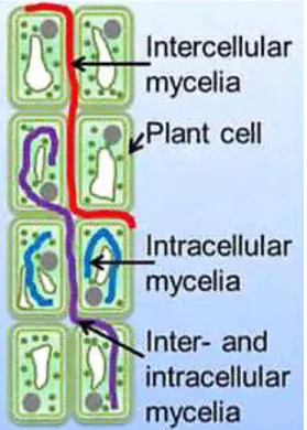
Figure 1 Different localization patterns of fungal endophytes within plant tissues 3 Figure 2 Location of the different classes of endophytes (according to Rodriguez
et al., 2009)
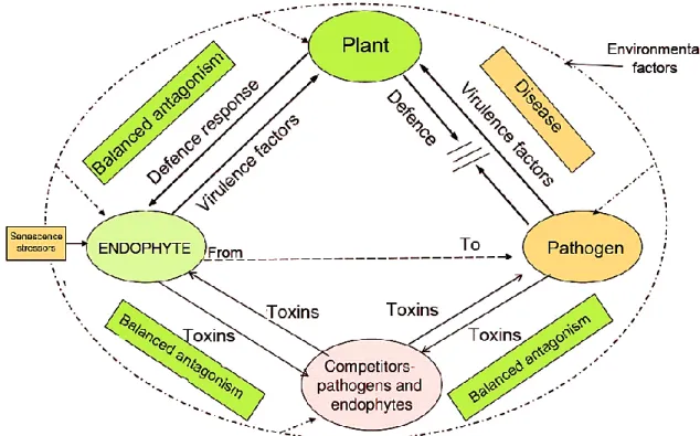
6 Figure 3 Paradigm of balanced antagonism as suggested by Schulz et al. (2015) 9 Figure 4 Schematic representation of endophyte-endophyte interspecies
crosstalk
12
Figure 5 Chemical structures of secondary metabolites isolated from the conglomerate Preussia/Sporormiella as indicated in Table 2
15
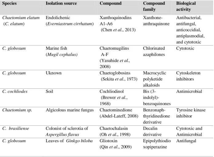
Figure 6 Chemical structures of selected secondary metabolites isolated from
Chaetomium spp. as indicated in Table 3.
18
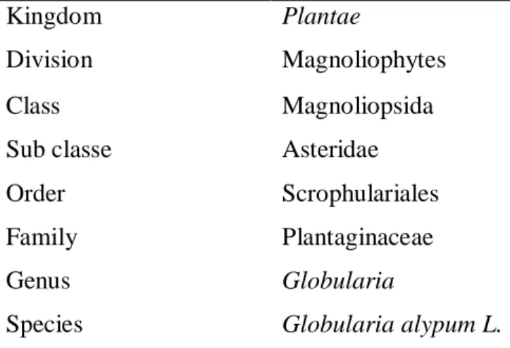
Figure 7 Globularia alypum 19
Figure 8 Globularia alypum used in this study 21
Figure 9 Root fragments on agar plates 22
Figure 10 Schematic diagram showing purification steps of metabolites isolated from mycelial and supernatant crude extracts of derived from fermentation culture of the fungal isolate DSM 10466 in 8L of ZM/2 liquid medium.
31
Figure 11 Schematic diagram showing purification steps of metabolites isolated from mycelial and supernatant crude extracts derived from
fermentation culture of fungal isolate STMA 16219 in 8L of Q6/2 liquid medium
32
Figure 12 Schematic diagram showing purification steps of metabolites isolated from mycelial and supernatant crude extracts derived from
fermentation culture of fungal isolate DSM 32328 in 5L of Q6/2 liquid medium.
33
Figure 13 Schematic diagram showing purification steps of metabolites isolated from flasks culture in 8L of YMG medium of the fungal isolate STMA 16226.
34
Figure 14 Schematic diagram showing purification steps of metabolites isolated from mycelial and supernatant crude extracts derived from bioreactor culture of the fungal isolate STMA 16226 in 10 L of YMG medium
35
Figure 15 Schematic diagram showing purification steps of metabolites isolated from the supernatant crude extract derived from fermentation culture of the fungal isolate STMA 16225 in 3L YMG medium
Figure 16 Macroscopic morphology of fungal endophytes on YMG agar medium on 9 cm dish.
44
Figure 17 Phylogenetic relationship of fungal endophytes harbored in the roots of
G. alypum based on ITS sequences with Paludomyces mangrovei as out
group.
46
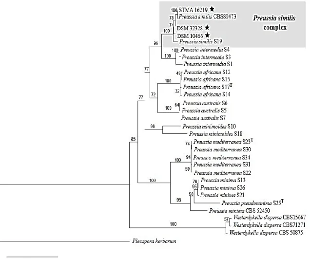
Figure 18 Phylogenetic tree of Preussia spp. based on combined data set of four markers. Multigenes alignment, PhyML (ITS, LSU, EF1-α).
48
Figure 19 Sterile mycelia of P. similis DSM32328 49
Figure 20 Teleomorph features of Preussia similis DSM 104666 50 Figure 21 Teleomorph characters of P. similis STMA 16219 51 Figure 22 Phylogenetic tree of Dendrothyrium spp. and the closely related genera
based on combined data set of four markers. Multigenes alignment, RxML (ITS, LSU, Tub and Actin)
53
Figure 23 Anamorph characteristics of Dendrothyrium variisporum 54
Figure 24 Teleomorph features of Chaetomium madrasense 56
Figure 25 Results of MIC assays of the most potent extracts 59 Figure 26 HPLC chromatogram of crude extracts from ZM/2 liquid culture of P.
similis DSM 104666
60
Figure 27 Chemical structures of preussildes A-F (1-6) 61
Figure 28 Antifungal activity of preussilides A (1) and C (3) against phytopathogen S. sclerotiorum determined by Agar diffusion at 100µg/disk
63
Figure 29 Results of bioassay-guided fractionation by RP-HPLC using B. subtilis as indicator organism.
64 Figure 30 Microscopic view of fragmented nuclei of preussilide C-treated L929
in MTT assay
66
Figure 31 Mono and co-cultures on YMG agar medium of P. similis and S.
sclerotiorum
66
Figure 32 HPLC chromatograms of individual axenic and co-culture of P. similis DSM 104666
67
Figure 33 Effects of preussilides A and C (1 and 3) on the Morphology of Human Osteosarcoma Cells (U2OS)
69-70 Figure 34 Preussilides treatment induces generation of small cells with reduced
nuclear size in the human Osteosarcoma Cell Line U2OS.
Figure 35 Chemical structures of the compounds (7-8) obtained from P. similis STMA 16219
73 Figure 36 HPLC chromatograms of crude extracts from Q6/2 liquid culture of P.
similis 16219
74 Figure 37 Mono and co-cultures of P. similis and S. sclerotiorum on YMG agar
medium
76 Figure 38 HPLC chromatograms of individual axenic and co-culture of Preussia
similis STMA 16219
76 Figure 39 Chemical structures of the compounds (10-13) isolated from Preussia
similis DSM 32328
77 Figure 40 HPLC chromatograms of both mycelial and supernanant crude extracts
derived from liquid culture in Q6/2 medium of Preussia similis DSM 32328
78
Figure 41 Microscopic view of fragmented nuclei of cytochalasin B-treated L929 in MTT assay
80
Figure 42 Effect of cytochalasin B on actin cytoskeleton of U2OS cells at concentrations of 1µg/mL and 5µg/mL for 24 hours
81 Figure 43 Phytotoxic test results of cytochalasin B on seedling growth. 82 Figure 44 Mono and co-cultures of Preussia similis DSM 32328 and S.
sclerotiorum on YMG agar medium
83 Figure 45 HPLC chromatograms of individual axenic and co-culture of P. similis
DSM 32328
83
Figure 46 Chemical structures of compounds (14-25) isolated from Dendrothyrium
variisporum
85 Figure 47 HPLC chromatograms of mycelial and supernanant crude extracts
derived from YMG liquid culture of Dendrothyrium variisporum in shake flasks and bioreactor
86
Figure 48 HPLC profile of mycelial crude extract derived from YMG liquid culture of Chaetomium madrasense
90
Figure 49 Chemical structure of compounds (26-28) isolated from Chaetomium
madrasense
List of tables
Table N° Title Page
Table 1 Symbiotic criteria used to characterize fungal endophytic classes criteria
8 Table 2 Compounds isolated from the conglomerate Preussia/Sporormiella 14 Table 3 Selected compounds isolated from Chaetomium spp. 17
Table 4 Botanical classification of Globularia alypum 19
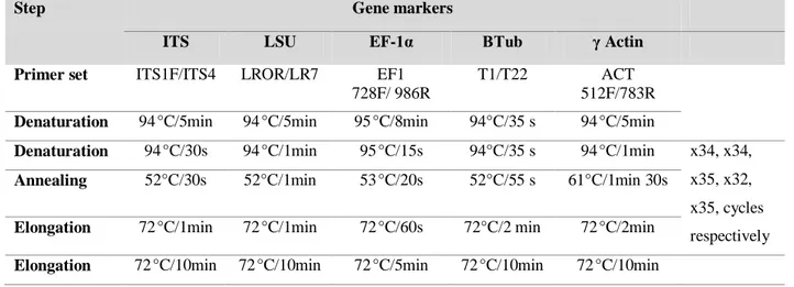
Table 5 Detailed PCR programs for the used marker genes amplification (ITS, LSU, EF-1α, Tub and Actin)
25 Table 6 Media used for large scale fermentation and cultivation period 30 Table 7 Fungal endophytes isolated from G. alypum and their closest hits
from BLAST research
43 Table 8 MIC values [µg/mL] of the crude extracts from small scale
fermentations of the screened fungi in various media
58-59 Table 9 LC-MS data of preussilides 1-6 and their physical properties 61 Table 10 MIC [µg/mL] values of the six preussilides against the tested
organisms.
62 Table 11 Cytotoxic effect (IC50) of preussilides A-F (1 - 6) against different
normal and cancer cell lines
65
Table 12 LC-MS data of three compounds of P. similis STMA 16219 and their physical properties
73
Table 13 MIC [µg/mL] values of P. similis STMA 16219 compounds 75 Table 14 LC-MS data of the four cytochalasins and their physical properties 78 Table 15 MIC [μg/mL] values of cytochalasin B and deoxaphomin against the
tested microorganisms
79 Table 16 Cytotoxic effect (IC50) of cytochalasin B and deoxaphomin against
different normal and cancer cell lines
79 Table 17 Suggested chemotypes for P. similis based on compounds isolated
from four strains in the current study
84 Table 18 LC-MS data of the twelve compounds and their physical properties 87 Table 19 MIC [μg/mL] values of compounds 14, 15, 17-25 against the tested
microorganisms
89 Table 20 Cytotoxic effect (IC50) of compounds 14, 15, 17-25 against two cancer
cell lines
Table 21 LC-MS data of the three known compounds and their physical properties
91
Table 22 MIC [µg/mL] values of compounds 26-28 92
Table 23 Cytotoxic effect (IC50) of compounds 26-28 against two cancer cell
lines
List of Publications
Noumeur S.R., Helaly S. E, Jansen R, Gereke M., Stradal T. E. B., Harzallah
D., Stadler M. 2017. Preussilides A–F, bicyclic polyketides from the endophytic
fungus Preussia similis with antiproliferative activity. Journal of natural
products 80 (5). 1531-1540.
Teponno R. B., Noumeur S. R., Helaly S. E, Hüttel S., Harzallah D., Stadler M.
2017. Furanones and anthranilic acid derivatives from the endophytic fungus
Table of Contents
Introduction………...…...…………...1
Literature Review
I. Endophytes I.1. Definition………..………...………...….…………...3I.2. Fungal endophytes.………..………...……….………4
I.3. Transmission………..………..……...………4
I.3.1. Vertical transmission………..………..4
I.3.2. Horizontal transmission………...………..……….……….….4
I.4. Colonisation of host plant by fungal endophytes…….……….…….………...………5
I.5. Adaptation………..……….5
I.6. Classes of fungal endophytes………..……...………….……….6
I.6.1. Clavicipitaceous fungal endophytes (Class 1)……..……….…...……….…….6
I.6.2. Non-Clavicipitaceous fungal endophytes…………..………...………..7
I.7. Balanced antagonism………...……...………...………..9
I.7.1. Plant- endophyte interactions………...………...…………...…………...10
I.7.2. Endophyte- competitors (endophytes, pathogens)……..………..…….……...10
I.8. Secondary metabolites production by endophytes………..………..…..……...12
I.8.1. Genus Preussia………..………....13
I.8.2. Genus Dendrothyrium………..………...………..15
I.8.3. Genus Chaetomium...………...……….16
II. Globularia alypum………...…….…...………...………..……….18
II.1. Taxonomy and botanical aspect of Globularia alypum……….……….18
Experimental Section
Chapter I: Material and methods
1. Plant material ..………...………...………..21
2. Isolation of the endophytic fungi………...………...………....………….21
3. Identification of fungal isolates ……….………..…….………..…22
3.1.Morphological studies………..……….... 22
3.1.1. Macroscopic features.……….………...………. 22
3.1.2. Microscopic features ....………..……..…...……... 23
3.1.3. Ultrastructure of ascospores by eletron microscopy……….…….….…23
3.2. Molecular studies………...……...………...23
3.2.1. DNA extraction………...………..………....23
3.2.2. Amplified regions used for the identification………..……….…. 24
3.2.3. Gel electrophoresis………...…………..…………...25
3.2.4. Purification of the PCR products and sequencing………...………….……25
3.3.5. Phylogenetic analysis………..…....…...26
4. Small scale fermentation and preparation of extracts from cultures………..………….……..…..27
4.1. Small scale fermentation………...…27
4.2. Extraction ………...……...….27
5. Generation of secondary Metabolite profiles of crude extracts………..…………28
6. Serial dilution assay………..………..…………...…28
7. Bioactive-guided fractionation of crude extracts………..……….29
8. Scale-up fermentation………...………..……..……….29
9. Isolation and purification of secondary metabolites………...………....30
10. Dual culture………..…...………….. 36
11. Structure elucidation ………....…………37
11.1. HRESIMS ………..………...…..……….37
11.2.NMR ………...……….………...37
11.3. Optical rotation and UV-spectra and CD spectroscopy……..………...….38
12. Biological assays of pure compounds………...………....….38
12.1. Antimicrobial activity ………...………..….…38
12.3. Cytotoxicity assay..……...………..………...39
12.4. Phytotoxicity test ...………...……….. 39
12.5. Nematicidal Activity Assay………..40
13. Cell biology experiments………...………..………..40
13.1. Effect on cells morphology………...………..…..…………....40
13.1.1. Immunofluorescence. ...……...………...………..………41
13.1.2. Flow cytometry ………..………...…...42
Chapter II: Results and discussion
1. Isolation and identification of fungal isolates………...…...…432. Small scale fermentation and screening for antimicrobial activity………...……...…57
3. Large scale fermentation, isolation and purification of bioactive metabolites…………..………..59
3.1. Compounds isolated from Preussia similis strains………..………....59
3.1.1. Compounds isolated from Preussia similis DSM 10466……….…...59
3.1.1.1. Fermentation and metabolites isolation...………...……..….59
3.1.1.2. Biological activity of preussilides……….………..……...61
3.1.1.3. Co-culture of P. similis with phytopathogen S. sclerotiorum………..66
3.1.1.4. Effects of preussilides on cell morphology………..……...68
3.2.2. Compounds isolated from Preussia similis STMA 16219………...73
3.1.2.1. Fermentation and metabolites isolation………...73
3.1.2.2. Biological activity……….………..…...…74
3.1.2.3. Dual culture...……….…..75
3.1.3. Compounds isolated from Preussia similis DSM 32328…….………..…..77
3.1.3.1. Fermentation and metabolites isolation………...…...77
3.1.3.1. Biological activity………...………...…78
3.1.3.2. Dual culture………...………...…....82
3.1.4. Chemotaxonomy of Preussia similis………...………....83
3.2. Compounds isolated from Dendrothyrium variisporum ..……….………85
3.2.1. Fermentation and metabolites isolation………..…..85
3.3. Compounds isolated from Chaetomium madrasense……..………..…………..90
3.3.1. Fermentation and metabolites isolation...………...………...90
3.3.2. Biological activity ………...…..………….91
Conclusion and perspectives……….…....94
References……….. 97 Appendices
Introduction
1 The need for new and beneficial therapeutic agents to cope with the health and environmental problems faced by society today is never ending (Li et al., 2015). The rising tide of bacteria resistance to antibiotic is one of the biggest issues to global health, due to the wide uses of antibiotics not only in human medicine but also in livestock and poultry farming in order to promote growth and prevent infection. The threat posed by the spread of antibiotic resistance is enhanced by an alarming decline in the discovery and development of new classes of antibiotic with new biologically active pharmacophores. In addition, the apparent increase in viral and fungal infections, the lack of anticancer agents and the search for alternatives to harmful synthetic pesticides are also among the problems that underscore the need of new effective drugs (Richter et al., 2016).
Fungi are a prolific source of secondary metabolites that may serve as leads for the development of novel badly needed new agents. In particular, certain ecological groups of fungi like the endophytic mycobiota have recently been proved to yield a plethora of novel metabolites exhibiting a variety of biological activities (Kusari et al., 2008). Endophytic fungi, which colonize their host plants without causing visible disease symptoms, can grow inter- or intracellularly, systemically or locally within their hosts. Up to date, no endophyte-free plant species has been reported (Arora and Ramawat, 2017). In fact, the endophytes are involved in multipartite interactions to maintain a balanced antagonism in planta, not only with their host plant, but also with other microbial competitors sharing the same habitat, both bacteria and fungi. Secondary metabolites are important factors for maintaining these equilibria. Through a long time communication, evolutionary adaptation of endophytes in plants to the complex environment enabled them to have a tremendous biosynthetic capability. The repertoire of their biosynthetic capacities over the co-evolutionary process as well as microbial biodiversity encompassing an arsenal and unique of chemical scaffolds, once isolated and characterized, may increase the chances to develop new pharmaceutical agents to prevent and cure human ailments (Aly et al., 2013; Schulz et al., 2015).
So far, many natural product researchers are attracted by the ubiquitous presence of this intriguing plant mycobiota and their ability in promoting plant growth as well as in improving plant defence systems in order to strive against abiotic and biotic stresses (Kusari et al., 2012; Schulz et al., 2015; Netzker et al., 2015; Deka et al., 2017).
Introduction
2 This thesis reports for the first time the diversity of fungal endophytes of the medicinal plant
Globularia alypum (Plantaginaceae, Scrophulariales), used in the Algerian traditional medicine as a
healing agent, such as hypoglycaemic agent, laxative, cholagogue, stomachic, purgative and sudorific (Boutiti et al., 2008). Furthermore, the isolated fungal endophytes were screened for their secondary metabolites production.
Aims of the thesis
The aims of the thesis can be divided into a taxonomic and a chemical part, and are as follows:
Isolation of several endophytic fungi inhabiting the Algerian plant Globuaria alypum collected from Ain Touta Batna (Algeria).
Identification of the fungal isolates by morphological description and by ITS sequencing. For some fungi, the phylogenetic position was further illustrated and discussed by using protein coding genes.
From our collection of endophytic fungi, some fungal strains were selected for further study (small-scale fermentations), based on a phylogenetic preselection in order to select the most promising fungal strains and search for novel and biologically active secondary metabolites.
HPLC/DAD/MS profiling and antimicrobial screening of extracts derived from small-scale fermentations.
Large-scale fermentations of the interesting strains were performed.
Isolation and structure elucidation of pure compounds.
Extensive biological screening of the identified compounds to explore their potential biological importance.
Literature review
3
I. Endophytes I.1. Definition
The term “endophyte” is derived from the Greek words “endon” meaning within, and “phyton” meaning plant, and originally introduced by de Bary in 1866 referred to any organism occurring within plant tissues, distinct from the epiphytes that live on plant surfaces (Schulz and Boyle, 2006). So far, the most accepted definition of endophyte is that of Petrini (1991) “all organisms inhabiting plant
organs that at some time in their life, can colonize internal plant tissues without causing apparent harm to the host” (Hyde and Soytong, 2008). They spend the whole or part of its life cycle colonizing
inter- and/or intracellularly (Figure 1), systemically or locally inside the healthy leaves, petioles, stems, twigs, bark, root, fruit, flower and seeds (Schroeckh et al., 2014). However, some endophytes remain as latent pathogens. This lifestyle as parasitism may occur either during host senescence or when the plant is under stress (Hyde and Soytong, 2008). The endophytes include both prokaryotic and eukaryotic microorgansims. The prokaryotic endophytes which are represented by bacteria and actinomycetes are often colonisers of vascular tissues of host plants. They play very important roles in the plant fitness such as nitrogen fixation. The eukaryotic endophytes are represented by fungi (Schulz and Boyle, 2006).
Literature review
4
I.2. Fungal endophytes
Endophytic fungi are a polyphyletic group of microoganisms that may present in almost all plants (Hodgson et al., 2014). Nowadays, it is a well-established fact that plants are hosts for many types of microbial endophytes residing in different tissues, no endophyte-free plant species has been reported (Sandhu et al., 2017). Also, many species of plants can harboured the same species of endophytes (Zabalgogeazcoa, 2008). The majority of endophytic fungi are members of Ascomycota and their anamorphs, only small community of the total number of fungal endophytes is represented by Basidiomycota (Hyde and Soytong, 2008; Martin et al., 2015; Das et al., 2017).
I.3. Transmission
The endophytes may originate from the outside environment; rhizosphere and phyllosphere. So far, there are two known transmission routes for fungal endophytes: vertical and horizontal transmission (Tintjer et al., 2008). In some cases the transmission takes place by mixed modes (Foster and Wenseleers, 2006).
I.3.1. Vertical transmission
The vertical transmission occurs from the parent to the offspring via fungal hyphae invading the host's seeds. The fungal endophytes being transmitted vertically are often mutualistic and referred to as “true” endophytes, since their reproductive success is completely dependent on host reproductivity. Such a way of transmission keeps the continuity of partnerships between plant and endophytes by ensuring accuracy transmission of the beneficial symbionts through generations (Tadych et al., 2014). This mode of transmission seems to be restricted to some asexual fungi in the family Claviceapataceae which inhabit grasses. The vertical transmission is limited only to asexual reproduction.
I.3.2. Horizontal transmission
Horizontally transmitted fungal endophytes are sexual and are transmitted by means of spores (mitotic asexual or meiotic sexual spores) or possibly hyphae. The spores can be disseminated by water or air movement and/or insect vectors. The sexual reproduction requires production of meiotic sexual spores and is therefore always horizontal (Kandel et al., 2017). Horizontal transmission is predited to enhance virulence whereas vertical transmission evolves benefic parters. The species of
Literature review
5 the genus Epichloe are the most studied fungi on this pheneomen. However, not all mutualists are mandatory transmitted vertically, and there are many examples of mutualistic horizontally transmitted symbioses (Schulz and Boyle, 2005; Foster and Wenseleers, 2006).
I.4. Colonisation of host plant by fungal endophytes
The fungal endophytes often originate from the soil. The roots are frequently the primary site for endophytic infection as the root exudates facilitate their attachment and consequently their entry into plant tissues. Eventually, other portal entries beyond roots are possibles, such as, the stems, leaves, flowers and seeds, however the endophytic growth have been found to be narrow (Schulz and Boyle, 2006; Andrade-Linares and Franken, 2013). In case of flowers and seeds colonisation, the endophytes are thereby transmitted vertically from the maternal endophyte community into the offspring (Arora and Ramawat, 2017). Up to now, it is not clear if fungal endophytes sense root exudates for colonization (Rodriguez et al., 2009). The endophytic Ascomycota penetrate the roots by different mechanisms. In addition to the adhesion tips, it was found that fungal endophytes grow around roots by forming a network and produce appressoria like-structure as well as swollen cells on hyphal tips. Afterwards, the runner hyphae penetrate through between the intercellular spaces. Later, each single hyphae grows along the longitudinal axis of the organ between epidermal and cortical cells, and intracellular colonization is visualized as formation of microsclerotia. Root colonisation can be both inter- and intra-cellular, the hyphae often forming intracellular coils. The fungal hyphae cross the cell wall to reach the intracellular space leaving a constriction at entry sites (Arora and Ramawat, 2017).
I.5. Adaptation
Endophytic communities inhabiting a particular plant host are frequently referred to host specificity (some authors use the terms host preference of host exclusivity). The latter is the result of adaptation success between the endophyte and the host. However, some fungi can be found as endophytes, but in reality, they are specific to another substrate, normally found growing on it. An example of these accidental opportunists might be coprophilous species, which are a type of saprobic fungi that grow on animal dung, are sometimes detected as endophytes (Schroeckh and Brakhage, 2014). Also, the insect associated fungus Daldinia hawksworthii, which is normally a symbiont with the willow woodwasp Xiphydria prolongata, was found as endophyte of Salix (Pažoutová et al., 2013).
Literature review
6
I.6. Classes of fungal endophytes
Endophytic fungi have been classified into two broad groups based on their phylogeny and life history traits, the clavicipitaceous and the non-clavicipitaceous fungal endophytes. Now, the fungal endophytes are classified as class 1 (clavicipitaceous endophyes), and three other classes (class 2, class 3 and class 4) representing the group of non-clavicipitaceous endophytes (Figure 2) (Table1) (Rodriguez et al., 2009).
Figure 2: Location of the different classes of endophytes (according to Rodriguez et al., 2009) (Kusari and Spiteller,
2012)
I.6.1. Clavicipitaceous fungal endophytes (Class 1)
The group of clavicipitaceous endophytes represents the class 1 of fungal endophytes that form a symbiotic relationship with a broad spectrum of grasses (especially the family of Poaceae). They are mostly obligate biotrophic ascomycetes of the family Clavicipitaceae that perennially and systemically colonise the intercellular spaces of leaf primordia, leaf sheaths and culms of vegetative tissue (Scott, 2001; Sánchez Márquez et al., 2012). Due to their lack of the biosynthetic capacity for the production of secondary metabolites, grasses cohabitate these fungi which are known as a good producers of a set of secondary metabolites. The advantages of this grasses-endophytes association appear practically in the higher performance of bioprotection by inducing eco-physiological changes that make plants more tolerant to different biotic and abiotic stresses (Kuldau and Bacon, 2008).
Literature review
7 Therefore, the infected grasses have been found to be very vigorous especially in enhancing plant biomass, drought tolerance, protection against herbivore and insects. Such a defensive mutualistic association is very important to the ecological fitness and species diversity of grasses (Sánchez Márquez et al., 2012; Kusari and Spiteller, 2012). On the other hand, mutualistic benefits for endophytes may involve access to nutrients from the host apoplast, abiotic and biotic stress avoidance and dissemination by seed (Scott, 2001). The genus Epichloë is the most studied group of these associated grasses endophytes. These grass-fungal endophyte symbioses have been showed a range from pathogenic to mutualistic. The species from the genus Epichloë are often considered to be pathogenic to host grasses as they may induce abortion of plant reproductive structures when reaching sexual reproduction state and horizontal spreading may cause partial or complete sterilization of the hosts due to the production of a fungal stroma on the flowering culms (choke disease) (Gundel et al., 2012). In mutualistic relationships, the Epichloë endophyte grows systematically within its host including the developing seeds, and it is completely dependent of the survival and growth of the grass host plant for its own growth (Scott, 2001; Schaechter, 2012).
I.6.2. Non-Clavicipitaceous fungal endophytes
The non-clavicipitaceous endophytes are polyphyletic fungi, often poorly defined or with unknown ecological roles. They traditionally have been treated as a single functional group forming the class 2 of fungal endophytes until Rodriguez et al. (2009), have differentiated them into three functional classes (class 2, class 3 and class 4) based on host colonization patterns, mechanism of transmission between host generations, in planta biodiversity levels, and ecological function (Table 1).
a. Class 2
The fungal endophytes belonging to the class 2 may grow in the aerial and ground parts of the plant host. Most of their species are members of Ascomycota (Pezizomycotina) with minority being Basidiomycota (Agaricomycotina and Pucciniomycotina). Transmission of fungal endophytes of class 2 can be vertical or horizontal.
b. Class 3
The occurrence of Class 3 fungal endophytes is strongly related to aerial tissues and they are transmitted horizontally. Thus, their entry portal to the host plant is always the aerial part, where their
Literature review
8 growth will be limited or they spread to the lower part. Due to the above-tissues dependence, these fungi induce the formation of highly localized infections.
These fungi are found within photosynthetic and herbaceous tissues in addition to flowers and fruits, as well as in asymptomatic wood and inner bark. The biodiversity of class 3 endophytes is extremely high in planta. They include the endophytic fungi associated with leaves of tropical trees, the highly diverse associates of above-ground tissues of nonvascular plants, seedless vascular plants, conifers, and woody and herbaceous angiosperms in biomes ranging from tropical forests to boreal and Arctic/Antarctic communities.
c. Class 4
The class 4 endophytes are distinguished from the other two classes of non clavicipitaceous fungi based on their exceptional nature of hyphae. The latter are septate and dark in color due to the presence of melanin. They are known as dark septate endophytes (DSE), which colonize only the roots of the plant, extensively. Theit mode of transmission is exclusively horizonal. In general, Class 4 endophytes are primarily ascomycetous fungi that are conidial or sterile and that form melanized structures such as inter- and intracellular hyphae and microsclerotia in the roots (Rodriguez et al., 2009).
Table 1: Symbiotic criteria used to characterize fungal endophytic classes criteria (Rodriguez et al., 2009)
Clavicipitaceous Non Clavicipitaceous
Criteria Class 1 Class 2 Class 3 Class 4
Host range Narrow Broad Broad Broad
Tissue(s) colonized Shoot and rhizome Shoot, root and rhizome
Shoot Root
In planta colonization Extensive Extensive Limited Extensive
In planta biodiversity Low Low High Unknown
Transmission Vertical and
horizontal
Vertical and horizontal
horizontal horizontal
Fitness benefits* NHA NHA and HA NHA NHA
*Nonhabitat-adapted (NHA) benefits such as drought tolerance and growth enhancement are common among endophytes regardless of the habitat of origin. Habitat-adapted (HA) benefits result from habitat-specific selective pressures such as pH, temperature and salinity.
Literature review
9
I.7. Balanced antagonism
The theory predicts that endophytes produce phytotoxic metabolites toward their host plant and the latter can reacts by inducing its defence responses. Questions have been raised about the asymptomatically growth and survive of endophytes inside the living tissues of the host plant and how can these two partners succeed in the establishment and maintenance of this symbiosis relationship. Some authors have mainly been interested in questions concerning the mysterious endophytes-plant interactions, Schulz et al. (1999, 2015) have hypothesized that the asymptomatically relashionship endophye-plant is a result of a balanced antogonsim in which virulence factors and defence response are balanced. However, the balanced antagonism is a transitory period and it can be destabilized. In the unbalanced antagonism, either the fungus is prished or the plant becomes diseased. Even so, the switch between a balanced and unbalanced interaction depends of the status of two partners. In fact, the plant defense and endophyte virulence can be modified by environmental factors as well as senescence stressors. In addition to their escape to host defences systems, fungal endophytes must in the same time avoid the toxins of other microbial inhabitants, including pathogens and other endophytes competitors. Therefore, they must be engaged in multipartite interactions (Schulz et al., 1999; Kusari et al., 2012; Schulz et al., 2015). The equilibrium established between fungal endophytes and their host as well as other competitors is shown in figure 3.
Literature review
10 The interactions of endophytes with their host plants and with other endophytes and pathogens microorganisms (both fungi and bacteria) are summarized as follows:
I. 7.1. Plant- endophyte interactions
Mutualistic interactions involving fungi and bacteria that endophytically colonise plant roots, benefit the microbial partner with a reliable supply of nutrients as well as protection from environmental stresses. The plant, in turn, reaps the benefit of nutrient uptake and protection against biotic (e.g. pathogens, herbivores) and abiotic (e.g. drought tolerance) stress factors. In fact, the fungal endophytes could confer deterrence to their host plants in different ways. Many endophytes due their ability to produce antibacterial and antimycotic metabolites, can suppress or reduce severity of plant invasion by pathogens sharing the same habitat (Arora and Ramawat, 2017). Very likely, the synergetic effect of the total of antimicrobial compounds secreted by endophytic microbiota could enhance the plant protection (Kusari et al., 2012).
Even through, the intimate and prolong relationship between plant and microbe observed as a genetic exchange among plant and microbes to transfer information inherent among both organisms. This co-evolution of endophytes and their host plants is believed to shape natural product patterns of endophytic fungi, which often contribute in multiple ways to endophyte- host communication as well as host fitness and adaptation to environmental challenges. However, little is known about this relashionship (Sandhu et al., 2017).
I.7.2. Endophyte- competitors (endophytes, pathogens)
One living tissue of a host plant harboring a single type of endophytes is an exotic exception. Usualy, the plant is inhabited by diverse taxa of endophytic microorganisms including bacteria and fungi. Thus, the endophytes must interact with their neighbours sharing the same habitat. The diverse intra- and interspecies cross-talk is accomplished by a huge number of metabolites, most of them belong to the group of secondary metabolites. This cross-talk often involve secretion of antibacterial and antifungal metabolites against competitors in order to maintain balanced antagonisms. It is mostly like chemical warfare in which each endophytes secrete toxic metabolite to the respective partner leading to a reciprocal antagonisms to ensure their maintenance in the plant host. Also, the interaction between the inhabitants of the same plant does not occur only metabolically by production of inhibitory metabolites, but also occurs in a chemical communication between symbionts partners by
Literature review
11 secreting signal molecules which might have a crucial role in environmental recognition (Kusari et
al., 2012). This view is supported by Schulz et al. (2015), who found that in co-culture experiments,
the microorganisms secrete metabolites even before they make intimate physically contact, suggesting that a number of signal molecules might be involved in order to inform the partners of the presence of each other before they engage in the transfer of molecular and genetic information that include many mechanisms and classes of molecules. On the other hand, these interactions could trigger silent biosynthetic pathways. Each compound released by microorganism could has an impact on the metabolic profiles of other microorganisms sharing the same ecological niche. Therefore, their mutual interplay could lead to a significant and chemical diversity in the natural compounds released (Figure 4). Such a chemical diversity is a results of the expression of genes clusters previously unexpressed in individual axenic culture under standard laboratory growth conditions. In this respect, many authors have mainly highlighted the importance of the co-cultivation to induce silent genes clusters under standard laboratory conditions or can allow also a tremendously enhanced production of already known natural compounds (Scherlach and Hertweck, 2009; Schroeckh and Brakhage, 2014; Ola et al, 2013; Netzker et al., 2015; Wakefield et al., 2017). Also, another example of fungus-bacterial endosymbiont crosstalk was found by Partida-Martinez and Hertweck (2005) reporting that the phytopathogenic fungus Rhizopus microsporus as causing rice seedling blight was found to not be responsible for the production of rhizoxin as thought previously, but rather an endosymbiotic bacterium belonging to the genus Burkholderia, occuping the intracellular space of Rhizopus (Partida-Martinez and Hertweck, 2005). Interestingly, the endosymbiont not only produces the phytotoxin but also controls the differentiation and sporulation of the fungal host (Partida-Martinez et al., 2007). This unexpected tripartite plant–symbionts revealed that there are many complex networks of chemical communications between plant and endophytes, mediating by a diversity of chemical molecules. However, little is known about these hidden intriguing mechanisms.
There are evidence now, that using the traditional approach by using individual culture to discover natural products is often frustrating and usually the producer organism is qualified as incompetent. Now, with the co-cultivation approach a valuable insights are provided and the respective microoganisms could become competent just their silent biosynthetic metabolisms need to be encrypted by exposing them to the appropriate conditions (Kusari et al., 2012; Wakefield et al., 2017).
Literature review
12
Figure 4: Schematic representation of endophyte-endophyte interspecies crosstalk (Kusari et al., 2012)
(A) Fungus-fungus crosstalk is illustrated, (B) Fungus-bacterial endosymbiont crosstalk is demonstrated, (C) Fungus-bacteria crosstalk is presented.
I.8. Secondary metabolites production by endophytes
Fungi can produce a large array of low-molecular-mass organic compounds called secondary metabolites. In contrast to primary metabolites, secondary metabolites are usualy regarded as non-essential for the survival of living organisms, while their role is quiet versatile. They often confer competitive outcomes to their producing organisms usualy as mediators in interactions with others organisms including plants, animals and microrganisms that may indirectly influenced their growth and development by increasing their fitness (Bills et al., 2013; Netzker et al., 2015). Up to date, many classes of secondary metabolism biosynthesis pathways have been characterized in fungi, especially filamentous Ascomycota which their genomes encode a broad spectrum of enzymes that synthesize secondary metabolites, including nonribosomal peptide synthetases, polyketide synthases and terpene synthases (Bills et al., 2013). Endophytic fungi are of biotechnological interest due to their potential as an excellent source of various bioactive compounds (chemically and in terms of their biological activities) that have evolved during their symbiotic plant relationship with plants. The quality and quantity of secondary metabolite produced is related to each fungal species. This specificity, which imparts a species-specific chemical marker, can be used as additional characters to distinguish closely related fungal species (chemotaxonomy).
Literature review
13
I.8.1. Genus Preussia
The genus Preussia is a member of the family of Sporormiaceae in the order Pleosporales, subclass Pleosporomycetidae, class Dothideomycetes, subphylum Pezizomycotina, and phylum Ascomycota. Its species are most commonly found on various types of animal dung, but they can also be isolated from plant debris, soil and wood (Arenal et al., 2005; Porras-alfaro and Sinsabaugh, 2011), or as endophytes in plants (Arenal et al., 2007; Herrera et al., 2010; Mapperson et al., 2014). This genus was proposed by Fuckel (1866) with Preussia funiculata (Preuss) Fuckel as type species (Abdullah et al., 1999). The genus morphologically most closely related to Preussia is Sporormiella Ellis & Everh. The two genera have been mostly treated as congeneric because their salient discriminatory morphological features are known to depend on culture conditions (Kruys and Wedin, 2009). Moreover, recent molecular phylogenetic studies have revealed that Preussia and Sporormiella do not form monophyletic clades, but rather their DNA sequences become intermingled in phylogenetic trees (Arenal et al., 2005; Kruys and Wedin 2009; Kruys, 2015; Mapperson et al., 2014). In the current taxonomy, the genus Preussia includes Sporormiella and contains more than 100 species (Index fungorum, 2018). The species are morphologically characterized by brown to black pseudothecia, bitunicate asci, and dark brown, septate ascospores. A gelatinous sheath covers each ascospore and in general, each cell has a germ slit (Khan and Cain, 1979; Arenal et al., 2007). Even though, Preussia species are common coprophilous fungi, many authors have isolated them from different plant species. The spores of coprophilous species stick easily to the plant surface because they are often surrounded by gelatinous sheath. When a plant is foraged by an herbivore, the spores pass in the herbivore gastrointernial tract and they are ending up in the new dung pile, the spores germinate and produce new fruit bodies. Again the spores attach to the surface plant and the cycle restarts. However, much uncertainty still exists about the life cycle of coprophilous fungi. One hypothesis to explain their endophytic state, might be that the spores remain on the surface of plant as epiphytes and due to their resistance to surface-sterilants, they could escape to the surface sterilization of plant tissues and are actually isolation artefacts, rather than the endophytes (Newcombe et al., 2016).
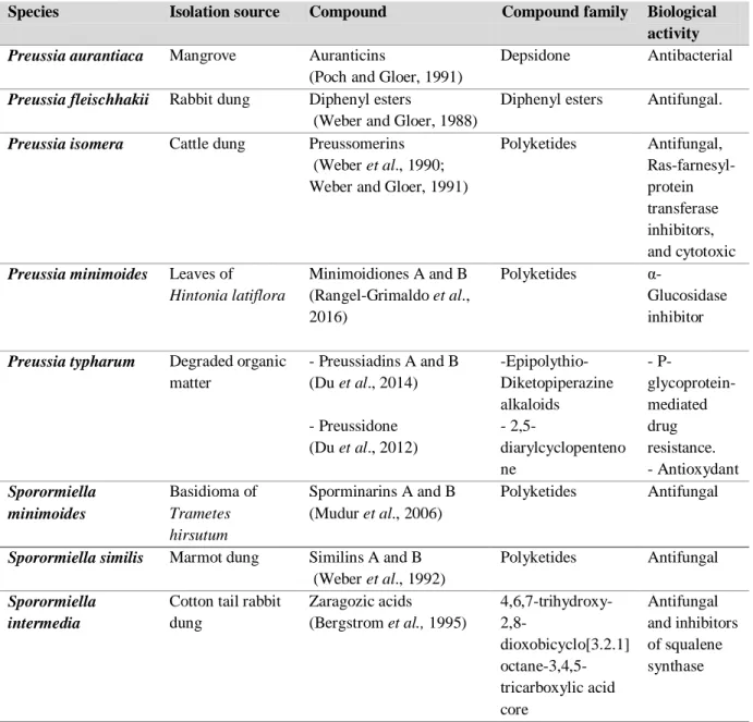
The conglomerate Preussia/Sporormiella has been known as potent producers of bioactive compounds, in particular polyketides (Du et al., 2012, 2014; Rangel-Grimaldo et al., 2016). Among them, Preussia similis (Sporormiella similis) (Khan & Cain) Arenal (Khan and Cain, 1979; Arenal et
Literature review
14 al., 2004) was found to be a rich source of antifungal compounds, e.g. preussomerin A (Weber and
Gloer, 1991), or similin A and B ( Weber et al., 1992) (Table 2 and Figure 5).
More recently, Gonzalez-Menendez et al. (2017), have reviewed the diversity of Preussia species and characterized for the first time the occurring chemotypes. In fact, thirty-one species have been divided into eleven chemotypes based on a combined data of morphological and phylogenetic analysis together with the chemical profile of the corresponding strains. The latter chemotaxonomic work did not attributed Preussia similis to any of these chemotypes.
Table 2. Compounds isolated from the conglomerate Preussia/Sporormiella
Species Isolation source Compound Compound family Biological
activity
Preussia aurantiaca Mangrove Auranticins
(Poch and Gloer, 1991)
Depsidone Antibacterial
Preussia fleischhakii Rabbit dung Diphenyl esters
(Weber and Gloer, 1988)
Diphenyl esters Antifungal.
Preussia isomera Cattle dung Preussomerins (Weber et al., 1990; Weber and Gloer, 1991)
Polyketides Antifungal, Ras-farnesyl-protein transferase inhibitors, and cytotoxic
Preussia minimoides Leaves of
Hintonia latiflora Minimoidiones A and B (Rangel-Grimaldo et al., 2016) Polyketides α-Glucosidase inhibitor
Preussia typharum Degraded organic matter - Preussiadins A and B (Du et al., 2014) - Preussidone (Du et al., 2012) -Epipolythio- Diketopiperazine alkaloids - 2,5-diarylcyclopenteno ne - P- glycoprotein-mediated drug resistance. - Antioxydant Sporormiella minimoides Basidioma of Trametes hirsutum Sporminarins A and B (Mudur et al., 2006) Polyketides Antifungal
Sporormiella similis Marmot dung Similins A and B (Weber et al., 1992)
Polyketides Antifungal
Sporormiella intermedia
Cotton tail rabbit dung Zaragozic acids (Bergstrom et al., 1995) 4,6,7-trihydroxy- 2,8-dioxobicyclo[3.2.1] octane-3,4,5-tricarboxylic acid core Antifungal and inhibitors of squalene synthase
Literature review
15
Figure 5: Chemical structures of secondary metabolites isolated from the conglomerate Preussia/Sporormiella as
indicated in Table 2 I.8.2. Genus Dendrothyrium
Dendrothyrium Verkley, Göker & Stielow, has been described recently as a new coelomycete
genus belonging to the family Montagnulaceae (Pleosporales, Pleosporomycetidae, Dothideomycetes, Pezizomycotina and Ascomycota). Dendrothyrium species were previously named as Coniothyrium spp. and their taxonomy was re-assessed based on analyses of concatenated ITS of the nrDNA operon,


![Table 10: MIC [µg/mL] values of the six preussilides against the tested organisms.](https://thumb-eu.123doks.com/thumbv2/123doknet/3433019.100277/85.918.124.818.342.968/table-mic-µg-ml-values-preussilides-tested-organisms.webp)