Université de Montréal
Novel surface-tethered estrogen polymeric platforms in
cardiovascular regenerative medicine
par Baowen Qi
Faculté de Pharmacie
Thèse présentée à la Faculté des études supérieures en vue de l’obtention du grade de Philosophiae Doctor (Ph.D.)
en Sciences Pharmaceutiques option Chimie Médicinale
July, 2015
Université de Montréal
Faculté des études supérieures et postdoctorales Cette thèse intitulée:
Novel surface-tethered estrogen polymeric platforms in
cardiovascular regenerative medicine
présentée par : Baowen Qi
a été évaluée par un jury composé des personnes suivantes :
Dr. Gaëlle Roullin, Président-rapporteur Dr. Françoise M. Winnik, Directeur Dr. Jean-François Tanguay, Co-directeur
Dr. Anna Ritcey, Examinateur externe Dr. Jean-François Masson, Membre du jury
i
Résumé
L’estradiol (E2) est une hormone femelle qui joue un rôle essentiel, à la fois dans la régulation et dans la détermination de certaines conditions physiologiques in vivo, telle que la différenciation et la prolifération cellulaire. Lorsque l’E2 est donné en supplément, par exemple dans le cas de thérapie hormonale, deux effets sont observés, un effet génomique et un effet non-génomique, de par son interaction avec les récepteurs à œstrogène du noyau ou de la membrane cellulaire, respectivement. L’effet non-génomique est plus difficile à étudier biologiquement parce que l’effet se produit sur une échelle de temps extrêmement courte et à cause de la nature hydrophobe de l’E2 qui réduit sa biodisponibilité et donc son accessibilité aux cellules cibles. C’est pourquoi il est nécessaire de développer des systèmes d’administration de l’E2 qui permettent de n’étudier que l’effet non-génomique de l’œstrogène. Une des stratégies employée consiste à greffer l’E2 à des macromolécules hydrophiles, comme de l’albumine de sérum bovin (BSA) ou des dendrimères de type poly(amido)amine, permettant de maintenir l’interaction de l’E2 avec les récepteurs d’œstrogène de la membrane cellulaire et d’éviter la pénétration de l’E2 dans le noyau des cellules. Toutefois, ces systèmes macromolécules-E2 sont critiquables car ils sont peu stables et l’E2 peut se retrouver sous forme libre, ce qui affecte sa localisation cellulaire. L’objectif de cette thèse est donc de développer de nouvelles plateformes fonctionnalisées avec de l’E2 en utilisant les approches de synthèses ascendantes et descendantes. Le but de ces plateformes est de permettre d’étudier le mécanisme de l’effet non-génomique de l’E2, ainsi que d’explorer des applications potentielles dans le domaine biomédical.
L’approche ascendante est basée sur un ligand d’E2 activé, l’acide 17,α-éthinylestradiol-benzoïque, attaché de façon covalente à un polymère de chitosan avec des substitutions de phosphorylcholine (CH-PC-E2). L’estradiol est sous forme de pro-drogue attachée au polymère qui s’auto-assembler pour former un film. L’effet biologique de la composition chimique du film de chitosan-phosphorylcholine a été étudié sur des cellules endothéliales. Les films de compositions chimiques différentes ont préalablement été caractérisés de façon physicochimique. La topographie de la surface, la charge de surface, ainsi que la rhéologie des différents films contenant 15, 25, ou 40% molaires de
ii
phosphorylcholine, ont été étudiés par microscopie à force atomique (AFM), potentiel zêta, résonance plasmonique de surface et par microbalance à cristal de quartz avec dissipation (QCM-D). Les résultats de QCM-D ont montré que plus la part molaire en phosphorylcholine est grande moins il y a de fibrinogène qui s’adsorbe sur le film de CH-PC. Des cellules humaines de veine ombilicale (HUVECs) cultivées sur des films de CH-PC25 et de CH-PC40 forment des amas cellulaire appelés sphéroïdes au bout de 4 jours, alors que ce n’est pas le cas lorsque ces cellules sont cultivées sur des films de CH-PC15. L’attachement de l’estradiol au polymère a été caractérisé par plusieurs techniques, telles que la résonance magnétique nucléaire de proton (1H NMR), la spectroscopie infrarouge avec transformée de Fourier à réfraction totale atténuée (FTIR-ATR) et la spectroscopie UV-visible. La nature hydrogel des films (sa capacité à retenir l’eau) ainsi que l’interaction des films avec des récepteurs à E2, ont été étudiés par la QCM-D. Des études d’imagerie cellulaires utilisant du diacétate de diaminofluoresceine-FM ont révélé que les films hydrogels de CH-PC-E2 stimulent la production d’oxyde nitrique par les cellules endothéliales, qui joue un rôle protecteur pour le système cardiovasculaire. L’ensemble de ces études met en valeur les rôles différents et les applications potentielles qu’ont les films de type CH-PC-E2 et CH-PC dans le cadre de la médecine cardiovasculaire régénérative. L’approche descendante est basée sur l’attachement de façon covalente d’E2 sur des ilots d’or de 2 m disposés en rangées et espacés par 12 m sur un substrat en verre. Les ilots ont été préparés par photolithographie. La surface du verre a quant à elle été modifiée à l’aide d’un tripeptide cyclique, le cRGD, favorisant l’adhésion cellulaire. L’attachement d’E2 sur les surfaces d’or a été suivi et confirmé par les techniques de SPR et de QCM-D. Des études d’ELISA ont montré une augmentation significative du niveau de phosphorylation de la kinase ERK (marqueur important de l’effet non-génomique) après 1 heure d’exposition des cellules endothéliales aux motifs alternant l’E2 et le cRGD. Par contre lorsque des cellules cancéreuses sont déposées sur les surfaces présentant des motifs d’E2, ces cellules ne croissent pas, ce qui suggère que l’E2 n’exerce pas d’effet génomique. Les résultats de l’approche descendante montrent le potentiel des surfaces présentant des motifs d’E2 pour l’étude des effets non-génomiques de l’E2 dans un modèle in vitro.
Mots-clés : estradiol, effet non-génomique, approches ascendantes et descendantes, substrats
en micromotifs, chitosan, phosphorylcholine, oxyde nitrique, médecine cardiovasculaire régénérative, kinase régulée par signal extracellulaire
iii
Abstract
Estradiol (E2) is an essential female hormone in the regulation and determination of various physiological conditions in vivo, such as cell proliferation and differentiation. When supplementing exogenous E2 as a clinical strategy for hormone therapy, it generates genomic and non-genomic effect simultaneously via binding to the estrogen receptors in the cell nucleus or membrane site. Compared to the genomic effect, it is quite difficult to monitor the E2-induced non-genomic biological behavior because this effect occurs in extremely transient time scale and the bioavailability and accessibility of E2 to target cells is very low due to the hydrophobic nature of E2. As a result, it is indispensable to develop E2 delivery systems to specifically understand estrogenic non-genomic nature. One of strategies is to graft E2 to the hydrophilic macromolecules, e.g. bovine serum albumin (BSA) or poly(amido)amine dendrimer, to maintain E2 interacting with membrane estrogen receptors instead of penetrating into the cell nucleus. However, the instability of those E2-macromolecules systems, either containing free E2 leaching or discrepancies of cellular localizations, led to controversies. Herein, the objective of present thesis is to develop novel E2-functionlized platforms by the principle of bottom-up and top-down approaches for understanding the mechanism of estrogenic non-genomic effect, and further, to explore their potential applications in the biomedicine.
As a bottom-up approach, an activated E2 ligand, 17α-ethinylestradiol-benzoic acid was covalently conjugated onto a phosphorylcholine substituted chitosan polymer (CH-PC-E2) as a prodrug strategy for the fabrication of self-assembled films. Through a series of combined physicochemical and cellular investigations, the relationship between various chemical compositions of chitosan-phosphorylcholine (CH-PC) films and cellular responses was also evaluated. Based on atomic force microscopy (AFM) examination, zeta-potential measurements, surface plasmon resonance (SPR), and quartz crystal microbalance with dissipation (QCM-D) measurements, surface topography, charge, and rheology of CH-PC films with 15, 25, and 40 mol% PC contents were characterized. Moreover, QCM-D measurements indicated that the amount of fibrinogen adsorbed on CH-PC films decreased significantly with increasing PC content. Finally, it was also showed that human umbilical vein endothelial cells (HUVECs) form spheroids on CH-PC25 and CH-PC40 films, but not on
iv
CH-PC15 films cultured over 4 days. In addition, the CH-PC-E2 polymer conjugates were prepared and characterized by several techniques, such as 1H nuclear magnetic resonance(1H NMR), Fourier transformed infrared-attenuated total refraction (FTIR-ATR) and UV/Vis spectra measurements. The hydrogel nature of CH-PC-E2 film as well as its interactions to estrogen receptors was further extensively investigated by QCM-D study. In the cellular study, CH-PC-E2 hydrogel films can significantly stimulate the production of nitric oxide, a protective molecule in the cardiovascular system, in the endothelial cells by a diaminofluorescein-FM diacetate imaging study. The studies above demonstrated the different roles and potential applications of CH-PC-E2 and CH-PC surfaces in the cardiovascular regenerative medicine. As a top-down approach, micropatterned substrates were used for E2 functionalization, which were prepared by photolithography via aligning ~ 2 m in diameter gold arrays onto a glass substrate. After that, a cell adhesive peptide, cyclic RGD was introduced to the glass surface in order to induce the attachment of cells. Meanwhile, estradiol was covalently immobilized on the gold surface and the process was monitored and validated by combining SPR and QCM-D studies. In the micropatterned substrate-coupled cell ELISA study, a phosphorylation level of extracellular signal-regulated kinase (ERK), which is an important non-genomic marker, was significantly elevated by this E2-functionalized micropatterned surface after 1 hour incubation. Furthermore, E2-functionalized micropatterned substrate didn’t proliferate cancer cells indicating the absence of genomic effect stimulation. Based on these results, our E2-functionalized micropatterned substrates can function as an in vitro model for the elucidation of estrogenic non-genomic behaviors.
Keywords : estradiol, non-genomic effect, top-down and bottom-up approaches,
micropatterned substrates, chitosan, phosphoylcholine, nitric oxide, cardiovascular regenerative medicine, extracellular signal-regulated kinase
v
Table of Contents
Résumé ... i Abstract ... i Table of Contents ... v List of figures ... xList of tables ... xiv
List of abbreviations ... i
Authors’ contributions ... iii
Acknowledgments ... iv
CHAPTER ONE ... 1
Introduction and Literature Review ... 1
1.1 Estrogen in cardiovascular medicine ... 1
1.1.1 Introduction to coronary artery heart disease ... 1
1.1.2 The anatomy and physiology of blood vessels ... 2
1.1.3 Estrogen effects on the cardiovascular system ... 3
1.1.4 Estrogen receptors and classical genomic effect ... 3
1.1.5 Estrogen non-genomic effect ... 4
1.1.6 Tools for estrogen non-genomic effect study ... 6
1.2 Polymeric biomaterials in cardiovascular medicine ... 7
1.2.1 Fundamental properties of polymeric biomaterials ... 7
1.2.2 Mechanical properties ... 7
1.2.3 Biodegradability ... 9
1.2.4 Cellular interactions ... 9
1.3 Non-fouling polymeric biomaterials in cardiovascular regenerative medicine ... 11
1.3.1 Overview of non-fouling polymeric biomaterials ... 11
1.3.2 Non-fouling polymeric biomaterials in cardiovascular regenerative medicine ... 16
1.4 Methods and Techniques ... 18
1.4.1 Surface plasmon resonance technology (SPR) ... 19
vi
1.4.3 Photolithgraphy ... 20
1.5 Project hypothesis, thesis rationale and research objectives ... 21
1.5.1 Project hypothesis ... 21
1.5.2 Thesis rationale ... 24
1.5.3 Research objectives ... 25
1.6 References ... 26
CHAPTER TWO ... 50
Phosphorylcholine-modified chitosan films as effective promoters of cell aggregation: correlation between the films properties and cellular response ... 50
2.1 Abstract ... 51
2.2 Author Keywords ... 51
2.3 Introduction ... 52
2.4Experimental Section ... 55
2.4.1 Materials ... 55
2.4.2 Synthesis and characterization of phosphorylcholine-chitosans (CH-PC) ... 55
2.4.3 Preparation of CH-PC solutions ... 56
2.4.4 Dynamic light scattering (DLS) and zeta potential measurements of CH-PC solutions. ... 56
2.4.5 Dynamic AFM measurements ... 56
2.4.6 Zeta potential measurements of CH-PC films ... 57
2.4.7 Surface plasmon resonance (SPR) measurements ... 57
2.4.8 Quartz crystal microbalance with dissipation (QCM-D) measurements ... 58
2.4.9 Ellipsometry measurements ... 59
2.4.10 Cell culture ... 59
2.4.11 Cell observation by optical microscopy ... 60
2.5Results and discussion... 60
2.5.1 Morphology and charge of the CH-PC Films ... 60
2.5.2 Hydration and mechanical properties of CH-PC films ... 63
2.5.3 Fibrinogen adsorption on CH-PC films. ... 66
2.5.4 Aggregation of human umbilical vein endothelial cells (HUVECs) and MCF-7 cells on CH-PC films... 68
vii
2.6Conclusions ... 70
2.7Acknowledgements ... 72
2.8References ... 72
2.9Appendix A. Supporting information ... 77
CHAPTER THREE ... 86
Synthesis, characterization and biological study of estrogen grafted chitosan phosphorylcholine polymer conjugates ... 86
3.1 Abstract ... 87
3.2 Author Keywords ... 87
3.3 Introduction ... 88
3.4Experimental section ... 89
3.4.1 Materials ... 89
3.4.2 Preparation of PC-substituted chitosan (CH-PC) ... 90
3.4.3 Conjugation 17α-ethinylestradiol to 4-iodobenzoic acid ... 90
3.4.4 Preparation and characterization of CH-PC-E2 conjugates ... 91
3.4.5 Dynamic light scattering (DLS) measurements ... 91
3.4.6 Spectroscopic ellipsometry analysis ... 92
3.4.7 Quartz crystal microbalance with dissipation (QCM-D) studies ... 92
3.4.8 Cell culture ... 94
3.4.9 DAF-FM fluorescence imaging study ... 94
3.4.10 Statistics ... 95
3.5Results and discussion... 96
3.5.1 Characterization of CH-PC-E2 conjugates ... 96
3.5.2 Viscoelastic properties of CH-PC-E2 and CH-PC formulated thin films... 98
3.5.3 The binding experiment of ER-α to CH-PC-E2 hydrogel film ... 100
3.5.4 CH-PC-E2 hydrogel film promotes intracellular nitric oxide (NO) production .. 102
3.6Conclusions ... 104
3.7Acknowledgements ... 104
3.8References ... 105
viii
CHAPTER FOUR ... 115
Estradiol-tethered micropatterned surfaces for the study of estrogenic non-genomic pathways ... 115
4.1 Abstract ... 116
4.2 Authors keywords ... 116
4.3 Introduction ... 117
4.4 Materials and methods ... 119
4.4.1 Materials ... 119
4.4.2 Cell culture ... 119
4.4.3 Glass surface modification and characterization ... 120
4.4.4 Gold surface modification monitored by XPS, SPR and QCM ... 120
4.4.5 Fabrication and modification of micropatterned surfaces ... 122
4.4.6 Detection of ERK phosphorylation by cell ELISA ... 123
4.4.7 MCF-7 cell proliferation assay ... 124
4.5 Results and discussions ... 124
4.5.1 Design of micropatterned substrates ... 124
4.5.2 Immobilization of cRGD onto glass substrates ... 126
4.5.3 XPS characterization of E2-PEG2k modified gold surfaces. ... 127
4.5.4 SPR evaluation of the interaction of ER-α with E2-modified gold substrates .... 128
4.5.5 QCM-D studies of the adsorption of ER-α on gold substrates modified with E2-PEG2k ... 130
4.5.6 Biological activity of E2-modified micropatterned substrates ... 132
4.6 Conclusions ... 134
4.7 Acknowledgements ... 134
4.8 References ... 135
4.9 Appendix C. Supporting information ... 140
CHAPTER FIVE ... 144
GENERAL DISCUSSION ... 144
5.1 Physical and biological properties of CH-PC polymer thin films ... 145
5.2 Synthesis, characterization and biological studies of CH-PC-E2 self-assembled films in the cardiovascular regenerative medicine ... 147
ix
5.3 E2-tethered micropatterned substrates selectively activate non-genomic signal pathways
... 149
5.4 References ... 150
CHAPTER SIX ... 154
CONCLUSIONS AND FUTURE WORKS ... 154
6.1 Conclusions ... 155
6.2 Future perspectives ... 155
x
List of figures
Figure 1.1 Schematic representation of the genomic and nongenomic estrogen pathways. For the genomic pathway, E2 (red triangle) crosses the cell membrane and binds to the estrogen receptor (ER) in the cell nucleus, inducing gene transcription. For the non-genomic pathway, E2 interacts with ER or ER/GPR30 on the cell membrane, which activates several transduction pathways. ... 4 Figure 1.2 Chemical structures of zwitterionic-based materials. (A) phosphorylcholine (PC),
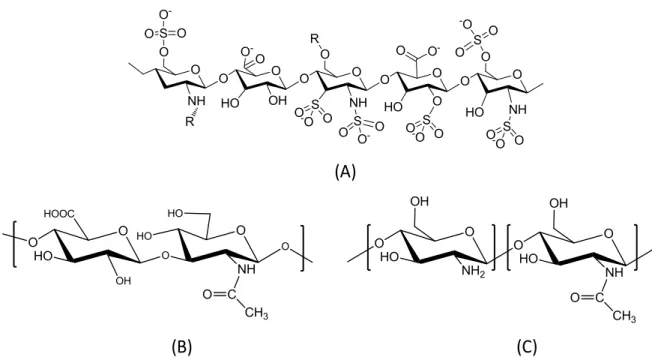
(B) sulfobetaine (SB), and (C) carboxybetaine (CB). ... 12 Figure 1.3 Chemical structures of natural polysaccharides. (A) heparin, (B) chitosan, and (C)
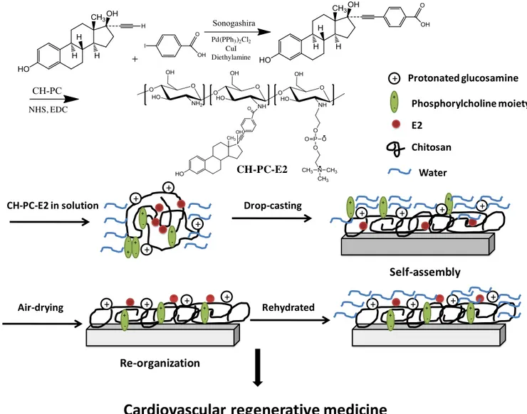
hyaluronic acid. ... 14 Figure 1.4 Tethering E2 to CH-PC polymers for the formulation of self-assembled thin films
by bottom-up approach. ... 22 Figure 1.5 Tethering E2 to photolithography-fabricated micropatterned subsrates by top-down
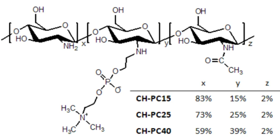
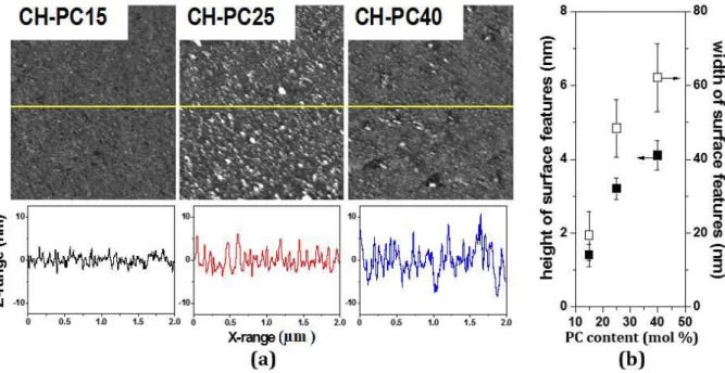
approach in order to specifically understand E2 non-genomic action. ... 23 Figure 2.1 Chemical structure of phosphorylcholine-chitosans ... 53 Figure 2.2 (a) AFM topography images of CH-PC films (2 m 2 m) acquired in a
phosphate buffer pH 6.8; maximum height (Z-range): 12 nm (CH-PC15 and CH-PC25) and 25 nm (CH-PC40) and cross-sectional profiles along the X axis corresponding to the yellow lines drawn through the AFM images; (b) Height and width of the surface features determined by AFM. ... 61 Figure 2.3 SPR and QCM responses of MPS-modified gold substrates when exposed to
solutions of CH-PC (1.0 g L- 1) in phosphate buffer, pH 6.8: a) changes in the SPR resonance angle (m) and b) changes in resonance frequency (f5/5, top) and dissipation
(D5, bottom) observed in the QCM-D experiments for the 5th overtone. ... 63
Figure 2.4 (A) Adsorption of fibrinogen on CH-PC films. Changes in resonance frequency and dissipation observed for the 5th overtone (upper and lower panels, respectively) for MPS-modified gold substrates coated with CH-PC upon exposure to a fibrinogen solution (0.1 g L-1, pH 6.8). The arrows indicate the time corresponding to the onset of the films rinse
xi
Figure 2.5 Optical micrographs of HUVECs on CH-PC coated surfaces 1 day and 4 days after seeding (micrographs A to C and E to G, respectively), and on polystyrene tissue culture plates (micrographs E and H); Scale barμ 200 m. ... 69 Figure 3.1 Synthetic route of CH-PC-E2 polymer conjugate ... 95 Figure 3.2 1H NMR spectrum of CH-PC-E2 conjugate in D2O at 70oC. ... 97
Figure 3.3 QCM-D study of in situ formed CH-PC-E2 and CH-PC hydrogel films on the silicon dioxide for overnight reaction at 25oC. (A) The shift of QCM-D frequency (Δf5/5)
and dissipation (ΔD5/5) showed the thin film formulations of CH-PC (black trace) and
CH-PC-E2 (red trace) onto the silicon dioxide surfaces as a function of time. (B) The storage modulus (squares) and loss modulus (circles) of CH-PC and CH-PC-E2 hydrogel films are shown in a frequency dependent manner. ... 99 Figure 3.4 ER-α adsorption experiment onto the CH-PC-E2 and CH-PC functionalized silicon
dioxide sensor by QCM-D at 25oC. The time of ER-α (concentration: 50 nmol L–1)
addition and PBS wash are indicated by the arrows. (A) The QCM-D frequency (Δf5 /5)
and dissipation (ΔD5 /5) shift showed the adsorption of ER-α onto the CH-PC film (black
trace) and CH-PC-E2 film (red trace) and as a function of time. (B) ΔD-Δf plots for ER-α adsorption corresponds to the ER-α non-specific adsorption of the CH-PC film (black trace) to the specific adsorption of the CH-PC-E2 film (red trace). ... 101 Figure 3.5 DAF-FM fluorescence imaging study of EA.hy926 cells incubated on the CH-PC
and CH-PC-E2 films for 1 hour. Nitric oxide production levels were determined by using DAF-FM for the CH-PC functionalized substrates (A&D), CH-PC-E2 functionalized substrates (B&E), and CH-PC-E2 functionalized substrates pretreated with ICI 182,780 (C&F) for 1 hour. The top panels correspond to phase contrast micrographs and the bottom panels correspond to DAF-FM fluorescence images from the identical sites. (Scale barμ 100 m). The quantitative NO production levels were also shown by analyzing the emission peak intensity (G). Data were calculated by three independent samples. Probability values were considered significant which are represented as *p < 0.05. ... 103 Figure 4.1 (A and B) Schematic representations of the micropatterned substrate employed.
cRGD (for the interaction with integrin) and E2 (for the interaction with mER) are co-immobilized onto a PEGylated micropatterned surface to ensure cell adhesion and E2 presentation to the cell membrane. (C) SEM image of the micropatterned surface
xii
consisting of gold circular regions (diameter: 2 m) arrayed with a 12- m pitch (scale bar: 5 m); and chemical sequences performed on glass surfaces (D) and on gold surfaces (E). ... 125 Figure 4.2 Cyclic RGD (cRGD) functionalized glass substrate enhanced MCF-7 cell adhesion.
(A) Optical images of MCF-7 cell onto the PEGylated glass substrate after 1 hour incubation at 37 ˚C, 5% CO2. (B) Optical images of MCF-7 cell onto the linear RGD
functionalized glass substrate after 1 hour incubation at 37 ˚C, 5% CO2. (C) Optical
images of MCF-7 cell onto the cRGD functionalized glass substrate after 1 hour incubation at 37 ˚C, 5% CO2. (D) Quantification analysis of cell numbers. All the data are
expressed as mean ±S.E. of triplicate determinations. P<0.05 is considered as the significantly statistical difference. ... 127 Figure 4.3 (A) SPR traces corresponding to the addition of ER-α solutions onto the E2-modified PEGylated gold substrate (red) and a control PEGylated gold substrate (black); temperature: 25oC. (B) ER-α coverage (ng/cm2) as a function of ER-α concentration of
the solution injected on the E2-modified PEGylated gold substrate (red symbols) and on the control PEGylated gold substrate (black symbols); the circles are the surface coverage values after rinsing the substrates with buffer. ... 129 Figure 4.4 (A) Frequency (Δf) and dissipation (ΔD) shifts as a function of the concentration of
the ER-α solution injected in the QCM-D cell compartment fitted an E2-modified substrate (red trace) and the control substrate (black trace); (B) ER-α coverage (ng/cm2)
as a function of ER-α concentration of the solution injected on the E2-modified PEGylated gold substrate (red symbols) and on the control PEGylated gold substrate (black symbols); the circles are the surface coverage values after rinsing the substrates with buffer. (C) ΔD-Δf plots for the ER-α adsorption onto the E2-modified surface (red trace) and the control surface (black trace). Different concentrations are also indicated by different colors for amino-PEG substrate and E2-PEG substrate. Amino-PEG substrate: 1 nM (black), 5 nM (green), 10 nM (cyan), 50 nM (yellow), and 100 nM (blue); E2-PEG substrate: 1 nM (red), 5 nM (navy), 10 nM (pink), 50 nM (brown), and 100 nM (purple). ... 131 Figure 4.5 Cellular response to E2-functionalized micropatterned substrates. Optical
xiii
areas functionalized with cRGD and gold dots modified with NH2 (A) or E2 (B)
incubated for 1 hr at 37 ˚C, 5% CO2; scale bars: 100 µm; the micrograph (C) is a
magnified view of (B); scale bar: 25 m; the arrows point to gold dots. (D) ERK phosphorylation of MCF-7 cells on each substrate; bars: mean ± S.E. of triplicate determinations. (E) MCF-7 cell proliferation on amino- or E2-immobilized micropatterned substrates after a 2-day incubation. ... 133
xiv
List of tables
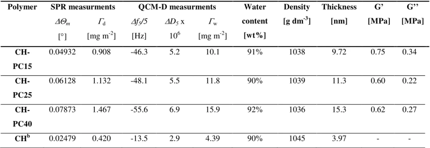
Table 1.1 Summary of the E2 non-genomic effects ... 5 Table 1.2 Summary of non-fouling materials ... 15 Table 2.1 Physico-chemical properties of films formed by CH-PC15, CH-PC25, CH-PC40,
and CH thin films formed on MPS-modified gold surfaces at pH 5.5. ... 65 Table 2.2 Overview of the properties of CH-PC films. ... 71 Table 3.1 Viscoelastic properties of CH-PC and CH-PC-E2 hydrogel films formed in situ.... 99
i
List of abbreviations
A Surface area
Å Angstrom
AFM Air force microscope
BMS Bare metal stents
BSA Bovine serum albumin
ºC Degree celsius
CABG Coronary artery bypass graft CAHD Coronary artery heart disease
CB Carboxylbetaine
CH Chitosan
CVDs Cardiovascular diseases
D2O Deuterium oxide
DLS Dynamic light scattering
DMSO Dimethyl sulfoxide
DS Substitution degree
E2 Estradiol
ECs Endothelial cells
ECMs Extra cellular matrices
EDC Estrogen dendrimer conjugate
EE2 17α-ethinylestradiol
ELISA Enzyme-linked immunosorbent assay eNOS Endothelial nitric oxide synthases
ER Estrogen receptor
ERK Extracellular signal-regulated kinase GPR G protein-coupled transmembrane receptor
h Hour
HPLC High performance liquid chromatography
hMSCs Human Mesenchymal Stem cells
1H NMR Proton nuclear magnetic resonance
HRT Hormonal replacement therapy
HUVEC Human umbilical vein endothelial cells iNOS Inducible nitric oxide synthases
ISR In-stent restenosis
kDa Kilo Dalton
ii
LBL Layer-by-layer
MAPK Mitogen-activated protein kinase
Nagg Aggregation number
NOs Nitric oxide
NOS Nitric oxide synthases
nNOS Neuronal nitric oxide synthases
PAMAM Polyamidoamine
PBMA Poly (butyl methacrylate)
PC Phosphorylcholine
PCI Percutaneous coronary intervention
PDMS Polydimethylsiloxane
PEG Poly(ethylene glycol)
PET Polyethylene terephthalate
PEVA Poly(ethylene vinyl acetate)
PI3K Phosphatidylinositol-4, 5-bisphosphate 3-kinase PKC Phospholipase C protein kinase C
PLA Polylactides
PLGA Poly lactic-co-glycolic Acid
PMMA Poly(methylmethacrylate)
PNIPAM Poly(N-isopropylacrylamide)
PTCA, Percutaneous transluminal coronary angioplasty
PTFE polytetrafluoroethylene
QCM-D Quartz-crystal microbalance
RH Hydrodynamic radius
SAT Subacute stent thrombosis
SB Sulfobetaine
SDS Sodium dodecyl sulfate
SPR Surface plasmon resonance
iii
Authors’ contributions
The present thesis is based on three articles. The current status of articles and authors’ contribution is described below:
Chapter 2:“Phosphorylcholine-modified chitosan films as effective promoters of cell
aggregation” has been published in Macromolecular Bioscience 15 (2015), 490–500. DOI: 10.1002/mabi.201400439. In this manuscript, the physical and biological properties of chitosan phosphorylcholine (CH-PC) polymers were systematically investigated. It explained the principles of developing appropriate CH-PC polymer backbones for the subsequent E2 immobilization study from a polymer perspective. My contribution in this project included carrying out cellular studies, interpreting the results and assisted Dr. Sayaka Toita for the synthesis of CH-PC polymers. Dr. Piotr Kujawa contributed characterization of polymer films, including SPR and QCM-D data modeling. Dr. Gregory Beaune helped to data interpretation and refined figures. All this work was supervised by Dr. Françoise M. Winnik. All authors agreed that this manuscript had been published.
Chapter 3: “Synthesis, characterization and biological study of estrogen grafted
chitosan phosphorylcholine polymer conjugates” is in preparation. In this manuscript, E2 was tethered onto a selected CH-PC polymer backbone as a functional film in the protection of cardiovascular system. My contribution in this project included synthesis and characterization of CH-PC-E2 polymers and cellular studies. Dr. Xingping Qiu helped to the E2-benzoic acid synthesis. All this work was supervised by Dr. Jun Nakanishi and Dr. Françoise M. Winnik. All authors agreed this manuscript to be published.
Chapter 4: “Estradiol-tethered micropatterned surfaces for the study of estrogenic
non-genomic pathways” is to be submitted to Chemical communication. In this manuscript, E2 was immobilized to the micropatterned substrates prepared by top-down approach for the activation of non-genomic signaling pathway. My contribution in this project included preparation and characterization of E2-tethered micropatterned surfaces, as well as estrogen receptor binding studies by SPR and QCM-D. The cell ELISA study was performed by Dr. Yoshihisa Shimizu. All this work was supervised by Dr. Jun Nakanishi and Dr. Françoise M. Winnik. All authors agreed this manuscript to be published.
iv
Acknowledgments
It would not have been possible to complete my PhD project without many people’s professional technical support, profound knowledge and warmly encouragement. To start with, I would like to express my greatest gratitude to my thesis supervisor, Professor Françoise Winnik, who directed me for the completion of all my research as well as trained me as an independent researcher for my future career. I appreciated a lot for her profound knowledge and valuable advices.
I would also like to thank Dr. Jun Nakanishi and Dr. Pitor Kujawa in International Center for Materials Nanoarchitectonics, National Institute for Material Science (MANA, NIMS) in Tsukuba, Japan. Their consistent experiences and efforts instructed my thesis in a well-ordered track. I also thank to my collaborator: Dr. Grégory Beaune, Dr. Sayaka Toita, Dr.Yoshihisa Shimizu, and Dr. Yiu-Ting Richard Lau in MANA. They gave me a huge amount of help in both technical and theoretical aspects. Moreover, I specially thank to staffs in MANA foundry: Ms.Tomoko Ohki, Mr. Akihiko Ohi, Mr. Hideki. Yoshikawa and Mr. Hidefumi Iga for the micropatterned surface preparation.
Meanwhile, I appreciated the great help and invaluable friendship from all my past and present colleagues in the University of Montreal. I will never forget their kindness and discussions during my PhD stage.My sincere thanks also go to Ms. Patricia Moraille and Dr. Cedric Malveau for the help of AFM and NMR measurement.
I am also thankful for my progress report committee members: Prof. Jean-Francois Tanguay and Prof. Grégoire Leclair for their availability and thoughtful discussions.
I also greatly appreciated all the financial support from following institutes and agencies: Faculty of Pharmacy, University of Montreal, China Scholarship Council (CSC), Natural Sciences and Engineering Research Council (NSERC), and World Premier International Research Center Initiative (WPI).
Finally, I would like to show my heartfelt thanks to my beloved family for their constant and unconditional love. Their sacrifice and dedication in the past few year would engrave on my memory forever.
CHAPTER ONE
Introduction and Literature Review
1.1 Estrogen in cardiovascular medicine
1.1.1 Introduction to coronary artery heart disease
Cardiovascular diseases (CVDs), which cover a variety of dysfunctions in tissues ranging from the cardiac muscle to the vascular system, are associated with a high rate of mortality and morbidity worldwide.[1, 2] Coronary artery heart disease (CAHD), also known as atherosclerosis, is one of the most severe CVDs, and is characterized by the deposition of cholesterol and accumulation of plaque within the arterial wall.[3] Epidemiological studies indicate that the causes of CAHD are diverse, including environmental influences (e.g. hormonal changes due to long-term smoking) and chronic physiological disorders (e.g. hypertension and diabetes).[4] The long-term consequences of atherosclerosis may include thrombus formation, which can lead to an acute myocardial infarction.[5]Acute myocardial infarction can be fatal, as it leads to a reduction in the oxygen supply (perfusion) to the heart, as well as creating an abnormality in myocardial energy metabolism.[6] To reduce the risk of myocardial infarction death, anticoagulant drugs, such as heparin[7] and warfarin[8], are widely prescribed in clinics. However, those drugs are associated with an increased risk of bleeding in some patients, which can lead to imbalanced hemostasis.[9] Thus, there is currently an incentive to develop novel medical intervention strategies that reduce the incidence of CAHD.[10]
Coronary artery bypass graft (CABG) surgery, percutaneous transluminal coronary angioplasty (PTCA), and endovascular stenting are current clinical strategies for the revascularization of atherosclerotic arteries and can significantly increase the life expectancy and quality of life for some patients.[11, 12]
For example, patients treated by CABG surgery had a significantly lower mortality than a control group, when comparing morality at 5 years (10.2% vs 15.8%), and 10 years (26.4% vs 30.5%) post-intervention.[13] At 12 months, the
2
rates of death and myocardial infarction were similar between the CABG and percutaneous coronary intervention (PCI), patients, but stroke was significantly more likely to occur with CABG (2.2% vs. 0.6% with PCI).[14] However, adverse cardiac complications were significantly higher in the PCI group (17.8%, vs. 12.4% for CABG), likely because of an increased rate of repeat revascularization. In conjunction with surgical treatment, novel medical devices (e.g. artificial heart valve or stent) are also being developed for the amelioration of these cardiovascular diseases, which will greatly enhance the quality of patients' lives.[15]
1.1.2 The anatomy and physiology of blood vessels
One of the therapeutic targets of coronary artery heart disease is the blood vessel, which is the main part of the circulatory system for nutrients and oxygen transportation throughout the body. They are classified as arteries, capillaries, and veins.[16] The inner wall of the blood vessels is lined with endothelial cells. These endothelial cells comprise the endothelium, which provides a mechanical barrier against the circulating blood. They also act as an interface that can sense alterations in blood flow and plasma constituents, as well as secrete a variety of powerful chemical mediators that regulate vascular remodeling.[17] In contrast, smooth muscle cells are located principally along the outer layer of the endothelium. They are also involved in biochemical vaso-regulatory processes, but in a far less efficient manner than endothelial cells.[18] Interactions between endothelial cells and smooth muscle cells are crucial to determine various pathophysiology of cardiovascular systems, including arteriogenesis and atherosclerosis[19]
Nitric oxide (NO) is a gaseous free radical cellular messenger generated by three types of nitric oxide synthases (NOS): neuronal (nNOS), inducible (iNOS) and endothelial NOS (eNOS).[20] NO is an important signal molecule produced by endothelial cells, serving to inhibit platelet aggregation, leukocyte adhesion, and smooth muscle cell proliferation.[21] Moreover, NO is recognized for its protective role within the vasculature by preventing biochemical cascades leading to thrombus formation.[22] For example, it has been shown that NO exerts a protective effect on vasculature through inhibiting platelet aggregation in a
3
transgenic mouse model. Blood clotting time was markedly decreased in eNOS-deficient versus wild-type mice (77±3 versus 133±3 seconds).[23]
1.1.3 Estrogen effects on the cardiovascular system
17β-estradiol (E2) is a female hormone produced by human ovaries that controls and regulates several physiological conditions. Synthetic estradiols are used in contraception and in hormonal replacement therapy (HRT) for menopausal women.[24] For example, ethinyl estradiol (EE2), which mimics the endocrine properties of E2, is used in oral contraceptive formulations such as Norlestrin, Brevicon, and Ortho–Novum.[25] As such, estradiol plays
critical roles in female sexual maturation and reproduction by initiating epithelial proliferation of the vagina, uterus, and breast.[26] It has been observed that the incidence of cardiovascular disease differs significantly between men and women, or between premenopausal women and postmenopausal women. This fact hinted that E2 may confer cardio-protective effects in addition to its hormonal functions.[27, 28] In cardiological studies, E2 has been shown to decrease the development of atherosclerosis and to elicit protection against ischemia by directly exerting the effects within the blood vessels.[29] Moreover, it has been reported that the local delivery of E2, via either an infusion catheter [30-32] or phosphorylcholine-coated stent, inhibited neointimal proliferation in coronary arteries following stent implantation.[33]
1.1.4 Estrogen receptors and classical genomic effect
E2 acts as a mediator via binding estrogen receptors (ERs).[34] ERs exist in two main forms, ERα and ERβ, encoded by two genes, ESR1 and ESR2, respectively, that are discovered at different chromosomal locations.[35] E2 passively diffuses into the cells where it binds to ERs in the nucleus.[36] Once bound, the estrogen receptor dimerizes and converts to an activated form. The dimerized ERs bind estrogen response elements (EREs) in the enhancer regions of responsive genes, thereby activating the transcription of estrogen-inducible genes.[37] This phenomenon is regarded as a genomic effect or nucleus effect.[38] It is also interesting to note that this effect has also been shown to promote the proliferation, angiogenesis, and metastasis of tumor cells, which causes non-negligible adverse effect.[39] There are multiple pathways to induce this genomic effect:[40] A) E2-nucleus ER complexes
4
bind to estrogen receptor elements (EREs) at the promoter regions of target gene, thereby inducing their expressions (ERE-dependent genomic pathway). B) E2-nucleus ERs complexes bind to transcription factor complexes (ERE-independent genomic pathway). C) Instead of E2, growth factors themselves are also able to activate multiple kinase cascades and induce phosphorylation of nuclear estrogen receptors (Growth factor-dependent genomic pathway).
1.1.5 Estrogen non-genomic effect
E2 can trigger several alternative pathways via direct or indirect signal transduction rather than through the genomic effect. These pathways are regarded as the non-genomic effects[41] that are initiated on either the membrane or the cytosolic sides of the cell (Figure
1.1).[38] E2-elicited non-genomic effects are very rapid (seconds to minutes) in comparison to the activation of genomic-mediated effects (hours to days).[42]
Figure 1.1 Schematic representation of the genomic and nongenomic estrogen pathways. For the genomic pathway, E2 (red triangle) crosses the cell membrane and binds to the estrogen receptor (ER) in the cell nucleus, inducing gene transcription. For the non-genomic pathway, E2 interacts with ER or ER/GPR30 on the cell membrane, which activates several transduction pathways (Figure taken from [43]).
5
Extracellular-signal-regulated kinase (ERK), a component of the mitogen-activated protein kinase (MAPK) family, and AKT kinase, a component of the phosphatidylinositol-4, 5-bisphosphate 3-kinase (PI3K) family, are involved in E2-mediated responses.[42, 44-47] In the ERK/MAPK pathway, the E2-induction is associated with several phosphorylation cascades.[48, 49] The majority of studies in MCF-7 cells reported the activation of ERK1/2 by E2 ranging from 2 to 15 min of its activity.[48, 50-53] But, some studies under similar conditions described E2 leading to elevations in ERK1/2 activity only after 2 h of induction, with persistent activation for up to 1 day.[54, 55] The difference in these results might originate from the differed cell culture methods. In the PI3K/AKT pathway, E2 leads to the release of nitric oxide (NO),[56] cyclic amines (cAMP, cGMP)[57, 58] and calcium[59] in a variety of cell types. Moreover, E2-induced non-genomic effects trigger the activation of phospholipase C (PLC)/protein kinase C (PKC) signal transduction pathways.[60] The cell signal pathways
triggered by E2 non-genomic effects are summarized in the Table 1.1. Table 1.1 Summary of the E2 non-genomic effects
Signal pathway Time E2 dose Analyzed
Protein
Cell line
PI3K/AKT[56] 5 to 30 mins 25 nM PI3K/AKT and eNOS
HUVEC
PI3K/AKT[61] 15 mins to 2 hrs 1 M PI3K/AKT Endometrial
cancer cells
PI3K/AKT[62] 0 to 60 mins 10 nM PI3K/AKT HepG2 cells
ERK/MAPK[50] 15 min to 1 day 1 M pERK 1/2 HepG2 cells
ERK/MAPK[51] 2 min to 2 hrs 10 nM pERK 1/2 MCF-7 cells
6
It has also been hypothesized that E2 exerts non-genomic actions by interacting directly with growth factor receptors.[63] This process is directed through E2-mediated tyrosine kinase’s activation of growth factor receptors, such as the epidermal growth factor receptor (EGFR).[64, 65] The activation of growth factor receptors, in turn, triggers downstream cell signal transduction cascades such as MAPK.[49, 66]
Other studies reported that the E2-induced non-genomic effect was mediated through a G protein-coupled transmembrane receptor, GPR30.[67] Recent studies have also implicated GPR30 as a potential candidate for the activation of membrane-initiated estrogen signaling.[68] Supporting this claim, it has been demonstrated that GPR30 increased both ER-α and EGFR activities in patients undergoing clinical treatment for breast cancer.[69] However, the assumption, that GPR30 is responsible for an E2-mediated non-genomic effect is still under dispute.[63]
1.1. 6 Tools for estrogen non-genomic effect study
Estrogen conjugated to membrane-impermeable proteins, e.g. E2-BSA, have been used to determine the non-nuclear ER actions in various cell types.[70-72] However, freshly prepared solutions of E2-BSA always contain free E2 which cannot be fully removed during the synthesis process. Moreover, E2-BSA binds to the ER only poorly because certain E2-BSA preparations are of very high molecular weight, suggesting extreme high steric hindrance.[73] Last, but maybe not least, E2-BSA is readily degraded by the cell, making long-term experiments problematic and in vivo studies impossible.[74]
Recently, an estrogen dendrimer conjugate (EDC) has been prepared for the stimulation of membrane-mediated non-genomic effects.[75] Such E2-dendrimer systems also activated vascular endothelial cell migration, which could regulate the processes of cardiovascular protection without enhancing growth of breast cancer cells.[76] Nonetheless, confocal microscopic studies showed that EDC can also penetrate into cytoplasmic sites at MCF-7 cells. As a result, a detailed profile of an estrogen-initiated short-term signaling pathway has not been elucidated yet as using the estrogen conjugate systems.
7
1.2 Polymeric biomaterials in cardiovascular medicine
1.2.1 Fundamental properties of polymeric biomaterials
Polymeric biomaterials are widely developed for in vitro and in vivo applications to study complex biological phenomena.[77] The essential design criteria for polymeric
biomaterials are related to their high-quality performance, structural and functional stability, and physiological compatibility.[78] To date, various polymeric materials have been tested for the preparation of biomaterials, these include polystyrene,[79] poly(methyl methacrylate) (PMMA),[80] polydimethylsiloxane (PDMS),[81] and polyurethane.[82] For example, PMMA is a transparent polymer commonly known as Plexiglass.[83] Thin films of PMMA can be modified by aminolysis reaction to introduce amine groups,[84] which can confer resistance against non-specific protein adsorption.[80] PDMS is the most commonly used material for the preparation
of microfluidic devices due to its elastomeric properties and ease in molding.[85] PDMS
micro-fabricated surfaces were prepared as a synthetic cornea in an attempt to stimulate stromal restoration.[86] Once the polymeric biomaterials are placed into the biological environment, many factors should be considered: surface charge, surface topology, surface energy and chemistry, to name a few.[87] Therefore, it is critical to develop cost-effective and efficient approaches to understand the physical properties of polymeric biomaterials, and to widen the design parameters for suitable constructs.
1.2.2 Mechanical properties
In order to provide an appropriate microenvironment for supporting cells or entire tissues in vitro and in vivo, biomaterials should have reliable mechanical properties.[87, 88]
Generally, once biomaterials enter in contact with living organisms, deformation of biomaterials or cells will occur due to forces exerted by external environment, e.g. blood stream. The elastic modulus (or modulus of elasticity) is a constant that define quantitatively the ability of a biomaterial to resist deformation under external force as well as to recover the initial state after removal of the force, including Young's modulus and shear modulus. Young’s modulus is usually expressed as E, representing the elastic resistance of objects to
8
tensile strength e.g., elongation or compression. Young’s modulus can be calculated by following equation (1):
� =
��
(1)
where E is the Young's modulus, � is the tensile strength, � is the extensional strain.
Shear modulus is usually expressed as G, representing the resistance of objects to deformation in shear. The relation between shear modulus (G) and Young’s modulus can be expressed as following equation (2):
� =
�+ѵ
(2)
where E is Young’s modulus, ѵ is Poisson's ratio.[89-92]
Moreover, some soft biomaterials, such as hydrogels, exhibit both viscous and elastic characteristics, which are recognized as viscoelasticity. Viscoelasticity is composed of the storage modulus G’, which defines the elastic property of materials and the loss modulus and G”, which governs the viscous property of materials.[93-96] Both G and G can be
characterized by a power-law frequency dependence with critical exponents α and β, respectively (G ∝ f α and G ∝ f β). Knowing the viscosity of material η(f), the loss modulus can be calculated based on the equation (3):
G f = 2πf η f .[97] (3) Polymeric biomaterials have a wide range of elasticity values (E or G’) from a few Pa to hundred MPa in a similar range of tissue or organs in vivo, which could offer various choices for the biomedical applications.[98] For example, the elastic modulus of synthetic PEG is in a range of a few kPa, and could be used to prepare extra cellular matrices (ECMs) to support cell and tissue growth in a three dimensional manner.[99, 100]. By tuning the elastic modulus of polyacrylamide gel based substrate from 1 kPa to 100 kPa,adult human mesenchymal stem cells (hMSCs) differentiate into neuron, muscle, or osteogenic cells, respectively.[101] Thus, a substantial understanding of the intrinsic mechanical properties of
9
polymeric biomaterials will facilitate the design and development of functional biomaterials more efficiently.
1.2.3 Biodegradability
The term of biodegradation is generally defined as the breakdown of the polymer materials by the hydrolytic or enzymatic cleavage reactions.[102] For tissue engineering applications, it is desirable to develop biodegradable polymer materials with predictable erosion kinetics. Hydrolysis is a major degradation mechanism in vivo that degrades larger polymer chains to smaller chains or monomers.[103] The effect of pH is critical in this
hydrolytic process.[104] For example, polylactide (PLA) polymers have the slowest hydrolysis
rate at pH~4.[105] Enzymatic cleavage is another important mechanism for polymer degradation.[106] The rate of in vivo enzymatic degradation of polymers depends on the distribution and concentration of the cleavage enzymes.[107] In most of cases, hydrolytic and enzymatic cleavage reaction occurs simultaneously.[108]
Both natural and synthetic polymers have been extensively investigated as biodegradable biomaterials. Natural polymers are promising due to their susceptibility to in vivo degradation.[109] For example, cellulose is a main composition of plants and natural fibers, which is degradable by several microorganisms. Cellulose-based hydrogels can facilitate the scarless healing processes upon degradation.[110] Synthetic polymers also demonstrate biodegradability and easily undergo chemically-tailored reactions for specific biomedical applications.[111] For example, by grafting PEG to a synthetic biodegradable polycarbonate, the degradation takes place within one day. It may be useful in clinical applications ranging from cosmetic surgery to cancer treatment.[112] As a consequence, investigations into polymeric materials will help to develop safe and environment friendly products by taking advantage of their biodegradable properties.
1.2.4 Cellular interactions
The interactions between cells and implanted biomaterials, especially cell adhesion to surfaces, are determined by the nature of biomaterials.[113] When deposited on an adhesive substrate, cells are favorable to be spread out. As given a certain time, the cells proliferate and
10
form a two-dimensional monolayer. However, when the spreading of cells is unfavorable, e.g. in the case of non-adhesive surfaces, cells spontaneously move by active cell motility and eventually meet to form compact clusters and three-dimensional aggregates (spheroid).[114, 115] The forces influencing cell adhesion include:
(1) Surface energy interaction. The primary factor of cell adhesion to the surface relies on the balance of surface energy, e.g. surface hydrophobicity and hydrophilicity.[116] Generally, surfaces with high-energies are able to enhance cell adhesion compared with surfaces with low-energies.[117] As a result, surfaces with high-energies are usually used for the cellular adhesion.
(2) Electrostatic interaction. Because a major part of the cell membrane is negatively charged, the surface charges of the biomaterial surfaces are usually designed to be positive to ensure cell adhesion.[118]
(3) ECM protein interaction. ECM proteins, such as fibronectin, can easily adsorb onto the biomaterial surfaces due to their wide-spread existence in vitro and in vivo environments, suggesting that the influence of these proteins is also predominant in the process of cell adhesion.[119, 120]
Functionalized surfaces that are able to induce, in a predictable way, cell adhesion will be useful. For example, a thermosensitive polymer poly (N-isopropylacrylamide) (PNIPAM) is able to undergo phase transition from hydrophilic to hydrophobic by tuning the temperature from room temperature to physiological conditions (~37 °C). [121] PNIPAM was recently used to prepare thermosensitive functionalized surfaces for harvesting cells in a confluent monolayer. This novel cell culture platform allows formation of cell monolayers in an environment that is more relevant to the real-life situation and hence transplantation-favorable to host tissues.[122, 123] To sum up, controlling the cellular responses, e.g. cell adhesion or cell spread, to biomaterials is a fundamental step in biomaterial research.
11
1.3 Non-fouling polymeric biomaterials in cardiovascular
regenerative medicine
1.3.1 Overview of non-fouling polymeric biomaterials
Non-specific protein (e.g. fibrinogen, fibronectin, and vitronectin) adsorption [124] and blood cell adhesion[116] onto a biomaterial surface triggers a foreign body reaction which can lead to detrimental consequences.[125] This process is known as biofouling or fouling.[116] Non-fouling is the term used to define the ability of a material to resist non-specific protein adsorption and blood cell adhesion in vivo and hence to not cause adverse effects when they are placed in contact with living organisms.[126] Several properties of polymeric materials, such as their composition, hydrophobicity/hydrophilicity, charge, and topology are responsible for biocompatibility.[127] The origin of the non-fouling characteristic of a polymeric biomaterial is due to the formation of a highly hydrated layer near the surface, which generates an energetic barrier to prohibit immune responses occurrence.[128, 129] The mechanism of non-fouling characteristic of polymeric materials can be explained as follows. Briefly, the polar groups of proteins can form hydrogen bonds with the surrounding water medium. However, if the surface of polymeric materials (e.g. PEG) is very hydrophilic, the water molecules can penetrate into the polymers to form a net-like structure, which is regarded as the “surface-bound” water. Meanwhile, both the protein and material surface tend to expulse water molecules to facilitate protein adsorption, which generate physical and energetic barriers (also known as dehydration entropic effects).[126, 130] The non-fouling characteristic of polymeric materials is fundamentally determined by numerous factors, i.e. film thickness and packing density or chain conformation and flexibility.[130] As a result, non-fouling characteristic is certainly one of the most significant characteristics of biomaterials, when considering their practical biomedical applications.
Poly (ethylene glycol) (PEG), zwitterionic-based materials, and some polysaccharides are among the most widely used non-fouling materials (summarized in the Table 1.2).[131, 132] PEGylated materials are the most commonly used biomaterials due to their highly hydrophilic and flexible nature. As introduced previously, a water barrier can be generated between PEG and proteins to entropically impede non-specific adsorption occurrence.[128] Moreover, the
12
high chain flexibility of PEG yields compression of chains and hence sterically repulses protein adsorption.[133] The end of PEG can be tailored by different functional moieties, such as thiol, amine, and carboxylic acid, for the different biological applications.[130] For example, thiol end functionalized PEG enables its immobilization onto gold surfaces and the thickness of the PEG layer is correlated with the response to the biological environment.[134] Zwitterionic-based materials, such as phosphorylcholine, sulfobetaine, and carboxybetaine derivatives (structures shown in Fig. 1.2), possessing covalently linked cationic and anionic groups,[135] are known to reduce non-specific protein adsorption and inhibit cell adhesion.[136,
137] Phosphorylcholine (PC) is present on phospholipids of the outer layer of the cell
membrane. It has an excellent ability to reduce nonspecific protein adsorption.[138] The key factors, e.g. hydration and ionic solvation, leading to the non-fouling behavior of PC were determined by molecular simulation techniques.[139] Similar to phosphorylcholine, both
sulfobetaine (SB) and carboxybetaine (CB) also showed strong anti-fouling capacities.[140, 141]
When poly-(sulfobetaine) was grafted onto a poly-(ether urethane) surface by atom-transfer radical-polymerization(ATRP), the surface exhibited extremely low fibrinogen adsorption.[142]
Figure 1.2 Chemical structures of zwitterionic-based materials. (A) phosphorylcholine (PC), (B) sulfobetaine (SB), and (C) carboxybetaine (CB).
The natural polysaccharide heparin, a potent anticoagulant, has shown promising biomedical applications due to its antimicrobial and anti-inflammatory capacities.[143-145] It is a
glycosaminoglycan consisting of a sulfated disaccharide repeat unit (Fig.1.3A). The most common disaccharide unit is composed of a 2-O-sulfated iduronic acid and 6-O-sulfated, or N-sulfated, glucosamine.[146] The molecular weight of heparin ranges from 4 to 40 kDa.[147] Heparin, which is a polyanion, adopts an extended conformation in vivo.[148, 149] Heparin-coated surfaces showed a good biocompatibility, which decreases the incidence of restenosis
13
and reduces thrombogenesis and inflammatory reactions.[150] Hyaluronic acid (HA) is anionic polysaccharide, composed of D-glucuronic acid and D-N-acetylglucosamine, linked via alternating β-1,4 and β-1,3 glycosidic bonds (Fig. 1.3B).[151] The molecular weight of HA
ranges between 100 kDa to 8000 kDa in vivo.[152] The glucuronic acid, carboxylic acid, the primary and secondary hydroxyl, and the N-acetyl groups of HA can be chemically modified for conjugating different moieties in order to prepare HA-based hydrogels.[153] HA hydrogels can promote the proliferation and migration of mesenchymal cells and enhance the endogenous tissue repair process.[154] The ultralow protein adsorption of HA is also demonstrated by surface plasmon resonance (SPR) (only 0.6−16.1 ng/cm2).[155] Chitosan is a cationic polysaccharide extracted from N-acetylation of chitin.[156] It has been studied for applications in several fields,[157] such as wound healing,[158] drug delivery,[159] gene therapy,[160] and tissue engineering.[161] It is a linear polycation composed of β-D-glucosamine
and β-D-N-acetylglucosamine residues in 1→4 linkages (Fig. 1.3C). [162] It is soluble in acidic
solutions, but it is poorly soluble in neutral pH conditions.[163] Moreover, the solubility of chitosan is highly associated with degradability, which is decided by its physical and chemical properties, such as deacetylation and molecule weight.[164] For instance, deacetylation can not only affects the degradation rate, but also enhance the solubility of chitosan at neutral pH.[164,
165] The molecular weight also affects chitosan solubility.[166] If chitosan was degraded into
low molecular weight chitosan (LMWC) with the molecular weight below 10Da, the water solubility of chitosan largely increased.[167] The non-fouling nature of chitosan was also widely acknowledged. In a previous study, it was also demonstrated that chitosan nanocoating protects against adsorption of BSA, TGF-β1 and human plasma due to its surface hydrophilicity.[168] Chitosan has also been developed as scaffold for wound healing purposes because of its antibacterial activity.[169] By using heparin /chitosan blended films, such antibacterial activity was further enhanced.[170] Besides its biodegradability and non-fouling features, chitosan also has cell adhesive properties as a artificial component of the extracellular matrix.[171] Chitosan films are known to support cell adhesion, as has been observed in the cases of dorsal root ganglions,[172] keratinocytes, and fibroblasts.[173]
14
Take these facts together, non-fouling materials should be extensively used in the cardiovascular regenerative medicine in order to produce neither inflammatory nor immune responses, while retaining the capacity of cell adhesion.
Figure 1.3 Chemical structures of natural polysaccharides. (A) heparin, (B) chitosan, and (C) hyaluronic acid.
(A)
15
Table 1.2 Summary of non-fouling materials
Polymer Preparation method Surface characterization method Method to assess biocompatibility
PEG[134] Self assembled
monolayer
Ellipsometry, AFM Endothelial cells adhesion kinetics study PEG-block-
polycarbonate[174]
Covalently immobilization
QCM-D, SEM Bacterial viability assay, platelet adhesion study,
static hemolysis assay
Catecholates anchored PEG[175]
Chemical deposition
Ellipsometry Human blood assay, bone-derived stromal cells,
bacterial adhesion PC[135] Self assembled
monolayer
XPS, ellipsometry, SPR
Human Fg and BSA adsorption study Poly(sulfobetaine methacrylate)[176] Chemical deposition ATR/FT-IR, XPS, SEM
BSA adsorption study
Carboxybetaine[177] Self assembled monolayer Contact angle measurement, XPS,QCM-D
BSA adsorption and blood cell adhesion study
Carboxybetaine graft polysiloxanes (PDMS-g-CB)[178] Chemical deposition ATR/FT-IR, XPS, SEM, contact angle
measurement
BSA, Hgb and lysozyme adsorption study HA[155] Covalently immobilization Contact angle measurement, AFM, SPR
BSA, lysozyme and soybean milk adsorption
study Chitosan[168] Physical
adsorption
SEM, AFM, MTT viability test.
BSA, human blood adsorption study
16
1.3.2 Non-fouling polymeric biomaterials in cardiovascular regenerative
medicine
Implantable biomaterials or blood contact devices are widely used to cure cardiovascular diseases or alleviate symptoms.[179] One such example is the cardiovascular
stent. Revascularization procedures, such as a coronary balloon angioplasty, are often accompanied by various complications (e.g. restenosis).[180] The introduction of a stent into an
artery after angioplastic surgery can expand blood vessels and restore blood flow through narrowed arteries.[181]. Silicone rubber or plastic was initially used to support the blood vessels
in order to maintain the intravascular blood flow.[182] Stainless steel AISI 316L[183] or titanium (Ti)[184], possessing exceptional corrosion resistance and mechanical properties, are the commonly used medical metals for cardiovascular stent nowadays. However, it is widely acknowledged that dominant side effect can be induced by these bare metal stents (BMS), e.g., late in-stent restenosis, a phenomenon that reoccurs of angina.[185, 186] Recently, synthetic or
natural polymer coated drug-eluting stents have proven to be superior to BMS in many respects.[187, 188] They are able to reduce significantly the incidence of restenosis by
locally-controlled release of drugs to the neointimal and inflammatory areas.[189] In order to be usable,
the polymer coating material must support the vessel lumen to maintain its patency, and naturally degrade within a certain time. Nowadays, two types of commercially available DESs are available (Cypher® and Taxus®) which are the most widely used in the clinical setting. They have embedded anti-proliferative agents (Rapamycin in the case of Cypher®[190] and paclitaxel in the case of Taxus®[191] ) with hydrophobic polymer surfaces. However, both Cypher® and Taxus® have some drawbacks. For example, they use durable thick polymers ((polyethylene vinyl acetate (PEVA)/ poly(butyl methacrylate (PBMA) for Cypher® and poly(styrene-b-isobutylene-b-styrene) for Taxus®).[192] As a result, new generation of polymer
coated stents are still highly in demand. A PEG-coated titanium surface shows excellent blood compatibility by resisting platelets and leukocytes adhesions.[193] In a porcine coronary
restenosis model, grafting PEG to the biodegradable polymer poly(D, L-lactide-co-glycolide) (PLGA), significantly inhibited neointimal hyperplasia 1 month after of implantation.[194] Phosphorylcholine (PC) and sulfobetaine (SB) are also attracting attention as new generation of stent coating materials. Recently, a PC and SB derivative materials coated magnesium alloy
17
surface demonstrated promising results in the reducing acute thrombotic responses.[195] Especially, PC-coated stent is extensively studied both in pre-clinical and clinical trials. In the mid-term (6 months) results, the symptoms of acute myocardial infarction of the patients were also significantly alleviated after implanting PC-coated stent.[196] The reliability of PC-coated stents was also validated in the long-term assessment.[197]
Another example of polymeric materials being used in cardiovascular medicine is vascular grafting. In arterial bypass procedures, synthetic polymers are widely used to redirect blood flow in the specific site of the body and have shown an excellent 5 year survival rate for the patients (exceeding 95 %).[198] The approach for reconstructing and remodeling tissues is to recruit autologous endothelial cells (EC) or endothelial progenitor cells (EPC) after implantation of the graft and to promote similar neovascularization to a native vessel by supporting local angiogenesis.[199, 200] This in situ endothelialization of a vascular graft following implantation is a promising solution for enhancing graft patency rates.[201] Commercially available synthetic vascular graft materials are either expanded polytetrafluoroethylene (ePTFE) or poly(ethylene terephthalate) (Dacron).[202] In some cases, the diameter of grafts should be as small as possible, in order to reach the target vessel site through narrow vasculature. However, these materials are not suitable for smaller-diameter (with internal <6 mm) tubes used in vascular bypass surgery, resulting in a high rate of graft failure.[203] The major reason is that they increase the risk of thrombus due to their low biocompatibility.[204] In this aspect, non-fouling polymer coating surfaces can be utilized as an alternative. For example, the PEG was grafted onto poly-(ethylene terephthalate) (PET) and greatly improved hemo-compatibility of the PET surfaces.[205] In addition, the PEG grafted PET was prepared to generate polymer brushes via a surface-initiated atom-transfer radical polymerization method and showed an excellent anti-fouling effect during the platelet adhesion assays.[206] In a porcine model, grafting PEG to poly (propylene sulphide) effectively reduced thrombogenicity and enhanced the graft patency of small diameter vascular grafts.[207] In the clinical trial, PEG-hirudin/iloprost coated PTFE based small diameter grafts inhibited the progress of pseudointima and intimal hyperplasia in the patient as well.[208] However, in vitro regular 2-dimensional vascular graft based endothelialization is usually initiated by using cellular monolayer.[209] Unfortunately, the efficacy of 2-dimensional vascular graft for the