Gian Luigi Canata • Pieter d
,
Hooghe
Kenneth J. Hunt • Gino M. M. J. Kerkhoffs
Umile Giuseppe Longo
Editors
Sports Injuries of the
Foot and Ankle
A Focus on Advanced Surgical
Techniques
ISBN 978-3-662-58703-4 ISBN 978-3-662-58704-1 (eBook) https://doi.org/10.1007/978-3-662-58704-1
Library of Congress Control Number: 2019935194 © ISAKOS 2019
This work is subject to copyright. All rights are reserved by the Publisher, whether the whole or part of the material is concerned, specifically the rights of translation, reprinting, reuse of illustrations, recitation, broadcasting, reproduction on microfilms or in any other physical way, and transmission or information storage and retrieval, electronic adaptation, computer software, or by similar or dissimilar methodology now known or hereafter developed.
The use of general descriptive names, registered names, trademarks, service marks, etc. in this publication does not imply, even in the absence of a specific statement, that such names are exempt from the relevant protective laws and regulations and therefore free for general use. The publisher, the authors, and the editors are safe to assume that the advice and information in this book are believed to be true and accurate at the date of publication. Neither the publisher nor the authors or the editors give a warranty, express or implied, with respect to the material contained herein or for any errors or omissions that may have been made. The publisher remains neutral with regard to jurisdictional claims in published maps and institutional affiliations. This Springer imprint is published by the registered company Springer-Verlag GmbH, DE part of Springer Nature.
The registered company address is: Heidelberger Platz 3, 14197 Berlin, Germany
Gian Luigi Canata Koelliker Hospital
Centre of Sports Traumatology Koelliker Hospital
Torino Italy
Kenneth J. Hunt
Department of Orthopedics University of Colorado Hospital Aurora, CO
USA
Umile GiuseppeLongo
Orthopaedics and Traumatology Unit University Campus Biomedico Roma
Italy
Department of Orthopaedic Surgery Aspetar Hospital
Doha Qatar
Gino M. M. J. Kerkhoffs Department of Orthopedic Surgery University of Amsterdam Academic Medical Center Amsterdam Zuidoost Noord-Holland The Netherlands
v
This book, Sports Injuries of the Foot and Ankle: A Focus on Advanced
Surgical Techniques by the Leg, Ankle and Foot (LAF) Committee of ISAKOS, is quintessential ISAKOS. This is a comprehensive book that should be in the library of every surgeon who operates on injuries to the foot and ankle, athlete or not. It is edited by an internationally acclaimed group of sports foot and ankle surgeons, Gian-Luigi Canata from Italy, Pieter D’Hooghe from Qatar, Ken Hunt from the USA, Giuseppe Longo from Italy, and Gino Kerkhoff from the Netherlands, and includes contributions from esteemed and internationally recognized experts from all over the world— true international authorities on the management of foot and ankle problems in athletes. The work and effort put into this book by the editors and authors, along with beautiful original illustrations, will make it the new standard by which foot and ankle surgical technique books will be measured.
This book represents some of what is best about ISAKOS—international experts, leaders in the field, not just in their respective country, but recog-nized all over the world, coming together to share thoughts, ideas, and con-cepts, to help advance the field and to make the world just a bit closer. Collaboration, whether at the biennial meeting, at committee meetings, and at interim offsite meetings, with research, state-of-the-art papers, and publica-tions, like this one, is the heart of ISAKOS. It is a brotherhood (for lack of a better gender neutral term), friendship, and sort of family.
The authors provide a comprehensive look at the different parts of the foot and ankle from a variety of perspectives. They introduce newer, cutting-edge techniques, along with the standard “tried and true” surgeries. They span the gamut of techniques, from the basics to complex, in a comprehensive, well- illustrated methodology. Each technique is described in detail to help the orthopedic surgeon perform it accurately and safely.
But more than just a surgical technique book, they discuss important top-ics such as the anatomy, biology, surgical outcomes, and footwear consider-ations. They also discuss newer areas in evolution, such as tissue engineering, and the ever important area of rehabilitation following these surgeries.
Drs. Canata, D’Hooghe, Hunt, Longo, and Kerkhoff, as members of the Leg, Ankle and Foot Committee of ISAKOS, have brought together a talented and respected group of foot and ankle specialists from the LAF Committee and other ISAKOS members and must be commended for the exceptional quality of this book and be congratulated for a job well done. This book will
be valuable to all surgeons who care for foot and ankle problems, particularly those who care for athletes with foot and ankle problems.
With the help and guidance of João Espregueira-Mendes, the head of the Publications Committee, it has been a huge endeavor. I am honored that it has been initiated and completed during my tenure as president. The authors, and editors, are to be congratulated for a Herculean effort and a book that sets a new standard.
Marc R. Safran ISAKOS Stanford, CA, USA 2017–2019
vii
This book is an update on current techniques for the treatment of foot and ankle injuries and conditions in the athletic patient. It is meant to serve as a current and comprehensive review of the state of the art, with an international perspective. Each chapter is written by orthopedic surgeons expert in the field, sharing their experience treating specific injuries and conditions, cutting- edge surgical procedures, and injury management strategies. Several different techniques are described step-by-step, easing the reader to thor-oughly understand what the surgeon is doing getting information on details.
ISAKOS is devoted to its mission of disseminating knowledge to the world of orthopedics and sports medicine. Through committees like the Leg, Ankle, and Foot (LAF) Committee represented herein, ISAKOS continu-ously works to help its membership, and the orthopedic and sports medicine communities it serves, to strive to improve the art, optimize the delivery of care worldwide, and seek at all times the best interest and outcomes of the patient.
The editors extend a sincere thanks to all the authors for their outstanding contributions and to ISAKOS for its steadfast and unwavering support for this project.
Torino, Italy Gian Luigi Canata
Doha, Qatar Pieter d’Hooghe
Aurora, CO Kenneth J. Hunt
Amsterdam, Netherlands Gino M. M. J. Kerkhoffs
Rome, Italy Umile Giuseppe Longo
ix
We are extremely grateful for the contribution of the authors who dedicated their time, knowledge, and expertise to this project.
We owe a debt of gratitude to:
Pontus Andersson for his outstanding illustrations.
Catena Cottone who kept close and continuous contacts with the authors and Springer.
ISAKOS leadership and staff for consistent, constant, and extraordinary support and encouragement. We wish to extend a special thank you to Marc Safran, current ISAKOS President; João Espregueira-Mendes, Publications Committee Chair; Jon Karlsson, Secretary; and Michele Johnson, Executive Director.
Dhanapal Palanisamy, Gabriele Schroeder, and Springer for their profes-sional work and dedication.
xi
Part I Ligament Injuries
1 Acute Ankle Ligament Injuries . . . 3
Kenneth J. Hunt and Peter Lawson
2 Lateral Endoscopy of the Ankle. . . 13
Stéphane Guillo
3 All-Inside Endoscopic Broström- Gould Procedure
for Chronic Ankle Instability . . . 21
Haruki Odagiri, Stéphane Guillo, and Thomas Bauer
4 Arthroscopic Ligament Repair and Reconstruction . . . 29
Masato Takao, Mai Katakura, and Yasuyuki Jujo
5 Mini-Incision Technique for Lateral Ankle
Ligament Repair in Chronic Instability . . . 45
Gian Luigi Canata, Valentina Casale, and Luca Pulici
6 Syndesmosis Injuries . . . 57
Pieter D’Hooghe
7 Subtalar Joint Instability . . . 77
Vincenzo Candela, Umile Giuseppe Longo,
Giuseppe Salvatore, Alessandra Berton, Nicola Maffulli, and Vincenzo Denaro
8 Lisfranc Complex Injuries . . . 85
Samuel O. Ewalefo, Stephanie M. Jones, Lorraine Boakye, Arthur R. McDowell, Scott Nimmons, Jorge L. Rocha, Humza Shaik, and MaCalus V. Hogan
Part II Cartilage
9 Cartilage Techniques for Osteochondral Lesions
of the Talus . . . 105
Eoghan T. Hurley, Yoshiharu Shimozono, and John G. Kennedy
10 Tissue Engineering for the Cartilage Repair of the Ankle . . . 119
Alberto Gobbi, Stefan Nehrer, Markus Neubauer, and Katarzyna Herman
11 New and Emerging Techniques in Cartilage Repair: Matrix-Induced Autologous Chondrocyte
Implantation (MACI) . . . 125
Jonathan J. Berkowitz and Richard D. Ferkel
12 Osteochondral Lesions of the Talus . . . 133
P. A. D. van Dijk and C. N. van Dijk
13 Lift, Drill, Fill, and Fix (LDFF): A New Arthroscopic
Treatment for Talar Osteochondral Defects . . . 141
Jari Dahmen, J. Nienke Altink, Mikel L. Reilingh, and Gino M. M. J. Kerkhoffs
14 One-Stage Treatment for Osteochondral Lesion of the Talus . . . 149
Bogusław Sadlik, Alberto Gobbi, Karol Pałka, and Katarzyna Herman
Part III Bone and Joint Injuries
15 Ankle Fractures . . . 161
Shinji Isomoto, Kazuya Sugimoto, and Yasuhito Tanaka
16 Ankle Fractures and Return to Sports in Athletes:
“Does Arthroscopy Add Value to the Treatment?” . . . 167
Pieter D’Hooghe, Fadi Bouri, Akis Eleftheriou, Thomas P. A. Baltes, and Khalid Alkhelaifi
17 Arthroscopic Treatment of Anterior
Ankle Impingement . . . 183
Thomas Bauer
18 Posterior Impingement and Os Trigonum . . . 191
Hélder Pereira, Jorge Batista, Duarte Sousa, Sérgio Gomes, J. P. Pereira, and Pedro L. Ripoll
19 Advanced Techniques in Arthroscopy of the Foot . . . 207
Alastair Younger, Andrea Veljkovic, Michael Symes, and Wafa Al Baluki
20 Ankle Alignment Procedures . . . 223
F. Vannini, A. Mazzotti, A. Panciera, B. D. Bulzacki Bogucki, S. Giannini, and C. Faldini
21 Current Concepts in the Treatment of Osteoarthritis
of the Ankle . . . 237
22 Jones Fractures . . . 249
K. C. Doan and Kenneth J. Hunt
23 Hallux Rigidus . . . 259
Stephanie L. Logterman and Kenneth J. Hunt
24 Hallux Valgus for Athletes . . . 265
Yasuhito Tanaka
25 Special Consideration and Perioperative Management
for Turf Toe Injuries . . . 273
Monique C. Chambers, Lorraine Boakye,
Arthur R. McDowell, Stephanie M. Jones, Alan Y. Yan, and MaCalus V. Hogan
26 Ankle Arthroplasty . . . 281
Jin Woo Lee and Kwang Hwan Park Part IV Tendons and Biology
27 Biologics in the Foot and Ankle . . . 305
Kimberly Allen, Enrique Feria-Arias, Christopher Kreulen, and Eric Giza
28 Peroneal Tendon Injuries . . . 317
P. A. D. van Dijk, G. M. M. J. Kerkhoffs, and C. N. van Dijk
29 Concept of the Hindfoot Endoscopy . . . 327
Jin Woo Lee and Bom Soo Kim
30 Foot and Ankle Tendoscopy . . . 337
Phinit Phisitkul, Chris C. Cychosz, and Craig C. Akoh
31 Insertional Achilles Tendinopathy . . . 349
Gian Luigi Canata and Valentina Casale
32 Non-insertional Achilles Tendinopathy:
State of the Art . . . 359
R. Aicale, D. Tarantino, and N. Maffulli
33 Achilles Tendon Ruptures . . . 369
Jon Karlsson, Olof Westin, Mike Carmont, and Katarina Nilsson-Helander
34 Minimally Invasive Repair of Acute Achilles Tendon Ruptures Using the Percutaneous Achilles
Repair System (PARS) Arthrex Device . . . 377
Part V Special Considerations
35 Outcome . . . 387
J. Nienke Altink, Jari Dahmen, Gwendolyn Vuurberg, and Gino M. M. J. Kerkhoffs
36 Outcomes Assessment for the Athlete . . . 393
J. Nienke Altink, Jari Dahmen, and Gino M. M. J. Kerkhoffs
37 Advances in Rehabilitation Techniques . . . 399
Konstantinos Epameinontidis, Mohsen Abassi, and Pieter D’Hooghe
38 Foot Orthotic Advances for the Athlete . . . 407
Author Queries
Chapter No.: 0004275971Queries Details Required Author's Response
AU1 Please check and confirm if the author affiliation is correct.
AU2 Please check and confirm whether the author names “Pieter d’Hooghe” and “Umile Giuseppe Longo” are presented correctly.
3 © ISAKOS 2019
G. L. Canata et al. (eds.), Sports Injuries of the Foot and Ankle,
https://doi.org/10.1007/978-3-662-58704-1_1
Acute Ankle Ligament Injuries
Kenneth J. Hunt and Peter Lawson
1.1
Introduction
Acute lateral ligament sprains of the ankle are a common injury for patients and athletes and a bur-densome healthcare issue for hospitals. While in general a relatively well-understood and treatable injury, lateral ankle sprains and recurrent injuries are very common and predictive and prognostic factors are still not entirely understood. In the treatment of these injuries, it is important for pro-viders to have a clear understanding of injury mechanisms and identify patients at risk for recur-rent injury and chronic instability. In order to select appropriate treatment strategies, the pro-vider must understand not only the severity of the injury but also the mechanisms and contributing factors that lead to lateral ankle ligament injuries and chronic instability. As new surgical and reha-bilitation techniques evolve, understanding of both natural and injury mechanics is critical.
1.2
Epidemiology
Ankle sprains continue to be a prevalent and costly healthcare issue with estimates suggesting that ankle sprains account for 7–10% of emergency
department admissions [1]. By other estimates, injury to the lateral ligaments of the ankle joint can account for about 1 in 10,000 people a day [2]. Generally, lateral ankle sprains are much more common than syndesmotic and medial ankle sprains [3].
Rates of incidence vary across gender, race, and age—black and white adolescent females are recognized as the populations most at risk for ankle sprains. Racially, black patients and white patients have shown incidence rates three times greater than Hispanic patients [4]. Generally, females have shown to be at a higher risk for ankle sprain injury than males, reporting 13.6 vs. 6.94 ankle sprain injuries per 1000 exposures [3]. However, there is some evidence that suggests that among patients 15–24 years, males present with higher rates of ankle sprains than females, but among patients older than 30 years, females have higher incidence rates than males [4]. It is important to take into consideration that while lateral ankle sprains are more common among female patients, medial and high ankle sprains generally show lesser or no gender differences [5]. Among youth, children are at higher risk than adolescents, and adolescents at higher risk than adults, reporting 2.85, 1.94, and 0.72 ankle sprain injuries per 1000 exposures, respectively [3].
Sports activities are widely recognized as the environment where participants are most prone to ankle injuries. This is particularly true of sports that involve jumping and changes in direction.
K. J. Hunt (*) · P. Lawson
Department of Orthopaedic Surgery, University of Colorado School of Medicine, Aurora, CO, USA e-mail: kenneth.j.hunt@ucdenver.edu
1
1 2 3 4 5 6 7 8 9 10 11 12 13 14 15 16 17 18 19 20 21 22 23 24 25 26 27 28 29 30 31 32 33 34 35 36 37 38 39 40 41 42 43 44 45 46 47 48 49 50 51 52 53 54 55 56 57Among youth sports, injury to the lateral liga-ments of the ankle joint accounts for approxi-mately one-fourth of all sports-related injuries [2]. Half (49.3%) of ankle sprains occur during athletic activity, 41.1% of which are associated specifically with basketball [4]. Analysis of injury risk by gender and sport has shown that female basketball athletes are considered most prone to first-time inversion ankle ligament injury [6]. Male basketball athletes and female lacrosse athletes are also considered high risk to injury [6]. Football and soccer also have high associa-tions with ankle sprains [4].
Collegiate sports are a more unique domain of interest as it includes large athlete populations commonly recognized for the higher stakes of competition, levels of training, greater athlete size, strength and speed, and demand to return to play. It is estimated that among the 25 most com-mon NCAA sports in the United States, there are over 16,000 lateral ligament complex (LLC) ankle sprains each year—accounting for approxi-mately 7.3% of all collegiate sport injuries [7]. LLC sprains are regarded as the most common injury in college sports in the United States, occurring at a frequent rate of 1/2020 (4.95/10,000) athlete exposures—more specifi-cally, the most frequent LLC sprain rates are in men’s and women’s basketball, which report at 1/836 (11.96/10,000) and 1/1052 (9.5/10,000), respectively [7]. Recurrence of LLC sprains is well recognized as an area of importance when monitoring and treating athletes. Studies have shown that among collegiate athletes 11.9% of LLC sprains are attributed to recurrence. Recurrent injuries are most frequent in women’s sports—specifically basketball (21.1%), outdoor track (21.1%), field hockey (20.0%), and tennis (18.2%) [7]. The sports with the most frequent recurrence rate among males include basketball (19.1%), tennis (14.3%), outdoor track (14.3%), and soccer (14.0%) [7]. Rapid identification and treatment of the competitive athlete is paramount. Reassuringly, 44.4% of athletes who suffer an LLC sprain are able to return to play within
24 hours [7]. Alternatively, 3.6% of athletes have higher grade injuries and require more than 21 days before returning to play, with some unable to return [7]. Thus, it is very important to reduce the incidence, severity, and recurrence of LLC sprains [7].
1.3
Anatomy
The ankle joint complex is multiplanar and is made up of the subtalar (talocalcaneal) joint, the tibiotalar joint, and the transverse-tarsal joint [8]. Each of these joints has a particular plane of motion and a specific function associated with it. The subtalar joint allows for ankle inversion and eversion, and the joint is primarily linked via the interosseous talocalcaneal ligament which connects the inferior articular facet of the talus to the articulating facet on the superior sur-face of the calcaneus [8]. The tibiotalar joint pri-marily functions is a hinge joint, in the plantarflexion and dorsiflexion movements of the foot [8]. The motion of this joint is limited by three groups of ligaments—the tibiofibular syndesmosis, the medial collateral ligaments, and the lateral collateral ligaments [8]. The transverse-tarsal joint is a combination of articu-lations between the talus, the calcaneus, and the navicular, and shares an inversion-eversion axis of motion in the foot [8].
Ligaments are an essential structural feature in the ankle joints, providing stability and con-trolled range of motion across each specific joint. The lateral ligament complex of the ankle is made up of the anterior talofibular ligament (ATFL), the calcaneofibular ligament (CFL), and the posterior talofibular ligament (PTFL) [1]. The medial (deltoid) ligament complex of the ankle is made up of the deep components—the anterior tibiotalar ligament (ATTL) and the posterior tib-iotalar ligament (PTTL)—and the superficial components—the tibionavicular ligament (TNL), the tibiospring ligament (TSL), and the tibiocal-caneal ligament (TCL) [9] (Fig. 1.1).
58 59 60 61 62 63 64 65 66 67 68 69 70 71 72 73 74 75 76 77 78 79 80 81 82 83 84 85 86 87 88 89 90 91 92 93 94 95 96 97 98 99 100 101 102 103 104 105 106 107 108 109 110 111 112 113 114 115 116 117 118 119 120 121 122 123 124 125 126 127 128 129 130 131 132 133 134 135 136 137 138 139 140 141 142 143 144
Anterior lateral malleolus ligament Posterior talotibeal ligament Deltoid ligament Talonavicular ligament
Second dorsal cuneonavicular ligament
First dorsal cuneonavicular ligament
Articular capsule
Medial cuneonavicular ligament Medial cuneonavicular ligament
Calcaneocuboid ligament
Medial talocalcaneal ligament
Posterior talocalcaneal ligament
Dorsal talonavicular ligament Calcaneonavicular part ligament Calcaneocuboid part ligament
Dorsal cuboideonavicular ligament Dorsal cuneonavicular ligament
Dorsal intercuneiform ligament Dorsal tarsometatarsal ligament
Bifurcated ligament Posterior lateral malleolus ligament Posterior talofibular ligament Calcaneofibular ligament
Interrosseous talocalcaneal ligament Anterior talofibular ligament
Long plantar ligament Long plantar ligament
Dorsal calcaneocuboid ligament
Dorsal intermetatarsal ligament Dorsal tarsometatarsal ligament
The stability of the ankle joint is multifacto-rial—many intrinsic elements (articular geome-try) and extrinsic elements (ligaments) contribute to supporting the ankle joint [10]. These primary contributing elements also depend upon other factors such as ground condition, loading level, and the direction and magnitude of applied forces when loading and unloading [10]. When consid-ering the articular geometry of the ankle, it is important to recognize that the talus has the bone morphology of a truncated cone, where the medial radius of curvature is lesser to the lateral radius of curvature, but there is variance in the medial-lateral distribution [11]. These structural variances explain the occurrence of high-risk ankles via their alteration in joint mechanics within the ankle [11]. A relatively high bone con-gruency across the tibiotalar joint distributes the applied loads across the large load-bearing sur-face area to mitigate impact stress on the ankle— some theorize even more effectively than the hip or knee [8]. Regarding stability of the ankle joint, the value of the high bone congruency has shown that when loaded (1 BW), articular geometry contributes 100% to translational stability and 60% to rotational stability [10]. Ligamentous sta-bility is recognized as the other primary element contributing to ankle joint stability. The anterior stability of the ankle is approximately 70–80% dependent upon the lateral ligaments, when unloaded [10]. The posterior stability of the ankle is approximately 50–80% dependent upon the deltoid ligaments, when unloaded [10]. The rota-tional stability is 50–80% dependent upon both the lateral and deltoid ligaments, but medial- lateral stability is not primarily dependent upon these ligaments [10]. Due to the unique geometry of the tibiotalar joint, it is recognized that the ankle is more stable in dorsiflexion and less sta-ble in plantarflexion [10, 12].
1.4
Ankle Joint Complex
Biomechanics
Direction and ranges of motion of the ankle joint are complex. The ankle joint primarily moves in plantarflexion-dorsiflexion, with the addition of
variable amounts of inversion-eversion (and abduction-adduction), allowing for more com-plex motions like supination and pronation [8]. The degree of multi-axial motion throughout the tibiotalar, subtalar, and transverse-tarsal joints varies depending primarily on the variance in talar anatomy and tissue stiffness [8]. However, typical range of motion for these joints from a neutral stance has been shown to be as much as 20° of dorsiflexion, 55° of plantarflexion, 23° of inversion, and 12° of eversion [8].
When assessing athletes with acute or chronic ligament injuries, it is important to understand the fundamentals of a gait mechanics to appreci-ate the impact, distribution of force, and flexion of muscles throughout the gait phases. A normal gait is comprised of a stance phase—which is further subdivided into heel rocker, ankle rocker, and forefoot rocker subphases—and a swing phase [8]. The heel rocker phase begins when the heel strikes the ground and ends when the foot is flat—during which the ankle is in a slight plan-tarflexed position, and the dorsiflexor muscles exhibit eccentric contraction [8]. The ankle rocker phase is transitional phase from plan-tarflexion to dorsiflexion about the tibiotalar joint [8]. The forefoot rocker phase begins when the heel of the calcaneus lifts off of the ground and ends when there is toe-off from the ground—this is marked at 50% of the gait cycle during which active plantarflexion generates the maximal joint power that propels the walker forward [8]. The swing phase activates slight dorsiflexion to better ensure foot clearance of the ground, before returning to plantarflexion in the heel rocker phase [8]. Inversion complements the plantarflex-ion at heel strike, and eversplantarflex-ion compliments the plantarflexion throughout the forefoot rocker phase, as both biplanar motions are enabled by the subtalar joint [8].
The load and applied forces are skillfully dis-tributed about the ankle joint throughout ones walking gait. The amplitude of the vertical com-ponent of the ground reaction force peaks at approximately 1.0–1.5 body weight, with slight proportional increase depending on walking speed [13]. On the superior surface of the talus, the tibiotalar joint bears 83% of the load and the 145 146 147 148 149 150 151 152 153 154 155 156 157 158 159 160 161 162 163 164 165 166 167 168 169 170 171 172 173 174 175 176 177 178 179 180 181 182 183 184 185 186 187 188 189 190 191 192 193 194 195 196 197 198 199 200 201 202 203 204 205 206 207 208 209 210 211 212 213 214 215 216 217 218 219 220 221 222 223 224 225 226 227 228 229 230 231 232 233 234 235 236 237
fibulotalar joint bears 17% of the load [14]. Seventy-seven to ninety percent of the load on the tibiotalar joint is applied on the surface of the talar dome with an appreciable loss across the medial and lateral gutter surfaces [15]. The rela-tively high bone congruency across the tibiotalar joint is credited for guiding the distribution of loading forces primarily through the tibiotalar joint to mitigate irregular location and magnitude of impact stress on the ankle [8].
1.5
Mechanism of Injury
The most common mechanism of injury to the lateral ligament complex is inversion of the ankle with the foot in plantarflexion [1, 11]. Of the lateral ligaments, a tear of the anterior tibiofibular ligament (ATFL) is most common, particularly in athletes, followed by the calca-neofibular Ligament (CFL) [16]. Other common ligaments injured include PTFL, the cervical ligament and the talocalcaneal ligament—which is more commonly injured when in dorsal-varus-flexion [11]. Common symptoms associated with the acute ankle sprains include pain, range of motion deficit, postural control deficit, and muscle weakness [17].
Ankle sprains are graded, and treated, based on their severity, and the treatment protocol is guided by grading. Severity of ankle sprains is graded I—mild, II—moderate, III—severe [16]. Grade I and II injuries are typically successfully treated by nonoperative management and func-tional treatments—this includes the use of RICE (rest, ice, compression, elevation), brief immobi-lization and protection, early range of motion, neuromuscular training, proprioceptive training, balance, and weight-bearing exercise [16]. Treatment of grade III injuries can be more com-plicated [16]. Grade III “sprains” involve com-plete tearing of the ATFL and CFL ligaments and much or all of the PTFL. Since the ligamentous complex is completely ruptured, these injuries must necessarily be managed differently. Immobilization, swelling reduction, and func-tional rehabilitation are initiated to help the ankle recover more quickly while avoiding risks of
other complications and sequelae [16]. However the use of surgical repair techniques for primary treatment is growing in popularity given the effectiveness of modern rehabilitation tech-niques, and the lost time and recurrent injury rates associated with high-grade ligament tears [16, 18].
1.6
Concomitant Injury
Considerations
Further complications stemming from injury to the lateral ligaments of the ankle joint often include acute pain local to the site of injury, residual complications such as joint instability, stiffness, swelling, peroneal tendon injury, avul-sion fractures, cartilage damage, and recurrent injury that increases the risk of long-term joint degeneration [2]. Common sequelae that occur in 10–30% of patients with chronic lateral ligament injuries include synovitis, tendinitis, ankle stiff-ness, swelling, pain, nerve stretch injury, and muscle weakness [16]. Pain in the limb, sprain of the foot, and abrasion of the hip/leg are complica-tions that have been found to be more common in lateral ankle sprain events than medial joint injury [5].
1.7
Chronic Ankle Instability
Chronic ankle instability (CAI) is classified by the persistence of lateral ankle sprain symp-toms—including pain, range of motion deficit, postural control deficit, and muscle weakness— however the true cause remains controversial [17]. Chronic mechanical instability is character-ized by general laxity which is associated with ligament lesions and other complications includ-ing impinclud-ingement, osteochondral lesions, and fibular tendon pathology [11]. Postural factors and proprioceptive deficiencies also favor func-tional instability and should be evaluated and considered during treatment of chronic ankle instability [11].
There remains debate and uncertainty regard-ing the factors and mechanisms that contribute to 238 239 240 241 242 243 244 245 246 247 248 249 250 251 252 253 254 255 256 257 258 259 260 261 262 263 264 265 266 267 268 269 270 271 272 273 274 275 276 277 278 279 280 281 282 283 284 285 286 287 288 289 290 291 292 293 294 295 296 297 298 299 300 301 302 303 304 305 306 307 308 309 310 311 312 313 314 315 316 317 318 319 320 321 322 323 324
chronic ankle instability. Some challenge the theory that kinematic variations are a significant mechanism contributing to CAI—as a study showed lower limb kinematics during forward and side jump landing tasks were not different when comparing CAI to healthy subjects [19]. Other studies suggest that while proprioceptive deficits, neuromuscular changes, muscle strength, postural changes, and central adaptations have been shown to contribute towards CAI, the direct mechanism by which these factors lead to CAI remains poorly understood [19–21].
1.8
Risk Factors
Given the ubiquitous nature of ankle ligament injury, and differences in study populations, there are an array of risk factors for recurrent injury and CAI. These include, but are not likely limited to, sex, weight, height, limb dominance, ankle joint laxity, anatomical alignment, strength, reaction time, and postural sway [22]. Factors that have been shown to correlate with an increased risk of lateral ankle sprain include increased body mass index, muscle strength (slow eccentric inversion strength, and fast con-centric plantarflexion), proprioception (passive inversion joint position sense), and muscle reac-tion time (earlier reacreac-tion time of the peroneus brevis) [23]. There is inconclusive evidence regarding the associations between decreased ankle eversion strength and delayed ankle ever-tor reaction time, and lateral ankle ligamentous sprains [23].
Generalized ligamentous laxity is considered a risk factor for instability recurrence following modified Broström procedure for chronic ankle instability [24]. Other metrics that have been shown to be associated with clinical failure fol-lowing use of the modified Broström procedure for chronic ankle instability include syndesmo-sis widening, osteochondral lesion of the talus, high preoperative talar tilt angle (>15°), and
high preoperative anterior displacement of the talus (>10 mm) [24]. Further research suggests determining additional predictive factors and grading chronic ankle instability to improve patient outcomes, and to better evaluate better treatment options to prevent early failure, including anatomic ligament reconstructions, nonanatomic ligament reconstructions, addi-tional augmentations, tendon grafts, and suture tape [24].
1.9
Evaluations and Diagnosis
Prompt and thorough examination of the ankle is of great importance when assessing ankle sprain injuries. Physical examination within 4–5 days of traumatic injury provides the highest quality diagnosis [1]. Diagnostic features often include swelling, hematoma, local pain on palpation, and a positive anterior door test [1]. When assessing a patient with an ankle sprain, it is important to test for ligamentous disruption and ligament function [16]. There are two main clin-ical stability tests used—these include the ante-rior drawer test, which tests ATFL function, and the inversion tilt test, which tests ATFL and CFL function [16]. Further assessment may include radiographic imaging to assess ligament injuries [16]. It is important to be cognizant of the situa-tional needs of your patient. While the Ottawa rules may be applied, weight-bearing ankle radiographs are very helpful to obtain in athletes with higher grade injuries since assessing align-ment and identifying fracture, articular or other bony injury can be very useful for treatment. While less common in the lay person, ultrasound and MRI are more commonly used to diagnose associated injury and are routine evaluations in athletes [1]. As always, it is important to con-sider and balance both the timeliness and accu-racy of these evaluations as patients’ risks, benefits, costs, and desires vary by injury and by individual [1]. 325 326 327 328 329 330 331 332 333 334 335 336 337 338 339 340 341 342 343 344 345 346 347 348 349 350 351 352 353 354 355 356 357 358 359 360 361 362 363 364 365 366 367 368 369 370 371 372 373 374 375 376 377 378 379 380 381 382 383 384 385 386 387 388 389 390 391 392 393 394 395 396 397 398 399 400 401 402 403 404 405 4061.10 Treatment
Beneficial treatment methods for acute lateral ligament injuries in the ankle joint include func-tional treatment, immobilization, NSAIDs, and sometimes surgery [2, 25]. The majority of acute lateral ankle ligament injuries can be managed without surgery, most commonly protected by a semi-rigid ankle brace [26]. Braces have been shown to reduce risk of reinjury following an ankle sprain [22].
Initially, nonsurgical treatment is used to treat mild, moderate, and severe ankle sprains. RICE (rest, ice, compression, and elevation) therapy is commonly used as it is beneficial in reducing pain and swelling in the first 4–5 days following injury [1]. Beyond immediate treat-ment, immobilization (below knee cast or removable boot) provides treatment of pain for 5–10 days [1]. It is important to note that while immobilization is a common and effective treat-ment in reducing pain in swelling in the first 7–10 days, it can worsen symptoms if used for more than 4 weeks [2, 25]. RICE, ankle braces, and immobilization remain the most common and effective nonsurgical treatments; however questions still remain concerning which nonsur-gical treatments are associated with the lowest re-sprain rates [26].
Surgical treatment is recommended for severe ankle sprain injuries that do not resolve with the initially conservative nonsurgical treatment methods, chronic ankle instability, and injureis with certain associated injuries or pathology. The details of these procedures are explored in later chapters. The goal of ankle ligament repair or reconstruction is to restore soft tissues to the ana-tomic condition prior to their instability, trauma, or arthritis [10]. Modifications of the Broström procedure are the primary technique used for sur-gical treatment of lateral ankle instability, specifi-cally ATFL repair; however surgical techniques continue to warrant need for improvement [27]. Surgery may provide increased joint stability, but
it is important to consider potential risks of each surgical approach [2, 25]. Surgical repair should be considered on an individual basis, particularly for patients with chronic instability and grade III injuries [26].
Beyond surgical reconstruction and traditional nonsurgical treatment, a few alternative treatment methods are used but effectiveness in improving symptoms remains poorly understood—these treatments include cold treatment, diathermy, homeopathic ointment, physical therapy, and ultrasound [2, 25]. Additionally, neuromuscular balance training has shown to be an effective pre-ventative treatment for patients with previous sprains [26].
When treating athletes, there is a trend toward more aggressive treatments such as surgery for professional athletes with acute grade II or III injuries, as this may provide better long-term stability and mitigate risk of recurrent injury and asociated injur, or prolonged missed time from sports participation [1].
1.11 Prognosis
The vast majority of patients do well following lateral ligament injury and following lateral liga-ment repair. Barring major concomitant injury (e.g., osteochondral injury), most are able to return to their previous level of function. Prognostic factors for acute lateral ankle sprains remain somewhat elusive in aggregate [28]. Age has demonstrated prognostic value in some stud-ies, but not all [28]. Independent predictors of poor recovery may include but are not limited to female gender, swelling, pain, limited range of motion and ability, injury severity rating, and MRI determined sprained ligaments [28]. Recent work suggests that generalized ligamentous lax-ity may be an independent predictor of clinical failures and poor radiological outcomes follow-ing modified Broström procedure for chronic ankle instability [24]. 407 408 409 410 411 412 413 414 415 416 417 418 419 420 421 422 423 424 425 426 427 428 429 430 431 432 433 434 435 436 437 438 439 440 441 442 443 444 445 446 447 448 449 450 451 452 453 454 455 456 457 458 459 460 461 462 463 464 465 466 467 468 469 470 471 472 473 474 475 476 477 478 479 480 481 482 483 484 485 486 487 488 489 490
1.12 Ankle Arthritis and Salvage
Strategies
Post-traumatic osteoarthritis, and other degenera-tive processes, can negadegenera-tively impact the biome-chanical functions of the foot and ankle [29, 30]. Furthermore, a decrease in muscular strength associated with increasing age demonstrates a reduction in the range of motion in the ankle joint across both genders [12]. However, while younger age females (20–39 years) have a higher range of motion than males, elderly women (70– 79 years) demonstrate less dorsiflexion and greater plantarflexion comparatively to elder men [12]. These changes in bone strength, muscle strength, and range of motion are important con-siderations to take particularly when treating more elderly patients.
More complex surgical treatment methods arise for patients whose lateral ankle sprain or chronic ankle instability may be complicated by other factors such as age and arthritis. Total ankle joint replacement is a common surgical interven-tion considered for end-stage ankle osteoarthritis, as total ankle replacements have shown improve-ments in walking speed, spatio-temporal function, and range of motion, in exchange for reductions in ankle joint moments and power [29, 30]. Ankle arthrodesis via fusion of the tibiotalar joint into a fixed position is another surgical consideration— this treatment option has been shown to improve walking speed and spatio-temporal function, but a reduction in the range of motion of the joint may result in adjacent joint osteoarthritis and other complications including malalignment, non-union, dysfunction, and pain [31, 32].
1.13 Economics
Ankle sprain emergency department admissions can be costly for both the patient and the hospital [1]. A very high recurrence rate of lateral ankle sprains contributes to significant medical expenses—mainly attributed to care, prevention, and secondary disability [17]. There are nuances that differentiate the costs and related care between various ankle sprain injuries and their
treatments. Emergency room treatment of lateral ankle sprains (US $1025) are relatively more costly than medial ankle sprains (US $912), but are comparable in costs for high ankle sprains (US $1034) [5]. These numbers do not include subsequent visits to a specialist, physiotherapy, and related treatments, let alone the costs of those that become chronic and/or require surgical repair. Among sources of expenses, medial ankle sprains are more likely to include diagnostic radiology, lateral ankle sprains are more likely to include medications, and high ankle sprains are more likely to include hospital-izations [5].
When treating patients with an ankle sprain, it is important to consider cost-effective treatment options. One study suggests using the Ottawa ankle rules diagnostic decision aid to exclude fractures of the ankle and mid-foot, rather than using radiographs, as a means of reducing radio-graph expenses [33]. Furthermore, semi-rigid ankle braces worn during sports activities have shown to be a more cost-effective secondary intervention for preventing recurrence of ankle sprains than neuromuscular exercise training [34]. Additionally, proprioceptive balance board training programs targeted at players with previ-ous ankle sprains that are prone to recurrence may prove to be a cost-effective long-term inter-vention [35]. It has been suggested that preventa-tive intervention via use of proprioceppreventa-tive balance training programs targeted at athletes with previ-ous ankle sprains may reduce costs per player up to $56 USD [7, 35]. More general estimates sug-gest that the cost of preventing one ankle sprain has been estimated at $483 USD [7]. Overall, preventative and cost-effective treatments for ankle sprain injuries particularly among patients at risk for recurrence can prove to be effective in reducing the financial burden of ankle sprain injuries.
1.14 Summary
Lateral ankle sprains are a very common and often troublesome injuries in athletes and nonath-letes alike. There is substantial existing evidence 491 492 493 494 495 496 497 498 499 500 501 502 503 504 505 506 507 508 509 510 511 512 513 514 515 516 517 518 519 520 521 522 523 524 525 526 527 528 529 530 531 532 533 534 535 536 537 538 539 540 541 542 543 544 545 546 547 548 549 550 551 552 553 554 555 556 557 558 559 560 561 562 563 564 565 566 567 568 569 570 571 572 573 574 575 576 577 578 579
of anatomic, biomechanical, and ligamentous tis-sue qualities that provide an explanation for lat-eral ankle sprain injuries; however predictive and prognostic factors remain incompletely under-stood. Conservative treatment, such as RICE and semi-rigid ankle braces, are common and effec-tive initial treatments for ankle sprain injuries. Surgical treatment considerations are reserved for more severe injuries that do not resolve and athletes that demand more stable treatment but should be used cautiously among elderly patients that present risks of other ankle complications. Risk for recurrence is important to consider as recurrent injuries can be damaging and costly for the patient and can be indicative of greater chronic instability issues at hand. Ultimately, it is important to treat these patients, but also to iden-tify patients at risk for injury recurrence to miti-gate the patient’s potential losses and to ultimately improve their outcome, performance, and quality of life.
References
1. van den Bekerom MP, et al. Management of acute lat-eral ankle ligament injury in the athlete. Knee Surg Sports Traumatol Arthrosc. 2013;21(6):1390–5. 2. Struijs PA, Kerkhoffs GM. Ankle sprain. BMJ Clin
Evid. 2010;2010:1115.
3. Doherty C, et al. The incidence and prevalence of ankle sprain injury: a systematic review and meta- analysis of prospective epidemiological studies. Sports Med. 2014;44(1):123–40.
4. Waterman BR, et al. The epidemiology of ankle sprains in the United States. J Bone Joint Surg Am. 2010;92(13):2279–84.
5. Shah S, et al. Incidence and cost of ankle sprains in United States emergency departments. Sports Health. 2016;8(6):547–52.
6. Beynnon BD, et al. First-time inversion ankle liga-ment trauma: the effects of sex, level of competition, and sport on the incidence of injury. Am J Sports Med. 2005;33(10):1485–91.
7. Roos KG, et al. The epidemiology of lateral liga-ment complex ankle sprains in national colle-giate athletic association sports. Am J Sports Med. 2017;45(1):201–9.
8. Brockett CL, Chapman GJ. Biomechanics of the ankle. Orthop Traumatol. 2016;30(3):232–8.
9. Mengiardi B, et al. Medial collateral ligament com-plex of the ankle: MR appearance in asymptomatic subjects. Radiology. 2007;242(3):817–24.
10. Watanabe K, et al. The role of ankle ligaments and articular geometry in stabilizing the ankle. Clin Biomech (Bristol, Avon). 2012;27(2):189–95. 11. Bonnel F, et al. Chronic ankle instability:
biome-chanics and pathomebiome-chanics of ligaments injury and associated lesions. Orthop Traumatol Surg Res. 2010;96(4):424–32.
12. Nigg BM, Fisher V, Ronsky JL. Gait characteris-tics as a function of age and gender. Gait Posture. 1994;2(4):213–20.
13. Nilsson J, Thorstensson A. Ground reaction forces at different speeds of human walking and running. Acta Physiol Scand. 1989;136(2):217–27.
14. Calhoun JH, et al. A comprehensive study of pressure distribution in the ankle joint with inversion and ever-sion. Foot Ankle Int. 1994;15:125–33.
15. Michael JM, et al. Biomechanics of the ankle joint and clinical outcomes of total ankle replacement. J Mech Behav Biomed Mater. 2008;1(4):276–94. 16. Lynch SA, Renstrom PA. Treatment of acute lateral
ankle ligament rupture in the athlete. Conservative versus surgical treatment. Sports Med. 1999;27(1): 61–71.
17. Kobayashi T, Gamada K. Lateral ankle sprain and chronic ankle instability: a critical review. Foot Ankle Spec. 2014;7(4):298–326.
18. White W, McCollum G, Calder J. Return to sport fol-lowing acute lateral ligament repair of the ankle in professional athletes. Knee Surg Sports Traumatol Arthrosc. 2016;24(4):1124–9.
19. De Ridder R, et al. Multi-segment foot landing kine-matics in subjects with chronic ankle instability. Clin Biomech (Bristol, Avon). 2015;30(6):585–92. 20. Hertel J. Functional anatomy, pathomechanics, and
pathophysiology of lateral ankle instability. J Athl Train. 2002;37(4):364–75.
21. Hass CJ, et al. Chronic ankle instability alters cen-tral organization of movement. Am J Sports Med. 2010;38(4):829–34.
22. Beynnon BD, Murphy DF, Alosa DM. Predictive fac-tors for lateral ankle sprains: a literature review. J Athl Train. 2002;37(4):376–80.
23. Kobayashi T, Tanaka M, Shida M. Intrinsic risk fac-tors of lateral ankle sprain: a systematic review and meta-analysis. Sports Health. 2016;8(2):190–3. 24. Park KH, et al. Generalized ligamentous laxity is
an independent predictor of poor outcomes after the modified brostrom procedure for chronic lateral ankle instability. Am J Sports Med. 2016;44(11): 2975–83.
25. Struijs PA, Kerkhoffs GM. Ankle sprain: the effects of non-steroidal anti-inflammatory drugs. BMJ Clin Evid. 2015;2015:1115.
26. Petersen W, et al. Treatment of acute ankle ligament injuries: a systematic review. Arch Orthop Trauma Surg. 2013;133(8):1129–41.
27. Cao Y, et al. Surgical management of chronic lateral ankle instability: a meta-analysis. J Orthop Surg Res. 2018;13(1):159. 580 581 582 583 584 585 586 587 588 589 590 591 592 593 594 595 596 597 598 599 600 601 602 603 604 605 606 607 608 609 610 611 612 613 614 615 616 617 618 619 620 621 622 623 624 625 626 627 628 629 630 631 632 633 634 635 636 637 638 639 640 641 642 643 644 645 646 647 648 649 650 651 652 653 654 655 656 657 658 659 660 661 662 663 664 665 666 667 668 669 670 671 672 673 674 675 676 677 678 679 680 681 682 683 684 685 686 687 688
28. Thompson JY, et al. Prognostic factors for recov-ery following acute lateral ankle ligament sprain: a systematic review. BMC Musculoskelet Disord. 2017;18(1):421.
29. Brodsky JW, et al. Changes in gait following the Scandinavian total ankle replacement. J Bone Joint Surg Am. 2011;93(20):1890–6.
30. Rouhani H, et al. Multi-segment foot kinematics after total ankle replacement and ankle arthrodesis during relatively long-distance gait. Gait Posture. 2012;36(3):561–6.
31. Chopra S, et al. Outcome of unilateral ankle arthrodesis and total ankle replacement in terms of bilateral gait mechanics. J Orthop Res. 2014;32(3):377–84.
32. Cenni F, et al. Functional performance of a total ankle replacement: thorough assessment by combining gait and fluoroscopic analyses. Clin Biomech. 2013;28(1):79–87.
33. Bachmann LM, et al. Accuracy of Ottawa ankle rules to exclude fractures of the ankle and mid-foot: sys-tematic review. BMJ. 2003;326(7386):417.
34. Janssen KW, et al. The cost-effectiveness of mea-sures to prevent recurrent ankle sprains: results of a 3-Arm randomized controlled trial. Am J Sports Med. 2014;42(7):1534–41.
35. Verhagen EA, et al. An economic evaluation of a pro-prioceptive balance board training programme for the prevention of ankle sprains in volleyball. Br J Sports Med. 2005;39(2):111–5. 689 690 691 692 693 694 695 696 697 698 699 700 701 702 703 704 705 706 707 708 709 710 711 712 713 714 715 716 717 718
13 © ISAKOS 2019
G. L. Canata et al. (eds.), Sports Injuries of the Foot and Ankle,
https://doi.org/10.1007/978-3-662-58704-1_2
Lateral Endoscopy of the Ankle
Stéphane Guillo
2.1
Introduction
Endoscopy of the hindfoot has long been limited to arthroscopy of the anterior part of the ankle. More recently, the posterior route described 15 years ago [1] has resulted in a great step for-ward by making it possible to reach the posterior intra-articular as well as the extra-articular struc-tures. Tendoscopy of the peroneal tendons has by now been described more than 10 years ago [2] for the treatment of tendinopathies. This tech-nique has, however, been used very little to date. Nonetheless, it offers an exceptionally good view of the lateral part of the hindfoot. By following the peroneal tendons, and using accessory por-tals, it can be used to find, explore, and reach the lateral ligaments of the ankle, the rear side of the lateral malleolus, the entire lateral side of the anterior and posterior subtalar joints, the sinus tarsi, as well as the upper side of the calcaneus to its apophyseal.
Building on tendoscopy, by considering the container but not the content, this new concept of lateral ankle endoscopy hence emerged that now-adays constitutes one of the foremost tools for investigation when treating a greater number of pathologies of the hindfoot. Just like endoscopy of the shoulder, it allows a bona fide endoscopic
dissection of the extra-articular structures of the lateral side and it makes endoscopic treatment of chronic lateral instability of the ankle a potential option.
2.2
Indications
Tendoscopy has first of all been described to treat tendinopathies. Adhesions linked with inflamma-tory phenomena are readily treated by simple passage of the trocar of the optical device (can-dlelight effect). Other than this can(can-dlelight effect, tendinopathy of the peroneal tendon can be treated by straightforward debridement using a shaver. By means of a supplemental mini-open, one can perform a repair of a possible fissuration.
Tendoscopy of the peroneal tendons is also a way to reach the lateral side of the calcaneus, as well as the lateral side of the subtalar joint. In addition to the treatment of ligament pathologies, it therefore allows treatment of possible lateral impingement by the bony spur as well as rectifi-cation of certain fragmented fractures of this region (lateral tubercle of the talus, calcaneal apophyseal edge,…).
Tendoscopy also allows peroneal tendon instability to be treated [3, 4]. Lastly, it can con-stitute the first part of exploration or a procedure at the level of the sinus tarsi. It then allows systemization of the dissection.
S. Guillo (*)
Orthopaedic Surgeon, Bordeaux-Mérignac Sports Clinic, Bordeaux, France
2
1 2 3 4 5 6 7 8 9 10 11 12 13 14 15 16 17 18 19 20 21 22 23 24 25 26 27 28 29 30 31 32 33 34 35 36 37 38 39 40 41 42 43 44 45 46 47 48 49 50 51 52 53 54 55 56 57 582.3
Surgical Anatomy
The peroneus longus tendon inserts on the proxi-mal two-thirds of the lateral side of the fibula, while the peroneus brevis tendon emerges at the level of the distal third and on the adjacent inter-osseous membrane. The peroneus longus tendon extends the fleshy body of the muscle 3–4 cm above the malleolus while the muscle fibres of the peroneus brevis tendon very often descend up to fibula tip. This feature can be the basis for gen-uine impingements between the two peroneal tendons [5].
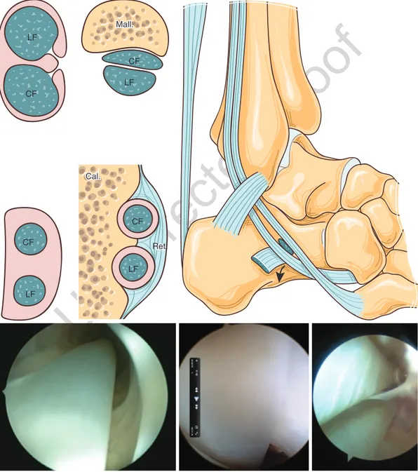
They are generally described as having three different areas (A, B, and C) [6] to which Sammarco [7] has added a fourth (D) (Fig. 2.1).
Area A corresponds with the posterior side of the malleolus, featuring a gutter in 8 out of 10. The absence of a gutter at this level is recognized as being a risk of dislocation of the peroneal ten-dons [2]. In this part, the tendons are held back by their sheath, which provides a reinforcement that provides a great deal of stability: the superior peroneal retinaculum, distinct and wide along its entire retromalleolar trajectory (Fig. 2.2). The peroneus brevis tendon is anterior and flattened distally, while the peroneus longus tendon behind has a more round cross-section.
Area B corresponds with the part comprised between the malleolus at the level of the lateral side of the calcaneus and the cuboid bone. At this level, the two tendons are at first free and their
a b 1 2 3 5 6 7 4 a b c d
Fig. 2.1 The four areas of the peroneal tendons. (a) Lateral view. (b) Plantar view
1 2 3
Fig. 2.2 Retinaculum of the peroneal tendon (lateral view): (a) Superior retinaculum. (b) Inferior retinaculum. (c) Tubercle of the peroneal tendons
59 60 61 62 63 64 65 66 67 68 69 70 71 72 73 74 75 76 77 78 79 80 81 82 83 84 85 86 87 88 89
trajectory crosses the calcaneofibular ligament (which stands out in tendoscopy) while following the edge of the posterior subtalar joint. In this part, the peroneus brevis tendon is on top and the peroneus longus tendon underneath. More dis-tally, the two tendons each enter into their own tunnel. This very special area is situated at the level of the peroneal tubercle (PT). The tunnels are separated by a septum that arises from the
PT. In this trajectory, each tendon marks a furrow at the lateral side of the calcaneus. The inferior retinacular ligament marks the end of these osteofibrous gutters (Fig. 2.3).
Area C: Situated facing the cuboid bone, this area is that of the plantar crossing of the peroneus longus tendon, while the peroneus brevis tendon remains on the lateral side. In 20% of cases, there is an accessory fibular bone in this area.
Fig. 2.3 Arthroscopic anatomy. Layered sections of the different areas CF LF CF Mall. LF CF LF Cal. CF LF va Ret. 90 91 92 93 94 95 96 97 98 99 100 101 102 103 104 105 106 107
Area D corresponds to the trajectory of the peroneus longus tendon.
Tendoscopy is made possible thanks to the presence of a synovial sheath. The sheath is a single entity from the proximal part up to the peroneal tubercle (Fig. 2.4). While this does not have genuine therapeutic implications at present, it should be noted that on this entire trajectory, the two tendons remain connected first to the pos-terior side of the malleolus, then to the lateral side of the calcaneus, each by their own vincula. It lies in alignment with the muscle fibres and represents the vinculum of the tendons.
The main neurological risk is in regard to the sural nerve, which after having crossed the superficial aponeurosis, typically in the upper third of the leg, rejoins the lesser saphenous vein in the lateral third of the leg, between the fibula and the calcaneal tendon. It crosses the trajectory of the peroneal tendons in area B to then inner-vate the dorsolateral skin of the foot and the toes. At the level of the malleolus, it gives rise to a cutaneous branch that is important for innerva-tion of the heel (the calcaneal branch). The superficial fibular nerve does not constitute a risk. It runs in the lateral side of the leg, in front of the peroneal tendons, but typically pierces the superficial fascia 7–8 cm above the malleolus. Its superficial trajectory is then more forward, in front of the malleolus, constituting a risk primar-ily with the anterolateral route for arthroscopy of the ankle.
2.4
Technique
2.4.1 Setup
A tourniquet is placed above the knee, so as to take the path of the tendons into account. Rather than the supine position with a cushion under the prone buttock that is used by some, we preferentially use a sideways recumbent position with the foot raised. Nonetheless, it is sometimes useful to have an intermediate setup in the case where arthroscopy of the ankle is to be undertaken, so as to allow a sideways and an anterior position [7, 8]. The patient is placed lying on their side with their pel-vis tilted slightly backward by approximately 30°. The hip and the knee are free. The ankle is held in line with the hip by support placed 10–20 cm more proximal. It is important to carefully verify the setup of the patient that by means of three different positions needs to allow anterior arthroscopy of the ankle (position 1), a lateral endoscopy of the ankle (position 2), and possibly removal of the gracilis (position 3) to be performed.
Position 2 is obtained by performing an exter-nal rotation of the hip to place the anterior side of the ankle as the highest point. Position 3 is obtained by resting the ankle on the support. Position 1 is obtained by performing a flexion and an external rotation of the hip (Fig. 2.5).
2.4.2 The Instruments
The instrumentation is conventional with an arthroscope of 4 mm and an arthroscopy shaver of 3.5–4 mm. It is not essential to use an arthro-pump or even electrocoagulation as the interven-tion is carried out using a tourniquet.
A basket forceps is very useful to start debrid-ing a fissure tendinopathy. Among the small instruments, we prefer a N°15 scalpel blade, safer and less traumatizing than a blade of 11, and we recommend generating the first portal by employing two small Gillies hooks. It is further-more indispensable to have a small curved Halstead forceps. This allows trauma to the sub-cutaneous nerves to be avoided after incision of the skin.
1
Fig. 2.4 Synovial sheath of the peroneal tendons
108 109 110 111 112 113 114 115 116 117 118 119 120 121 122 123 124 125 126 127 128 129 130 131 132 133 134 135 136 137 138 139 140 141 142 143 144 145 146 147 148 149 150 151 152 153 154 155 156 157 158 159 160 161 162 163 164 165 166 167 168 169 170 171 172 173 174 175 176 177 178 179 180 181 182
2.4.3 The Actual Tendoscopy Technique for the Peroneal Tendons
The intervention can generally be performed under general or locoregional anaesthesia. Performing the procedure under local anaesthesia is also an option, with the major advantage of being able to carry out a dynamic test, which is useful in the diagnosis of certain forms of pero-neal instability [2].
2.4.3.1 The Approach Routes
It is possible to generate the portals along the full length of the tendon behind the fibula but also distally on the lateral side of the hindfoot. In the vast majority of cases, however, two portals, one 3 cm above and the other 3 cm below the malleo-lus, are sufficient. The proximal portal is per-formed first. It offers the advantage of allowing easier identification of the sheath of the peroneal
tendons, which is thicker at this level. The risk of nerve injury is much less, it is not necessary to dilate the peritendinous space, and the descent of the arthroscope in the sheath of the peroneal ten-dons is easier than when going up as the wall becomes thinner distally and there is more room.
A subcentimeter longitudinal incision using a blade of 15 therefore only opens the skin 2.5–3 cm above the malleolar tip for the sheath of the peroneal tendons. We recommend going down 1 cm when the intervention is in regard to the sinus tarsi (Fig. 2.6).
Using Gillies hook-type spreaders, and under visual control, the sheath of the peroneal tendons is then exposed for the longitudinal incision. It is then very easy to control and then to introduce the soft arthroscopy trocar into the sheath. The arthroscope is then pushed distally, beyond the tip of the malleolus. It is then possible to position the second portal using a needle. Transillumination allows the sural nerve to be avoided (Fig. 2.7).
a b c
Fig. 2.5 The three positions for the setup. (a) Position 1; (b) position 2; (c) position 3
a b
Fig. 2.6 Initial performance of the proximal portal between 2.5 and 3 cm above the malleolar tip. (a) Landmarks. (b) Opening of the sheath under visual control, equipped with Gillies hooks
183 184 185 186 187 188 189 190 191 192 193 194 195 196 197 198 199 200 201 202 203 204 205 206 207 208 209 210 211 212 213 214 215 216 217 218 219 220 221 222
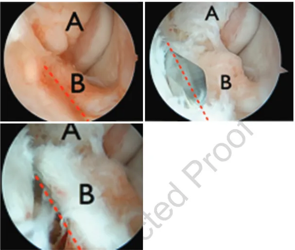
An initial inspection can then start from the distal emergence of the tendons, each from their own groove, up to the posterior side of the mal-leolus. It allows nearly all of the area to be visualized.
The vast majority of fissure tendinopathies are situated in the tendon reflection areas, under the malleolar tip.
By distally continuing the exploration after the malleolar groove, the base of the calcaneo-fibular ligament can be visualized. Its debride-ment with a shaver allows the posterior subtalar articulation to be visualized on its lateral and anterior side. It is then possible to perfectly con-trol the resection of small fragments or exostoses of this region by this arthroscopic portal. As was shown recently, arthroscopic treatment of the lat-eral impingement, proposed by Lui [9], particu-larly after fracture of the calcaneus, has proven to be an interesting conservative alternative both as a result of its efficacy and of its absence of mor-bidity [10].
This same route moreover allows access to the sinus tarsi to be fully secured: it suffices to perfo-rate the adipose tissue right after the base of the calcaneofibular ligament. It amounts to a bona fide conversion of a tendoscopy into subtalar arthroscopy since one can thereby reach the ante-rior part of this joint, as well as the calcaneal apophyseal edge and even the calcaneocuboid joint.
2.4.4 The Actual Lateral Endoscopy Technique
The intervention takes place under general anaes-thesia only because locoregional anaesanaes-thesia does not allow for easing of the external rotation of the hip necessary for performing the anterior arthroscopy.
2.4.4.1 Placement of the Portals
Three portals are required to perform this sur-gery. The conventional anteromedial portal is called portal N° 1. The second portal (route N° 2) is not drawn on the skin; it is performed using transillumination after having placed the arthro-scope. The third portal (route N° 3) is that of the sinus tarsi. It is necessary to draw two lines on the skin: The upper edge of the peroneus brevis is a line passing through the malleolar insertion point of the anterior talofibular ligaments (ATFL) and of the calcaneofibular ligament (CFL) and ori-ented at 10° relative to the axis of the malleolus. Portal N° 3 is situated at the intersection of these two lines (Fig. 2.8).
2.4.4.2 Stage N° 1
The arthroscope is placed in the anteromedial portal (N° 1). In order to obtain a good view of the lateral talofibular gutter, it is very important to position portal N° 1 correctly, that is to say, in dorsal hyperflexion and as close as possible to the anterior tendon. The positioning of the view
Fig. 2.7 Performance of the distal portal by
transillumination Fig. 2.8
Performance of the sinus tarsi portal
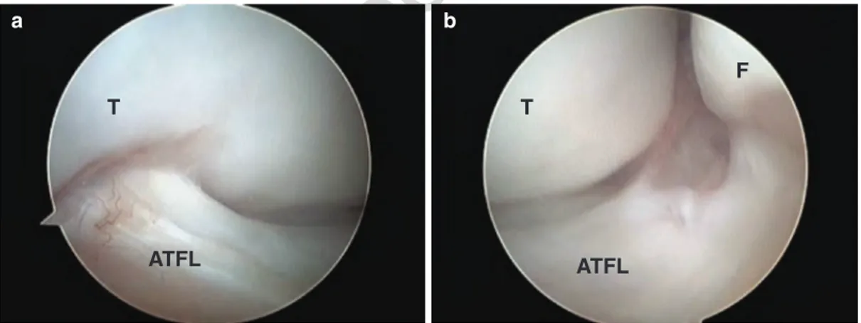
223 224 225 226 227 228 229 230 231 232 233 234 235 236 237 238 239 240 241 242 243 244 245 246 247 248 249 250 251 252 253 254 255 256 257 258 259 260 261 262 263 264 265 266 267 268 269 270 271 272 273 274 275 276 277 278 279 280 281 282
spot needs to allow the anterolateral gutter to be seen (Fig. 2.9). The luminous spot generated by the arthroscope on the skin then allows the anterolateral approach to be performed (portal N° 2). Using a shaver placed in this portal, debridement of all of the lateral gutter is per-formed. This preparation needs to allow all of the scar tissue between the anterior tibiofibular liga-ment and the anterior talofibular ligaliga-ment (ATFL) to be withdrawn. The preparation contin-ues with the release of the ATFL on its malleolar insertion. It is then possible to fully expose the ATFL by preparing it in the same way as a ten-don of the cuff on its upper side but also on its lateral edge (Fig. 2.10).
2.4.4.3 Stage N° 2
The arthroscope is placed in portal N° 2. An instrumental portal (portal N° 3) is performed at the level of the sinus tarsi using previously drawn cutaneous marks. A shaver is then intro-duced through this portal to complete the prepa-ration at the level of the malleolar insertion of the ATFL and of all of its lateral side and its lower edge. The dissection is then pursued by following the lateral articular surface of the talus until encountering the subtalar joint. The lateral edge of the calcaneus is identified below the joint. By staying in contact with the calca-neus with the shaver, the calcaneal insert of the
calcaneofibular ligament (CFL) is sought behind and within the fibular tendons while taking good care to remain in contact with the lateral cortex of the calcaneus and by moving from the front to the back. This stage needs to be done carefully in order to identify the CFL at its insertion.
2.4.4.4 Stage N° 3
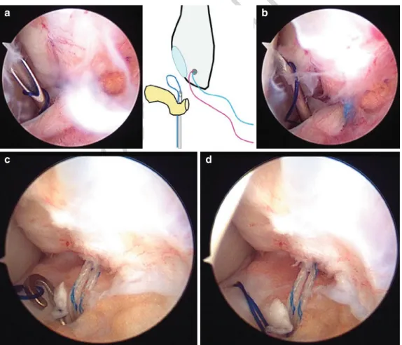
The arthroscope is introduced in N° 3. Using a shaver placed in portal N° 2 it is possible to pur-sue the dissection and full visualization of the talar insertion of the ATFL (Fig. 2.11).
2.5
Conclusion
In addition to a lateral approach of the bone and joint structures of the hindfoot, lateral endoscopy allows for full exposure of the lateral ligamen-tous apparatus and of the tendons. It hence con-stitutes a minimally invasive way to treat a considerable number of pathologies of this region. It allows a targeted treatment by à la carte endoscopic dissection. The indications are broader nowadays with the treatment of lateral impingement, fragment fractures (resection), subtalar arthrodesis, instability of the peroneal tendons, and above all treatment of instability of the ankle.
Fig. 2.9 View of the lateral gutter with the talus to the
right and the malleolus to the left Fig. 2.10 View of the lateral gutter after preparation
283 284 285 286 287 288 289 290 291 292 293 294 295 296 297 298 299 300 301 302 303 304 305 306 307 308 309 310 311 312 313 314 315 316 317 318 319 320 321 322 323 324 325 326 327 328 329 330 331 332 333 334 335 336 337
References
1. van Dijk CN, Kort N, Scholten PE. Tendoscopy of the posterior tibial tendon. Arthroscopy. 1997;13:692–8. 2. van Dijk CN, Kort N. Tendoscopy of the peroneal
ten-dons. Arthroscopy. 1998;14:471–8.
3. Guillo S, Calder JD. Treatment of recurring peroneal tendon subluxation in athletes: endoscopic repair of the retinaculum. Foot Ankle Clin. 2013;18:293–300. 4. Vega J, Batista JP, Golano P, Dalmau A, Viladot R.
Tendoscopic groove deepening for chronic subluxation of the peroneal tendons. Foot Ankle Int. 2013;34:832–40. 5. Michels F, Jambou S, Guillo S, Van Der Bauwhede
J. Endoscopic treatment of intrasheath peroneal ten-don subluxation. Case Rep Med. 2013;2013:4.
6. Brandes CB, Smith RW. Characterization of patients with primary peroneus longus tendinopathy: a review of twenty-two cases. Foot Ankle Int. 2000;21: 462–8.
7. Sammarco VJ. Peroneal tendoscopy: indications and techniques. Sports Med Arthrosc Rev. 2009;17:94–9. 8. Guillo S, Archold P, Perera A, Bauer T, Sonnery-
Cottet B. Arthroscopic anatomic reconstruction of the lateral ligaments of the ankle with gracilis autograft. Arthrosc Tech. 2014;3(5):e593–8.
9. Lui TH. Endoscopic lateral calcaneal ostectomy for calcaneofibular impingement. Arch Orthop Trauma Surg. 2007;127:265–7.
10. Bauer T, Deranlot J, Hardy P. Endoscopic treatment of calcaneo-fibular impingement. Knee Surg Sports Traumatol Arthrosc. 2011;19:131–6.
ATFL
CFL
SUBTALAR JOINT
S tep ha n e G UI LLO S te ph an e G U IL LO
Fig. 2.11 Visualization after arthroscopic dissection of the anterior talofibular ligament and the calcaneofibular ligament 338 339 340 341 342 343 344 345 346 347 348 349 350 351 352 353 354 355 356 357 358 359 360 361 362 363 364 365 366 367