Université de Montréal
Uncovering the role of misfolded SOD1 in the pathogenesis
of Amyotrophic Lateral Sclerosis
par Sarah Pickles
Département de Biochimie et médicine moléculaire, Université de Montréal Faculté de médicine
Thèse présentée à la Faculté médicine en vue de l’obtention du grade de doctorat
en Biochimie
Université de Montréal
Cette thèse intitulée:
Uncovering the role of misfolded SOD1 in the pathogenesis
of Amyotrophic Lateral Sclerosis
Présentée par Sarah Pickles
a été évaluée par un jury composé des personnes suivantes:
Dr. Luis Rokeach, Président rapporteur Dr. Christine Vande Velde, Directeur de recherché
Dr. Christopher Rose, Membre du jury Dr. Daryl Bosco, Examinateur externe Dr. Jean-Claude Labbé, Représentant du doyen
Résumé
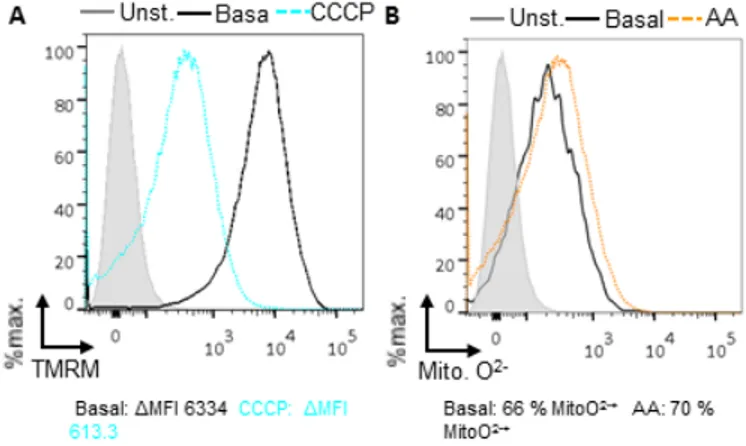
La sclérose latérale amyothrophique (SLA) est une maladie neurodégénérative charactérisée par la perte des neurones moteurs menant à la paralysie et à la mort. Environ 20% des cas familiaux de la SLA sont causés par des mutations de la superoxyde dismutase 1 (SOD1), conduisant vers un mauvais repliement de la protéine SOD1, ce qui a comme conséquence un gain de fonction toxique. Plusieurs anticorps spécifiques pour la forme mal repliée de la protéine ont été générés et utilisés comme agent thérapeutique dans des modèles précliniques. Comment le mauvais repliement de SOD1 provoque la perte sélective des neurones moteurs demeure non résolu. La morphologie, le bilan énergétique et le transport mitochondrial sont tous documentés dans les modèles de la SLA basés sur SOD1, la détérioration des mitochondries joue un rôle clé dans la dégénération des neurones moteurs. De plus, la protéine SOD1 mal repliée s’associe sélectivement sur la surface des mitochondries de la moelle épinière chez les modèles de rongeurs de la SLA. Notre hypothèse est que l’accumulation de la protéine SOD1 mal repliée sur les mitochondries pourrait nuire aux fonctions mitochondriales. À cette fin, nous avons développé un nouvel essai par cytométrie de flux afin d’isoler les mitochondries immunomarquées avec des anticorps spécifiques à la forme malrepliée de SOD1 tout en évaluant des aspects de la fonction mitochondriale. Cette méthode permettra de comparer les mitochondries portant la protéine SOD1 mal repliée à celles qui ne la portent pas. Nous avons utilisé un anticorps à conformation spécifique de SOD1, B8H10, pour démontrer que la protéine mal repliée SOD1 s’associe avec les mitochondries de la moelle épinière des rat SOD1G93A d’une manière dépendante du temps. Les mitochondries avec la
production excessive de superoxyde significativement plus grand, mais possèdent un potentiel transmembranaire comparable aux mitochondries B8H10-. En outre, la présence de la protéine
mal repliée SOD1 reconnue par B8H10 coïncide avec des niveaux plus élevés de la forme pro-apoptotique de Bcl-2. L’immunofluorescence de sections de moelle épinière du niveau lombaire avec l’anticorps spécifique à la conformation B8H10 et AMF7-63, un autre anticorps conformationnel spécifique de SOD1, démontre des motifs de localisations distincts. B8H10 a été trouvé principalement dans les neurones moteurs et dans plusieurs points lacrymaux dans tout le neuropile. Inversement, AMF7-63 a marqué les neurones moteurs ainsi qu’un réseau fibrillaire distinctif concentré dans la corne antérieure. Au niveau subcellulaire, SOD1 possèdant la conformation reconnu par AMF7-63 est aussi localisée sur la surface des mitochondries de la moelle épinière d’une manière dépendante du temps. Les mitochondries AMF7-63+ ont une
augmentation du volume comparé aux mitochondries B8H10+ et à la sous-population non
marquée. Cependant, elles produisent une quantité similaire de superoxyde. Ensemble, ces données suggèrent qu’il y a plusieurs types de protéines SOD1 mal repliées qui convergent vers les mitochondries et causent des dommages. De plus, différentes conformations de SOD1 apportent une toxicité variable vers les mitochondries. Les protéines SOD1 mal repliées réagissant à B8H10 et AMF7-63 sont présentes en agrégats dans les fractions mitochondriales, nous ne pouvons donc pas prendre en compte leurs différents effets sur le volume mitochondrial. Les anticorps conformationnels sont des outils précieux pour identifier et caractériser le continuum du mauvais repliement de SOD1 en ce qui concerne les caractéristiques
Abstract
Amyotrophic Lateral Sclerosis (ALS) is a neurodegenerative disorder characterized by the loss of motor neurons resulting in paralysis and death. Approximately 20% of familial ALS cases are caused by mutations in superoxide dismutase (SOD1), which leads to misfolding of the SOD1 protein, resulting in a toxic gain of function. Several antibodies have been generated that are specific for the misfolded form of the protein, and have been used as therapeutics in pre-clinical models. How misfolded SOD1 provokes a selective loss of motor neurons remains unresolved. Mitochondrial morphology, bioenergetics and transport are all documented is SOD1-mediated ALS models, thus mitochondrial impairment plays a key role in motor neuron degeneration. Moreover, misfolded SOD1 selectively associates with the surface of spinal cord mitochondria in ALS rodent models. We hypothesize that the accumulation of misfolded SOD1 on mitochondria could impair mitochondrial function. To this end, we developed a novel flow cytometric assay to immunolabel isolated mitochondria with misfolded SOD1 antibodies while also evaluating aspects of mitochondrial function. This method will allow for a comparison of mitochondria bearing misfolded SOD1 to those without. We utilized the B8H10 conformation specific SOD1 antibody to demonstrate that misfolded SOD1 associates with SOD1G93A rat
spinal cord mitochondria in a in a time dependent manner. Mitochondria with B8H10-reactive misfolded SOD1 associated with their surface (B8H10+) have a significantly larger volume and
produce excessive amounts of superoxide, but have a similar transmembrane potential compared to B8H10- mitochondria. In addition, the presence of B8H10-reactive misfolded SOD1
numerous puncta throughout the neuropil. Conversely, AMF7-63 marked motor neurons as well as a distinctive fibrillar network that was concentrated in the anterior horn. At the subcellular level, AMF7-63-reactive misfolded SOD1 also localized to the mitochondrial surface of spinal cord mitochondria in a time-dependent manner. AMF7-63+ mitochondria have an increased
volume compared to B8H10+ mitochondria and the unlabelled subpopulation. However, they
produce similar amounts of superoxide. Together, these data suggest that there are multiple species of misfolded SOD1 that converge at the mitochondria to cause damage. Moreover, different SOD1 conformations may ellicit varying toxicities towards mitochondria. Both B8H10 and AMF7-63-reactive misfolded SOD1 are present in aggregates in mitochondrial fractions and can therefore not account for any different effects produced in terms of mitochondrial volume. Conformational antibodies are invaluable tools to identify and characterize the continuum of misfolded SOD1 species with regards to biochemical characteristics and toxicity. The information presented in this thesis will be used in determining the future therapeutic potential of these antibodies.
Table of contents
Résumé……….i Abstract………...iv Table of contents………...vi List of tables……….xii List of figures………..xiiiList of acronymes and abbreviations……..………...………....xv
Dedication………....xxi
Acknowledgments………..xxii
Chapter 1: Introduction...1
1.1. Amyotrophic lateral sclerosis...1
1.2. Epidemiology………...1
1.3. Clinical presentation………...…3
1.4. Genetics………....4
1.4.1. Superoxide dismutase 1...4
1.4.2. Trans active response DNA binding protein 43………..7
1.4.3. Fused in sarcoma/Translocated in sarcoma………9
1.4.4. Chromosome 9 open reading frame 72………..10
1.4.5. Rare variants………12
1.6.1. Function……….15
1.6.2. Structure, folding and post-translational regulation………....17
1.6.2.1. SOD1 aggregation………...20
1.6.2.2. Additional post-translational modifications………...……...22
1.6.3. SOD1 animal models………....23
1.6.4. Misfolded SOD1………....29
1.6.4.1. Misfolded SOD1 in SOD1-mediated FALS...30
1.6.4.2. Misfolded SOD1-linked toxicity…………...32
1.6.4.3. Motor neuron vulnerability………...35
1.6.4.4. Modulating levels of misfolded SOD1………....36
1.6.5. Non-cell autonomous toxicity………..37
1.6.6. SOD1and sporadic ALS………..…39
1.6.7. Propagation of SOD1………...41
1.6.8. SOD1-mediated toxicity………..….43
1.6.8.1. Mitochondria as a target for SOD1 toxicity…………...43
1.6.8.1.1. Mitochondrial morphology………....45
1.6.8.1.2. Mitochondria transport………..…48
1.6.8.1.3. Mitochondrial calcium handling………...50
1.6.8.1.4. Mitochondrial bioenergetic defects………...51
1.6.8.1.5. Altered mitochondrial protein import………..…54
1.6.8.1.6. SOD1 at the mitochondria………...54
1.6.8.1.7. Mitochondrial aggregates………..……57
1.6.8.1.9. Mitochondrial targeted interventions………...…60
1.7. Therapeutics...61
1.7.1. Reducing levels of SOD1………..62
1.7.2. Reducing levels of misfolded SOD1………63
1.8. Overview and rational for thesis……….64
2. Immunodetection of outer membrane proteins by flow cytometry of isolated mitochondria………..…67 2.1. Contributions……….……68 2.2. Abstract………..68 2.3. Introduction...69 2.4. Protocol………..………70 2.5. Representative results………...79 2.6. Discussion………..….86 2.7. Acknowledgments……….……88
3. Mitochondrial damage revealed by immunoselection of ALS-linked misfolded SOD1 ...89
3.1. Contributions………...…..…90
3.2. Abstract………..90
3.3. Introduction………...91
3.4. Results………....93 3.4.1. Differential detections of misfolded SOD1 on mitochondria by
3.4.4. B8H10+ mitochondria have disrupted mitochondrial volume
homeostasis……….102
3.4.5. B8H10+ mitochondria produce excessive superoxide in the absence of depolarization……….104
3.4.6. Increased exposure of Bcl-2 BH3 domain in mitochondrial coated with misfolded SOD1……….107
3.4.7. B8H10 labels misfolded SOD1 within motor neurons prior to gliosis and clinical disease……….108
3.4.8. Dysfunction of B8H10+ mitochondria subset in a second model…110 3.4.9. B8H10 immunoreactivity in ALS patient cells...113
3.5. Discussion………..……...…117
3.6. Materials and methods………...123
3.7. Acknowledgments………...126
4. ALS-linked misfolded SOD1 species have divergent impacts on mitochondria…..…127
4.1. Contributions………...128
4.2. Abstract………128
4.3. Introduction…..………...129
4.4. Results………..132
4.4.1. Differential detection of misfolded SOD1 on mitochondria by conformation-restricted antibodies………..132
4.4.2. Immunodetection of mitochondrial-bound misfolded SOD1 by flow cytometry AMF7-63 detects misfolded SOD1G93A at the mitochondrial surface………138
4.4.3. Mitochondria with surface-bound AMF7-63-reactive SOD1 have an enlarged volume and produce increased amounts of superoxide. ...147
4.4.4. Misfolded SOD1 conformers are present in mitochondrial
aggregates………151
4.4.5. Preferential recognition of demetalated and reduced recombinant SOD1………..…..153
4.5. Discussion……….…156
4.5.1. Misfolded SOD1 specific antibodies recognize distinct non-native SOD1 confomers……….…156
4.5.2. AMF7-63-reactive misfolded SOD1 correlates with increased mitochondrial size/volume……….…157
4.5.3. Demetalated SOD1 is preferentially detected by misfolded SOD1 antibodies AMF7-63 and B8H10………...160
4.6. Conclusions………..160
4.7. Materials and methods………...…161
4.8. Acknowledgments………...164
5. Discussion………..165
5.1. Misfolded SOD1, Bcl-2 and VDAC1………...165
5.2. Selective accumulation of misfolded SOD1 in tissue and cells………..…...….166
5.3. A continuum of misfolded SOD1 species………167
5.4. Deficient mitochondrial import of SOD1………...171
5.5. Relevance of misfolded SOD1 specific antibodies B8H10 and AMF7-63 in ALS ………..172
5.6.5. Optineurin……….179
5.6.6. p62/Sequestosome 1………..179
5.6.7. Coiled-coil helix coiled-coil helix domain 10………...180
5.7. Novel avenues of research………...…181
5.7.1. Mitochondrial-associated membranes………..……181
5.7.2. Mitophagy………...183
5.7.3. M itochondrial unfolded protein response………...….185
5.8. Mitochondria in SALS……….189
6. Conclusion……….190
7. Bibliography………..i
Appendix 1:Misfolded SOD1 and ALS: zeroing in on mitochondria
Appendix 2: Endo-MitoEGFP mice: a novel transgenic mouse with fluorescently marked mitochondria in microvascular endothelial cells
List of tables
I. Introduction
Table 1: Genes implicated in ALS………..………...5
Table 2: Transgenic SOD1 mouse models………...…24
Table 3: Transgenic SOD1 rat models……….25
Table 4: Misfolded SOD1 specific antibodies………..31
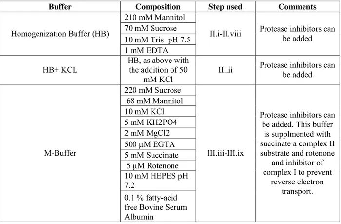
II. Chapter 2 : Immunodetection of outer membrane proteins by flow cytometry of isolated mitochondria Table 1: Buffer compositions………...…71
List of figures
I. Introduction
Figure 1: ALS-causing SOD1 mutations………...6
Figure 2: Timeline of selected histopathological and clinical changes relevant to disease in SOD1G93A mice………....27
Figure 3: Timeline of selected histopathological and clinical changes relevant to disease in SOD1G93A rats………...28
Figure 4: Misfolded SOD1 specific antibodies ………..………34
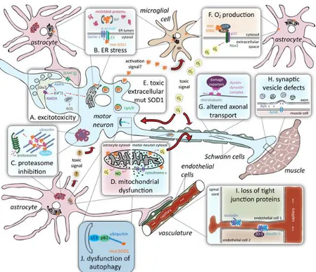
Figure 5: Proposed mechanisms of toxicity in SOD1-mediated ALS………...44
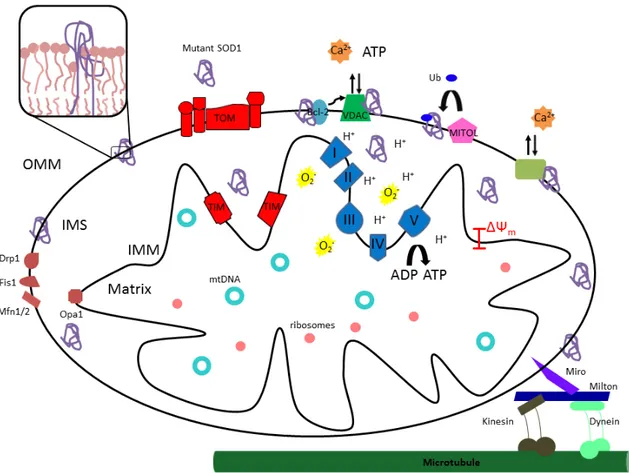
Figure 6: Mutant SOD1 and mitochondria in ALS………...46
II. Chapter 2 : Immunodetection of outer membrane proteins by flow cytometry of isolated mitochondria Figure 1: Schematic of isolation, immunolabelling and analysis of mitochondria...……….….80
Figure 2: Strategy for the analysis of isolated mitochondria by flow cytometry………....83
Figure 3: Assaying mitochondrial transmembrane potential (ΔΨm) and superoxide production in isolated mitochondria by flow cytometry………...85
III. Chapter 3: Mitochondrial damage revealed by immunoselection for ALS-linked misfolded SOD1. Figure 1: Preferential detection of B8H10 reactive misfolded SOD1 associated with mitochondria………..…95
Figure 2: Detection of mitochondrial-bound misfolded SOD1 by flow cytometry…………..100
Figure 3: Mitochondria with misfolded SOD1 associated have a greater mitochondrial volume ………..103
Figure 4: Mitochondria with misfolded SOD1 associated exhibit an increased production of mitochondrial superoxide but retain a normal transmembrane potential………..105
Figure 5: Increased Bcl-2 BH3 domain exposure on mitochondria bearing misfolded SOD1 ……….109
Figure 6: Accumulation of misfolded SOD1 in motor neurons begins prior to gliosis and motor
neuron loss………...111
Figure 7: Mitochondrial-associated misfolded SOD1 tracks with mitochondrial damage in
SOD1G37R mouse model………...114
Figure 8: Misfolded SOD1 detection in ALS patient cells………...115
IV. Chapter 4: ALS-linked misfolded SOD1 species have divergent impacts on
mitochondria
Figure 1: Misfolded SOD1 antibodies have distinct labelling patterns in SOD1G93A rat spinal
cords………...134
Figure 2: B8H10 and AMF7-63 reactive misfolded SOD1 is present in SOD1G93A spinal
mitochondrial fractions………..139
Figure 3: Four distinct mitochondrial subpopulations revealed by simultaneously
immunolabelling with AMF7-63 and B8H10 misfolded SOD1 antibodies………145
Figure 4: Presence of AMF7-63 reactive misfolded SOD1 at mitochondrial surface
coincides with increased mitochondrial volume……….149
Figure 5: AMF7-63 and B8H10 antibodies detect misfolded SOD1 in spinal cord
aggregates………..152
Figure 6: Misfolded SOD1 specific antibodies show preferential avidity for demetelated
(apo) SOD1………154
V. Discussion
List of acronyms and abbreviations
3’UTR: Three prime untranslated regionAA: Antimycin A
AAV: Adeno-associated virus
ALS Bi: Amyotrophic Lateral Sclerosis with behavioral impairment ALS/Ci: Amyotrophic Lateral Sclerosis with cognitive Impairment ALS: Amyotrophic Lateral Sclerosis
APOA2: ApolipoproteinA-II ASP: Antisense oligonucleotides
ATFS-1: Activating transcription factor associated with stress-1 ATP: Adenosine triphosphate
ATX2: Ataxin-2
Bcl-2: B cell lymphoma 2
BiP: Binding immunoglobulin protein
C. elegans: Caenorhabditis elegans
C9ORF72: Chromosome 9 open reading frame 72 CCCP: Carbonyl cyanide m-chlorophenyl hydrazone CCS: Copper chaperone for superoxide dismutase
CFTR: Cystic fibrosis transmembrane conductance regulator CHCHD10: Coiled-coil helix coiled-coil helix domain 10 CHOP: C/EBP homologous protein
CREST or SS18L1: Synovial sarcoma translocation gene on chromosome 18-like component of neuron-specific nBAF chromatin remodeling complex
CyPH: Cyclophilin D DCA: Dichloroacetate
DENN: Differentially expressed in normal neoplastic cells Derlin-1: Degradation in endoplasmic reticulum protein DNA: Deoxyribonucleic acid DNA
DRG: Dorsal root ganglia Drp1: Dynamin-related protein 1 DSE2: Disease specific epitope
EDTA: Ethylenediaminetetraacetic acid
eIF2A: Eukaryotic translation initiation factor 2A ER: Endoplasmic reticulum
ERAD: Endoplasmic-reticulum-associated protein degradation Erv1: Essential for respiration and vegetative growth
ERα: Estrogen receptor alpha ETC: Electron transport chain
FALS: Familial Amyotrophic Lateral Sclerosis Fis1: Fission 1
GEFs: Guanine Exchange Factors GRP-78: 78 kDa glucose-related protein Grx2: Dithiol glutaredoxin
hESC: Human embryonic stem cells HIV-1: Human immunodeficiency virus 1
hnRNP: Heterogeneous nuclear ribonucleoproteins IL-4: Interleukin 4
IMM: Inner mitochondrial membrane IMS: Intermembrane space
iPSC: Induced pluripotent stem cell JNK: c-Jun N-terminal kinases KARS: Lysyl-tRNA synthetase kDa: Kilo Dalton
LC3: Microtubule-associated protein 1A/1B-light chain 3 LC3-II: LC3-phosphatidylethanolamine conjugate (LC3-II) MAMs: Mitochondria-associated membranes
MAP: Mitogen activated protein MFI: Mean fluorescence intensity Mfn1/2: Mitofusin 1/2
Mia40: Mitochondrial intermembrane space assembly 40 MIF: Macrophage migration inhibitory factor
MS758: Misfolded SOD1 clone 758 MSC: Mesenchymal stem cells
MTG: MitoTracker Green mtDNA: mitochondrial DNA
MTS: Mitochondrial targeting sequence
NADPH: Nicotinamide adenine dinucleotide phosphate NEFL: Neurofilament
NES: Nuclear export sequence NF-ĸB: Nuclear factor-kappa B NLS: Nuclear localization sequence NMJ: Neuromuscular junction NRF1: Nuclear respiratory factor-1 NSC: Neural stem cells
OBMPFD: Inclusion body myopathy with early-onset Paget disease and Frontotemporal dementia
OMM: Outer mitochondrial membrane
OMMAD: Outer mitochondrial membrane associated degradation Opa1: Optic atrophy 1
PGC1-α: NAD-dependent protein deacetylase, or peroxisome proliferator-activated receptor gamma, coactivator 1 alpha
P: Post-natal day
QGSY: Gln-Gly-Ser-Tyr
RAN translation: Repeat associated non-ATG translation RGG: Arg-Gly-Gly
RIP-1: Receptor interacting-1 protein RNA: Ribonucleic acid
ROS: Reactive oxygen species RRM: RNA recognition motifs
SALS: Sporadic Amyotrophic Lateral Sclerosis SCA2: Spinocerebellar ataxia type 2
scFv: Secretable single-chain fragment variable SDS: Sodium dodecyl sulfate
SEDI: SOD1 exposed dimer interface shRNA: short hairpin RNA
SIRT3: Sirtuin 3 SNPH: Synaptaphilin
SOD1: Superoxide dismutase T cells: T lymphocytes
TALEN: Transcription activator-like effector nucleases TDP-43: Transactive response DNA binding protein 43 TMRM: Tetramethylrhodamine methyl ester
UPRmt: Mitochondrial unfolded protein response
USOD: Unfolded SOD1
VDAC1: Voltage dependent anion channel 1 VEGF: Vascular endothelial growth factor
Acknowledgements
It is said that it takes a village to raise a child. In my experience it takes a whole research community to graduate a PhD candidate.
On that note, I would like to thank the whole CRCHUM, old and new. I am blown away by the creativity, expertise and generosity of everyone at this research center, especially the Neuroscience Axis. In particular, I would like the thank Nathalie Arbour, for all her support and advice.
To my current and former lab mates, Laurie Destroismaisons, Sarah Peyrard, Yousra Khalfalah, Sabrina Semmler, Guillaume Caron and Jade-Emmanuelle Deshaies, Karli McDonald, Anais Aulas and Stephanie Stabile, it has been has been a pleasure and honor working with you day in and day out. We have shared successes, setbacks, expertise, advice, cocktails, jokes and occasionally some science. I wish you all nothing but success for the future and have the utmost confidence you will all do great things. To the Mito-Team, Sabrina and honorary member Laurie, thank you for all your hard work, I know I could not have done it without you both, keep up the good fight. To Christine, I most wholeheartedly want to thank you for this opportunity and your continued support. You have not just been my supervisor, but also my mentor, career adviser and cheerleader. I sincerely enjoyed my time in your lab and hope to be half the scientist you are.
To my family, Mom, Dad and Grandma, thank you for always being there, thank you for your support, and thank all your for all your love. All those poster projects finally paid off! I love all so much and am so grateful for having you in my life. To my husband, Matthew, thank you for your love, patience, counsel, and calmness. I love you and am excited to begin this next chapter in our lives.
1. Introduction
1.1. Amyotrophic lateral sclerosis
Amyotrophic Lateral Sclerosis (ALS) was first described by Jean-Martin Charcot as a progressive paralysis of voluntary muscles ultimately leading to death [1]. Charcot classified these symptoms under the name of ALS with amyotrophic meaning atrophy of muscles, and lateral sclerosis referring to the hardening of tissue in the lateral part of the spinal cord. In North America, ALS is also called Lou Gehrig’s disease, in honour of the New York Yankees player who died of this disease in 1941. In France, the disease is known as Charcot’s disease, named after its founder, whereas in the United Kingdom and Australia it is commonly referred to simply as Motor Neuron Disease.
1.2. Epidemiology
ALS is a late onset neurodegenerative disorder. The mean age of onset is 61.8 ± 3.8 years [2]. The range of disease onset is quite broad, with symptoms beginning from twenty to ninety years old [3]. Juvenile and early adult onset are occasionally reported and associated with specific genetic lesions [4, 5]. ALS is relatively rare with an incidence of 1.8/100, 000 in North America [2]. The incidence in men is slightly higher than woman [6].The prevalence of ALS disease is 3.4 per 100, 000 in North America [2]. These figures underscore the lifetime risk of
Although clinically identical, ALS patients can be segregated into two groups, sporadic (SALS) and familial ALS (FALS) [9]. SALS is used to describe individuals that have no family history of ALS, which is contrasted with FALS which is used to refer to individuals who have a first or second degree relative with ALS [10]. Approximately 5 to 10% of ALS cases are classified as familial [11], leaving the vast majority due to sporadic and not fully defined etiologies.
While the cause(s) of SALS is largely undefined, it is postulated that symptoms can result from genetic susceptibility in combination with environmental factors and time [12]. Increased age and tobacco use are associated with increased incidence of ALS [12]. Other proposed risk factors are athleticism [13], pesticide exposure [14], and trauma [15], however, the validity of these risk factors remains to be confirmed. The complexity and cost of determining environmental risk factors of SALS make it a daunting task, and thus limit the available strategies to model SALS in a research setting.
Classically, ALS is considered a disease with a purely motor phenotype, however this view is gradually changing. It is now appreciated that many patients exhibit cognitive defects, and in fact, ALS overlaps significantly with Frontotemporal Dementia (FTD). Following Alzheimer’s, FTD is the second most common form of dementia, and manifests as executive and language dysfunction with changes in behaviour and personality [16]. The prevalence of FTD is between 10 and 30 cases per 100,000 in people aged 45 to 60 years [17]. Approximately 50 % of ALS patients have a loss of function in neurological tests examining frontal lobe function. These patients are referred to as ALS with cognitive or behavioral impairment (ALS Ci/ALS Bi). Fifteen percent of ALS patients have a significant enough decline in frontal lobe function to be
officially diagnosed with FTD, and are classified as ALS-FTD [18]. ALS also clusters with certain neurodegenerative and neuropsychiatric diseases, for instance diagnosis of schizophrenia is higher in families with ALS, as well as rates of suicide [19]. Genetic susceptibility to neurological and neurophysiological factors may be at play for increasing risk in families with known ALS cases, however these factors are yet to be identified.
1.3. Clinical presentation
ALS is characterized by the degeneration of upper motor neurons in the cerebral cortex, and lower motor neurons in the brain stem and spinal cord [20]. Symptoms of lower motor neuron degeneration include muscle cramping and fasciculations (involuntary muscle contractions), muscle atrophy and corresponding weakness [20]. Symptoms caused by the loss of upper motor neurons include motor symptoms, such as uncoordinated and slow movement, spastic tone, as well as non-motor symptoms such as executive dysfunction [20]. Loss of upper motor neurons can also produce some Parkinson-like symptoms, including muscle rigidity and tremor [20].
In the majority of patients (65-75%), onset of disease begins in the limbs (spinal onset), usually unilateral [21]. About one-third of patients have a bulbar onset, where they experience speech and swallowing dysfunction first, characterized by flaccid or spastic dysarthria, dysphagia, hoarseness, tongue wasting, weakness and fasciculations [21]. Limb onset is associated with prolonged survival, as well as younger age at presentation of symptoms [20].
of onset, symptom presentation, and rate of disease progression, highlighting that ALS is clinically heterogeneous [25].
1.4. ALS Genetics (Table 1)
1.4.1. Superoxide Dismutase 1
In 1993, SOD1 (superoxide dismutase 1), found on chromosome 21q22.22 was identified as the first gene to cause FALS [26]. This finding was surprising given that SOD1 had been known as an abundant detoxifying enzyme for years [27]. There are over 170 reported mutations in SOD1 that cause ALS ([28], ALSoD: http://alsod.iop.kcl.ac.uk/) (Fig. 1). This is remarkable given that the entire gene encodes five exons and produces a protein of only 153 amino acids, even more surprising is that there are over 75 different amino acid mutations [29]. A major area of study in ALS research is determining what properties the SOD1 mutations have in common, and which of these are important for pathogenesis. Minor amino acid substitutions, such as G93A, (glycine to alanine) lead to disease, suggesting that almost any alteration in protein structure will result in ALS. The validity of some of the identified SOD1 mutations as causative of ALS remains contentious [30]. ALS causative mutations in SOD1, are found in all five exons of SOD1, with no clear hotspots [31]. However, six mutations are reported at residue G93: A, C, D, R, S or V. Each mutation produces a variable phenotype with G93V being most aggressive, G93A with intermediate aggressiveness (2 to 3 years from diagnosis to death), G93S, C and D being least severe, and G93R having a variable phenotype [32]. The majority of SOD1 mutations (80%) are missense with only a few insertions and deletions [33]. In almost all ALS kindreds, mutations in SOD1 are dominantly inherited with the exception of the D90A mutation, which is recessively inherited [34]. Mutations in SOD1 constitute approximately 12
Table 1:
Genes implicated in ALS
Gene Protein Function Inheritance Diagnosis Percentage (%) of cases References
FALS SALS
SOD1 Superoxide metabolism AD, AR ALS, PMA 12 3 Rosen et al., 1993
TARDP RNA metabolism AD ALS, ALS-FTD 4 1 Sreedharan et al.,2008 Kabashi et al., 2008;
FUS RNA metabolism AD, AR ALS, ALS-FTD 4 1 Kwiatkowski et al., 2009; Vance et al., 2009
C9ORF72 DENN protein, unknown function AD ALS, ALS-FTD, FTD 40 7
DeJesus-Hernandez et al., 2011; Renton et al., 2011
SQSTM1 Ubiquitination, autophagy AD ALS, ALS-FTD 1 <1 Fecto et al., 2011
VCP Proteasome AD ALS, ALS-FTD, FTD, IBMPFD 1 1 Johnson et al., 2010
OPTN Autophagy AD, AR ALS, POAG <1 <1 Maruyama et al., 2010
PFN1 Cytoskeletal dynamics AD ALS <1 <1 Wu et al., 2012
UBQLNS Ubiquitination, autophagy X-linked ALS, ALS-FTD <1 <1 Deng et al., 2011
VAPB Vesicular trafficking AD ALS, SMA <1 <1 Nishimura et al., 2004
hnRNPA2B1/A1 RNA metabolism AD ALS, IBMPFD, FTD <1 <1 Kim et al., 2013 Table 1: Genes implicated in ALS. Adapted from Renton et al., 2014 [35] and Leblond et al., 2014 [10]. Values represent the
percentage of ALS cases explained by each gene. AD, autosomal dominant; AR, autosomal recessive; XD, X-linked dominant; PMA, progressive muscular atrophy;; IBMPFD, inclusion body myopathy with Paget’s disease and frontotemporal dementia;; POAG, primary open-angle glaucoma; SMA, spinal muscular atrophy; DENN, differentially expressed in normal and neoplasia.
Figure 1: ALS-causing SOD1 mutations. Adapted from Fujisawa et al., 2012 [36]. Schematic
depicting SOD1 mutations. Primary sequence for wild-type SOD1 is depicted. Color change denotes exons, exon 1, 3 and 5 (black) and 2 and 4 (red). Secondary structure displayed below, β strands (light blue arrows) and α helix (dark blue hexagon).
to 20% of FALS, and 1-2% of SALS cases [37]. Due to a founder effect, about 50% of North American ALS patients carry the A4V mutation [38]. The second most prevalent mutation in North America is I113T. In contrast, the H46R mutation is more prevalent in Japan, with about 40% of SOD1-FALS patients carrying this particular mutation [38]. The population of Scandinavia has a high prevalence of the recessively inherited D90A mutation [12].
Genotype-phenotype predictions in ALS are difficult to make as the clinical presentation is quite heterogeneous. Exceptions include the A4V mutation, which is characterized by a short survival period, typically one year after diagnosis [39]. The H46R, D90A, G27R and D110Y mutations usually display slow disease progression with patients surviving 10 to 15 years after diagnosis [38-41]. Patients with SOD1 mutations usually have a younger age of onset, predominance of lower motor neuron involvement (limb-onset), and a lack of cognitive or behavioural difficulties [25].
1.4.2. Trans active response DNA binding protein 43
Identification of ubiquitinated transactive response DNA binding protein 43 (TDP-43) in cytoplasmic inclusions within spinal cords of ALS and FTD patients [42] led shortly thereafter to the discovery of mutations in TARDBP as causative for ALS [43-45] and FTD [46]. Prior to its association with ALS, TDP-43 was identified as a transcriptional repressor of human immunodeficiency virus 1 (HIV-1) [47]. Forty-seven TARDBP mutations have been described
TDP-43 is a highly conserved, ubiquitously expressed protein of 414 amino acids [48]. TDP-43 has a predominantly nuclear localization, although it can shuttle between the nucleus and cytoplasm [49]. Structurally, TDP-43 contains two ribonucleic acid (RNA) recognition motifs (RRM), that can bind RNA and deoxyribonucleic acid (DNA), a C-terminal glycine-rich prion-like domain [50], an N-terminal nuclear localization sequence (NLS), and a nuclear export sequence (NES) located in RRM2 [51]. Recently, TDP-43’s previously ignored N-terminus has garnered attention and is now considered important for TDP-43 aggregation [52-55].
TDP-43 is part of a family of proteins referred to as heterogeneous nuclear ribonucleoproteins (hnRNPs) that are involved in multiple stages of RNAprocessing. TDP-43 also binds DNA and is involved in transcription. TDP-43 functions in RNA splicing, regulating the splicing of cystic fibrosis transmembrane conductance regulator (CFTR) [56] and apolipoproteinA-II (APOA2) [57]. In addition, TDP-43 is involved in the generation of microRNA, RNA transport, translation and the formation of stress granules [49]. TDP-43 binds approximately 6000 mRNA transcripts in the brain and decreasing the level of TDP-43 expression affects the splicing and abundance of hundreds of RNAs [58]. Therefore the precise physiological role of TDP-43 in the central nervous system (CNS) is quite complex.
It is still unknown whether mutations in TARDBP result in disease by either a loss or a toxic gain of function, or even a combination of a nuclear loss of function and a cytoplasmic gain of function. In either case, TDP-43 levels are tightly regulated [59-61] via auto regulation by self-splicing and/decreased translation [58, 62].
1.4.3. Fused in Sarcoma/Translocated in Sarcoma
In 2009, mutations in the FUS gene, located on chromosome 16p11.2, were linked to ALS [63, 64]. The protein Fused in Sarcoma/Translocated in Sarcoma (FUS) was first investigated as an oncogene in liposarcoma [65]. The discovery of a second mRNA binding protein connected to ALS garnered much excitement, and confirmed the importance of mRNA processing in neurodegeneration.
FUS is a 526 amino acid protein. Structurally, it is composed of an N-terminal transcriptional activation domain, an RRM, three Arg-Gly-Gly (RGG1-3) repeat domains, a zinc-finger motif, a NES, and a C-terminal non-classical NLS [66, 67]. FUS also contains two predicted prion domains, in the N-terminal Gln-Gly-Ser-Tyr (QGSY) region and in the C-terminal RGG2 domain [50]. Under physiological conditions FUS is localized to the nucleus, although like TDP-43, it has the ability to shuttle between the nucleus and cytoplasm [68]. FUS functions in many cellular processes including DNA repair, transcriptional regulation, microRNA processing, splicing and stress response [67]. FUS, like TDP-43, also binds thousands of mRNA targets in the brain, and deletion of FUS leads to alterations in the splicing and abundance of hundreds of mRNAs [69]. Interestingly, the majority of binding targets of TDP-43 and FUS are distinct [69].
Of the approximately fifty reported dominantly inherited FUS mutations [25], the majority are missense, however deletions, insertions and mutations in the three prime
tremor and FTD [70-72]. Clinically, FUS mutations associate with a younger age of onset (less than 40 years old [25]), a bulbar presentation and shorter disease course. The P525L mutation has an extremely early onset, often in childhood, with an aggressive progression [73, 74]. Similar to TDP-43, whether FUS mutations cause ALS by a way of loss of function or gain of a toxic function remains an area of intense investigation.
1.4.4. Chromosome 9 open reading frame 72
In 2011, two groups independently identified a hexanucleotide (GGGGCC or G4C2)
repeat expansion in the non-coding region of the chromosome 9 open reading frame 72 (C9ORF72) gene on chromosome 9p21 [35, 75]. This finding sparked considerable excitement in the field, as the C9ORF72 expansion was responsible for over one third (~40%) of FALS cases, and about 7% of SALS cases [37], thereby constituting the most frequent cause of genetically inherited ALS. In addition, the C9ORF72 expansion is the first intronic expansion linked to ALS [37]. The C9ORF72 expansion is also associated with approximately 25% of familial FTD cases and 6% of sporadic FTD [76], again strengthening the premise that ALS and FTD constitute a disease spectrum.
Epidemiologically, the C9ORF72 expansion is most prevalent in European populations and correlates with early onset, typically bulbar, cognitive and behavioural changes, as well as increased incidence of neuropsychiatric illness [11, 20]. Similar to other nucleotide repeat expansion disorders like Huntington’s disease, ataxias, and myotonic dystrophy, the number of repeats is critical to pathogenesis [77]. Healthy people have between 2 to 30 hexanucleotide repeats in C9ORF72, whereas ALS patients have between 700 to 2400 repeats [20]. The number of repeats has been characterized in a number of tissues, and is larger in neuronal tissues than
in the blood [78, 79]. A firm correlation of expansion length with disease phenotype has not yet been established [80].
The function of chromosome 9 open reading frame 72 (C9ORF72) remains elusive. Unlike the other major FALS linked proteins, SOD1, TDP-43 or FUS, C9ORF72 was unknown before its association to ALS. Bioinformatic studies reveal homology to differentially expressed in normal neoplastic cells (DENN) domain proteins, which primarily function as Rab-Guanine Exchange Factors (GEFs) [81]. These proteins play a prominent role in membrane trafficking, including endocytosis and autophagocytosis [82]. C9ORF72 interacts with several Rabs (Rab1, Rab5, Rab9 and Rab11) as well as parts of the autophagic machinery, including ubiquilin-2 and microtubule-associated protein 1A/1B-light chain 3(LC3)-positive vesicles [83], supporting its
putative role in membrane trafficking.
Despite not knowing the precise physiological role of the C90RF72 gene product, there are three proposed mechanisms of pathogenicity. Collectively, decreased levels of the C9ORF72 transcript in ALS patient lymphoblasts [75] and the finding that Danio rerio and Caenorhabditis elegans loss of function models show axonal degeneration of motor neurons and locomotor
defects [84, 85], point towards haploinsufficency as a cause of disease. Conversely, gain-of-function models based on toxicity of RNA or dipeptide proteins generated by repeat associated non-ATG (RAN) translation are also suggested. In repeat diseases, translation can occur at various sites of the repeat, as opposed to the ATG initiation site, although the mechanisms are currently unclear, RNA structure likely plays a role in RAN-translation [86]. RNA foci are
are reversed when iPSC-differentiated neurons are treated with antisense oligonucleotides (ASO) against the G4C2 expansion [87]. RAN generated di-peptides containing (antisense:
Pro-Arg, Pro-Ala, Gly-Pro, and sense: Gly-Ala, Gly-Pro-Arg, Gly-Pro) cytoplasmic aggregates have also been detected in C9ORF72 patient brains [88, 90]. Recently, a study examined all three pathogenic hypotheses simultaneously. The Pro-Arg dipeptide, in particular, is extremely toxic to primary mouse cortical and motor neurons. Expression of the C9ORF72 expansion is also toxic to primary neurons, while knock-down of the C9ORF72 transcript had no effect on cell survival [91]. Interestingly, RNA and dipeptide related toxicity are linked, as the toxicity of Pro-Arg aggregates and the C9ORF72 expansion are synergistic [91]. The idea that these two mechanisms of toxicity may converge is in agreement with the finding that both RNA foci and dipeptide aggregates are present in post-mortem tissue of ALS patients [90].
1.4.5. Rare Variants
Reviews of ALS genetics are rapidly out-dated due to the high speed in which novel genes are discovered. Newly discovered genes include, OPTN [92], VCP [93], UBQLN2 [94],
DAO [95], hnRNPA2B1/A1 [96], CHCHD10 [96], MATR3 [97] and TBK1 [98], to name a few.
These genes and their protein products will be instrumental in revealing novel pathways involved in ALS, although they are rare causes of ALS [37]. Several of these genes are causative for other diseases including FTD, primary open angle glaucoma (POAG), and inclusion body myopathy with early-onset Paget disease and frontotemporal dementia (IBMPFD).
1.4.6. De novo mutations
De novo mutations are the subject of intense research as of late and are linked to many
neurological conditions, including autism [99]. De novo mutations are not inherited from parent to child, rather they occur through genetic copying error or during cell division [100]. De novo mutations of previously known ALS genes SOD1 [101] , and FUS [102-106] are found SALS cases. Exome sequencing of ALS trios (affected patients, and non-affected parents) identifies a mutation in SS18L1 (Synovial sarcoma translocation gene on chromosome 18-like component of neuron-specific nBAF chromatin remodeling complex) or CREST, a gene encoding a calcium-regulated transcriptional activator [107]. While this constitutes an interesting finding that implicates chromatin remodelling as a novel pathway in ALS, the pathogenicity of all de
novo mutations requires further validation [3].
1.4.7. Genetic risk factors
Twenty-three percent of SALS cases are proposed to be the result of a genetic defects [108]. Identified genetic modifiers include PGRN, KIFAP3, EPHA4 and UNC13A, although they require further validation [3]. A yeast screen of potential modifiers of TDP-43 toxicity finds poly-(A)-binding protein (Pab1p)-binding protein (PBP1) as a potent enhancer of toxicity [109]. The human orthologue is Ataxin-2. Mutations in ATX2 lead to a CAG expansion that causes Spinocerebellar ataxia type 2 (SCA2). ALS patients have a greater number ATX2 CAG repeats
1.5. Pathology
A pathological hallmark of ALS is the presence of intracellular protein inclusions in the soma and axons of neurons. Inclusions found in ALS patients can be placed into three categories, skein-like, bunina bodies, and hyaline, based on morphology and the identity of proteins found within the inclusion [110]. Skein-like and hyaline inclusions usually contain ubiquitin, while bunina bodies are ubiquitin negative [111]. Skein-like inclusions and bunina bodies are found in all ALS cases. SOD1, TDP-43 and FUS localize to skein-like inclusions while hyaline inclusions, containing SOD1, are mostly limited to SOD1-mediated FALS [112].
The protein inclusions found in the brain and spinal cords of ALS patients contains ubiquitinated proteins and the majority (~97%) of these inclusions are composed of TDP-43. Neurons from ALS patients have an accumulation of TDP-43 in the cytoplasm and a loss of nuclear TDP-43 [49]. TDP-43 present within inclusions is hyperphosphorylated, ubiquitinated and cleaved to generate C-terminal fragments [49] and recently found to be acetylated. TDP-43 inclusions are found in neurons and occasionally in glial cells in the brain (hippocampus and neocortex) as well as in spinal cord motor neurons [48]. That TDP-43 inclusions are found in the majority of ALS patients, even without TDP-43 mutations, suggest its involvement in almost all ALS cases. FUS immunoreactive cytoplasmic inclusions are present in less than 1% of ALS cases, and segregate with patients carrying mutations in FUS [63, 64]. TDP-43 inclusions are not found in these individuals, which may indicate that FUS acts downstream of TDP-43 [37] or by independent mechanisms. FUS inclusions are prominently cytoplasmic, although nuclear inclusions are also sometimes observed [113], and are present in neurons and less frequently in the glia [64]. Patients with the C9ORF72 expansion also have TDP-43 and sequestosome 1
(SQSTM1) orp62 inclusions. These inclusions are increasingly found in the frontal region, and in hippocampal neurons [114]. With the identification of new genes linked to ALS, their associated protein are increasingly studied for their presence in patient inclusions. Optineurin and ubiquilin-2 have been found within inclusions in some sporadic ALS cases [115]. The prevalence of these inclusions as well as their overlap with TDP-43 positive inclusions, or other protein inclusions remains to be fully characterized.
SOD1 inclusions are present in patients that carry a genetic defect in SOD1, which represents roughly 2% of all inclusions found in ALS patients [49]. SOD1 is found in both skein-like and hyaline inclusions [116, 117]. TDP-43 containing inclusions are not usually found in patients with SOD1-mediated ALS [118], implying SOD1-mediated FALS may be distinct. SOD1positive inclusions are reported in some sporadic ALS patients [116, 117], however the validity of this finding is currently under debate [119]. The incorporation of SOD1 into inclusions implies that it is aggregated and likely misfolded. In the following sections mutant SOD1 folding/misfolding and aggregation will be discussed in depth. Furthermore, the literature confirming the prescence of misfolded SOD1 in ALS patient inclusions will examined.
1.6. The role of SOD1 in ALS pathogenesis 1.6.1. Function
SOD1 is a ubiquitously expressed, well conserved metalloenzyme that binds copper and zinc [120]. Its principle function is to convert superoxide into hydrogen peroxide [27].
radical superoxide into hydrogen peroxide [27]. In addition to the cytosol and IMS, SOD1 is also reported to localize to the nucleus [123], peroxisomes [124], endoplasmic reticulum (ER) and Golgi apparatus [125]. SOD1 is an abundant protein, especially in the CNS where it constitutes 1-2% of total soluble protein [126]. SOD1 belongs to a family of three proteins (SOD1-3) with the same function. SOD2, is found in the mitochondrial matrix, and unlike SOD1, binds manganese instead of copper and zinc [127]. SOD3 is found in the extracellular matrix and like SOD1, also requires copper and zinc for its enzymatic activity [127].
SOD1 mutations were initially suspected as being causative for ALS due to a loss of function resulting in the accumulation of reactive oxygen species (ROS), however, the observation that many SOD1 mutants retain their dismutase activity at levels comparable to wild-type SOD1 refuted this claim [128]. Furthermore, SOD1 null mice do not develop symptoms resembling ALS [129]. In contrast, transgenic animals over-expressing mutant human SOD1 die early from a progressive paralytic disease [130], providing strong evidence that SOD1 mutations cause a toxic gain of function in ALS. Although SOD1 null mice do not develop ALS, and live to a reasonably old age, they are hypersensitive to axotomy [129] and cerebral ischemia [131] indicating that they have abnormal phenotypes associated with both motor neurons and neurons in general. A partial loss of SOD1 function may be involved in SOD1-linked ALS [29].
Recently, some non-canonical functions for SOD1 have come to light. Mutant SOD1 binds the 3’UTR of vascular endothelial growth factor (VEGF), a neurotrophic factor for motor neurons, causing destabilization of the transcript and loss of VEGF expression [132]. Similarly, mutant SOD1 regulates low molecular weight neurofilament (NEFL) mRNA through its 3’ UTR resulting in a loss of protein expression in neuronal cells [133]. Correction of the SOD1 mutation
in iPSC-derived motor neurons using transcription activator-like effector nucleases (TALEN)-mediated homologous recombination rescued the loss of NFL expression [134]. SOD1 is also reported to act as a transcription factor. In response to high levels of ROS, SOD1 translocates to the nucleus to increases the expression of genes related to oxidative stress resistance and cellular repair [135]. High levels of respiration causing production of oxygen and glucose, lead to SOD1-mediated stabilization of a group of proteins that downstream result in the repression of respiration [136]. These non-traditional functions of SOD1 clearly indicate there is much more to be discovered about the signalling functions of this molecule, as it regulates gene expression at multiple levels. Furthermore, how SOD1 is transcriptionally regulated remains to be fully explored, but uncovering the factors that modulate its expression could be vital for therapeutics aimed at changing SOD1 expression.
1.6.2. Structure, folding and post-translational modifications
Several post-translational modifications are required to form the mature SOD1 protein. Following translation, SOD1 is loaded with zinc by an unidentified mechanism, then copper by the copper chaperone for superoxide dismutase (CCS), which also facilitates the formation of an intramolecular disulfide bond between Cys57 and Cys146, and finally SOD1 forms a homodimer [137]. SOD1 can acquire copper by a CCS-independent pathway, involving reduced glutathione [138]. CCS may also regulate SOD1 activity in response to oxidative stress [139]. Zinc binding structurally organizes the immature polypeptide, while copper binding is
49-83) located between β-strands 4 and 5 and the electrostatic loop (residues 121-142) which is formed between β-strands 7 and 8. Both loops contribute to the active site, where the superoxide anion interacts with copper to be reduced [142].
SOD1 is an unusually stable protein capable of resisting denaturation at high heat (SOD1 has a melting temperature of approximately 92°C), whereas most proteins unfold between 40-60°C [143]. This stability is reflected in the ability of the mature SOD1 protein to withstand high concentrations of denaturants, 6M guanidine chloride and 10M urea, detergents, 4% sodium dodecyl sulfate (SDS), and treatment with 1 mg/mL proteinase K [144-147]. The stability of SOD1 is attributed to its metal occupancy, disulfide bond formation, and dimerization, which are all interdependent [110, 148]. Absence of metals will promote dissolution of the disulfide bond [149]. SOD1 without metals or an intact disulfide bond cannot form dimers [148, 150 {Arnesano, 2004 #703, 151], whereas presence of the metals and disulfide bond will promote dimerization [152].
The high stability of wild-type SOD1 is rather surprising given that almost any mutation leads to disease. Indeed, an intense area of investigation is to determine similarities and differences in the biochemical properties amongst the various mutant proteins, although it remains possible that there is no one common property [141]. Several mutants have a decreased thermostability, approximately 5-10°C lower than wild-type SOD1, comparable to apo SOD1 [153]. However, a number of mutants also have stabilities that are similar to wild-type SOD1 [154]. Furthermore, most mutants have wild-type-like activities [155, 156] and with the exception of the metal binding mutants, there are very few structural differences compared to wild-type SOD1 [157-160]. These findings suggests that the majority of mutant SOD1 is well folded. Examination of SOD1 folding in a cell-free rabbit reticulocyte lysate assay reveals
SOD1 mutants are unable to fold as efficiently as wild-type SOD1, although many mutants were able to eventually reach a well-folded state, as determined by resistance to proteinase k digestion [141]. This delay in folding kinetics leads to the accumulation of a pool of immature/intermediate SOD1 forms, which have decreased stability and may negatively affect a diverse array of cellular functions [141].
SOD1 lacking its complement of metals has a drastically reduced stability, as evidenced by a decreased melting temperature 50 to 59°C [161]. SOD1 with its proper complement of metals is referred to as holo, whereas SOD1 in the demetalated state is referred to as apo. Probing the metalation and activity status of recombinant mutant SOD1 proteins demonstrates that several mutants do not contain their full complement of metals and some lack dismutase activity, suggesting that copper is missing from the active site [156]. Soluble wild-type and mutant (G37R, H46R/H48Q and G93A) SOD1 from the brains and spinal cords of transgenic mice contain the proper complement of metals, except for H46R/H48Q, which lacks copper binding and has reduced zinc binding [162]. However, insoluble SOD1 had a low occupancy of metals [162]. Due to the high affinity of SOD1 for copper and zinc, the authors propose loss of metals was not probable in vivo and the origin of demetalated insoluble SOD1 originates from immature peptides which have yet to incorporate metals [162]. In support of this proposal, in-cell NMR experiments document that a portion of mutant SOD1 protein exists in an unstructured state incapable of zinc binding. Furthermore, toxic oligomers likely originate from this precursor [163]. Zinc can also aberrantly bind immature forms of SOD1 (apo SOD1 dimers, monomers
that in addition to its function in copper loading, it may also act as a chaperone [163] thereby decreasing levels of immature SOD1 [163].
Reduction of the disulfide bond in apo SOD1 substantially decreases the melting temperature from 52 °C [154] to 43°C [153], indicating the disulfide bond provides additional stability for SOD1 structure. Mutant SOD1 is more prone to reduction than wild-type SOD1 [149] and reduced SOD1 is destabilized compared to holo SOD1 [165].
As an alternative to the theory that SOD1 mutations retard protein folding kinetics leading to increased levels of unstable immature precursors, ALS mutations may predispose the protein to misfold in response to stress that the wild-type protein would normally withstand. One such stress is oxidation. SOD1 is exposed to ROS as part of its normal function and oxidative stress has been linked to ALS [166]. Increased levels of oxidative stress are documented in tissues from ALS patients [167, 168] as well as SOD1 mouse models [169]. Oxidation of SOD1 mutants causes destabilization of the protein mediated by oxidation of metal binding histidine residues [170]. Furthermore, oxidation leads to release of bound metals and exposure of hydrophobic residues, which is further enhanced when SOD1 is mutated [171]. Oxidation of either wild-type or mutant SOD1 leads to monomerization, followed by protein destabilization [172]. Treatment of SOD1 with denaturants causes unfolding with metal depleted monomers as an intermediate, thus confirming the plausibility of this precursor as an intermediate in SOD1 unfolding [173, 174].
1.6.2.1 SOD1 aggregation
Immature forms of SOD1 caused by the loss of one or more post-translational modifications, or genetic mutation, destabilizes the protein structure and likely underlies the
formation of aggregates [110]. Indeed, mutant SOD1 forms inclusions within spinal cord motor neurons in both FALS patients with SOD1 mutations and SOD1 rodent animal models [175-179]. One such subset of aggregates have a fibrillar appearance and are composed of β-sheets [180]. Interestingly, fibrils have the ability to self-regulate, once a fibril is formed it has the ability to nucleate or seed fibrils from properly folded protein [181, 182].
Fibrils are found in numerous neurodegenerative diseases including Parkinson’s, Huntington’s and Alzheimer’s disease [182]. The presence of fibrils in SOD1-mutated FALS remains debated, as reports of fibrils in spinal cords are conflicting [119, 183]. In ALS rodent models, SOD1 inclusions stain positive for Thioflavin T, a molecule that upon binding to β-sheets exhibits enhanced fluorescence [184]. Based on Thioflavin T fluorescence, mutant and apo SOD1 readily form fibrils in vitro [152, 185]. Moreover, structural studies demonstrate ALS-linked mutations and apo SOD1 form fibrils [158, 186, 187]. In silico analysis of the SOD1 sequence reveals four regions predicted to fibrillize [188]. Two C-terminal segments of SOD1, residues 101-107 and 147-153, accelerate fibril formation of apo wild-type and mutant SOD1
in vitro [188]. It has been proposed that metal deficient and disulfide reduced SOD1 would only
be required for the nucleation of fibril formation, followed by incorporation of more stable forms of SOD1, due to the self-seeding property of fibrils [110]. Thus, implying an initially small pool of unstructured SOD1 could spontaneously increase over time. Treatment of apo reduced wild-type SOD1 aggregates with proteases and subsequent analysis by mass spectrometry reveals three protease resistant peptides making up the core of the SOD1 aggregates. SOD1 mutants use
Whether or not SOD1 aggregates are toxic and by what mechanisms remains undefined. SOD1 monomers [190], oligomers [191] and aggregates [192] have all been proposed to mediate toxicity. Similarly, soluble SOD1 versus insoluble SOD1 is suggested to underlie SOD1-linked toxicity [193]. Regardless of what the toxic species is in vivo, there is consensus that misfolding of SOD1 protein is the initiating event.
1.6.2.2. Additional post-translational modifications
In addition to the post-translation modification required for the proper folding of SOD1, several other modifications have been described that can modify SOD1 aggregation propensity or SOD1 protein levels. The maturation of SOD1 consists of removal of the initiating methionine and acetylation of the N-terminal residue [194]. A recent study suggest that acetylsalicylic acid acetylates several lysine residues in mutant apo SOD1 which prevents its association into amyloid like fibrils [195]. Whether SOD1 is acetylated within cell culture, animal models, or in ALS patients remained unknown. In response to increased oxidative stress, wild-type and mutant SOD1 are glutathionylated at Cys111, which causes destabilization of the SOD1 dimer [196, 197]. Phosphorylation at Thr2 and Thr58 or Ser59 in SOD1 is reported in human erythrocytes [197]. SUMO (small ubiquitin-like modifier) proteins are a family of protein that are post-translationally and reversibly attached to proteins to modify their transport, regulation, stability, and response to stress [198]. SOD1 is sumoylated at Lys9 and Lys75 by both SUMO1 and SUMO2/3 [199, 200]. Sumoylation increases SOD1 stability and the formation of aggregates [199, 200]. Taken together, these findings provide evidence that SOD1 is post-translationally regulated in multiple ways, however the relevance of many of these modifications in ALS pathogenesis remains uninvestigated.
Insoluble SOD1 from the spinal cords of symptomatic SOD1G93A mice was found to be
mono and oligoubiquitinated [201]. Ubiquitination was observed in the spinal cord but not hippocampus, and is confined to Lys48, suggesting proteasome-mediated degradation of SOD1 [201]. Several E3 ubiquitin ligases are reported to ubiquitinate SOD1 for degradation, including E6-AP [202], Dorfin [203], neuronal homologous to E6AP carboxyl terminus (HECT)-type ubiquitin-protein isopeptide ligase (NEDL1) [204], and the ER-associated E3 ubiquitin ligase Gp78 [205]. Several of these E3 ubiquitin ligases, such as E6-AP and Dorfin, are found within mutant-SOD1 inclusions [202, 203].
1.6.3. SOD1 animal models
The first transgenic mouse model of ALS (SOD1G93A) was developed by expressing
SOD1 mini-genes, containing human genomic fragments driven by the endogenous SOD1 promoter and regulatory regions. This mouse develops a progressive and fatal paralysis that closely resembles ALS [130]. Many other transgenic models over-expressing human SOD1 missense mutations have been developed, as well as three C-terminal truncation mutations and several experimental mutations [130, 177, 179, 184, 206-215] (Table 2). The SOD1G93A mouse
is the most extensively studied, and for the time being is the gold-standard for testing therapeutic agents [25]. The SOD1G37R and SOD1G85R models are the second most frequently used mouse
models. In addition two rat models exist, expressing the G93A [216] and H46R [217] mutation in human SOD1 (Table 3).
Table 2
Transgenic SOD1 mouse models
Mutation Disease onset
(months) Reference
A4V a 8 Deng et al., 2006
G37R 4 to 6 Wong et al., 1995 G37R b * 11 to 13 Boillee et al., 2006 H46R 5 Chang-Hong et al., 2005 H46R/H48Q 4 to 6 Wang et al., 2002 H46R/H48Q/H63G/H120G 8 to 12 Wang et al., 2003 L84V 5 to 6 Tobisawa et al., 2003 G85R 8 to 14 Brujin et al., 1997 G86R c 3 to 4 Ripps et al., 1996
D90A 12 Jonsonn et al., 2006
G93A 3 to 4 Gurney et al., 1994
L126X 7 to 9 Wang et al., 2005
L126X 11 Deng et al., 2006
L126delTT 15 Watanabe et al., 2005
G127X d 8 Jonsson et al., 2004
Wild-type * non symptomatic Tu et al., 1998
Table 2: Transgenic SOD1 mouse models. Adapted from Turner and Talbot 2008 [206]. .
Double transgenic for SOD1WT (a). SOD1 gene floxed (b). Mouse gene (c). Homozygous for
Table 3
Transgenic SOD1 rat models
Mutation Disease onset
(months) Reference
G93A * 3.5 to 4 Howland et al., 2002
H46R 4 to 5 Nagai et al., 2001
Wild-type * non-symptomatic Chan et al., 1998
Table 3: Transgenic SOD1 rats models. Adapted from Turner and Talbot 2008 [206].
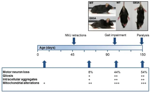
Characterization of SOD1G93A mice reveals disease onset at 3 months (higher levels of
transgene expression lead to earlier disease onset) with weakness, and weight loss due to muscle atrophy that progresses to paralysis and then death at about 4 months [130] (Fig. 2). Denervation of neuromuscular junctions begins around 47 days, with axon loss at 80 days and motor neuron loss at 100 days [218, 219]. Other prominent pathological features include fragmentation of the Golgi apparatus [219], mitochondrial morphological alterations [220, 221], SOD1 positive aggregates [176], gliosis of astrocytes and microglia [222]. Although the SOD1G93A rats have
not been characterized as extensively as the mouse modeld, they share many common features including an age dependent muscle atrophy progressing to paralysis (~3.5-4 months), motor neuron loss and gliosis [216, 223, 224] (Figure 3). Characterization of disease progression in the SOD1G93A rat model is challenging as there exist colony differences [224, 225]. Therefore
careful reporting of disease onset, surivial and major pathological hallmarks are required for each colony.
Small SOD1 animal models exist, however they receive far less attention than rodent models. Expression of mutant SOD1 in Danio Rerio causes dose dependent axonopathy, motor neuron loss, muscle atrophy, and these fish have less endurance and decreased survival, compared to wild-type controls [226, 227]. Expression of mutant or wild-type SOD1 in
Drosophila motor neurons decreases the flies’ ability to climb, causes defects in neural
electrophysiology, accumulation of SOD1 in neurons, and induces stress in surrounding glial cells, but does not lead to motor neuron loss or death [228]. The lack of specificity of phenotype between mutant and wild-type could indicate toxicity of wild-type SOD1. C. elegans models of ALS expressing mutant SOD1 selectively in neurons or muscle demonstrate modest locomotion defects and aggregation of SOD1, but normal survival [229, 230].
Figure 2: Timeline of selected histopathological and clinical changes relevant to disease
in SOD1G93A mice. Adapted from Bendotti and Carri 2004 [231], Fischer et al., 2004 [218]
and Turner and Talbot et al., 2008 [206]. Mitochondrial morphology and structure are altered prior to motor neuron loss and other hallmarks of disease.
Figure 3: Timeline of selected histopathological and clinical changes relevant to disease
in SOD1G93A rats. Data pooled from Howland et al., 2002, [216] (orange), Matsumoto et al.,
2006 [223] (purple) and Thompsen et al., 2014 [224] (red). Peak body weight: the point at which rats begin to loose weight due to muscle atrophy; Gait-impairment: the point at which animals being to limp; End-stage: the point at which the rat cannot right itself; NMJ:
1.6.4. Misfolded SOD1
Mutant SOD1 adopts a similar structure to wild-type SOD1 [157-160], however a portion of mutant SOD1 adopt a non-native “misfolded” conformation, as demonstrated by loss of Proteinase K resistance [232, 233{Vande Velde, 2008 #2, 234]. It is increasingly appreciated that SOD1-mediated disease pathogenesis is a result of misfolded SOD1. In an effort to specifically study misfolded SOD1, several groups have developed conformation specific antibodies that selectively target certain conformations of SOD1 (reviewed in [33, 235]). These antibodies were generated by immunizing animals with either apo SOD1G93A
protein(A5C3, B8H10, C4F6 and D3H5) or SOD1 peptides consisting of residues that are normally buried within the folded protein, Disease specific epitope (DSE2 3H1 and DSE1a 10C12), SOD1 exposed dimer interface (SEDI), unfolded SOD1 (USOD), mutant SOD1 specific antibody clone (MS758), AJ10 and a series of polyclonal peptide antibodies produced by Forsberg and colleagues (Fig. 3). Collectively, these antibodies recognize epitopes only available when SOD1 adopts a non-native conformation. These tools have allowed the field to probe the spatial and temporal localization of misfolded SOD1 as well as examine
mechanisms to modulate its abundance. While initially designed as therapeutics, their potential is just now emerging. Hopefully these reagents will allow the field to answer why motor neurons selectively degenerate in SOD1-mediated FALS and in SALS as a whole, and by what mechanisms.
![Figure 1: ALS-causing SOD1 mutations. Adapted from Fujisawa et al., 2012 [36]. Schematic](https://thumb-eu.123doks.com/thumbv2/123doknet/2063806.6223/33.918.141.775.205.718/figure-als-causing-sod-mutations-adapted-fujisawa-schematic.webp)

![Figure 4: Misfolded SOD1 specific antibodies. Adapted from Pickles et al., 2012 [235]](https://thumb-eu.123doks.com/thumbv2/123doknet/2063806.6223/61.918.181.727.151.601/figure-misfolded-sod-specific-antibodies-adapted-pickles-et.webp)