Université de Montréal
The effect of Nystatin on the inner ear
An experimental guinea pig study
by
Owen Woods, M.D.
Department of Biomedical Science Faculty of Medicine
Memoir presented to the Faculty of Postgraduate Studies to obtain the status of M. Sc,
in Biomedical Sciences
August 2011
This memoir entitled:
The effect of Nystatin on the inner ear An experimental guinea pig study
Presented by: Owen Woods
Was evaluated by a jury composed of:
Louis Guertin, M.D., F.R.C.S.C., president Issam Saliba, M.D., F.R.C.S.C., research director
Résumé
Objectifs
Le Nystatin est un antibiotique efficace pour le traitement d’otomycose. Bien que sa sécurité au niveau de l’oreille externe soit bien établie, son utilisation n’est pas recommandée lorsqu’il y a une perforation tympanique. L’objectif de cette étude est d’évaluer le potentiel ototoxique du Nystatin lorsque celui-ci est appliqué directement au niveau de l’oreille moyenne.
Méthodes
Nous avons fait une étude expérimentale avec 18 cochons d’Indes de souche Hartley que nous avons divisés en deux groupes. En exposant l’oreille moyenne de chaque animal au Nystatin (groupe I) ou à la néomycine (groupe II) et chaque oreille controlatérale à une solution physiologique (NaCl), la fonction auditive a été évaluée avec un test de potentiels évoqués auditif du tronc cérébral avant et après les injections. Une étude par microscopie électronique a permis une comparaison histologique de l’état des cellules ciliées cochléaires entre les 2 groupes.
Résultats
Les pertes auditives moyennes du groupe « Nystatin » étaient de 13.0 dB et comparables aux pertes moyennes observées dans les oreilles ayant été injectées avec du NaCl (4.0 dB dans le groupe I et 15.1 dB dans le groupe II). Le groupe de contrôle « néomycine » a subi une perte auditive moyenne de 39.3 dB, ce qui représente une différence cliniquement et statistiquement significative (p<0.001). L’étude histologique avec une microscopie à balayage électronique a démontré une conservation de l’architecture des cellules ciliées cochléaires dans les groupe Nystatin et NaCl. La néomycine a causé une destruction marquée de ces structures.
Conclusions
Le Nystatin ne provoque pas d’atteinte auditive ni de destruction des cellules ciliées externes après injection directe dans l’oreille moyenne chez le cochon d’Inde.
iii
Abstract
Objective
Nystatin is an effective topical antifungal agent widely used in the treatment of otomycosis. Though it is safe for external ear use, current recommendations are to avoid its use in cases of tympanic membrane perforation. The objective of our study was to test the security of Nystatin when applied directly to the middle ear of a guinea pig model.
Methods
We performed an experimental study with 18 Hartley guinea pigs that were divided into two groups. Exposing middle ears from one group to Nystatin (group I) and from the other to the ototoxic neomycin (group II), we compared results of auditory brainstem response (ABR) testing at three intervals during the study. Each animal’s contralateral ear was injected with a physiological solution (NaCl). At the end of the study, we performed a histological analysis of the animals’ cochleae using a scanning electron microscope.
Results
Average hearing loss in the Nystatin group was 13.0 dB which was similar to the results obtained in the NaCl-exposed ears (4.0 dB in group I and 15.1 dB in group II). Average hearing loss in the neomycin group was 39.3 dB, which represents a clinically significant difference (p<0.001). Scanning electron microscope evaluation revealed intact cochlear hair cell architecture in the Nystatin and normal saline groups, compared to important destruction in the neomycin group.
Conclusion
Nystatin does not cause hearing impairment or cochlear hair cell damage when exposed directly to the middle ear of a guinea pig model.
List of abbreviations
ABR: Auditory brainstem response Da: DaltonsdB: Decibel Hz: Hertz
OAE: Oto-acoustic emissions OHC: Outer hair cell
RWM: Round window membrane RWN: Round window niche
SEM: Scanning electron microscopy TM: Tympanic membrane
v
Table of contents
Résumé français ... i
Abstract... ii
List of abbreviations ... iii
Table of contents ... iv List of tables...v List of figures... vi Dedications... vii Acknowledgements ... viii Introduction...1 Objectives...3
Review of the literature ...4
Anatomy and physiology...4
Antibiotic ototoxicity ...8
Inner ear evaluation ...12
Methods...14
Animals ...14
General procedures and group assignment...14
Anesthesia protocol ...15
Tympanic membrane perforation and injection ...15
Auditory brainstem response...16
Animal sacrifice and preparation ...17
Scanning electron microscopy...18
Results ...19
Auditory Brainstem Response...19
Descriptive results...19 Statistical analysis ...21 Electron Microscopy ...23 Discussion ...25 Nystatin ...25 Results analysis ...27
Pathophysiology of the safety of Nystatin ...28
Possible implications in humans ...29
Study limits and future outlook ...31
Electron microscopy ... 31
Auditory brainstem response ... 31
Conclusion ...33
vii
List of tables
Table 1: Antibiotic Ototoxicity Literature Review Table 2: Description of Injection Materials Table 3: Auditory Brainstem Response Results
List of figures
Figure 1: Transverse section of the cochlea Figure 2: Group I Auditory Brainstem Response Figure 3: Group II Auditory Brainstem Response Figure 4: Scanning Electron Microscopy
ix
To my family and professors, who have guided me through every challenge, without whom such a project would have been impossible.
Acknowledgements
A special thank you to Dr. Saliba for his incredible dedication and guidance. To my professors and colleagues at Université de Montréal, thank you for your support and inspiration.
I would also like to recognize three teams at Sainte Justine Hospital and Université de Montréal who were essential to the completion of this project. First, Denise Carrier and the staff of the Sainte Justine Hospital Animal Care Center took the time to teach me the ins and outs of animal care in research. Second, Valerie Ouellette and the audiology team gave me a crash course in guinea pig ABR. And last but certainly not least Dr. Antonio Nanci, Sylvia Zalzal and their staff at the department of Dentistry provided their equipment and expertise making the scanning electron microscopy possible.
Finally, I would also like to extend my gratitude to Professor Miguel Chagnon, who once again guided me through the statistical analyses necessary for my project
Introduction
Otomycosis is a common pathology among children and adults. Initially defined as fungal infection of the external ear, this definition has evolved in recent years to include infection extending to the middle ear and open mastoid cavities(1). This type of infection has been associated with chronic otitis media(2). Some suggest that a recent increase in the prevalence of otomycosis is due to increasing use of topical quinolones, which increase risk of opportunistic infection.
Like with other fungal infections, exposure to a hot and humid environment is an important risk factor. Common presentation includes unilateral ear pain with persistent discharge and mild hearing loss. Physical examination reveals a typical white crust lining the outer ear. When culture is performed, Candida albicans, Candida parapsilosis and
Aspergillus fumigatus are the pathogens found in over 95% of cases(3). Proper treatment requires keeping the ear sufficiently dry and application of a local topical antifungal cream or ointment.
Studies measuring the safety of these agents in animal models have yielded mixed results. Acetic acid and gentian violet have demonstrated a potential for ototoxicity (4, 5). Other common agents including clotrimazole, ciclopirox and miconazole had no deleterious effect on cochlear outer hair cell architecture(5-7).
In 2000, Tom performed an experiment using electron microscopy to study the effect of several known anti-mycotic agents on guinea pig outer hair cell (OHC) structure(5). Though he was able to prove histologically an absence of toxicity from clotrimazole,
miconazone and tolnaftate, no conclusive results were obtained with regards to Nystatin. Furthermore, no correlation between histological analysis and auditory brainstem response (ABR) was made.
3
Objective
The objective of this study is to evaluate the toxic potential of Nystatin when applied directly into the middle ear of an animal model. In order to do so, we have performed the first study evaluating the effect of Nystatin on both the microscopic integrity of the OHCs of their cochlea and the results obtained on ABR evaluation.
Review of the literature
Anatomy and PhysiologyAn in-depth understanding of the anatomy and physiology of the auditory system is essential for such a project to be successful. A brief overview of the key concepts will be discussed here, with specific emphasis on ABR.
Before reaching the inner ear, a sound wave must travel through the external ear, the middle ear with its three ossicles and the oval window(8). Each of these structures has a specific role in sound amplification.
The external ear is composed of the auricle, the external auditory canal and the tympanic membrane. These structures combine to amplify sound by a factor of 30 to 100 at 3000 Hz(9). They also serve as a frequency filter, helping with sound localization since sources located at a higher location transmit high frequencies more readily.
The middle ear allows a transition from a low-impedance environment (air) to a high-impedance environment (fluid). This situation would normally result in a reflection of 99% of sound energy, but the middle ear compensates with two separate mechanisms. First and foremost, the large surface area of the tympanic membrane vibrates and concentrates its energy on the relatively small surface area of the oval window, increasing the amplitude by a factor of 17-20. Second, the lever effect created by the ossicles creates a slight amplitude gain (about 1.3 times)(10).
5
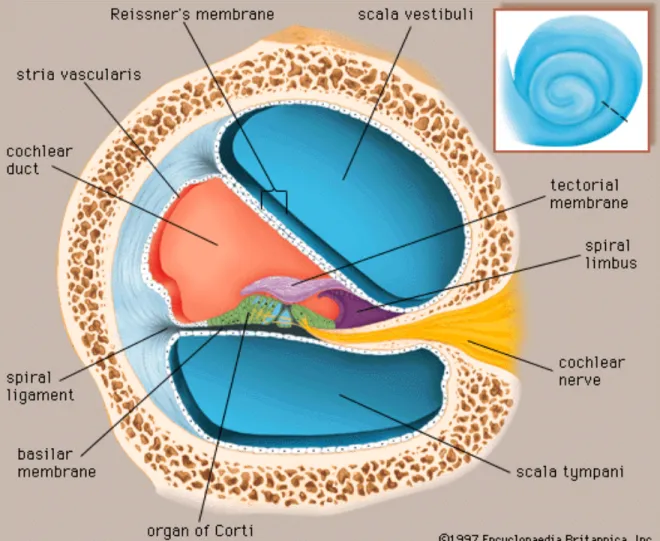
Figure 1: Transverse section of the cochlea
Source: 1997 Encyclopaedia Britannica, Inc.
The principal acoustic structure of the inner ear, the cochlea, has three functions: sound amplification, breakdown of complex acoustic waves into simpler ones, and conversion of sound waves into nerve effluxes. The cochlea is divided into three compartments, as shown on the transverse section of Figure 1. Note the basilar membrane, which separates the scala tympani from the cochlear duct. Also shown is the organ of Corti, the neurosensory unit of hearing, composed mainly of the tectorial membrane and inner and
outer hair cells. When the stapes vibrates, it causes a movement of footplate in the oval window and an increase in the pressure of the perilymph of the scala vestibuli. This pressure gradient is transmitted to the scala tympani via a communication at the cochlea’s apex, the helicotrema. From this point, a vibration of the basilar membrane instigates an approximation of the tectorial membrane to the hair cells. This begins the process known as sound transduction. Each section of the cochlea is responsible for sound transduction for a specific frequency, with low frequencies at the apex and high frequencies at the base. This tonotopic organization is maintained throughout the auditory pathway culminating in the primary auditory cortex.
Neural input leaves the cochlea via the cochlear nerve. The great majority (95%) of the nerve fibers that make up the cochlear nerve origin from the single row of inner hair cells, whereas the rest origin from the three rows of OHCs. The auditory nerve pathway continues toward the cochlear nucleus, the superior olivary nuclei, the lateral lemniscus and the inferior colliculus of the midbrain before reaching its terminal fibers in the acoustic area of the temporal lobe cortex(11). This pathway is the subject of testing when performing ABR tests. This clinical exam measures electrical activity along the auditory pathway using earphones and strategically placed electrodes. This exam does not require patient collaboration, making it ideal for testing in young children and animals. Hearing thresholds are determined mainly by identifying reproducible I, III, and V waves on two or more occasions beyond a reasonable doubt. These three waves measure activity in the distal cochlear nerve, the superior olivary complex, and the inferior colliculus, respectively(12).
7
Guinea pigs:
Most studies researching ototoxic agents are performed on animals. Though mice, rats and chinchillas have all been studied, guinea pigs are the most popular choice among researchers owing to their readily accessible round window niche (RWN). It is worth underlining some of the anatomical and physiological differences found between human and guinea pig auditory systems.
The inner ear anatomy of the guinea pig resembles that of a human with a few notable differences. In 2005, Wysocki reviewed the topographical anatomy of the guinea pig temporal bone(13). The three most important specific characteristics of the cochlea were its 3.5 turns, its thin bony cover and the unique position of the round window. Despite a study by Counter using magnetic resonance imaging claiming a guinea pig cochlea has 2.5 turns, Wysocki supported the popular belief that it actually ranges from 3.5 to 4 turns(14-17). The thin bony cover is found in all rodent species, in great contrast to human anatomy. Finally, the round window is found in the posterio-superior end of the basal turn of the cochlea. Although this finding does not have physiological implications, it makes access to the cochlea via the middle ear much easier in the setting of an experimental study.
Perhaps the most important functional difference between the two species is their hearing range. While humans can hear sound ranging from 20 to 20 000 Hz, guinea pigs have been shown in behavioral studies to respond to sounds up to 50 000 Hz(18). There appears to be a relationship between this finding and the extra turn of the cochlea. Studying several different animal species, West successfully demonstrated a statistically significant positive correlation between number of turns of the cochlea and length of the basilar
membrane with the range of audible frequencies(19). In low frequencies, however, guinea pigs are less sensitive. In a 2010 review Salt found that average hearing thresholds at 125 dB were 17.6 dB higher than the human average(20).
Despite these differences, humans and guinea pigs appear to respond similarly to known ototoxic agents. Blakley found dose-dependent hearing loss after Cisplatin treatment in guinea pigs comparable to levels reported in the literature for humans(21). Of the studies discussed in this paper, none have found major discrepancies between human and guinea pig response to ototoxic drugs.
Antibiotic Ototoxicity
Ototoxicity to antibiotic agents is not a new concept. Soon after gaining popularity in the early 1940’s, systemic gentamicin appeared to be responsible for permanent bilateral hearing loss with or without vestibular defects in a significant number of treated patients(22). Since that time, several studies have tested commonly used antibiotics for their potential short and long-term effects on hearing. For the purposes of this review, antibacterial agents will be discussed first followed by an in-depth look at anti-mycotic agents.
Aminoglycosides are bacteriostatic agents that exert their effect by inhibiting the 30S ribosomal subunits of targeted pathogens. Soon after an association between gentamicin treatment and hearing loss was established, cochlear hair cell damage was discovered on histological analysis. Further testing demonstrated intracochlear gentamicin concentrations were similar to systemic concentrations, but that the antibiotic was sequestered in the inner ear with a half life of up to 5 months(23). It appears to generate free
9 radicals which damage inner ear neurons, resulting in irreversible hearing loss(24). Recent studies have shown that hair cells at the base of the cochlea are affected before those at the apex, explaining why resulting hearing loss in worst in high frequencies(25). Severe toxicity can also cause retrograde auditory nerve damage. This effect is dose-dependent and can be worsened by age or in combination with other ototoxic treatments such as loop diuretics. Other aminoglycosides appear to be just as harmful. Despite one study suggesting its safety(26), neomycin has proven ototoxic potential and should be avoided when possible(5, 27). While amikacin, kanamycin and neomycin have an important propensity for cochleotoxicity, streptomycin appears to have more vestibulotoxic properties(28). In a study by Song et al., iron chelators seemed to have protective properties with the use of gentamicin(29). Further evidence should be obtained, however, before such a treatment can be considered safe. Though efforts continue to find effective protective agents against aminoglycoside ototoxicity(25), for the moment it is best to limit long-term use of these agents whenever possible.
The obvious deleterious effects of aminoglycosides have led researchers to investigate the possible consequences of other antibiotics, notably ones used for local treatment of otitis externa. Fluoroquinolones have proven their safety in a large number of studies. Topical ciprofloxacin does not cause significant damage to cochlear hair cells(30-35). This is presumably because the molecule does not diffuse across the cochlear round window membrane even when applied directly into the middle ear space(36). Among topical quinolones, only moxifloxacin has demonstrated a potential for cochleotoxicity(37), although this study was performed exclusively using distortion product oto-acoustic emissions and results have not been duplicated.
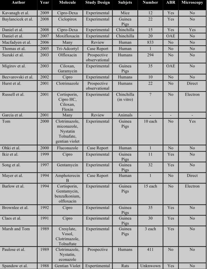
Antimycotic agents have been studied with great interest owing to the increasing worldwide rate of otomycosis. In 1974, Schonebeack and Zakrisson were the first to study the possibility of ototoxic antimycotics(38). They published a case report of two patients treated with 5-fluorocytosine and performed post-treatment audiograms, which were normal. Since that time, studies have greatly evolved. In order to test the effects of Nystatin, amphoterecin B, and Griseofulvin, Parker and James performed a standard microscopic evaluation of guinea pig cochleas treated with these agents(39). They concluded that while Nystatin and amphoterecin B seemed safe, Griseofulvin altered OHC architecture. Spandow was the first to utilize ABR testing post-antifungal treatment, finding a significant hearing loss in rats after gentian violet administration(40). Based on clinical symptoms in a prospective study of 411 patients, Paulose found no ototoxic reactions after topical treatments with clotrimazole, Nystatin and econazole(1). In an experimental guinea pig study using ABR testing, Marsh and Tom found solutions containing acetic acid and propylene glycol to be harmful. Clotrimazole and tolnaftate, on the other hand, had little or no impact on auditory thresholds(7). As a follow-up to this study, Tom used scanning electron microscopy (SEM) to verify the state of OHCs after exposure to six well-known agents(5). As expected, clotrimazole, miconazole and tolnaftate proved safe and gentian violet caused OHC destruction. Nysatin left an apparent residue in the cochlea impeding proper evaluation, which led the author to suggest avoiding Nystatin in cases of tympanic membrane perforation. Jinn reported modified OHC morphology and decreased time to cell death in guinea pig cochleas exposed to acetic acid, confirming earlier findings(4). Most recently, in 2008, Baylancicek found no significant impact of Ciclopirox treatment on ABR testing in guinea pigs(6). Table 1 summarizes our antibiotic ototoxicity literature review.
11
Table 1: Antibiotic Ototoxicity Literature Review
Author Year Molecule Study Design Subjets Number ABR Microscopy
Kavanagh et al. 2009 Cipro-Dexa Experimental Mice 12 Yes No Baylancicek et al. 2008 Ciclopirox Experimental Guinea
Pigs
22 Yes No
Daniel et al. 2008 Cipro-Dexa Experimental Chinchilla 15 Yes Yes Daniel et al. 2007 Moxifloxacin Experimental Chinchilla 20 OAE No
Macfadyen et al. 2006 Many Review Human 833 No No
Thomas et al. 2005 Tri-Adcortyl Case Report Human 1 No No Suzuki et al. 2003 Olfloxacin Prospective
observational
Humans 294 No No
Migirov et al. 2003 Ciloxan, Garamycin
Experimental Guinea Pigs
35 OAE No
Becvarovski et al. 2002 Cipro Experimental Humans 10 No No Hurst et al. 2001 Clotrimazole Prospective
observational
Humans 22 No Direct
Russell et al. 2001 Cortisporin, Cipro HC, Ciloxan, Floxin Experimental Chinchilla (in vitro) ? No Electron
Garcia et al. 2001 Many Review Animals - - -
Tom 2000 Clotrimazole, miconazole, Nystatin Tolnafate, gentian violet Experimental Guinea Pigs 10 each No Yes
Ohki et al. 2000 Fluconazole Case Report Human 1 No No Ikiz et al. 1999 Cipro Experimental Guinea
Pigs
11 Yes No
Song et al. 1997 Gentamycin Experimental Guinea Pigs
32 Yes No
Mayer et al. 1994 Amphoterecin B
Case Report Human 1 No Direct Barlow et al. 1994 Cortisporin,
Gentamycin, benzalkonium, olfloxacin Experimental Guinea Pigs 15 each No Electron
Brownlee et al. 1992 Cipro Experimental Guinea Pigs
35 Yes No
Claes et al. 1991 Cipro Experimental Guinea Pigs
30 Yes No
Marsh and Tom 1989 Cresylate, Vosol, Clotrimazole, Tolnaftate Experimental Guinea Pigs 3 each Yes No
Paulose et al. 1989 Clotrimazole, Nystatin, econazole
Prospective Humans 411 No No
Stone et al. 1975 Systemic Tobramycin RCT Humans 116 No No Schonebeck and Zakrisson 1974 5-fluorocytosine
Case Report Humans 2 No No
Inner Ear Evaluation
Testing inner ear function in an animal model is challenging, and each technique has its advantages and drawbacks. Here we will briefly review four well-described techniques: cochleogram plotting, SEM, otoacoustic emissions (OAE) and ABR.
Early studies used contrast microscopy to establish a mapping of apparent cochlear hair cell damage named a cochleogram(39). This technique has the benefit of studying the entire cochlear structure, thereby comparing basal and apical cell damage. Although this technique has been used in experimental studies since 1966, only recently has a review been published with a proposed standardized cochleogram technique(41). The authors suggest plotting basilar membrane length as a percent instead of a length, stating equations used for frequency-place maps and normalizing different basilar membrane lengths to percentaged before making average cochleograms. There are several limitations to this technique. First, no clear correlation has been demonstrated between electrocochleogram findings and frequency-specific hearing evaluation. Second, it is a technically difficult procedure due to tectorial membrance adhesion, which often limits the view of a significant portion of cochlear hair cells.
With the availability of SEM, preservation of cochlear hair cell architecture and the length of the basilar membrane are now used to evaluate potential damage(42). Basilar membrane length should be standardized and expressed as a percentage rather than a length(41). The limitations of this technique are twofold. First, proper visualisation of the
13 entire cochlear ramp and its inner and outer hair cells is extremely challenging. Details on cochlear dissection and preparation have been described elsewhere(43). Second, the results obtained are purely histological and may not accurately reflect in vivo inner ear function.
Two techniques are potentially effective in measuring inner ear function in an animal model. Oto-acoustic emissions are the easiest and fastest to perform, and have been used in a number of studies(44-51). Their reliability has been challenged in a study by Migirov et al., who found similar distortion products in guinea pigs treated with normal saline and topical gentamicin(44). He argues that results reflect the degree of inflammation of the middle ear more than actual inner ear function.
Auditory brainstem response is a more complex yet potentially more reliable and widely-used measure of animal inner ear function(6, 30-32, 34, 52-71). We recommend a combination of ABR and electron microscopy to correlate functional status with histological analysis. A recent study using these techniques demonstrated the protective effect of Ringer’s Lactate solution in guinea pigs who receive Cisplatin compared to a control group(72).
Methods
AnimalsWe used a total of 18 Hartley Guinea Pigs. We chose this species because of their readily accessible cochlea, their functional anatomy which resembles that of humans, their widespread use in similar studies and finally because of our significant experience with this species in our laboratory.
Animals were cared for in accordance with the guidelines of the Canadian Council of Animal Care and the standards of the institutional animal care committee.
General Procedures and Group Assignment
The experiment was performed over a span of 13 weeks, beginning with group assignation and ending with SEM interpretation.
Each guinea pig was randomly assigned to one of two groups: an experimental group (Group I) and a positive control group (Group II). Animals from Group I received Nystatin injections in one randomly selected ear and physiological saline (0.9% NaCl) injections in the other. Animals from Group II received neomycin injections in one ear and physiological saline in the other. Each contralateral ear was therefore used as a negative control, which allowed us to reduce the number of animals required and minimize the effect of variability between animals.
15
Anesthesia protocol
Anesthesia was induced by placing the animals in a plastic container with two entrance ports and one exit port. Oxygen and Isoflurane 1% were administered in parallel systems and were ventilated out through the exit port. All manipulations, including tympanic membrane (TM) perforation, middle ear injections, and animal sacrifice were performed under general anesthesia. The guinea pigs had spontaneous breathing but did not react to stimulus. ABR recordings required us to maintain anesthesia with a mask outside of the container. Each animal’s adequate recovery from anesthesia was assured before leaving it unsupervised.
Tympanic Membrane Perforation and Injection
On Day 1 of the experiment, we perforated the posterior superior quadrant of each TM under general anesthesia using an operative microscope. Approximately 60% of the surface of the TM was left intact in each case. This perforation provided an adequate exposure of the round window niche (RWN), where the assigned product would later be applied. Each animal then passed an ABR test within the first week. ABR protocol details are described below.
On Day 5, we injected approximately 0.1 cc of the experimental solution into each ear. The injection was performed through the perforation using a long 20-gauge needle. The solution was deposited directly over the round window niche and filled the entire middle ear cavity. A concern for the animals in Group II was that the neomycin, since it is a liquid and much less consistent than Nystatin, would not remain in the middle ear long enough to exert an effect. We therefore added 0.1 cc of Neosporin 0.25% cream to emulate the
conditions in Group I. Each injection as described above was repeated once a week for a total of three weeks.
At this point we waited a total of four weeks before proceeding with further tests. This waiting time was considered necessary in order to minimize the amount of residue present at the RWN, which if considerable could interfere with ABR readings as well as SEM analysis.
Our second ABR recordings were performed during week 8. A thorough cleaning and drying of each ear was performed the day before the recordings. The tympanic membrane was reperforated in the ears if closure had occurred.
Because the first set of injections did not cause adequate hearing loss as demonstrated by ABR results in the positive control neomycin group (group II), we repeated a series of three injections in each group at closer intervals (48 hours between injections). Four weeks later, we repeated all ABR testing.
Table 2: Description of Injection Materials
Drug Manufacturer Concentration Type
Nystatin Ratiopharm 100 000 U/g Ointment
Neomycin Ratiopharm 1% Aqueous
Neosporin GlaxoSmithKline 0.25% Cream
17
Auditory Brainstem Response
All ABR tests were performed with the same Nicolet Bravo™ device (Nicolet Bravo System; Nicolet Biomedical, Madison, WI, USA). The same room of the animal care center was used for all tests. Electrodes were inserted subcutaneously. The non-inverting electrodes were placed directly above the left and right mastoid processes, the inverting electrode was placed at the apex and the ground electrode was placed in the middle of the animal’s back.
We used tone burst stimuli to record differences across a range of frequencies. Tone bursts at 2000, 3000, 4000, 6000 and 8000 Hz were emitted. Amplitude thresholds were found to the nearest 5 dB. A minimum of 300 repetitions were averaged out with each recording. To be considered a positive reading, a visible wave V needed to be reproducible beyond a doubt on a minimum of two stimulations with the same amplitude. ABR values are provided as absolute decibel (dB) levels.
Animal Sacrifice and Preparation
All animals were sacrificed under general anesthesia. For initial dissection we focused on exposing the middle ear by removing the residual TM and the cartilaginous portion of the external auditory canal. Fixation was accomplished by immersing the guinea pig heads in a 2.5% Gluteraldehyde solution for 5 days.
Decalcification was perfomed using a concentrated Plank solution, consisting of Aluminum Chloride and Hydrochloric Acid. This solution was changed three times per week for two weeks leading up to electron microscopy.
Temporal bone dissection was performed at the end of this process. The bone was considerably softened and easy to section with a standard 15-gauge scalpel. We began by
dissection was required to locate the cochlea and separate it from the temporal bone without damaging it. In order to view rows of ciliated hair cells, transverse cuts through the cochlea were made. One section was obtained per turn of the cochlea, giving us four specimens to analyse from each ear.
Scanning Electron Microscopy
These sections of each cochlea were then analyzed with our research center’s scanning electron microscope (JEOL JSM-6460LV). All sections were evaluated for preservation or destruction of cochlear hair cells.
Statistical Analysis
Before beginning our study, we determined the number of animals necessary to find a clinically significant difference (10 dB) with sufficient statistical power using the method of Snedecor and Cochran(73).
To interpret ABR results, we performed a variance analysis with repeated measures. Results were compared between groups, ears injected and frequencies at each of three pre-determined time intervals: before injection, after one series of injections and after two series of injections.
19
Results
We excluded one animal from the Nystatin group at the beginning of the study due to profound bilateral hearing loss. Eight guinea pigs were male and nine were female, all weighing between 257 and 323 grams at the beginning of the experiment.
Auditory Brainstem Response Descriptive results:
After tympanic membrane perforation, mean ABR thresholds among groups ranged from 49.3 ± 15.1 dB to 54.3 ± 16.8 dB. After our first set of three injections, we noted a similar mild increase in the ABR threshold in all groups, ranging from 57.1 ± 9.4 dB to 66.3 ± 15.2 dB. At this point we repeated a set of three injections in order to reach a toxic level of neomycin.
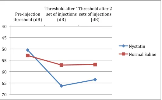
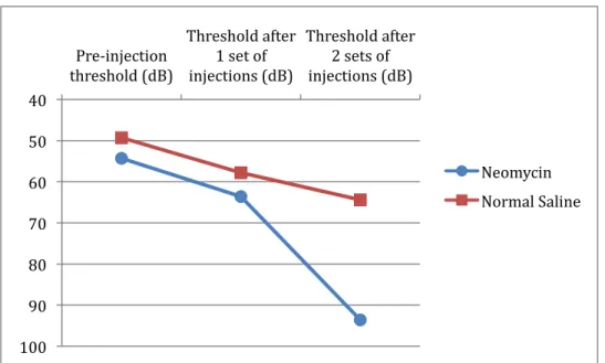
A third and final ABR evaluation was completed after the six injections. In Group I, similar thresholds were noted when comparing Nystatin-treated ears with our negative control group (63.5 ± 11.5 vs. 56.9 ± 8.6 dB). In Group II, the neomycin-treated ears had highly increased ABR threshold (93.6 ± 18.1 dB) compared with the control group (64.4 ± 16.7 dB). All findings are summarized in Table 3 and in Figures 2 and 3(74).
Table 3: Auditory Brainstem Response Results
Pre-injection
threshold (dB) Threshold after 1 set of injections (dB) Threshold after 2 sets of injections (dB) Nystatin 50.5 66.3 63.5 Group 1 Normal Saline 52.9 57.1 56.9 Neomycin 54.3 63.6 93.6 Group 2 Normal Saline 49.3 57.8 64.4
Figure 2: Group I Auditory Brainstem Response
40 45 50 55 60 65 70 Pre-‐injection threshold (dB) Threshold after 1 set of injections
(dB)
Threshold after 2 sets of injections
(dB)
Nystatin Normal Saline
21
Figure 3: Group II Auditory Brainstem Response
Statistical Analysis
Initial analysis was performed using a four-way ANOVA with the following independent variables: group (experimental vs. positive control), treatment (Nystatin or neomycin vs. normal saline), time (pre-injection, after 1 set of injections, after 2 sets of injections) and frequency (2000, 3000, 4000, 6000 and 8000Hz). An interaction between the frequency, time and treatment variables (F=2.68, p=0.01) forced us to perform independent three-way ANOVA tests for each recorded frequency. These tests did not reveal any statistically significant differences of injection product effect between frequencies. 40 50 60 70 80 90 100 Pre-‐injection threshold (dB) Threshold after 1 set of injections (dB) Threshold after 2 sets of injections (dB) Neomycin Normal Saline
on results, we proceeded with a three-way ANOVA with the remaining variables. This test revealed an interaction between these variables (F=4.97; p=0.017), leading us to separate our analysis into Nystatin and neomycin groups, and perform independent two-way ANOVA analyses for each.
Nystatin group:
We found mean ABR values to be statistically significant in at least one of the measured times (F=11.39, p=0.01). A Bonferoni multiple comparison test revealed that pre-injection thresholds were better compared to thresholds after two series of injections by an average of 10.1 dB (p=0.003). All other thresholds were similar, including comparisons between test ears and control ears.
Neomycin group:
Once again, an interaction was discovered between the time and ear injected variables (F=9.17, p=0.02). A Bonferoni multiple comparison test revealed that the only significant differences between ears were after 2 series of injections. At this moment, an average difference of 29.1 dB (p<0.0001) was found between test ears and control ears
Electron Microscopy
The inner ears exposed to physiological saline solution had no discernible hair cell loss and were used as a basis for comparison. Neomycin-treated ears demonstrated partial to total destruction of all three layers of outer hair cells. Images from the Nystatin group
23 were similar to the negative control group, with preserved outer hair cell architecture. Examples of images obtained are shown in Figure 4.
A cochlea exposed to normal saline. Note the three rows of outer hair cells and the row of inner hair cells with normal architecture. b Cochlea exposed to neomycin. Note the important outer hair cell destruction. c Cochlea exposed to nystatin. Preserved outer hair cell architecture is evident.
25
Discussion
Nystatin
Successful treatment of otomycosis begins with a correct diagnosis and requires proper cleansing of the external ear canal, optimal environmental factors and an effective antifungal agent. Recognized risk factors include immunosuppression, recent acute otitis media, presence of cerumen and recent use of topical antibiotic and steroid preparations(75). Patients will typically complain of otalgia, pruritis, persistant discharge and mild hearing loss.
Evaluation with binocular vision is crucial and therefore all patients should be examined with a microscope. Physical exam findings are characteristic and include a blotting paper appearance of the matted mycelia and fruiting bodies or conidiphores(76). Proper cleansing and drying are necessary to exclude complications such as TM perforation or associated middle ear pathology. Malignant otitis externa can also result from otomycosis in immunocompromised individuals, most often when Aspergillus fumigatus is responsible(77).
Topical antimicrobial preparations are the mainstay of otomycosis treatment. The ideal topical antifungal should not be ototoxic and should be effective against the most commonly isolated organism. Nystatin is macrolid antibiotic agent that exerts its effect by inhibiting sterol synthesis and increasing permeability of the cell membrane.(78) It is available as ointment and cream preparations, ophthalmic drops, liquid suspension and oral pills. Despite previous efforts to demonstrate its safety when exposed to the middle ear, no study had thus far excluded its potential for ototoxicity.
study, Lawrence et al. demonstrated that Nystatin has specific anti-fungal activity in contrast to Merthiolate and Cresylate, which have non-specific anti-microbial activity(79). In a series in Burma, Than et al. reported curing 80% of his patients within a week of initiating Nystatin therapy(80). In a series of 411 patients, Paulose et al. found that Nystatin was more effective when combined with a steroid or another antimicrobial agent(1). Resistance to Nystatin has been reported. In a patient diagnosed with otomycosis induced by topical antimicrobials, Jackman reported a case of a patient requiring oral Fluconazole after unsuccessful Nystatin treatment(81). Based on clinical experience, Besbes claims that Nystatin is the most effective anti-mycotic agent avaible(82). It covers a wide spectrum of fungal infections, including the very common Candida and Aspergillus species. Using 15 species of fungi and yeast, Stern et al. demonstrated in an in vitro study that Nystatin had the widest spectrum of activity among antifungals(83). Among the currently used antimycotic agents, Nystatin is also the least expensive agent available in our tertiary care center.
Since no otic Nystatin preparation is currently available, different options must be considered. Regular application of ophthalmic drops is an effective solution, but requires treatment for at least seven days and efficacy varies with patient compliance. We recommend generous application of ointment in a clean external auditory canal or mastoid cavity with follow-up at seven days to clean residue and confirm infection resolution.
27 Results analysis
Our results support the predominant belief that Nystatin is free of ototoxic potential, even when exposed directly to the middle ear. When we look at our data in depth, however, some of the findings were unexpected.
Our first observation was that initial ABR recordings yielded thresholds ranging from 49.3 to 54.3 dB after TM perforation. Although data for guinea pigs is not available, a 2005 review of simple TM perforation in children noted possible hearing loss up to 35 dB attributable to the perforation(84). Many factors may explain why our observed hearing loss was greater. First, the mechanism of perforation was highly traumatic, using a surgical pick to tear the TM and an alligator forcep to remove 30 to 50 percent of the TM. Such manipulation could potentially cause damage to the underlying ossicles. We considered at the beginning of the study placing trans-tympanic ventilation tubes in all animals to minimize trauma and perforation closure. We decided against it due to the probable difficulty we would experience with tube obstruction. Next, the unit of measure in our study is the absolute dB, whereas hearing tests in humans most often utilize hearing level dB. Independently of the cause of our findings, they underline the importance of obtaining a pre-treatment ABR recording in the setting of such an experiment.
After our first set of injections, thresholds increased to 57.1 to 66.3 dB, without clinically or statistically significant differences between the groups. Despite three treatments of neomycin in three weeks, no evidence of significant hearing loss was found. Most agree that toxicity level depends on the time of exposure and the dosage, but no clear guidelines exist for animal studies. In a study of similar design, Marsh injected anti-mycotics into guinea pig middle ears and left each substance in place for one hour(7). He then obtained ABR recordings one hour later and found significant hearing loss (over 40
therefore scheduled three more injections at two-day intervals. This strategy was presumably responsible for obtaining a toxic level of neomycin, with measured thresholds averaging 93.6 dB.
The use of burst tone stimuli in our ABR testing allowed us to compare the impact of injections between frequencies. Theoretically we would have expected neomycin to have a greater impact on the higher frequencies, as is known of aminoglycoside toxicity in humans. Our results, however, did not support this hypothesis. One possible explanation for this finding is that we selectively tested frequencies beginning at 2000 Hz, expecting little or no impact in the range of 250 to 1000 Hz.
Pathophysiology of the safety of Nystatin
In our study, there was no clinically or statistically significant difference between the effect of Nystatin and Sodium Chloride on guinea pig ear function. Based on our SEM images showing what appears to be Nystatin residue (Figure 3C), which supports Tom’s findings, it appears evident that the anti-mycotic is not harmful to OHCs. There is however another possibility that deserves special consideration: the Nystatin molecule may not penetrate into the cochlea in the first place. Experts agree that the most likely route of access to the cochlea from the middle ear is the round window membrane (RWM). This membrane is the only soft tissue communication between the two spaces. The permeability of the RWM depends on molecular size and configuration, liposolubility, concentration gradient and particle electrical charge(85). Much like the tympanic membrane, the RWM is composed of two mucous membranes and one central fibrous layer(86). In an experimental
29 guinea pig study, Mikulec demonstrated that factors such as middle ear inflammation and suction around the RWM could increase its permeability by a factor of 10-15(87). In humans, this membrane is known to be permeable to gentamicin and steroid solutions, as they are effective treatments for Menière’s disease and other inner ear pathologies. These clinical observations have been confirmed with in vitro experimental studies(88, 89). While the molecular weight of gentamicin is 477.6 Da (C21H43N5O7), Nystatin’s is considerably greater at 926.1 Da (C47H75NO17). This does not appear to be the limiting factor in this case, however, since Chelikh demonstrated that even Inulin (molecular weight 7,000 Da) crossed the RWM in guinea pigs after prolonged delivery(90). With these findings suggesting that Nystatin does in fact permeate into cochlear perilymph, it is reasonable to assume that the molecule itself is not harmful to guinea pig OHCs.
Possible implications in humans
In a previous section, we discussed some of the differences between human and guinea pig anatomy. Though it has never been proven that human ears respond differently to ototoxic agents, it is important to consider the variables between the two models before extrapolating our results.
Let us begin by considering the factors that would potentially increase exposure to the tested agent in the human. First, the maximum quantity of ointment that can be used in a human ear greatly exceeds that of a guinea pig. While we used less than 0.1 cc of Nystatin for guinea pigs to fill up their middle ear, the quantity used on an adult patient with a mastoid cavity could easily be 10 times greater. Second, our experiment was performed on ears that were devoid of infection and consequent inflammation. Goycoolea demonstrated that inflammation causes an acute phase of increased RWM permeability followed by
reproduced using neomycin in guinea pigs(91). In humans with acute otomycosis, it would be possible for inflammation to extend to the middle ear and temporarily increase RWM permeability. Finally, Ghiz found the average surface area of this membrane in guinea pigs to be 1.18 mm2, compared with 2.98 mm2 found in humans by Takahashi(92, 93).
A strong argument can also be made for less Nystatin reaching the perilymph in a context of otomycosis in humans. First and foremost, Nystatin application in humans is topical in the external auditory canal. Even in the presence of a TM perforation or a ventilation tube, the actual quantity of product exposed to the middle ear would range from negligible to moderate. In our study, we deliberately filled the middle ear space with ointment and left it in place for several weeks. Also, two anatomical factors protect the human OHCs better than guinea pigs: a thicker RWM and a deeper and less accessible round window niche. Average human RWM thickness is 5 to 6 times that of the guinea pig and in 70% of cases is covered by mucosal folds, or a “false round window membrane”, further protecting the cochlea(5). The depth of the round window niche in humans is variable and acts as another anatomical barrier to ototoxic agents(39).
General consensus among experts is that human OHCs are more protected than their guinea pig counterparts, but further studies confirming this belief for the Nystatin molecule are necessary.
31 Study limits and future outlook
Electron microscopy:
Our greatest challenge for this study was obtaining consistent images of the organ of Corti allowing us to evaluate OHC architecture at every level of the cochlea. The principal problem was unpredictable tectorial membrane adhesion. Though different preparation techniques have been described, none seem to address this problem specifically. A recent study published in May 2011 describes a new technique whereby the RWN is perforated and perfused with 2% gluteraldehyde in a phosphate-buffered solution (PBS), and subsequently perfused with 1% osmium tetroxyde(94). Exposure of the organ of Corti was obtained by lateral wall dissection and therefore did not depend on tectorial membrane removal. Though much more complex and time-consuming, this technique yielded impressive images and is well worth attempting. With such evolving techniques and higher resolution microscopes, it will be possible to evaluate the ototoxic potential of antibiotics and other agents in much greater detail.
Auditory Brainstem Response:
As previously discussed, auditory brainstem response appears to be the most reliable tool to evaluate hearing function in animals. This technique, however, remains far from ideal. It is a device designed for use in human beings with our relatively limited frequency range. What would we observe if we tested ototoxic agents in guinea pigs in frequencies as high as 40 or 50 kHz instead of stopping at 8 kHz? Would those results have any implications in humans? An argument could be made that our equipment does not evaluate the entire basal turn of the guinea pig cochlea. A second limitation with our ABR testing was the environment in which the tests were carried out, which was not up to the
quiet but not soundproof. Other instruments in the surrounding environment caused visible interference, which may have potentially influenced our results. Future studies correlating ABR results with histological findings may lead to specific parameters for animal testing, thereby increasing the internal and external validity of these studies.
33
Conclusion
Nystatin is a large-spectrum and inexpensive anti-mycotic agent effective in the treament of otomycosis. Its direct application in the middle ear of guinea pigs does not influence inner ear function as measured by auditory brainstem response, nor does it affect cochlear outer cell hair architecture observed with a scanning electron microscope. Further studies will be required to confirm Nystatin as a safe option for treatment of otomycosis in presence of a ventilation tube or tympanic membrane perforation in humans.
References
1. Paulose KO, Al Khalifa S, Shenoy P, Sharma RK. Mycotic infection of the ear (otomycosis): a prospective study. J Laryngol Otol 1989;103(1):30-5.
2. Vennewald I, Schonlebe J, Klemm E. Mycological and histological investigations in humans with middle ear infections. Mycoses 2003;46(1-2):12-8.
3. Martin TJ, Kerschner JE, Flanary VA. Fungal causes of otitis externa and tympanostomy tube otorrhea. Int J Pediatr Otorhinolaryngol 2005;69(11):1503-8. 4. Jinn TH, Kim PD, Russell PT, Church CA, John EO, Jung TT. Determination of ototoxicity of common otic drops using isolated cochlear outer hair cells. Laryngoscope 2001;111(12):2105-8.
5. Tom LW. Ototoxicity of common topical antimycotic preparations. Laryngoscope 2000;110(4):509-16.
6. Baylancicek S, Serin GM, Ciprut A, Sari M, Akdas F, Tutkun A. Ototoxic effect of topical ciclopirox as an antimycotic preparation. Otol Neurotol 2008;29(7):910-3.
7. Marsh RR, Tom LW. Ototoxicity of antimycotics. Otolaryngol Head Neck Surg 1989;100(2):134-6.
8. Paff GH. Anatomy of the head and neck. Philadelphia,: Saunders; 1973. x, 235 p. p.
9. Purves D, Williams SM. Neuroscience. 2nd ed. Sunderland, Mass.: Sinauer Associates; 2001. xviii, 681, [16, 3, 25] p. p.
10. Bailey BJ, Johnson JT, Newlands SD. Head & neck surgery--otolaryngology. 4th ed. Philadelphia, PA: Lippincott Williams & Wilkins; 2006. 2 v. (xxviii, 2826, I-62 p., [24] p. of plates) p.
11. Pasha R. Otolaryngology : head & neck surgery : clinical reference guide. 2nd ed. San Diego: Plural Pub.; 2006. xviii, 535 p. p.
12. Hall JW. New handbook of auditory evoked responses. Boston, Mass.: Pearson; 2006.
13. Wysocki J. Topographical anatomy of the guinea pig temporal bone. Hear Res 2005;199(1-2):103-10.
34
14. Goksu N, Haziroglu R, Kemaloglu Y, Karademir N, Bayramoglu I, Akyildiz N. Anatomy of the guinea pig temporal bone. Ann Otol Rhinol Laryngol 1992;101(8):699-704.
15. Asarch R, Abramson M, Litton WB. Surgical anatomy of the guinea pig ear. Ann Otol Rhinol Laryngol 1975;84(2 PART 1):250-5.
16. Wells JR, Gernon WH, Ward G, Davis RK, Hays LL. Otosurgical model in the guinea pig (Cavia porcellus). Otolaryngol Head Neck Surg 1986;95(4):450-7.
17. Counter SA, Bjelke B, Klason T, Chen Z, Borg E. Magnetic resonance imaging of the cochlea, spiral ganglia and eighth nerve of the guinea pig. Neuroreport 1999;10(3):473-9.
18. Heffner R, Heffner H, Masterton B. Behavioral measurements of absolute and frequency-difference thresholds in guinea pig. J Acoust Soc Am 1971;49(6):1888-95. 19. West CD. The relationship of the spiral turns of the cochlea and the length of the basilar membrane to the range of audible frequencies in ground dwelling mammals. J Acoust Soc Am 1985;77(3):1091-101.
20. Salt AN, Hullar TE. Responses of the ear to low frequency sounds, infrasound and wind turbines. Hear Res;268(1-2):12-21.
21. Blakley BW, Hochman J, Wellman M, Gooi A, Hussain AE. Differences in Ototoxicity across Species. J Otolaryngol Head Neck Surg 2008;37(5):700-3.
22. Warchol ME. Cellular mechanisms of aminoglycoside ototoxicity. Curr Opin Otolaryngol Head Neck Surg;18(5):454-8.
23. Dulon D, Hiel H, Aurousseau C, Erre JP, Aran JM. Pharmacokinetics of gentamicin in the sensory hair cells of the organ of Corti: rapid uptake and long term persistence. C R Acad Sci III 1993;316(7):682-7.
24. Selimoglu E. Aminoglycoside-induced ototoxicity. Curr Pharm Des 2007;13(1):119-26.
25. Chen Y, Huang WG, Zha DJ, et al. Aspirin attenuates gentamicin ototoxicity: from the laboratory to the clinic. Hear Res 2007;226(1-2):178-82.
26. Fairbanks DN. Otic topical agents. Otolaryngol Head Neck Surg 1980;88(4):327-31.
27. Matz G, Rybak L, Roland PS, et al. Ototoxicity of ototopical antibiotic drops in humans. Otolaryngol Head Neck Surg 2004;130(3 Suppl):S79-82.
30. Ikiz AO, Serbetcioglu B, Guneri EA, Sutay S, Ceryan K. Investigation of topical ciprofloxacin ototoxicity in guinea pigs. Acta Otolaryngol 1998;118(6):808-12. 31. Brownlee RE, Hulka GF, Prazma J, Pillsbury HC, 3rd. Ciprofloxacin. Use as a topical otic preparation. Arch Otolaryngol Head Neck Surg 1992;118(4):392-6.
32. Claes J, Govaerts PJ, Van de Heyning PH, Peeters S. Lack of ciprofloxacin ototoxicity after repeated ototopical application. Antimicrob Agents Chemother 1991;35(5):1014-6.
33. Kavanagh KR, Parham K, Schoem SR. Auditory function after a prolonged course of ciprofloxacin-dexamethasone otic suspension in a murine model. Arch Otolaryngol Head Neck Surg 2009;135(3):238-41.
34. Daniel SJ, Munguia R. Ototoxicity of topical ciprofloxacin/dexamethasone otic suspension in a chinchilla animal model. Otolaryngol Head Neck Surg 2008;139(6):840-5.
35. Russell PT, Church CA, Jinn TH, Kim DJ, John EO, Jung TT. Effects of common topical otic preparations on the morphology of isolated cochlear outer hair cells. Acta Otolaryngol 2001;121(2):135-9.
36. Becvarovski Z, Kartush JM, Bojrab DI. Intratympanic ciprofloxacin and the human labyrinthine sampling model. Laryngoscope 2002;112(4):686-8.
37. Daniel SJ, Duval M, Sahmkow S, Akache F. Ototoxicity of topical moxifloxacin in a chinchilla animal model. Laryngoscope 2007;117(12):2201-5.
38. Schonebeck J, Zakrisson JE. Topical 5-fluorocytosine therapy in otomycosis. J Laryngol Otol 1974;88(3):227-31.
39. Parker FL, James GW. The effect of various topical antibiotic and antibacterial agents on the middle and inner ear of the guinea-pig. J Pharm Pharmacol 1978;30(4):236-9.
40. Spandow O, Anniko M, Moller AR. The round window as access route for agents injurious to the inner ear. Am J Otolaryngol 1988;9(6):327-35.
41. Viberg A, Canlon B. The guide to plotting a cochleogram. Hear Res 2004;197(1-2):1-10.
42. Linss V, Linss W, Emmerich E, Richter F. The cochleogram of the guinea pig. Eur Arch Otorhinolaryngol 2007;264(4):369-75.
43. Yung MW, Wright A. An improved morphological technique for the study of cochleotoxicity in guinea pigs. J Laryngol Otol 1986;100(11):1235-44.
36
44. Migirov L, Himmelfarb M. Methodology for studying the effects of topically applied ear drops on otoacoustic emissions in guinea pigs. J Laryngol Otol 2003;117(9):696-9.
45. Gultekin E, Yener M, Ozdemir I. The effect of topical Castellani solution on outer hair cell function of rats. Laryngoscope;120(4):808-12.
46. Moody MW, Lang H, Spiess AC, Smythe N, Lambert PR, Schmiedt RA. Topical application of mitomycin C to the middle ear is ototoxic in the gerbil. In: Otol Neurotol; 2006. p. 1186-92.
47. Arikan OK, Muluk NB, Budak B, Apan A, Budak G, Koc C. Effects of ropivacaine on transient-evoked otoacoustic emissions: a rabbit model. Eur Arch Otorhinolaryngol 2006;263(5):421-5.
48. Wimmer C, Mees K, Stumpf P, Welsch U, Reichel O, Suckfull M. Round window application of D-methionine, sodium thiosulfate, brain-derived neurotrophic factor, and fibroblast growth factor-2 in cisplatin-induced ototoxicity. Otol Neurotol 2004;25(1):33-40.
49. Morawski K, Telischi FF, Merchant F, Abiy LW, Lisowska G, Namyslowski G. Role of mannitol in reducing postischemic changes in distortion-product otoacoustic emissions (DPOAEs): a rabbit model. Laryngoscope 2003;113(9):1615-22.
50. Yagiz R, Tas A, Uzun C, Adali MK, Koten M, Karasalihoglu AR. Effect of topically applied povidone-iodine on transient evoked otoacoustic emissions in guinea pigs. J Laryngol Otol 2003;117(9):700-3.
51. Jassir D, Buchman CA, Gomez-Marin O. Safety and efficacy of topical mitomycin C in myringotomy patency. Otolaryngol Head Neck Surg 2001;124(4):368-73.
52. Murillo-Cuesta S, Garcia-Alcantara F, Vacas E, et al. Direct drug application to the round window: a comparative study of ototoxicity in rats. Otolaryngol Head Neck Surg 2009;141(5):584-90.
53. Jang CH, Park H, Cho YB, Choi CH. Evaluating the ototoxicity of topical piperacillin-tazobactam. Int J Pediatr Otorhinolaryngol 2008;72(12):1815-21.
54. Chadwick GM, Asher AL, Van Der Veer CA, Pollard RJ. Adverse effects of topical papaverine on auditory nerve function. Acta Neurochir (Wien) 2008;150(9):901-9; discussion 909.
55. Suzuki M, Ushio M, Yamasoba T. Time course of apoptotic cell death in guinea pig cochlea following intratympanic gentamicin application. Acta Otolaryngol 2008;128(7):724-31.
2007;28(5):605-8.
57. Radeloff A, Smolders JW. Brain-derived neurotrophic factor treatment does not improve functional recovery after hair cell regeneration in the pigeon. Acta Otolaryngol 2006;126(5):452-9.
58. Iwai K, Nakagawa T, Endo T, et al. Cochlear protection by local insulin-like growth factor-1 application using biodegradable hydrogel. Laryngoscope 2006;116(4):529-33.
59. Mills PC, Ahlstrom L, Wilson WJ. Ototoxicity and tolerance assessment of a TrisEDTA and polyhexamethylene biguanide ear flush formulation in dogs. J Vet Pharmacol Ther 2005;28(4):391-7.
60. Okuda T, Sugahara K, Takemoto T, Shimogori H, Yamashita H. Inhibition of caspases alleviates gentamicin-induced cochlear damage in guinea pigs. Auris Nasus Larynx 2005;32(1):33-7.
61. Babu SC, Kartush JM, Patni A. Otologic effects of topical mitomycin C: phase I-evaluation of ototoxicity. Otol Neurotol 2005;26(2):140-4.
62. Perez R, Freeman S, Sohmer H, Sichel JY. Vestibular and cochlear ototoxicity of topical antiseptics assessed by evoked potentials. Laryngoscope 2000;110(9):1522-7. 63. Chen Z, Ulfendahl M, Ruan R, Tan L, Duan M. Acute treatment of noise trauma with local caroverine application in the guinea pig. Acta Otolaryngol 2003;123(8):905-9.
64. Teranishi MA, Nakashima T. Effects of trolox, locally applied on round windows, on cisplatin-induced ototoxicity in guinea pigs. Int J Pediatr Otorhinolaryngol 2003;67(2):133-9.
65. Tsukasaki N, Whitworth CA, Rybak LP. Acute changes in cochlear potentials due to cisplatin. Hear Res 2000;149(1-2):189-98.
66. Jiang Z, Zhang L. [Ototoxicity of gentamicin assessed by the vestibular evoked potentials]. Zhongguo Yi Xue Ke Xue Yuan Xue Bao 2000;22(5):460-2.
67. Conlon BJ, Smith DW. Topical aminoglycoside ototoxicity: attempting to protect the cochlea. Acta Otolaryngol 2000;120(5):596-9.
68. Conlon BJ, McSwain SD, Smith DW. Topical gentamicin and ethacrynic acid: effects on cochlear function. Laryngoscope 1998;108(7):1087-9.
69. Janning MH, Whitworth CA, Rybak LP. Experimental model of cisplatin ototoxicity in chinchillas. Otolaryngol Head Neck Surg 1998;119(6):574-80.
38
70. Strain GM, Merchant SR, Neer TM, Tedford BL. Ototoxicity assessment of a gentamicin sulfate otic preparation in dogs. Am J Vet Res 1995;56(4):532-8.
71. Tan U, Senyuva F, Marangoz C. Electrocorticographic effects of topically applied scopolamine. Epilepsia 1978;19(3):223-32.
72. Nader ME, Theoret Y, Saliba I. The role of intratympanic lactate injection in the prevention of cisplatin-induced ototoxicity. Laryngoscope;120(6):1208-13.
73. Snedecor GW, Cochran WG. Statistical methods. 8th ed. Ames: Iowa State University Press; 1989. xx, 503 p. p.
74. Woods O, Saliba I. Effect of Nystatin on Guinea Pigs' Inner Ear. Neurotox Res. 75. Ho T, Vrabec JT, Yoo D, Coker NJ. Otomycosis: clinical features and treatment implications. Otolaryngol Head Neck Surg 2006;135(5):787-91.
76. Hurst WB. Outcome of 22 cases of perforated tympanic membrane caused by otomycosis. J Laryngol Otol 2001;115(11):879-80.
77. Carfrae MJ, Kesser BW. Malignant otitis externa. Otolaryngol Clin North Am 2008;41(3):537-49, viii-ix.
78. Munguia R, Daniel SJ. Ototopical antifungals and otomycosis: a review. Int J Pediatr Otorhinolaryngol 2008;72(4):453-9.
79. Lawrence TL, Ayers LW, Saunders WH. Drug therapy in otomycosis: an in vitro study. Laryngoscope 1978;88(11):1755-60.
80. Than KM, Naing KS, Min M. Otomycosis in Burma, and its treatment. Am J Trop Med Hyg 1980;29(4):620-3.
81. Jackman A, Ward R, April M, Bent J. Topical antibiotic induced otomycosis. Int J Pediatr Otorhinolaryngol 2005;69(6):857-60.
82. Besbes M, Makni F, Cheikh-Rouhou F, Sellami H, Kharrat K, Ayadi A. [Otomycosis due to Scopulariopsis brevicaulis]. Rev Laryngol Otol Rhinol (Bord) 2002;123(2):77-8.
83. Stern JC, Shah MK, Lucente FE. In vitro effectiveness of 13 agents in otomycosis and review of the literature. Laryngoscope 1988;98(11):1173-7.
84. [Surgical treatment of tympanic perforation in children]. Arch Pediatr 2005;12(3):372-6.
85. Pickett BP, Shinn JB, Smith MF. Ear drop ototoxicity: reality or myth? Am J Otol 1997;18(6):782-9; discussion 789-91.
44):1-20.
87. Mikulec AA, Hartsock JJ, Salt AN. Permeability of the round window membrane is influenced by the composition of applied drug solutions and by common surgical procedures. Otol Neurotol 2008;29(7):1020-6.
88. Plontke SK, Mynatt R, Gill RM, Borgmann S, Salt AN. Concentration gradient along the scala tympani after local application of gentamicin to the round window membrane. Laryngoscope 2007;117(7):1191-8.
89. Yang J, Wu H, Zhang P, Hou DM, Chen J, Zhang SG. The pharmacokinetic profiles of dexamethasone and methylprednisolone concentration in perilymph and plasma following systemic and local administration. Acta Otolaryngol 2008;128(5):496-504.
90. Chelikh L, Teixeira M, Martin C, Sterkers O, Ferrary E, Couloigner V. High variability of perilymphatic entry of neutral molecules through the round window. Acta Otolaryngol 2003;123(2):199-202.
91. Nomura Y, Okuno T, Kawabata I. The round window membrane. Adv Otorhinolaryngol 1983;31:50-8.
92. Takahashi H, Takagi A, Sando I. Computer-aided three-dimensional reconstruction and measurement of the round window and its membrane. Otolaryngol Head Neck Surg 1989;101(5):517-21.
93. Ghiz AF, Salt AN, DeMott JE, Henson MM, Henson OW, Jr., Gewalt SL. Quantitative anatomy of the round window and cochlear aqueduct in guinea pigs. Hear Res 2001;162(1-2):105-12.
94. Choung YH, Kim SW, Tian C, et al. Korean red ginseng prevents gentamicin-induced hearing loss in rats. Laryngoscope;121(6):1294-302.