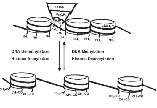
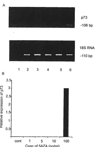
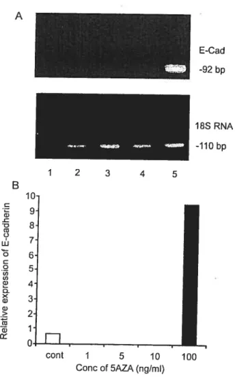
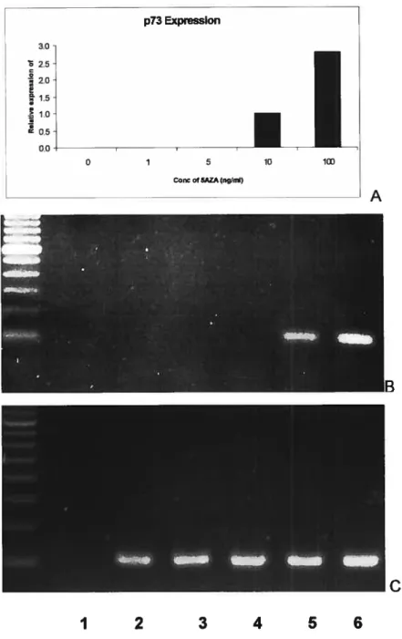
Activation of expression of p15, p73 and E-cadherin in myeloid leukemia cells by different concentrations of 5-aza-2'-deoxycytidine
98
0
0
Texte intégral
Figure


+7

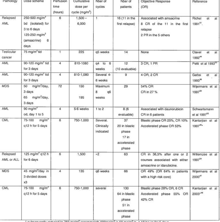
Documents relatifs
