ISSN: 1524-4628
Copyright © 2010 American Heart Association. All rights reserved. Print ISSN: 0039-2499. Online Stroke is published by the American Heart Association. 7272 Greenville Avenue, Dallas, TX 72514
DOI: 10.1161/STROKEAHA.110.579409
2010;41;863-868; originally published online Apr 1, 2010;
Stroke
Peter Paul De Deyn and for the BeFaS Investigators
Fumal, Sandrine Jeangette, Werner Verslegers, Robert Baker, Derralynn Hughes,
Belachew, Christine Van Broeckhoven, Patricia Redondo, Dimitri Hemelsoet, Arnaud
Raf Brouns, Vincent Thijs, François Eyskens, Marleen Van den Broeck, Shibeshih
Patients With Cerebrovascular Disease
Belgian Fabry Study: Prevalence of Fabry Disease in a Cohort of 1000 Young
http://stroke.ahajournals.org/cgi/content/full/41/5/863
located on the World Wide Web at:
The online version of this article, along with updated information and services, is
http://www.lww.com/reprints
Reprints: Information about reprints can be found online at
journalpermissions@lww.com
410-528-8550. E-mail: Fax:
Kluwer Health, 351 West Camden Street, Baltimore, MD 21202-2436. Phone: 410-528-4050. Permissions: Permissions & Rights Desk, Lippincott Williams & Wilkins, a division of Wolters
http://stroke.ahajournals.org/subscriptions/
Prevalence of Fabry Disease in a Cohort of 1000 Young Patients With
Cerebrovascular Disease
Raf Brouns, MD, PhD; Vincent Thijs, MD, PhD; Franc¸ois Eyskens, MD, PhD;
Marleen Van den Broeck, BSc; Shibeshih Belachew, MD, PhD; Christine Van Broeckhoven, PhD, DSc;
Patricia Redondo, MD, PhD; Dimitri Hemelsoet, MD; Arnaud Fumal, MD, PhD;
Sandrine Jeangette, MD, PhD; Werner Verslegers, MD; Robert Baker, BSc, MSc AIB, MS;
Derralynn Hughes, MD, PhD; Peter Paul De Deyn, MD, PhD; for the BeFaS Investigators*
Background and Purpose—Data on the prevalence of Fabry disease in patients with central nervous system pathology arelimited and controversial. In this study, we assessed the prevalence of Fabry disease in young patients presenting with cerebrovascular disease in Belgium.
Methods—In this national, prospective, multicenter study, we screened for Fabry disease in 1000 patients presenting with ischemic stroke, transient ischemic attack, or intracranial hemorrhage; unexplained white matter lesions; or vertebro-basilar dolichoectasia. In male patients, we measured !-galactosidase A (!-GAL A) activity in dried blood spots. Female patients were screened for mutations by exonic DNA sequencing of the !-GAL A gene.
Results—!-GAL A activity was deficient in 19 men (3.5%), although all had normal !-GAL A gene sequences. Enzymatic deficiency was confirmed on repeat assessment in 2 male patients (0.4%). We identified missense mutations in 8 unrelated female patients (1.8%): Asp313Tyr (n!5), Ala143Thr (n!2), and Ser126Gly (n!1). The pathogenicity of the 2 former missense mutations is controversial. Ser126Gly is a novel mutation that can be linked to late-onset Fabry disease.
Conclusion—!-GAL A deficiency may play a role in up to 1% of young patients presenting with cerebrovascular disease. These findings suggest that atypical variants of Fabry disease with late-onset cerebrovascular disease exist, although the clinical relevance is unclear in all cases. (Stroke. 2010;41:863-868.)
Key Words: Fabry disease ! cerebrovascular accident ! white matter lesions ! dolichoectasia ! !-galactosidase A
! lysosomal storage disorders ! Belgium
F
abry disease (Anderson-Fabry disease; Online MendelianInheritance in Man No. 301500) is an X-linked lysosomal storage disorder caused by mutations in the !-galactosidase A (!-GAL A) gene (GLA), leading to !-GAL A deficiencies.1
Consequently, neutral glycosphingolipids accumulate in many tissues and cell types, including the vascular epithe-lium, cornea, kidneys, and heart. Male hemizygotes are generally more severely affected than are heterozygous fe-males, but disabling clinical features and disease progression often also occur in female patients.2Patients with the classic
phenotype of Fabry disease usually present in childhood with pain crises, acroparesthesia, hypohidrosis, gastrointestinal symptoms, angiokeratoma, and corneal abnormalities. Severe morbidity and mortality follow in adult life due to renal failure, cardiac involvement, and stroke.3 Atypical variants
with late-onset manifestations in the cardiac, neurologic, and renal systems have been described.4 –13
Estimates on the prevalence of classic Fabry disease vary from 1 in 40 000 to 1 in 117 000.14 Given the nonspecific
presenting symptoms and delayed presentation, atypical
vari-Continuing medical education (CME) credit is available for this article. Go to http://cme.ahajournals.org to take the quiz.
Received January 17, 2010; accepted January 26, 2010.
From the Laboratory for Neurochemistry and Behaviour, Institute Born-Bunge, and Department of Biomedical Sciences (R. Brouns, P.D.D.), University of Antwerp; and Department of Neurology and Memory Clinic (R. Brouns, P.D.D.), ZNA Middelheim General Hospital, Antwerp, Belgium; Department of Neurology (R. Brouns), University Hospital Brussels, Vrije Universiteit Brussel, Brussels, Belgium; Department of Neurology (V.T.), University Hospitals Leuven and Vesalius Research Center, VIB3, Leuven, Belgium; ZNA Queen Paola Child Hospital and Provincial Centre for Metabolic Disorders (F.E.), University of Antwerp, Antwerp, Belgium; Laboratory of Neurogenetics, Institute Born-Bunge; University of Antwerp; and Neurodegenerative Brain Diseases Group (M.V.d.B., C.V.B.), Department of Molecular Genetics, VIB, Antwerp, Belgium; Department of Neurology (S.B.), Centre Hospitalier Universitaire de Lie`ge, Lie`ge, Belgium; Department of Neurology (P.R.), Centre Hospitalier Universitaire de Tivoli, La Louvie`re, Belgium; Department of Neurology (D.H.), University Hospital Ghent, Ghent, Belgium; Departments of Neurology and Functional Neuroanatomy (A.F.), Headache Research Unit, University of Lie`ge, Lie`ge, Belgium; Department of Neurology (S.J.), Centre Hospitalier Universitaire de Charleroi, Charleroi, Belgium; Department of Neurology (W.V.), ZNA Jan Palfijn General Hospital, Merksem, Belgium; and Lysosomal Storage Disorders Unit (R. Baker, D.H.), Royal Free Hampstead NHS Trust, London, England.
*The BeFaS Investigators are listed in the online Appendix.
Correspondence to Prof Dr P.P. De Deyn, Laboratory of Neurochemistry and Behaviour, Institute Born-Bunge, and Department of Biomedical Sciences, University of Antwerp-CDE, Universiteitsplein 1, 2610 Antwerp, Belgium. E-mail peter.dedeyn@ua.ac.be
© 2010 American Heart Association, Inc.
Stroke is available at http://stroke.ahajournals.org DOI: 10.1161/STROKEAHA.110.579409
ants of Fabry disease might be underdiagnosed.15
Epidemio-logic studies have reported the condition in 0.2% to 1.2% of patients with end-stage renal disease4 – 8and in 1% to 6.3% of
patients with unexplained hypertrophic cardiomyopathy.9 –11
The prevalence of Fabry disease in young patients with cryptogenic stroke was reported to be as high as 4.9% in men and 2.4% in women.13 This finding, however, was not
reproduced in a smaller, retrospective study.16White matter
lesions and vertebrobasilar dolichoectasia are commonly found in patients with Fabry disease,17but literature data on
the prevalence of Fabry disease in patients with these neuro-imaging findings are nonexistent.
The aim of the Belgian Fabry Study (BeFaS) was to prospectively assess the frequency of Fabry disease in young Belgian patients with neurologic hallmarks of this disease, namely, stroke, unexplained white matter lesions, and verte-brobasilar dolichoectasia.
Methods
Patients and ProceduresThirty-three clinical neurology departments throughout Belgium participated in this national, prospective, multicenter study, BeFaS. From March 2007 to October 2008, 1000 patients aged 18 to 60 years, consecutively presenting at a participating neurology depart-ment with stroke (ischemic stroke, transient ischemic attack, or intracranial hemorrhage), unexplained white matter lesions, or ver-tebrobasilar dolichoectasia were enrolled. Unexplained white matter lesions were defined as the presence of lesions in the deep or subcortical white matter on magnetic resonance imaging or com-puted tomography for which no etiology was found after expert neurologist evaluation. Patients who were unable to provide in-formed consent, who were already diagnosed with Fabry disease, or in whom this disease was previously excluded by appropriate enzymatic or genetic analyses were excluded from participation in BeFaS. The study was conducted in accordance with the revised Declaration of Helsinki (1998) and in agreement with the guidelines of the ethics committees of ZNA Antwerp, the University of Antwerp, and the ethics committees of each of the participating clinical neurologic centers.
After informed consent was obtained, demographic data, cardio-vascular risk factors, presence of signs and symptoms of Fabry disease, and clinical and neuroimaging data were registered in a database by using web-based case report forms. Assessment of clinical signs suggestive of Fabry disease was optional. Diagnosis or exclusion of cornea verticillata was performed by ophthalmologists, screening for angiokeratoma was done by routine clinical examina-tion, and presence of acroparesthesia was obtained by anamnesis. This symptom was regarded to be present if a history of burning or tingling sensation in the extremities was reported by the patient or a proxy.
Enzymatic and Genetic Testing
A blood sample was obtained for production of blood spots for measurement of !-GAL A activity in male patients and for genetic analyses of GAL in female patients, as described previously.18In
men, screening for Fabry disease can reliably be performed by enzymatic analysis, but this technique is affected by a high risk of false-negative results in women. Genetic testing therefore is the method of choice in the latter.2All diagnostic tests were performed
blinded to case identity. Male patients with abnormal enzymatic activity ("3.0 ng/h per mL) were additionally examined by gene sequencing and repeat analysis of !-GAL A activity in a subset of patients. In female patients with an abnormal result on gene sequencing, repeat gene sequencing was performed.
Statistical Analysis
Data were analyzed with the use of Microsoft Excel (version 2007; Microsoft Corp, Redmond, Wash) and the SPSS 15.0 software package for Windows (SPSS Inc, Chicago, Ill). The 2-tailed unpaired
ttest was used to compare continuous variables between 2 groups. A 1-way ANOVA was applied for comparison of continuous variables when there were #2 groups, and the "2test was used for noncon-tinuous variables.
Role of the Funding Source
The sponsor of the study had no role in the study design, data collection, data analysis, data interpretation, or the writing of the report. The corresponding author had full access to all data and had final responsibility for the decision to submit for publication.
Results
Clinical DataThe mean age ($SD) in the cohort of 1000 patients partici-pating in BeFaS was 47.7$9.1 years, and 547 participants (54.7%) were male. Magnetic resonance imaging of the brain was obtained in 80.2% of patients. Cerebral computed tomog-raphy was performed in the remaining patients. Classic cerebrovascular risk factors, including arterial hypertension, diabetes mellitus, dyslipidemia, smoking, and coronary dis-ease, were present in 38.7%, 9.7%, 38.2%, 55.9%, and 7.6% of patients, respectively. In 16.3% and 4.4% of participants, a history of previous stroke or renal disease was found. Family history for stroke, myocardial infarction before the age of 60 years, and dialysis was present in 19.7%, 13.0%, and 1.3%. Clinical signs suggestive of Fabry disease were reported in a minority of patients: cornea verticillata was found in 0.1% and angiokeratoma in 0.9%. Dyshidrosis, episodic pain, and acroparesthesia were reported by 4.6%, 7.0%, and 13.3% of patients. Evidence of hypertrophic cardiomyopathy was found on echocardiography in 12.0%. White matter lesions were found in 35.0% of patients.
The main clinical criterion for inclusion in BeFaS was stroke (n!842 patients), 573 of whom were diagnosed with ischemic stroke, 220 with transient ischemic attack, and 49 with intracranial hemorrhage (33 and 16 patients with intra-cerebral or subarachnoid hemorrhage, respectively). One hundred fifty-three patients were included for unexplained white matter lesions on neuroimaging but only 2 patients for vertebrobasilar dolichoectasia. Specification of the inclusion criterion was missing on the web-based inclusion form for 3 patients.
Evidence of acute cerebral infarction was present on neuroimaging in 570 patients with ischemic stroke (99.5%). The infarct was located in the anterior circulation in 335 patients (58.8%), in the posterior circulation in 152 patients (26.7%), and in both the anterior and posterior circulation in 83 patients (14.6%). Stroke etiology was classified according to TOAST criteria19 as atherothrombotic in 143 patients
(25.1%), cardioembolic in 125 (21.9%), and lacunar in 99 (17.4%). A specific cause for the ischemic stroke was present in 69 patients (12.1%): cervicocephalic arterial dissection (n!37), vasculitis (n!11), hereditary coagulopathy (n!6), antiphospholipid syndrome (n!3), radiotherapy-induced vas-culopathy (n!2), amphetamine-induced vasvas-culopathy (n!2), moyamoya syndrome (n!2), migrainous stroke (n!2),
bral venous thrombosis (n!2), hemolytic uremic syndrome (n!1), and paraneoplastic coagulopathy (n!1). In 134 pa-tients (23.5%), stroke etiology was cryptogenic. The preva-lence of intracranial hemorrhage in the reported population was only 5.8% of all stroke patients. This may be explained by the fact that for patients with intracranial hemorrhage, routine medical practice in many participating centers is often provided by neurosurgeons rather than neurologists. Sixteen of 49 patients with intracranial hemorrhage (32.7%) had subarachnoid hemorrhage. The location of intracerebral hem-orrhage was lobar in 13 patients (39.4%), involved basal ganglia in 11 (33.3%), brain stem in 5 (15.2%), and thalamus in 4 (12.1%). A cerebral vascular anomaly was present in 21 patients (aneurysm, cavernous malformation, and arterio-venous malformation, respectively, in 15, 4, and 2 patients). Other causes of intracranial hemorrhage included alcohol abuse in 2 patients and hypertension in 1 individual. Unex-plained white matter lesions and dolichoectasia were found in patients who underwent neuroimaging for indications like headache, dizziness, seizures, or syncope. Table 1 shows baseline clinical data and signs and symptoms of Fabry disease, according to the inclusion criterion. In contrast to patients who were enrolled because of unexplained white
matter lesions, stroke patients were predominantly male, less frequently received magnetic resonance– based neuroimag-ing, displayed more classic cerebrovascular risk factors, and more often had hypertrophic cardiomyopathy and a positive family history of stroke or myocardial infarction at a young age. A detailed stratification of the study population by entry condition, sex, age, and cerebrovascular history is presented in Table 2.
Diagnostic Test Results
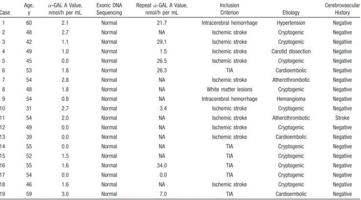
As shown in the Figure, !-GAL A activity was assessed on dried blood spot in 545 of 547 male patients and was below the normal cutpoint of 3.0 nmol/h per mL in 19 patients. Exonic DNA sequencing of GLA was performed in all these patients but failed to identify a mutation. Additionally, a repeat dried blood spot was obtained for 9 patients; !-GAL A activity was found to be normal in 7. However, in 1 patient, a complete enzyme deficiency was observed, and a severe deficiency (0.3 ng/h per mL) was found in another (Table 3).
GLA sequencing results were available for 448 of 453 female patients. Two and 3 samples were lost due to a technical error or during shipment, respectively. A missense mutation was identified in 8 unrelated female patients:
Table 1. Baseline Clinical Data, Cerebrovascular Risk Factors, and Signs and Symptoms Suggestive of Fabry Disease, According to the Inclusion Criterion
Stroke
Unexplained White Matter Lesions
(n!153) Vertebrobasilar Dolichoectasia (n!2) P Value Ischemic Stroke (n!573) TIA (n!220) Intracranial Hemorrhage (n!49) General data Age at presentation, y 48.2 (9.0) 47.0 (9.3) 48.8 (7.9) 46.4 (9.8) 56.0 (2.8) 0.065 Male 350 (61.1%) 122 (55.5%) 122 (55.5%) 44 (28.8%) 1 (50.0%) "0.001 MRI-based neuroimaging 451 (78.7%) 182 (82.7%) 20 (40.8%) 147 (93.6%) 2 (100%) "0.001 Classic cerebrovascular risk
factors Arterial hypertension 241 (42.1%) 91 (41.4%) 19 (38.8%) 34 (22.2%) 2 (100.0%) "0.001 Diabetes mellitus 72 (12.6%) 18 (8.2%) 1 (2.0%) 5 (3.3%) 1 (50.0%) "0.001 Dyslipidemia 235 (41.0%) 106 (48.2%) 6 (12.3%) 34 (22.2%) 1 (50.0%) "0.001 Smoking 353 (61.6%) 119 (54.1%) 31 (63.3%) 55 (36.0%) 1 (50.0%) "0.001 Coronary disease 48 (8.4%) 18 (8.2%) 2 (4.1%) 7 (4.6%) 1 (50.0%) 0.075 Signs and symptoms
suggestive of Fabry disease
White matter lesions 153 (26.7%) 31 (14.1%) 12 (24.5%) 153 (100.0%) 1 (50.0%) "0.001 Previous stroke 87 (15.2%) 44 (20.0%) 9 (18.4%) 21 (13.7%) 0 (0.0%) 0.442
Renal disease 26 (4.5%) 8 (3.6%) 3 (6.1%) 6 (3.9%) 0 (0.0%) 0.930
Hypertrophic cardiomyopathy 75 (13.1%) 34 (15.5%) 5 (10.2%) 6 (3.9%) 0 (0.0%) "0.001 Family history of stroke 103 (18.0%) 59 (26.8%) 6 (12.3%) 25 (16.3%) 1 (50.0%) 0.017 Family history of MI "60 y 69 (12.0%) 41 (18.6%) 4 (8.2%) 13 (8.5%) 1 (50.0%) 0.012 Family history of dialysis 8 (1.4%) 3 (1.4%) 1 (2.0%) 1 (0.7%) 0 (0.0%) 0.942
Cornea verticillata 0 (0%) 0 (0%) 0 (0%) 1 (0.7%) 0 (0.0%) 0.002
Angiokeratoma 7 (1.2%) 2 (0.9%) 0 (0%) 0 (0%) 0 (0.0%) 0.061
Dyshidrosis 24 (4.2%) 11 (5.0%) 6 (12.2%) 5 (3.3%) 0 (0.0%) 0.119
Pain episodes 23 (4.0%) 18 (8.2%) 2 (4.1%) 26 (17.0%) 1 (50.0%) "0.001 Acroparesthesia 54 (9.4%) 34 (15.5%) 8 (16.3%) 37 (24.2%) 0 (0.0%) "0.001 TIA indicates transient ischemic attack; MRI, magnetic resonance imaging; and MI, myocardial infarction.
Asp313Tyr in 5, Ala143Thr in 2 subjects, and a novel mutation, Ser126Gly, in 1 subject (Table 4). The presence of the mutation was confirmed by repeating the exonic DNA sequencing.
Discussion
Cerebral micro- and macroangiopathies are hallmarks of Fabry disease.17,20 It is hypothesized that lipid deposits in
vascular endothelial and smooth muscle cells cause oxidative stress, vascular dysfunction, vessel occlusion, and tissue ischemia.21These pathophysiological mechanisms are
asso-ciated with an increased risk of premature stroke, progressive white matter lesions, and dolichoectasia as major clinical and neuroimaging correlates.17Recent literature data indicate that
stroke frequently occurs before the diagnosis of Fabry disease has been made and in the absence of other key signs of this disease.22
In contrast to the considerable number of studies on the presence of Fabry disease in patients with renal or cardiac
disorders,4 –11only 2 studies reported on the prevalence of
Fabry disease with neurologic hallmarks for this condi-tion.13,16 Both studies focused on young patients with
cryp-togenic stroke. In a large cohort of 721 patients diagnosed with cryptogenic stroke, the prevalence of Fabry disease was found to be as high as 4.9% in men and 2.4% in women.13We
were unable, however, to reproduce this finding in a retro-spective study of 103 cryptogenic stroke patients.16 Fabry
disease is associated with cerebral micro- and macroangiopa-thy.17,20 Moreover, cardioembolic phenomena23 and
coagu-lopathy24may be common in this disorder. Limiting
screen-ing for Fabry disease to patients without these conditions might therefore induce selection bias. For this reason, inclu-sion criteria in BeFaS were not restricted to patients pres-enting with cryptogenic stroke. Other screening projects for Fabry disease in ischemic stroke patients have recently been finalized (M.A. Wozniak et al and M. Viana-Baptista et al; personal communications) or are currently ongoing. Despite being identified as relevant indicators for cerebral vasculopa-thy in Fabry disease, epidemiologic studies in patients with stroke of all causes, unexplained white matter lesions, or dolichoectasia have not been done before. Assessment of the clinical signs suggestive of Fabry disease was not performed in a standardized manner and may limit generalizability of these findings. For instance, the remarkably high prevalence of acroparesthesia in our population may be secondary to overreporting.
In the present study, deficient !-GAL A activity was present in 3.5% of male patients, and a mutation in GAL was found in 1.8% of female patients. Diagnosis of Fabry disease, however, is not straightforward in all patients. The absence of a mutation in all male patients with this enzyme deficiency suggests that the dried blood spot analysis may have been false-positive in some patients. However, repeat assessment !-GAL A activity in a subset of 9 patients confirmed the enzyme deficiency in a patient aged 49 years with ischemic stroke secondary to carotid artery dissection and in another patient aged 54 years with a cryptogenic transient ischemic
Table 2. Study Population Stratified by Entry Condition, Sex, Age, and Cerebrovascular History
Entry Condition Sex
Age at Presentation, Mean$SD, y Cerebrovascular History Stroke Male 49.2 (8.5) 81 (16.2%) Female 46.0 (9.4) 59 (17.5%) Ischemic stroke Male 49.6 (8.6) 52 (14.9%) Female 45.9 (9.1) 35 (15.7%) TIA Male 48.4 (8.2) 20 (16.4%) Female 45.3 (10.3) 24 (24.5%) Intracranial hemorrhage Male 47.6 (8.5) 9 (32.1%) Female 50.4 (6.7) 0 (0.0%) Unexplained white matter
lesions Male 47.7 (9.3) 10 (22.7%) Female 45.9 (10.0) 11 (10.1%) Vertebrobasilar dolichoectasia Male 57.0 (0.0) 0 (0.0%) Female 55.0 (0.0) 0 (0.0%) TIA indicates transient ischemic attack.
Figure. Flowchart for the BeFaS.
attack (cases 4 and 17, Table 3). Magnetic resonance imaging showed acute cerebral ischemia in the anterior circulation in both patients, and the family history was remarkable for stroke in 1 patient. An atypical variant of Fabry disease cannot be excluded in these patients, and additional diagnos-tic assessments are ongoing. We found the Asp313Tyr mutation in 5 female patients. This mutation has been identified in classically affected males as the single muta-tion25 or in the cis position with another missense
muta-tion.26,27On the other hand, this mutation was also reported in
0.45% of normal X chromosomes, and in vitro studies favor the classification of Asp 313Tyr as a polymorphism.27 The
mutation Ala143Thr has previously been reported in patients with the late-onset variant of Fabry disease,5,15but Ser126Gly
appears to be a novel mutation that may also be linked to late-onset Fabry disease. The discovery of a previously unreported mutation is not surprising, because most families have a private mutation and new mutations are frequently found with the diagnosis of an index case.28
Our results suggest that Fabry disease may play a role in up to 1% of young patients presenting with cerebrovascular disease. However, current knowledge on the complex patho-genesis of this condition is incomplete and does not always allow a definitive diagnosis in every patient, especially in cases of late-onset Fabry disease. This illustrates the need for additional basic and epidemiologic research. In an ongoing research project, we are focusing on patients with abnormal screening results identified in the BeFaS and aim to obtain more certainty about the diagnosis of Fabry disease through careful clinical, biochemical, and histological examinations. In addition, studies with larger cohorts of high-risk patients are ongoing and may help to formulate definite answers for these patients.
Summary
We report on a national, prospective, multicenter study on the prevalence of Fabry disease in 1000 young patients
pres-Table 3. Diagnostic Assessments in 19 Male Patients With !-GAL A Deficiency
Case Age, y !-GAL A Value, nmol/h per mL Exonic DNA Sequencing
Repeat !-GAL A Value, nmol/h per mL
Inclusion
Criterion Etiology
Cerebrovascular History 1 60 2.1 Normal 21.7 Intracerebral hemorrhage Hypertension Negative
2 48 2.7 Normal NA Ischemic stroke Cryptogenic Negative
3 42 1.1 Normal 29.1 Ischemic stroke Cryptogenic Negative
4 49 1.0 Normal 1.5 Ischemic stroke Carotid dissection Negative
5 45 0.0 Normal 26.5 Ischemic stroke Cryptogenic Negative
6 53 1.8 Normal 26.3 TIA Cardioembolic Negative
7 54 2.8 Normal NA Ischemic stroke Atherothrombotic Negative
8 48 1.8 Normal NA White matter lesions Cryptogenic Negative
9 54 0.8 Normal NA Intracerebral hemorrhage Hemangioma Negative
10 31 2.7 Normal 3.4 Ischemic stroke Cryptogenic Negative
11 54 2.0 Normal NA Ischemic stroke Atherothrombotic Stroke
12 49 0.0 Normal NA Ischemic stroke Cryptogenic Negative
13 39 0.0 Normal NA Ischemic stroke Cardioembolic Negative
14 55 0.0 Normal NA TIA Cryptogenic Negative
15 52 1.5 Normal NA TIA Cryptogenic Negative
16 55 1.6 Normal 34.0 TIA Cryptogenic Negative
17 54 0.0 Normal 0.0 TIA Cryptogenic Negative
18 46 1.6 Normal NA Ischemic stroke Cryptogenic Negative
19 59 3.0 Normal 7.0 TIA Cardioembolic Negative
NA indicates not available; TIA, transient ischemic attack.
Table 4. Diagnostic Assessments in 8 Female Patients With a Mutation in the GAL
Case Age, y Gene Sequencing Inclusion Criterion Etiology Cerebrovascular History
1 43 Asp313Tyr TIA Cryptogenic Negative
2 52 Asp313Tyr TIA Cryptogenic Negative
3 41 Asp313Tyr White matter lesions Cryptogenic Negative 4 35 Asp313Tyr White matter lesions Cryptogenic Negative
5 53 Asp313Tyr TIA Cryptogenic Stroke
6 43 Ala143Thr Ischemic stroke Carotid dissection Negative 7 51 Ala143Thr Ischemic stroke Carotid dissection Negative
8 41 Ser126Gly Ischemic stroke Cryptogenic Negative
enting with cerebrovascular disease. Our results suggest that !-GAL A deficiency may play a role in up to 1% of young patients presenting with cerebrovascular disease. Although the clinical relevance is not straightforward in all cases, these findings suggest that atypical variants of Fabry disease with late-onset cerebrovascular disease exist and that their genetic background is poorly understood.
Acknowledgments
We thank the participating patients and all BeFaS investigators for their commitment to the study (see supplemental Appendix, avail-able online at http://stroke.ahajournals.org) and the VIB Genetic Service Facility for the genetic services. BeFaS was funded by an unrestricted grant from Shire Belgium.
Disclosures
P.P. De Deyn, R. Brouns, F. Eyskens, and V. Thijs have received compensation from Shire Belgium for serving on the BeFaS scien-tific advisory committee. V. Thijs is a Clinical Investigator of the FWO Flanders. The other authors have no conflicts of interest to declare.
References
1. Fabry H. An historical overview of Fabry disease. J Inherit Metab Dis. 2001;24(suppl 2):3–7.
2. Zarate YA, Hopkin RJ. Fabry’s disease. Lancet. 2008;372:1427–1435. 3. Brady RO, Schiffmann R. Clinical features of and recent advances in
therapy for Fabry disease. J Am Med Assoc. 2000;284:2771–2775. 4. Kotanko P, Kramar R, Devrnja D, Paschke E, Voigtlander T, Auinger M,
Pagliardini S, Spada M, Demmelbauer K, Lorenz M, Hauser AC, Kofler HJ, Lhotta K, Neyer U, Pronai W, Wallner M, Wieser C, Wiesholzer M, Zodl H, Fodinger M, Sunder-Plassmann G. Results of a nationwide screening for Anderson-Fabry disease among dialysis patients. J Am Soc
Nephrol. 2004;15:1323–1329.
5. Terryn W, Poppe B, Wuyts B, Claes K, Maes B, Verbeelen D, Vanholder R, De BK, Lameire N, De PA, De SG. Two-tier approach for the detection of !-galactosidase A deficiency in a predominantly female haemodialysis population. Nephrol Dial Transplant. 2008;23:294 –300. 6. Nakao S, Kodama C, Takenaka T, Tanaka A, Yasumoto Y, Yoshida A,
Kanzaki T, Enriquez AL, Eng CM, Tanaka H, Tei C, Desnick RJ. Fabry disease: detection of undiagnosed hemodialysis patients and identification of a ‘renal variant’ phenotype. Kidney Int. 2003;64:801– 807. 7. Kleinert J, Kotanko P, Spada M, Pagliardini S, Paschke E, Paul K,
Voigtlander T, Wallner M, Kramar R, Stummvoll HK, Schwarz C, Horn S, Holzer H, Fodinger M, Sunder-Plassmann G. Anderson-Fabry disease: a case-finding study among male kidney transplant recipients in Austria.
Transplant Int. 2009;22:287–292.
8. Tsakiris D, Simpson HK, Jones EH, Briggs JD, Elinder CG, Mendel S, Piccoli G, dos Santos JP, Tognoni G, Vanrenterghem Y, Valderrabano F. Report on management of renale failure in Europe, XXVI, 1995: rare diseases in renal replacement therapy in the ERA-EDTA Registry.
Nephrol Dial Transplant.1996;11(suppl 7):4 –20.
9. Sachdev B, Takenaka T, Teraguchi H, Tei C, Lee P, McKenna WJ, Elliott PM. Prevalence of Anderson-Fabry disease in male patients with late onset hypertrophic cardiomyopathy. Circulation. 2002;105:1407–1411. 10. Nakao S, Takenaka T, Maeda M, Kodama C, Tanaka A, Tahara M,
Yoshida A, Kuriyama M, Hayashibe H, Sakuraba H. An atypical variant of Fabry’s disease in men with left ventricular hypertrophy. N Engl
J Med. 1995;333:288 –293.
11. Monserrat L, Gimeno-Blanes JR, Marin F, Hermida-Prieto M, Garcia-Honrubia A, Perez I, Fernandez X, de Nicolas R, de la Morena G, Paya E, Yague J, Egido J. Prevalence of Fabry disease in a cohort of 508 unrelated patients with hypertrophic cardiomyopathy. J Am Coll Cardiol. 2007;50:2399 –2403.
12. Pierre-Louis B, Kumar A, Frishman WH. Fabry disease: cardiac mani-festations and therapeutic options. Cardiol Rev. 2009;17:31–35. 13. Rolfs A, Bottcher T, Zschiesche M, Morris P, Winchester B, Bauer P,
Walter U, Mix E, Lohr M, Harzer K, Strauss U, Pahnke J, Grossmann A, Benecke R. Prevalence of Fabry disease in patients with cryptogenic stroke: a prospective study. Lancet. 2005;366:1794 –1796.
14. Mehta A, Ricci R, Widmer U, Dehout F, Garcia de Lorenzo A, Kampmann C, Linhart A, Sunder-Plassmann G, Ries M, Beck M. Fabry disease defined: baseline clinical manifestations of 366 patients in the Fabry Outcome Survey. Eur J Clin Invest. 2004;34:236 –242. 15. Spada M, Pagliardini S, Yasuda M, Tukel T, Thiagarajan G, Sakuraba H,
Ponzone A, Desnick RJ. High incidence of later-onset Fabry disease revealed by newborn screening. Am J Hum Genet. 2006;79:31– 40. 16. Brouns R, Sheorajpanday R, Braxel E, Eyskens F, Baker R, Hughes D,
Mehta A, Timmerman T, Vincent MF, De Deyn PP. Middelheim Fabry Study (MiFaS): a retrospective Belgian study on the prevalence of Fabry disease in young patients with cryptogenic stroke. Clin Neurol Neurosurg. 2007;109:479–484.
17. Fellgiebel A, Muller MJ, Ginsberg L. CNS manifestations of Fabry’s disease. Lancet Neurol. 2006;5:791–795.
18. Chamoles NA, Blanco M, Gaggioli D. Fabry disease: enzymatic diagnosis in dried blood spots on filter paper. Clin Chim Acta. 2001;308: 195–196.
19. Adams HP Jr, Bendixen BH, Kappelle LJ, Biller J, Love BB, Gordon DL, Marsh EE III. Classification of subtype of acute ischemic stroke: defi-nitions for use in a multicenter clinical trial. TOAST. Trial of Org 10172 in Acute Stroke Treatment. Stroke. 1993;24:35– 41.
20. Fellgiebel A, Keller I, Marin D, Muller MJ, Schermuly I, Yakushev I, Albrecht J, Bellhauser H, Kinateder M, Beck M, Stoeter P. Diagnostic utility of different MRI and MR angiography measures in Fabry disease.
Neurology. 2009;72:63– 68.
21. Shen JS, Meng XL, Moore DF, Quirk JM, Shayman JA, Schiffmann R, Kaneski CR. Globotriaosylceramide induces oxidative stress and up-regulates cell adhesion molecule expression in Fabry disease endo-thelial cells. Mol Genet Metab. 2008;95:163–168.
22. Sims K, Politei J, Banikazemi M, Lee P. Stroke in Fabry disease fre-quently occurs before diagnosis and in the absence of other clinical events: natural history data from the Fabry Registry. Stroke. 2009;40: 788 –794.
23. Shah JS, Hughes DA, Sachdev B, Tome M, Ward D, Lee P, Mehta AB, Elliott PM. Prevalence and clinical significance of cardiac arrhythmia in Anderson-Fabry disease. Am J Cardiol. 2005;96:842– 846.
24. Friedman GS, Wik D, Silva L, Abdou JC, Meier-Kriesche HU, Kaplan B, Bonomini L, DeFranco P, Lyman N, Mulgaonkar S, Jacobs M. Allograft loss in renal transplant recipients with Fabry’s disease and activated protein C resistance. Transplantation. 2000;69:2099 –2102.
25. Eng CM, Resnick-Silverman LA, Niehaus DJ, Astrin KH, Desnick RJ. Nature and frequency of mutations in the !-galactosidase A gene that cause Fabry disease. Am J Hum Genet. 1993;53:1186 –1197.
26. Guffon N, Froissart R, Chevalier-Porst F, Maire I. Mutation analysis in 11 French patients with Fabry disease. Hum Mutat. 1998;(suppl 1):S288 –S290.
27. Yasuda M, Shabbeer J, Benson SD, Maire I, Burnett RM, Desnick RJ. Fabry disease: characterization of !-galactosidase A double mutations and the D313Y plasma enzyme pseudodeficiency allele. Hum Mutat. 2003;22:486 – 492.
28. Chien YH. Novel human pathological mutations: gene symbol: GLA. disease: Fabry disease. Hum Genet. 2009;125:336.