En vue de l’obtention du
DOCTORAT DE L’UNIVERSITE DE TOULOUSE
Délivré par :
Université Toulouse 3 Paul Sabatier (UT3 Paul Sabatier)
Cotutelle internationale avec « Universidade Estadual Paulista Júlio Mesquita Filho »
Présentée et soutenue par Rafael MIGUEL SÁBIO
Le 13 octobre 2016
NANOHYBRIDES LUMINESCENTS A BASE DE SILICE ET DE
COMPLEXES HETEROBIMETALLIQUES d-f SILYLES
ED SDM : Sciences et Génie des Matériaux – CO034
Unité de recherche :
Centre Interuniversitaire de Recherche et d’Ingénierie des Matériaux – UMR 5085
Directeurs de thèse :
Pr. Marie-Joëlle MENU CIRIMAT, Université de Toulouse 3, France
Pr. Sidney José Lima RIBEIRO Universidade Estadual Paulista, IQ Araraquara, Brésil
Autres membres du jury :
Dr. Michel WONG CHI MAN Institut C. Gerhardt, ENSCM Montpellier2, France Rapporteur Pr. Lauro JUNE QUEIROZ MAIA Instituto de Física, U. F. de Goiás, Goiânia, GO, BR Rapporteur Pr. Isabelle GAUTIER LUNEAU Institut NEEL UPR 2940, Grenoble, France Examinateur
Pr. Marc VERELST CEMES, Université Toulouse3, France Examinateur
Dr. J. Maurício ALMEIDA CAIUT Universidade de São Paulo, Ribeirão Preto, SP, BR Examinateur Dr. Lucas ALONSO ROCHA Universidade de Franca, Franca, SP, BR Examinateur
Luminescent nanohybrids based on silica and d-f heterobimetallic silylated complexes: new tools for biological media analysis
Nanohybrides luminescents à base de silice et de complexe hétérobimétalliques d-f silylés:
de nouveaux outils d'analyse des milieux biologiques
Thesis in Co-title with the Université de Toulouse 3 – Paul Sabatier – France presented to the Intitut of Chemistry, Universidade Estadual Paulista, to obtain the degree of Doctor in Chemistry.
Brazilian Advisor: Prof. Dr. Sidney José Lima Ribeiro (UNESP-BR) French Advisor: Profa. Dra. Marie-Joëlle Menu (UPS-FR)
Co advisor: Prof. Dr. José Maurício Almeida Caiut (USP-BR)
Araraquara 2016
FICHA CATALOGRÁFICA
Sábio, Rafael Miguel
S116n Luminescent nanohybrids based on silica and d-f heterobimetallic silylated complexes: new tools for biological media analysis / Rafael Miguel Sábio. – Araraquara: [s.n.], 2016
244 p.: il.
Thesis (doctor) – Universidade Estadual Paulista, Instituto de Química
Advisor: Sidney José Lima Ribeiro Advisor: Marie-Joëlle Menu
Co-advisor: José Maurício Almeida Caiut
1. Metallic complexes. 2. Rare earth metals. 3. Mesoporous materials. 4. Silica. 5. Luminescence. I. Title.
Elaboração: Seção Técnica de Aquisição e Tratamento da Informação Biblioteca do Instituto de Química, Unesp, câmpus de Araraquara
CURRICULUM VITAE
PERSONAL DETAILS
Name: Rafael Miguel Sábio
Name in citations: SÁBIO, RAFAEL MIGUEL; SÁBIO, R. M.
PROFESSIONAL ADDRESS
1) Universidade Estadual Paulista “Júlio de Mesquita Filho” - UNESP, Instituto de Química, Departamento de Química Geral e Inorgânica, Laboratório de Materiais Fotônicos (Lamf) Rua Prof. Francisco Degni, 55. Jd. Quitandinha, CEP: 14800-900 – Araraquara – SP
Telefone: 16 3301-9765
E-mail: rafaelmsabio@gmail.com
ACADEMIC GRADUATION/TITLES
Undergraduate
Bachelor degree
Institution: Universidade Estadual Paulista “Júlio de Mesquita Filho” - UNESP, Instituto de Química, Campus Araraquara
Location: Araraquara – SP Degree: Bachelor in Chemistry Period: 2006-2009
Trabalho Orientado em Química – Departamento de Química Analítica: Monografia intitulada - “Avaliação de Preparo Alternativo de Amostra de Óleo Lubrificante na Determinação de Zn por Espectrometria de Absorção Atômica”, sob orientação do Prof. Dr. José Anchieta Gomes Neto.
Graduate
Master Degree
Institution: Universidade Estadual Paulista “Júlio de Mesquita Filho” - UNESP, Instituto de Química, Departamento de Química Analítica, Campus Araraquara
Location: Araraquara – SP Period: 2010-2012
Scholarship: CNPq
Dissertation Title: Ancoragem de complexos de rutênio com ligantes sililados em sílica mesoporosa obtida via pirólise de spray.
Advisor: Sidney José Lima Ribeiro Co advisor: José Maurício Almeida Caiut
Internship Abroad:
Institution: Institut Carnot/CIRIMAT – Université de Toulouse 3 - Paul Sabatier Location: Toulouse, França
Period: Fevereiro/2011 – Maio/2011
Ph.D. Degree (co-title thesis)
Institution: Universidade Estadual Paulista “Júlio de Mesquita Filho” - UNESP, Instituto de Química e Institut Carnot/CIRIMAT – Université de Toulouse 3 - Paul Sabatier. Universidade Estadual Paulista “Júlio de Mesquita Filho” - UNESP, Instituto de Química, Departamento de Química Geral e Inorgânica, Campus Araraquara
Location: Araraquara – SP e Toulouse – FR Period: 2012-2016
Scholarship: CAPES/COFECUB
Thesis title: Luminescent nanohybrids based on silica and d-f heterobimetallic silylated complexes:new tools for biological media analysis.
French advisor: Prof.ª Dr.ª Marie-Joëlle Menu Brazilian advisor: Prof. Dr. Sidney José Lima Ribeiro Brazilian co advisor: Prof. Dr. José Maurício Almeida Caiut
BIBLIOGRAPHIC PRODUCTION
Papers published
1. SÁBIO, R. M.; GRESSIER, M.; CAIUT, J. M. A.; MENU, M.-J.; RIBEIRO, S. J. L. Luminescent multifunctional hybrids obtained by grafting of ruthenium complexes on mesoporous silica. Materials Letters, v. 174, p. 1-5, 2016.
2. SÁBIO, R. M.; OLIVEIRA, S. R.; TOGNOLLI, J. O.; GOMES NETO, J. A. Determination of zinc in lubricating oil by flame AAS employing ultrasonic extraction. Atomic Spectroscopy, v. 32, p. 240-245, 2011.
Papers accepted for publication
1. R. SILVA, R. R.; DUARTE, A. P.; SÁBIO, R. M.; CAIUT, J. M. A.; GRESSIER, M.; MENU, M.-J.; FRANCO JR, A.; RIBEIRO, S. J. L. Bifunctional magnetic luminescent particles based on iron oxide nanoparticles grafted with europium silylated bypiridine tris(β-diketonate) complex. ChemistrySelect, 2016.
CONFERENCE/CONGRESS
Conference proceedings (full paper)
1. SÁBIO, R. M.; DUARTE, A. P.; MESSADDEQ, Y.; GRESSIER, M.; MENU, M. J.; CAIUT, J. M. A.; RIBEIRO, S. J. L. Luminescence SiO2 Mesoporous Based in d-Block Chromophores as Antenna
Groups for System Ru(III)Nd(III) Binuclear Complex In: MRS SPRING MEETING, 2012, San Francisco. MRS SPRING MEETING, 2012.
2. STAIN, S. N.; SÁBIO, R. M.; ROCHA, L. A.; SARMENTO, V. H. V.; RIBEIRO, S. J. L.; CAIUT, J. M. A. Structural and Luminescence Properties of the SiO2/TiO2 Mesoporous System Doped with Europium Complex In: MRS SPRING MEETING, 2012, San Francisco. MRS SPRING MEETING, 2012.
Conference proceedings (abstract)
1. SÁBIO, R. M.; GRESSIER, M.; MENU, M. J.; CAIUT, J. M. A.; RIBEIRO, S. J. L. New near-infrared luminescent nanohybrids obtained by grafting of Ru, Ln complexes on mesoporous silica nanoparticles. In: GFL 2015 - Rénion du Groupe Français des Luminophores, 2015, Clermont-Ferrrand. GFL 2015 - Réunion du Groupe Français des Luminophores, 2015.
2. SÁBIO, R. M.; GRESSIER, M.; CAIUT, J. M. A.; RIBEIRO, S. J. L.; MENU, M. J. Preparação de complexos binucleares Ru2+-Nd3+: processos de sensibilização e transferência de energia d-f.
In: 6° Encontro Nacional sobre Terras Raras, 2014, Recife. 6° Encontro Nacional sobre Terras Raras, 2014.
3. SÁBIO, R. M.; DUARTE, A. P.; GRESSIER, M.; CAIUT, J. M. A.; RIBEIRO, S. J. L.; MENU, M. J. Study of d-f energy transfer in heteronuclear M-Ln dyads incorporated in silica nanoparticles obtained by sol-gel method In: XVII International Sol-Gel Conference, 2013, Madrid. XVII International Sol-Gel Conference, 2013.
4. SÁBIO, R. M.; OLIVEIRA, S. R.; TOGNOLLI, J. O.; GOMES NETO, J. A. Avaliação da Extração Ultrassônica para Determinação de Zinco em Óleo Lubrificante por Espectrometria de Absorção Atômica em Chama In: 2° Congresso Analitica Latin America, 2011, São Paulo. 2° Congresso Analitica Latin America, 2011.
5. SÁBIO, R. M.; CAIUT, J. M. A.; Rocha, L. A.; DUARTE, A. P.; RIBEIRO, S. J. L.; MENU, M. J. Luminescents Mesoporous Materials Obtained by Aerosols Pyrolysis - Supramolecular Arrays and Energy Transfers. In: X Encontro Anual da Sociedade Brasileira de Pesquisa em Materiais (SBPMat), 2011, Gramado. X Encontro Anual da Sociedade Brasileira de Pesquisa em Materiais (SBPMat), 2011.
6. SÁBIO, R. M.; CAIUT, J. M. A.; Rocha, L. A.; RIBEIRO, S. J. L.; MENU, M. J. Materiais Mesoporosos Luminescentes Obtidos Via Pirólise de Aerossóis In: 34ª Sociedade Brasileira de Quimica (SBQ Nacional), 2011, Florianópolis. 34ª Sociedade Brasileira de Química (SBQ Nacional), 2011.
7. SÁBIO, R. M.; CAIUT, J. M. A.; Rocha, L. A.; RIBEIRO, S. J. L.; MENU, M. J. Materiais Mesoporosos Luminescentes Obtidos via Pirólise de Aerossol In: 18 Encontro da Sociedade Brasileira de Química (SBQ Regional), 2011, São José do Rio Preto. 18° Encontro da Sociedade Brasileira de Química (SBQ Regional), 2011.
8. ROCHA, L. A.; CAIUT, J. M. A.; RIBEIRO, S. J. L.; MESSADDEQ, Y.; SÁBIO, R. M.; DUARTE, A. P.; DEXPERT-GHYS, J. Advanced Functional Materials Obtained By Spray Pirolysis Process In: IX Encontro Anual da Sociedade Brasileira de Pesquisa em Materiais (SBPMat), 2010, Ouro Preto. IX Encontro Anual da Sociedade Brasileira de Pesquisa em Materiais (SBPMat), 2010.
Conference proceedings (expanded abstract)
1. SÁBIO, R. M.; CAIUT, J. M. A.; GRESSIER, M.; RIBEIRO, S. J. L.; MENU, M. J. Nouveaux complexes binucléaires d-f à fonction alkoxysilane pour l'obtention de nanomarqueurs luminescents dans l'IR In: Materiaux 2014, 2014, Montpellier. Materiaux 2014, 2014.
2. LAHOUD, M. G.; MUNIZ, E. C.; SÁBIO, R. M.; JAVIER, E.; DAVOLOS, M. R.; FREM, R. C. G. Investigation of the relationship between thermal treatment and emission intensity of thulium(III) complexes In: Third International Conference on Multifunctional, Hybrid and Nanomaterials, 2013, Sorrento. Third International Conference on Multifunctional, Hybrid and Nanomaterials, 2013.
3. SÁBIO, R. M.; LAHOUD, M. G.; CAIUT, J. M. A.; RIBEIRO, S. J. L.; DAVOLOS, M. R.; FREM, R. C. G. Luminescent Hybrids obtained by mesoporous silica doped with new terbium complex. In: III International Conference on Multifunctional Hybrid and Nanomaterials In: Third International Conference on Multifunctional, Hybrid and Nanomaterials, 2013, Sorrento. Third International Conference on Multifunctional, Hybrid and Nanomaterials, 2013.
4. LAHOUD, M. G.; MATURI, F. E.; SÁBIO, R. M.; RIBEIRO, S. J. L.; DAVOLOS, M. R.; FREM, R. C. G. PMMA-[Tb2(dcpz)2(suc)] organic inorganic hybrids In: Third International Conference on
Multifunctional, Hybrid and Nanomaterials, 2013, Sorrento. Third International Conference on Multifunctional, Hybrid and Nanomaterials, 2013.
Events
Oral presentations / events
1. Apresentação oral no GFL 2015 - Réunion du Groupe Français des Luminophores, 2015. (Encontro). New near-infrared luminescent nanohybrids obtained by grafting of Ru, Ln complexes on mesoporous silica nanoparticles.
2. Workshop: Ano Internacional da Luz - IYL 2015, 2015.
3. Apresentação oral no Materiaux 2014, 2014. (Conferência) Nouveaux complexes binucléaires d-f à fonction alkoxysilane pour l'obtention de nanomarqueurs luminescents dans l'IR.
4. 1st SAMPA - Advanced School on Materials for Photonic Applications, 2012.
OTHER EXPERIENCE ACADEMIC
- Teaching:
Química Inorgânica Estrutura e Propriedades Teórico e Experimental – 120 horas: Universidade Estadual Paulista “Júlio de Mesquita Filho” (UNESP) - Araraquara, Brasil. Supervisor: Prof. Dr. Sidney José Lima Ribeiro.
Responsável pela disciplina de Química para o curso de Técnico em Automação Industrial ministrada no CETEC (Centro Educacional Técnico) período de 08/2014 a 02/2015 - Catanduva-SP
DEDICO esta Tese aos meus amados pais José e Marlei,
pelo carinho, apoio e confiança em todos os momentos!
E principalmente por me proporcionarem uma das coisas mais
importantes na vida: a educação!
REMERCIEMENTS
Les travaux de recherche présentés dans ce mémoire ont été réalisés au Laboratório de Materiais Fotônicos de l’Universidade Estadual Paulista (UNESP) à Araraquara et au Centre Interuniversitaire de Recherche et d’Ingénierie des Matériaux de l’Université Paul Sabatier à Toulouse. Je remercie Monsieur Philippe Tailhades, Directeur de recherche CNRS, pour m’avoir permis de mener à bien ce travail.
Je remercie Monsieur Michel Wong Chi Man, Directeur de Recherche au CNRS, Institut Charles Gerhardt à ENSCM de Montpellier et Monsieur Lauro June Queiroz Maia, Professeur à l’Universidade Federal de Goias du Brésil pour avoir accepté de juger ce mémoire en qualité de rapporteur et pour participer à ce jury. Je tiens également à remercier Madame Isabelle Gautier Luneau Professeur à l’Université de Grenoble, Monsieur Marc Vereslt Professeur à l’Université de Toulouse 3 – Paul Sabatier et Monsieur Lucas Alonso Rocha Professeur de l’Universidade de Franca, Brésil, pour avoir accepté de siéger à ce jury.
Je remercie mes directeurs de thèse Prof. Dr. Sidney J. L. Ribeiro et Prof Dra. Marie-Joëlle Menu. Je leur témoigne toute ma reconnaissance pour la disponibilité, les conseils et l’aide dont ils ont su me faire part et pour la confiance qu’ils ont su m’accorder.
Je tiens à remercier mes co-encadrants de thèse José Mauricio A. Caiut et Dr. Marie Gressier, pour une participation effective dans cette thèse, la discussion des résultats, corrections des rapports, enfin, pour leur énorme disponibilité et attention.
Je tiens à remercier les enseignants, étudiants et employés du Département de Chimie Générale et Inorganique de l'Institut de Chimie à Araraquara qui ont, de quelques façons que ce soit, contribué à ma formation et à l'exécution de cette thèse.
Je tiens à remercier tout les membres du Laboratório de Materiais Fotônicos, Karina, Gustavo, Robson, Fernando, Hernane, Laís, Silvia, Juliane, Marcelo, entre autres pour l’amitié et aussi les moments à Araraquara.
Je tiens à remercier tout particulièrement tous les membres du RMN Nivaldo e Silvia pour les mesures realisés et aussi les discussions.
Je tiens à remercier tout particulièrement tous les membres du CIRIMAT pour l’accueil qu’ils m’ont réservé lors de mon arrivée au laboratoire. Je tiens à remercier Lucien Datas, Marie
Claire Barthelemy, Stephane Le Blond du Plouy et Jean Jacques Demay pour leurs contributions dans les diverses analyses (Microscopie et surface spécifique).
Je tiens à remercier le programme de coopération entre le Brésil et la France - CAPES-COFECUB, par l'échange de formation et de soutien en France.
Je tiens à remercier mes parents pour l'amour, particulièrement José et Marlei, Natalie, Vanessa, Wilma, João e João Victor pour leur soutien inconditionnel. Vous êtes mon exemple de vie. Sans oublier toute ma famille qui sera toujours mon refuge.
Je remercie mes amies de colocation Coti et Noé, pour les moments de détente, de soutien, et les nombreux souvenirs qui seront toujours inscrits dans ma mémoire.
Enfin je tiens à remercier tous mes amis : André, Carol, Adriana, Léo, Luiza, Lucianna, Fabio, Alex, Wan-Yu, Laurent, Leila, Cédric, entre autres. Merci à vous pour tous les moments en France.
Je remercie mes amis de Catanduva et Araraquara, Guilherme, Fernanda, Jaque, Daiane, Leandro, Izabela, Fred, Rodrigo, Marlon, Paulo, Jefferson et Harry. Merci à vous pour tous les moments au Brésil et les nombreux souvenirs qui seront toujours présents dans ma mémoire.
“Nossas dúvidas são traidoras e nos fazem
perder o que, com frequência, poderíamos
ganhar, por simples medo de arriscar.”
The design of heterobimetallic luminescent complexes has gained growing interest in recent years due to their unique photophysical properties. More specifically, the development of heterobimetallic complexes using d-block chromophores to sensitize the near-infrared (NIR) emission of lanthanide complexes (such as Nd(III) and Yb(III)) has received significant attention taking into account their longer emission wavelengths and the interest of the NIR emission which penetrates human tissue more effectively than UV light. These properties give them potential applications in medical diagnostics or biomedical assays. Transitions to excited state levels of transition metal complexes occurring in the visible and characterized by large absorption coefficients, could efficiently sensitize f-f levels of Ln(III) ions. In this work new d-f heterobimetallic complexes containing silylated ligands were prepared supported on silica materials. [Ru(bpy)2(bpmd)]Cl2 (labeled Ru), [Ru(bpy)(bpy-Si)(bpmd)]Cl2 (labeled RuL) and
[Ln(TTA-Si)3] (labeled LnL3) and d-f heterobimetallic complexes, Ru—LnL3 and Ln—RuL (Ln =
Nd3+, Yb3+) were prepared. Structural characterization was carried out by Raman Scattering, 1H and 13C NMR spectroscopies. Results obtained from 1H-13C HMBC and HSQC correlation
NMR spectra confirm the formation of proposed complexes. Photophysical properties studies highlight the efficiency of Ru—Ln energy transfer processes in NIR-emitting lanthanide complexes mediated by conjugated bridging ligand (2,2'-bipyrimidine). Lifetime measurements were carried out and values of quantum yield for energy transfer (ET)
between 30 and 84 % could be evaluated. ET of 73.4 % obtained for the Yb—RuL complex is
the largest value reported for Ru(II)—Yb(III) heterobimetallic complexes so far. Grafting on different silica matrix was also demonstrated. SiO2-Ru, SiO2-NdL3 and SiO2-YbL3 nanohybrids
were obtained with grafting efficiencies from 0.08 to 0.18 mmol g-1 of silica. SiO
2-RuNd and
SiO2-RuYb were performed from simultaneous grafting of ruthenium and lanthanides silylated
complexes. Grafting efficiencies from 0.10 to 0.16 were obtained. ET of 40 and 27.5 % were
obtained from SiO2-RuNd and SiO2-RuYb, respectively. The higher values observed for the
Nd(III) nanohybrid is well explained by the matching of donor and acceptor energy levels. SiO2
-RuYbL3, SiO2-YbRuL, SiO2 d-YbRuL and SiO2-NdRuL were carried out from grafting of d-f
heterobimetallic silylated complexes. Grafting efficiencies from 0.03 to 0.17 were obtained. Luminescent properties from these nanohybrids were similar to the free complexes. However, the SiO2-YbRuL and SiO2 d-YbRuL showed distinct luminescent profiles compared with the
free Yb—RuL. The grafting inside the mesoporous channels may prevent luminescent desactivation processes comparing to the dense silica matrix. The photophysical properties associated with the morphology and stability of the mesoporous silica matrix allow suggesting these new NIR luminescent nanohybrids as nanoprobes or nanomarkers in biomedicine.
Le design de complexes luminescents hétérobimétalliques a suscité ces dernières années un intérêt croissant en raison de leurs propriétés photophysiques uniques. Dans ces complexes de lanthanide (Nd (III) et Yb (III)) associé à des chromophores du bloc d, la forte émission des métaux de transition dans le visible est utilisée pour sensibiliser de façon efficace les niveaux f-f des lanthanides(III) qui émettent à leur tour dans le visible ou l’IR selon les terres rares. Plus spécifiquement l’attention s’est focalisée sur le développement de complexes hétérobimétalliques d-f pour l’émission dans le proche infrarouge (NIR). En effet le proche infrarouge, comparé à l’UV, pénètre plus facilement les tissus biologiques humains notamment la peau. Bien que de telles propriétés confèrent à ces complexes bimétalliques un fort potentiel pour le diagnostic médical, aucun complexe hétérobimétallique d-f greffé de façon covalente à une matrice de silice n’a été décrit.
Dans ce travail de nouveaux complexes hétérobimétalliques d-f contenant des ligands silylés ont été préparés et greffés sur la silice. Les complexes monomères [Ru(bpy)2(bpmd)]Cl2
(noté Ru), [Ru(bpy)(bpy-Si)(bpmd)]Cl2 (noté RuL) et [Ln(TTA-Si)3] (noté LnL3) et les complexes
hétérobimétalliques d-f Ru-LnL3 et Ln-RuL (Ln = Nd3+, Yb3+) ont été préparés. La
caractérisation des complexes a été effectuée par spectroscopie Raman, RMN 1H et 13C RMN.
Les spectres RMN 1D 1H et 13C NMR ainsi que 2D de corrélation HSQC confirment les structures
proposées. L’étude des propriétés photophysiques met en évidence l’émission de l’élément lanthanide dans le proche infrarouge ainsi que l'efficacité du processus de transfert d'énergie Ru-Ln qui est facilité par le ligand (2,2'-bipyrimidine). Les mesures de durée de vie et de rendement quantique (ET) pour le transfert d'énergie indiquent des valeurs remarquables
comprises entre 30 et 84 %. La valeur du rendement quantique (ET) du complexe d'Yb-RuL,
73,4 %, est à ce jour la plus grande valeur rapportée pour un complexe hétérobimétallique Ru (II)-Yb (III). Le greffage sur différentes matrices de silice, mésoporeuse SiO2 ou dense SiO2 d, a
été réalisé. Les nanohybrides SiO2-RuL, SiO2-NdL3 et SiO2-YbL3 ont été obtenus avec des taux
de greffage allant de 0,08 à 0,18 mmol de complexe par gramme de silice. SiO2-RuNd et SiO2
-RuYb ont été obtenus par greffage simultané des complexes silylés monomères de ruthénium et de lanthanide, des taux de greffage de 0,10 à 0,16 mmol.g-1 ont été obtenus,
respectivement. Les rendements quantiques ET de transfert d’énergie des nanohybrides
SiO2-RuNd et SiO2-RuYb sont respectivement de 40 and 27,5 %. La valeur remarquable
obtenue pour le nanohybride impliquant le néodyme, SiO2-RuNd, s’explique par bonne
adéquation entre les niveaux d’énergie du donneur et de l’accepteur. Les nanohybrides SiO2
-RuYbL3, SiO2-YbRuL, SiO2d-YbRuL et SiO2-NdRuL ont été obtenus par greffage des complexes
silylés hétérobimétallic d-f élaborés dans ce travail, les taux de greffage, de 0,03 à 0,17 mmol.g-1 permettent d’envisager une fonctionnalisation chimique ultérieure de ces
nanoobjets.
Les propriétés de luminescence de ces nanohybrides sont similaires à celles des complexes non greffés hormis pour SiO2-YbRuL and SiO2d-YbRuL qui présentent des profils de
luminescence différents comparés au complexe libre Yb—RuL. Le greffage à l'intérieur des pores de la silice pourrait éviter le processus de désactivation de la luminescent contrairement au greffage sur la matrice de silice dense. Les propriétés photophysiques associées à la
morphologie et à la stabilité de la matrice de silice mésoporeuse permettent d’envisager l’utilisation de ces nouveaux nanohybrides luminescents dans le proche infrarouge comme nanosondes ou nanomarqueurs de systèmes biologiques.
Figure 1. Schematic representation of the antenna effect from the organic ligand to the lanthanide(III) ion. Abbreviations: A = absorption; F = fluorescence; P = phosphorescence; ISC = intersystem crossing; ET = energy transfer; S = singlet; T = triplet. Full vertical lines indicate radiative transitions; dashed vertical lines indicate nonradiative transitions.4, 13, 15, 16 ... 50
Figure 2. Representation of the diagram of the f–f energy levels of Nd(III), Er(III) and Yb(III) up to ca. 20,000cm−1. The energy levels from which NIR luminescence originates are marked in
bold, and the main NIR emissions showed as descending arrows. Adapted from25. ... 53
Figure 3. Absorption spectrum of human skin (gray line) with light scattering contribution (dotted blue line), definition of biological windows, and emission wavelengths of some lanthanide(III) ions.14 ... 54
Figure 4. Chemical structures of some Ru(II) and dinuclear Ru(II)—Ru(II) complexes with Two-Photon Absorption (TPA).40–42 ... 58
Figure 5. Some examples of Ru(II)—Ln(III) heterobimetallic complexes displayed in the literature.13, 28, 44, 45 ... 59
Figure 6. Silylated Bidentade Ligands (Bpy-Si, TTA-Si) and Ru(II) and Eu(III) Complexes.70–76..
... 67
Figure 7. Synthetic route of the 4-methyl-4’-(n-triethoxysilylpropyl)amine-methyl-2,2’-bipyridine ligand (bpy-Si). ... 76
Figure 8. Synthetic route of the 4,4,4-Trifluoro-2-(3-(trimethoxysilyl)-propyl)-1-(2-thienyl)-1,3-butanedione ligand (TTA-Si). ... 76
Figure 9. Structural representation of RuCl2DMSO2(bpy-Si)silylated complex. ... 77
Figure 10. Structural representation of RuCl2(bpy)(bpy-Si)silylated complex. ... 78
Figure 11. Synthetic route of the [Ru(bpy)(bpy-Si)(bpmd)]Cl2 silylated complex (RuL). ... 79
Figure 12. Synthetic route of the [Ru(bpy)2(bpy-Si)]Cl2 silylated complex (RuL1). ... 80
Figure 14. Synthetic route of the Ln(TTA)3 2H2O complex (Ln). ... 82
Figure 15. Synthetic route of the [Ln(TTA-Si(OCH3)3] silylated complex (LnL3). ... 83
Figure 16. Synthetic route of the [Ru(bpy)2(bpmd)Ln(TTA-Si)3]Cl2 complex (Ru—LnL3). ... 84
Figure 17. Synthetic route of the [Ru(bpy)(bpy-Si)(bpmd)Ln(TTA)3]Cl2 complex (Ln—RuL)….
... 85
Figure 18. Representation of synthetic route of the mesoporous silica nanoparticles (MSNs).78
Reference: Adapted from Nandiyanto et al. (2019a) with permission. Copyright 2009 Elsevier n°3898390718024. ... 87
Figure 19. Representation of SiO2-Ru, SiO2-Nd and SiO2-Yb nanohybrids. ... 89
Figure 20. Representation of SiO2-RuNd and SiO2-RuYb nanohybrids. ... 89
Figure 21. Representation of SiO2-RuYbL3 nanohybrids. ... 91
Figure 22. Representation of SiO2-NdRuL nanohybrids. ... 91
Figure 23. Representation of SiO2-YbRuL nanohybrids. ... 92
Figure 24. Representation of SiO2 d-YbRuL nanohybrids. ... 92
Figure 25. Structure of the ligands bpy-Si (on the left) and TTA-Si (on the right). ... 101
Figure 26. Structure of the [Ru(bpy)2(bpy-Si)]Cl2 silylated complex (RuL1). ... 102
Figure 27. Structure of the RuCl2DMSO2(bpy-Si) (1), RuCl2(bpy)(bpy-Si) (2)
[Ru(bpy)(bpy-Si)(bpmd)]Cl2 (3) silylated complexes. ... 102
Figure 28. 13C CP MAS solid NMR spectra of RuCl
2DMSO2(bpy-Si) (1), RuCl2(bpy)(bpy-Si) (2)
and [Ru(bpy)(bpy-Si)(bpmd)]Cl2 (3) silylated complexes. The numbering of carbon atoms is
given on the figure 21. ... 103
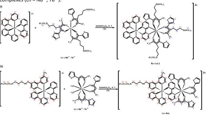
Figure 29. Synthesis routes of the Ru (i), RuL (ii) and LnL3 (Ln = Nd3+, Yb3+) complexes (iii)..
Figure 30. Synthesis routes of the heterobimetallic Ru—LnL3 (i) and Ln—RuL (ii) silylated complexes (Ln = Nd3+, Yb3+). ... 105
Figure 31. 1H NMR spectrum of the Ru complex (MeOD-d
4, 600 MHz). ... 106
Figure 32. TOCSY1D NMR spectra of the Ru complex (MeOD-d4, 600 MHz) irradiated at (a) 9.16
ppm, (b) 8.02 ppm and (c) 7.78 ppm. ... 107
Figure 33. 1H{13C} HSQC NMR spectra of the Ru complex (MeOD-d
4, 600 MHz) in the aromatic
region. The numbering of C and H atoms is given for the complex and present in both spectra. ... 108
Figure 34. 1H{13C}-HMBC NMR spectrum of the Ru complex in the aromatic region (MeOD-d
4,
600 MHz). ... 109
Figure 35. 1H NMR spectrum of the NdL3 complex (CDCl
3-d, 600 MHz). ... 110
Figure 36. TOCSY1D NMR spectra of the NdL3 complex (CDCl3-d, 600 MHz) irradiated at (a)
7.19 ppm and (b) 1.90 ppm. ... 111
Figure 37. 1H{13C}-HSQC NMR spectra of the NdL3 complex (CDCl
3-d, 600 MHz): (a) aliphatic
region; (b) aromatic region. The numbering of H and C atoms is given for the complex and present in both spectra. ... 113
Figure 38. 1H NMR spectrum of the Ru—NdL3 complex (MeOD-d
4, 600 MHz). ... 114
Figure 39. TOCSY1D NMR spectra of the Ru—NdL3 complex (MeOD-d4, 600 MHz) irradiated
at (a) 7.97 ppm, (b) 7.80 ppm, (c) 7.70 ppm, (d) 7.65 ppm and (e) 0.85 ppm. ... 115
Figure 40. 1H{13C}-HSQC NMR spectra of the Ru—NdL3 complex (MeOD-d
4, 600 MHz): (a)
aliphatic region; (b) aromatic region. The numbering of H and C atoms is given for the complex and present in both spectra. ... 117
Figure 41. 1H{13C}-HMBC NMR spectrum of the Ru—NdL3 complex in the aromatic region
(MeOD-d4, 600 MHz). ... 118
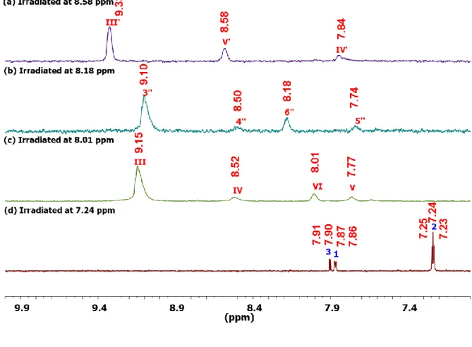
Figure 42. 1H NMR spectrum of the Yb—RuL complex (MeOD-d
Figure 43. TOCSY1D NMR spectra of the Yb—RuL complex (MeOD-d4, 600 MHz) irradiated at
(a) 8.58 ppm, (b) 8.18 ppm, (c) 8.01 ppm, (d) 7.24 ppm, (e) 2.68 ppm, (f) 1.68 ppm and (g) 1.18 ppm. ... 120
Figure 44. 1H{13C}-HSQC NMR spectra of the Yb—RuL complex (MeOD-d
4, 600 MHz): (a)
aliphatic region; (b) aromatic region. The numbering of H and C atoms is given for the complex and present in both spectra. ... 123
Figure 45. 1H{13C}-HMBC NMR spectrum of the Yb—RuL complex in the aromatic region
(MeOD-d4, 600 MHz). ... 124
Figure 46. 1H{13C}-HMBC NMR spectrum of the Yb—RuL complex (MeOD-d
4, 600 MHz)……
... 124
Figure 47. FTIR spectra for the samples (a) LnL3 (green line), Ru—LnL3 (blue line), Ru (red line) and (b) Ln (green line), Ln—RuL (blue line), RuL (red line), Nd(III) complex at left and Yb(III) at right, all analysis were carried out at KBr pellets. ... 126
Figure 48. FT-Raman spectra of (a) LnL3 (green line), Ru—LnL3 (blue line), Ru (red line) and (b) Ln (green line), Ln—RuL (blue line), RuL (red line). ... 128
Figure 49. Absorption spectra (2,5 x 10⁻⁵ mol/L, ethanol) of (a) NdL3 (green line), Ru—NdL3
(blue line), Ru (red line); (b) YbL3 (green line), Ru—YbL3 (blue line), Ru (red line); (c) Nd (green line), Nd—RuL (blue line), RuL (red line) and (d) Yb (green line), Yb—RuL (blue line), RuL (red line). ... 129
Figure 50. Room temperature excitation (on the left, monitoring at emission = 660 nm) and
emission (on the right, excitation at excitation = 455 nm) spectra of the RuL1 complex in solid
state. ... 131
Figure 51. Room temperature excitation (on the left, monitoring at emission = 670 nm) and
emission (on the right, monitoring at excitation = 475 nm) spectra of the Ru complex in solid
Figure 52. Room temperature excitation (on the left, monitoring at emission = 620 nm) and
emission (on the right, monitoring atexcitation = 465 nm) spectra of the RuL complex in solid
state. ... 133
Figure 53. Room temperature excitation (on the left, monitoring at emission = 1062 nm) and
emission (on the right, monitoring atexcitation = 395 nm) spectra of the Nd complex in solid
state. ... 133
Figure 54. Room temperature excitation (on the left, monitoring atemission = 977 nm) and
emission (on the right, monitoring atexcitation = 395 nm) spectra of the Yb complex in solid
state. ... 134
Figure 55. Room temperature excitation (on the left, monitoring at emission = 1063 nm) and
emission (on the right, monitoring atexcitation = 391 nm) spectra of the NdL3 complex in solid
state. ... 135
Figure 56. Room temperature excitation (on the left, monitoring atemission = 980 nm) and
emission (on the right, monitoring at excitation = 395 nm) spectra of the YbL3 complex in solid
state. ... 135
Figure 57. Room temperature excitation ((a), emission: 665 nm and (c), emission: 1065 nm) and
emission ((b), excitation: 450 nm; and (d), excitation: 375, 450 and 801 nm) spectra of the Ru—
NdL3 complex in solid state... 137
Figure 58. Room temperature excitation ((a), emission: 670 nm and (c), emission: 978 nm) and
emission ((b), excitation: 455 nm; and (d), excitation: 370 and 455 nm) spectra of the Ru—YbL3
complex in solid state. ... 138
Figure 59. Room temperature excitation ((a), emission: 610 nm and (c), emission: 1063 nm) and
emission ((b), excitation: 465 nm; and (d), excitation: 335, 370, 455 and 802 nm) spectra of the
Figure 60. Room temperature excitation ((a), emission: 610 nm and (c), emission: 980 nm) and
emission ((b), excitation: 450 nm; and (d), excitation: 370 and 450 nm) spectra of the Yb—RuL
complex in solid state. ... 140
Figure 61. Assumed schematic energy transfer processes from Ru and RuL complexes (Ru(II)) to Nd3+ and Yb3+ (A = Absorption; ISC = Inter-System Cross; P = phosphorescence; ET = Energy
Transfer; F = Fluorescence) in the Ru—LnL3 and Ln—RuL complexes. ... 143
Figure 62. Size distribution for the MSNs. Number of nanoparticles = 400. SEM images of MSNs used in size distribution measurements. ... 147
Figure 63. SEM images of mesoporous silica nanoparticles (MSNs). ... 148
Figure 64. TEM images of mesoporous silica nanoparticles (MSNs). ... 149
Figure 65. STEM images of mesoporous silica nanoparticles (MSNs). On the left: bright field images; on the right: dark field images. ... 150
Figure 66. DRIFT spectrum of the MSNs ... 151
Figure 67. TGA/DTA curves of the MSNs... 152
Figure 68. Nitrogen adsorption/desorption isotherms of the MSNs. ... 152
Figure 69. 29Si-MAS NMR spectrum (black line) and its respective deconvolution (red line) of
the MSNs. ... 153
Figure 70. Representation of grafting reactions of silylated complexes onto MSNs. ... 155
Figure 71. DRIFT spectra of (a) MSNs and different nanohybrids: (b) SiO2-Ru, (c) SiO2-Nd, (d)
SiO2-Yb, (e) SiO2-RuNd and (f) SiO2-RuYb. ... 156
Figure 72. FT-Raman spectra of (a) YbL3 (green line), RuL1 (red line) and SiO2-RuYb (blue line);
(b) NdL3 (green line), RuL1 (red line) and SiO2-RuNd (blue line). ... 158
Figure 73. 13C{1H}CP-MAS NMR spectra (I) and 29Si{1H}CP-MAS NMR spectra (II) of (a) SiO
2-Ru,
groups. 12: carbon atoms in methoxysilyl groups not grafted. The numbering of carbon atoms is given on the left side with Roman figures for the bipyridine ligands; Arabic numerals for the alkoxysilane groups (methoxysilyl and ethoxysilyl)... 161
Figure 74. FEG-SEM images of (a) MSNs and different nanohybrids: (b) SiO2-Ru, (c) SiO2-Nd,
(d) SiO2-Yb, (e) SiO2-RuYb and (f) SiO2-RuNd... 162
Figure 75. HR-TEM images of (a) MSNsand different nanohybrids: (b) SiO2-Ru, (c) SiO2-Nd, (d)
SiO2-Yb, (e) SiO2-RuYb and (f) SiO2-RuNd. ... 163
Figure 76. Electron microscope images and elemental cartography of the SiO2-Ru: (a) STEM
image, (b) Si cartography, (c) Ru cartography and (d) Si (blue color) and Ru (red color) cartographies. ... 164
Figure 77. Electron microscope images and elemental cartography of the SiO2-Nd: (a) STEM
image, (b) Si cartography, (c) Nd cartography and (d) Si (blue color) and Nd (green color) cartographies. ... 165
Figure 78. Electron microscope images and elemental cartography of the SiO2-Yb: (a) STEM
image, (b) Si cartography, (c) Yb cartography and (d) Si (blue color) and Yb (green color) cartographies. ... 166
Figure 79. Electron microscope images and elemental cartography of the SiO2-RuYb: (a) STEM
image, (b) Si cartography, (c) Ru cartography, (d) Yb cartography, (e) Si (blue color), Ru (red color) and Yb (green color) cartographies. (f) STEM image with a line profile of Ru (red line) and Yb (green line) atoms in the selected area (blue line). ... 168
Figure 80. Electron microscope images and elemental cartography of the SiO2-RuNd: (a) STEM
image, (b) Si cartography, (c) Ru cartography, (d) Nd cartography and (e) Si (blue color), Ru (red color) and Nd (green color) cartographies. (f) STEM image with a line profile of Ru (red line) and Nd (green line) atoms in the selected area (blue line). ... 169
Figure 81. Room temperature excitation (emission = 643 nm, on the left) and emission (excitation
= 455 nm, on the right) spectra of the SiO2-Ru nanohybrid in solid state. ... 171
Figure 82. Room temperature excitation (emission = 1063 nm; on the left) and emission
(excitation = 330 nm; on the right) spectra of the SiO2-Nd nanohybrid in solid state. ... 172
Figure 83. Room temperature excitation (on the left; emission = 981 nm) and emission (on the
right; excitation = 330 and 388 nm; green and red lines, respectively) spectra of the SiO2-Yb
nanohybrid in solid state. ... 172
Figure 84. Room temperature excitation ((a), emission: 610 nm and (c), emission: 1061.5 nm) and
emission ((b), excitation: 455 nm; and (d), excitation: 365 and 455 nm) spectra of the SiO2-RuNd
nanohybrid in solid state. ... 173
Figure 85. Room temperature excitation ((a), emission: 610 nm and (c), emission: 980 nm) and
emission ((b), excitation: 370 and 455; and (d), excitation: 365 and 475 nm) spectra of the SiO2
-RuYb nanohybrid in solid state. ... 175
Figure 86. Schematic energy transfer processes (Ru(II)) to Nd3+ and Yb3+ (A = Absorption; ISC =
Inter-System Cross; P = phosphorescence; ET = Energy Transfer; F = Fluorescence) in the SiO2
-Ru, SiO2-RuNd and SiO2-RuYb nanohybrids. ... 176
Figure 87. Representation of grafting reactions of (a) Ru—LnL3 complex onto MSNs and (b) Ln—RuL complexes onto MSNs and DSNs. ... 177
Figure 88. DRIFT spectra of (a) SiO2-RuYbL3, (b) SiO2-NdRuL, (c) SiO2-YbRuL and (d) SiO2
d-YbRuL nanohybrids. ... 178
Figure 89. FT-Raman spectra of (a) Ru—YbL3 (blue line) and SiO2-RuYbL3 (black line), (b) Nd—
RuL (blue line) and SiO2-NdRuL (black line), (c) Yb—RuL (blue line) and SiO2-YbRuL (black line)
and (d) Yb—RuL (blue line) and SiO2 d-YbRuL (black line). ... 180
Figure 90. 13C {1H}CP-MAS NMR spectra (I) and 29Si {1H}CP-MAS NMR spectra (II) of (a) SiO
2
-RuYbL3, (b) SiO2-NdRuL, (c) SiO2-YbRuL and (d) SiO2 d-YbRuL. *: carbon atoms in residual
M1: —O—SiR3. The numbering of carbon atoms is given on the figure 30 with Roman figures
for the bpy ligand; Arabic numerals for the bpy-Si and TTA-Si ligands. ... 182
Figure 91. FEG-SEM images of (a) SiO2-RuYbL3, (b) SiO2-NdRuL, (c) SiO2-YbRuL and (d) SiO2
d-YbRuL nanohybrids. ... 183
Figure 92. HR-TEM bright field (above) and dark field (below) images of (a) SiO2-RuYbL3, (b)
SiO2-NdRuL and (c) SiO2-YbRuL nanohybrids. ... 184
Figure 93. Electron microscope images and elemental cartography of the SiO2-RuYbL3: (a)
STEM image, (b) Si cartography, (c) Ru cartography, (d) Yb cartography, (e) Si (blue color), Ru (red color) and Yb (green color) cartographies. (f) STEM image with a line profile of Ru (red line) and Yb (green line) atoms in the selected area (blue line). ... 186
Figure 94. Electron microscope images and elemental cartography of the SiO2-NdRuL: (a)
STEM image, (b) Si cartography, (c) Ru cartography, (d) Nd cartography, (e) Si (blue color), Ru (red color) and Nd (green color) cartographies. (f) STEM image with a line profile of Ru (red line) and Nd (green line) atoms in the selected area (blue line). ... 187
Figure 95. Electron microscope images and elemental cartography of the SiO2-YbRuL: (a)
STEM image, (b) Si cartography, (c) Ru cartography, (d) Yb cartography, (e) Si (blue color), Ru (red color) and Yb (green color) cartographies. (f) STEM image with a line profile of Ru (red line) and Yb (green line) atoms in the selected area (blue line). ... 189
Figure 96. Electron microscope images and elemental cartography of the SiO2 d-YbRuL: (a)
STEM image, (b) Si cartography, (c) Ru cartography, (d) Yb cartography, (e) Si (blue color), Ru (red color) and Yb (green color) cartographies. (f) STEM image with a line profile of Ru (red line) and Yb (green line) atoms in the selected area (blue line). ... 190
Figure 97. Room temperature excitation ((a), emission: 664 nm and (c), emission: 979 nm) and
emission ((b), excitation: 455 nm; and (d), excitation: 395 and 455 nm) spectra of the SiO2-RuYbL3
Figure 98. Room temperature excitation ((a), emission: 614 nm and (c), emission: 1063 nm) and
emission ((b), excitation: 455 nm; and (d), excitation: 337 and 455 nm) spectra of the SiO2-NdRuL
nanohybrid in solid state. ... 194
Figure 99. Room temperature excitation ((a), emission: 610 nm and (c), emission: 980 nm) and
emission ((b), excitation: 455 nm; and (d), excitation: 377 and 455 nm) spectra of the SiO2-YbRuL
nanohybrid in solid state. ... 195
Figure 100. Room temperature excitation ((a), emission: 614 nm and (c), emission: 979 nm) and
emission ((b), excitation: 455 nm; and (d), excitation: 370 and 455 nm) spectra of the SiO2 d-YbRuL
nanohybrid in solid state. ... 196
LIST OF TABLES
Table 1. Photophysical properties for MLCT-based visible emission and Ln(III) based NIR…. ... 142
Table 2. Chemical shift (ppm), percentage of species and condensation degree obtained from deconvolution of 29Si MAS spectrum from MSNs. ... 154
Table 3. Morphological properties of commercial Ludox silica AS-40 and MSNs. ... 155
Table 4. DRIFT data for the nanohybrids. ... 158
Table 5. Tn and Qn chemical shifts of the nanohybrids. ... 160
Table 6. Grafting efficiencies, in mmol of ruthenium and lanthanide complexes.g-1 of silica,
and amount of ruthenium and lanthanide complexes.nm-2 of silica for the nanohybrids…..
... 170
Table 7. Photophysical properties for MLCT-based visible emission and Ln(III) based NIR…. ... 176
Table 8. DRIFT data for the nanohybrids. ... 179
Table 9. Tn and Qn chemical shifts of the nanohybrids. ... 181
Table 10. Grafting efficiencies, in mmol of ruthenium and lanthanide complexes.g-1 of silica,
LIST OF ABBREVIATIONS AND ACRONYMS
OLEDs: Organic Light Emitting Diodes A: Absorption
P: Phosphorescence IC: Internal Conversion ISC: Intersystem crossing ET: Energy Transfer S0 or S1: singlet
T1 or T2: triplet
φ: quantum yield
φ’: relative quantum yield Abs: Absorbance
Em: Emission R: Reference S: Unknown
ANMTTA: [4′-(4-amino-3-nitrophenoxy)methylene-2,2′:6′,2″-terpyridine-6,6″-diyl] bis(methylenenitrilo) tetrakis(acetic acid)
HOCl: hypochlorous acid
HTTA: (4′-hydroxymethyl-2,2′:6′,2″-terpyridine-6,6″-diyl) bis(methylenenitrilo) tetrakis(acetic acid)
TTA: 2-thenoyltrifluoroacetonate DBSO: dibenzyl sulfoxide
TFAC: trifluoroacetylacetonate DBM: 1,3-diphenylpropane-1,3-dione PTSO: p-tolylsulfoxide
NIR: Near-Infrared
hfaa = anion of hexafluoroacetylacetone pz = pyrazole BT: Back Transfer 1P: one photon 2P: two photons H2pdo: 5,6-dihydroxyphenanthroline dppz: 2,3-bis(2-pyridyl)pyrazine dik: 1,3-diketonate
bppz: 2,3-bis(2-pyridyl)pyrazine MLCT: metal-to-ligand charge transfer LC: ligand centered H2L: 2-(1H-benzo[d]imidazol-2-yl)-6-methoxyohenol ppy: 2-phenylpyridine dcbpy: 2,2’-bipyridine-4,4’-dicarboxylate bpmc: 5-bromopyrimidine-2-carboxylic acid pdt: 1,3-dimethyl-5-phenyl-1H-[1,2,4]triazole
PLIM: Phosphorescence Lifetime Imaging Microscopy TPA: two-photons absorption
OPA: one-photon absorption
NA = nicotinamide (pyridine-3-carboxamide)
tpphz: tetrapyrido[3,2-a:2′,3′- c:3′′,2′′-h:3′′′,4′′′-j] phenazine
EnT: quantum yield of energy transfer
kEnT: energy transfer rate constant
AFP: alpha-fetal protein BibzImH2: Bibenzimidazole
bpmd: 2,2'-bipyrimidine bpy: 2,2’-bipyridine
LUMO: Lowest Unoccupied Molecular Orbital
fod: 6,6,7,7,8,8,8-heptafluoro-2,2-dimethyl-3,5-octanedionato LED: Light Emitting Diode
MSNs: mesoporous silica nanoparticles DSNs: dense silica nanoparticles
APTES: 3-aminotriethoxysilane DBM: dibenzoylmethane HMDSA: hexamethyldisilazane
phen-Si: 5-(N,N-bis-3-(triethoxysilyl)-propyl)ureyl-1,10-phenanthroline bpy-Si: 4-methyl-4’-(n-triethoxysilylpropyl)amine- methyl- 2,2’-bipyridine
TTA-Si: 4,4,4-Trifluoro-2-(3-(trimethoxysilyl)-propyl)-1-(2-thienyl)-1,3-butanedione Ru: [Ru(bpy)2(bpmd)]Cl2
RuL: [Ru(bpy)(bpy-Si)(bpmd)]Cl2
RuL1: [Ru(bpy)2(bpy-Si)]Cl2
Nd: Nd(TTA)3 2H2O
Yb: Yb(TTA)3 2H2O
NdL3: [Nd(TTA-Si)3]
YbL3: [Yb(TTA-Si)3]
Ru—NdL3: [Ru(bpy)2(bpmd)Nd(TTA-Si)3]Cl2
Ru—YbL3: [Ru(bpy)2(bpmd)Yb(TTA-Si)3]Cl2
Nd—RuL: [Ru(bpy)(bpy-Si)(bpmd)Nd(TTA)3]Cl2
Yb—RuL: [Ru(bpy)(bpy-Si)(bpmd)Yb(TTA)3]Cl2
TEOS: tetraethylorthosilicate
CTAB: cetyltrimethylammonium bromide
AIBA: 2,2’-Azobis (2-methylpropionamide) dihydrochloride EA: Elemental Analysis
FTIR: Fourier Transformed Infra-Red Spectroscopy
DRIFT: Diffuse Reflectance Infra-Red Fourier Transformed Spectroscopy FT-Raman: Fourier Transformed Raman Spectroscopy
UV-Vis: Ultraviolet-Visible Spectroscopy NMR: Nuclear Magnetic Resonance
MAS NMR: Magic Angle Spinning Nuclear Magnetic Resonance
CP-MAS NMR: Cross-Polarization and Magic Angle Spinning Nuclear Magnetic Resonance TOCSY: Total Correlation Spectroscopy
HSQC: Heteronuclear Single Quantum Coherence HMBC: Heteronuclear Multiple Bond Correlation TMS: Tetramethylsilane
MS: Mass Spectrometry ESI: Electrospray
DCI: Desorption Chemical Ionization FAB: Fast Atom Bombardment FWHM: Full Width at Half Maximum
STEM: Transmission and Scanning Transmission Electron Microscopy TEM: Transmission Electron Microscopy
EDX or EDS: Energy Dispersive X-ray spectroscopy TGA: Thermogravimetric Analysis
DTA: Differetial Thermal Analysis
J: coupling constant wavelength MeOH: methanol EtOH: ethanol THF: tetrahydrofuran CH2Cl2: dichloromethane DMF: dimethylformamide MeOD: deuterated methanol CDCl3: deuterated cloroform
CONTENTS
1 – INTRODUCTION... 47
1.2. Luminescent silylated complexes and silica-based nanohybrids ... 63 2 – OBJECTIVES ... 69 2.1. General Objective ... 71 2.2. Specific Objectives ... 71 3 – EXPERIMENTAL SECTION ... 73 3.1 Synthesis ... 75
3.1.1. Ligands modified by alkoxysilyl groups ... 75
3.1.1.1. Synthesis of 4-methyl-4’-(n-triethoxysilylpropyl)amine- methyl- 2,2’-bipyridine ligand (bpy-Si). ... 75 3.1.1.2. Synthesis of 4,4,4-Trifluoro-2-(3-(trimethoxysilyl)-propyl)-1-(2-thienyl)-1,3-butanedione ligand (TTA-Si). ... 76
3.1.2. Synthesis of Ruthenium(II) complexes ... 77
3.1.3. Synthesis of Lanthanide(III) complexes ... 82
3.1.4. Synthesis of d-f heterobimetallic (Ru(II)—Ln(III)) silylated complexes ... 84
3.1.5. Mesoporous silica nanoparticles (MSNs) ... 87
3.1.6. Luminescent silica-based nanohybrids obtained by grafting of ruthenium(II) and lanthanide(II) silylated complexes onto MSNs ... 88
3.1.6.1. SiO2-Ru, SiO2-Nd and SiO2-Yb ... 89
3.1.6.2. SiO2-RuNd and SiO2-RuYb ... 89
3.1.7. Luminescent silica-based nanohybrids obtained by grafting of d-f heterobimetallic silylated complexes onto mesoporous silica nanoparticles (MSNs) and dense silica nanoparticles Ludox AS40 (DSNs) ... 90
3.1.7.1. SiO2-RuYbL3 ... 90
3.1.7.2. SiO2-NdRuL ... 91
3.1.7.3. SiO2-YbRuL ... 92
3.1.7.4. SiO2 d-YbRuL ... 92 3.2. Characterization Methods ... 93
3.2.1. Elemental Analysis ... 93
3.2.2. Fourier Transformed Infra-Red Spectroscopy... 93
3.2.3. Fourier Transformed Raman Spectroscopy ... 93
3.2.4. Ultraviolet-Visible Spectroscopy ... 94
3.2.5. Nuclear Magnetic Resonance... 94
3.2.6. Mass Spectrometry…. ... 95
3.2.7. Luminescence ... 95
3.2.8. Scanning Electron Microscopy ... 96
3.2.9. Transmission and Scanning Transmission Electron Microscopy ... 96
3.2.10. N2 Adsorption and Desorption ... 97
3.2.11. Thermogravimetric Analysis ... 97
4 – LUMINESCENT RUTHENIUM(II), LANTHANIDE(III) AND d-f HETEROBIMETALLIC SILYLATED COMPLEXES ... 99
4.1. Characterization of the ligands modified by alkoxysilyl groups (bpy-Si and TTA-Si)… ... 101
4.2. Synthesis of d-f heterobimetallic silylated complexes ... 101
4.3. Spectroscopic characterization of the ruthenium(II), lanthanide(III) and d-f heterobimetallic silylated complexes ... 105
4.4. Luminescent properties of ruthenium(II), ytterbium(III) and neodymium(III) complexes... 131
4.5. Photophysical properties of heterobimetallic Ru(II)—Yb/Nd(III) silylated complexes. ... 136
5 – LUMINESCENT NANOHYBRIDS OBTAINED BY GRAFTING OF RUTHENIUM(II), LANTHANIDE(III) AND d-f HETEROBIMETALLIC SILYLATED COMPLEXES ... 145
5.1. Characterization of mesoporous silica nanoparticles (MSNs) ... 147
5.3. Synthesis and characterization of silica-based nanohybrids obtained by grafting of silylated ruthenium(II) and lanthanide (III) complexes onto MSNs ... 155
5.3.1. Spectroscopic characterization of silica-based nanohybrids obtained by grafting of silylated ruthenium(II) and lanthanide (III) complexes onto MSNs ... 156
5.3.2. Microstructural characterization of silica-based nanohybrids obtained by grafting of silylated ruthenium (II) and lanthanide (III) complexes onto MSNs ... 162
5.3.3. Photophysical properties of silica-based nanohybrids obtained by grafting of ruthenium(II) and lanthanide(III) silylated complexes ... 171
5.4. Synthesis and characterization of silica-based nanohybrids obtained by grafting of
d-f heterobimetallic silylated complexes onto MSNs and Ludox silica AS-40 (DSNs) ... 177
5.4.1. Spectroscopic characterization of the silica-based nanohybrids obtained by grafting of d-f heterobimetallic silylated complexes onto MSNs and Ludox silica AS-40 (DSNs).. ... 178
5.4.2. Microstructural characterization of silica-based nanohybrids obtained by grafting of d-f heterobimetallic silylated complexes onto MSNs and Ludox silica AS-40 (DSNs).. ... 183
5.4.3. Luminescent properties of silica-based nanohybrids obtained by grafting of d-f heterobimetallic silylated complexes onto MSNs and Ludox silica AS-40 (DSNs) ... 192
6 – CONCUSION AND PERSPECTIVES ... 197
REFERENCES ... 203
1. INTRODUCTION
The group of elements known as "Rare Earths" includes lanthanide metals (elements with atomic number between Z = 57, the lanthanum, and Z = 71, lutetium), and scandium (Z = 21) and yttrium (Z = 39). Lanthanides share similar chemical and physical properties, existing mainly in their trivalent state LnIII ([Xe]4fn, n = 0-14).1–3 Shielding of the 4f orbitals by the outer
5s and 5p sub-shells results in weak ligand field effects and consequently, characteristic narrow-line emission and long lifetimes of the excited states are observed from the visible to the near-infrared region.1–3 Lanthanide based materials have been widely employed as
analytical sensors, imunoassays, bioimaging device, OLEDs, optical communication devices and in solar energy conversion, among others.3–7 For example, Eu3+ ions provide suitable red
luminescence for biological detection and organic electroluminescence.8, 9 Nd3+ ions and Yb3+,
emiting in the range of 900 to 1400 nm, are good candidates for biological luminescence imaging, laser systems and luminescent nanothermometers.7, 10 Pr3+ ions (emission at 1300
nm) and Er3+ ions (emission at 1500 nm) have been mainly used in optical amplifiers for
fiber-optic networks.7, 11–13
Although there are a number of energy levels available for the f-f transitions of lanthanide ions the very low molar absorption coefficients for UV-visible light (< 10 M) is a serious drawback.4, 14 This problem has been circumvented by using organic chromophores.
These ligands exhibit strong absorption in the UV region (~106 M) and subsequent ligand to
Lanthanide energy transfer can take place in the so-called “antenna effect”.4, 14 The figure 1
shows the schematic representation of the energy transfer mechanism between organic ligands and lanthanide ion.
Figure 1. Schematic representation of the antenna effect from the organic ligand to the lanthanide(III) ion. Abbreviations: A = absorption; F = fluorescence; P = phosphorescence; ISC = intersystem crossing; ET = energy transfer; S = singlet; T = triplet. Full vertical lines indicate radiative transitions; dashed vertical lines indicate nonradiative transitions.4, 13, 15, 16
radiative transition nonradiative transition Lanthanide ion emission IC IC 4f 4f ET F S0 T1 T2 S1 A Organic ligand ISC P Lanthanide (III)
Upon irradiation in UV the organic ligands are excited to a vibrational level of the first excited singlet state (S0 S1). The molecule undergoes fast internal conversion (IC) to lower
vibrational levels of the S1 state, for instance, through interaction with solvent molecules. The
excited singlet state can be deactived radiatively to the ground state (fluorescence, S1 S0)
or can undergo nonradiative intersystem crossing (ISC) from the singlet state (S1) to the triplet
state (T1 and T2). The triplet state (T1 and T2) can be deactivated radiatively to the ground state
(molecular phosphorescence (P)). Alternatively, the energy transfer (ET, nonradiative transition) takes place from the triplet state of the ligand to the central lanthanide ions (excited 4f state) in the so-called “antenna effect”.4, 13, 15, 16 After this indirect excitation (ET),
the lanthanide ion may undergo a radiative transition to a lower 4f state by characteristic line-like photoluminescence or may be deactivated by nonradiative processes.4, 13, 15, 16
In this way, the luminescent quantum yield (φ) is an important parameter for evaluation of the efficiency of the emission process in luminescent compounds, for instance,
luminescent complexes. The quantum yield is defined as the ratio of the number of emitted to absorbed photons according to the equation 1.16, 17
One method to determine the luminescent quantum yield consists on determine the relative quantum yield (φ’). In this case, the quantum yield of unknown sample is compared with that of the reference sample, in solution, according to the equation 2.16, 17
Where, Abs is the absorbance at the excitation wavelength, Em is emission intensity, and n is the refractive index of the solvent used. Subscripts R and S refer to the reference and the unknown, respectively.16, 17
The energy gap between the excited state of the ligand and that of the lanthanide ion determines whether or not effective energy transfer takes place. For efficient energy transfer, the lowest energy triplet state of the sensitiser must be higher than (1000-2000 cm-1) the
lowest excited state (emitting state) of the lanthanide ions.13, 18 Botelho and co-workers
reported the synthesis, characterization and study of photophysical properties of a nine-coordinate Eu3+ complex based on the tridentate
4-(4’-tert-butylbiphenyl-4-yl)-2,2’-bipyridine-6-carboxylic acid ligand. This complex contains three tridentate ligands acting as an efficient sensitizer avoiding the coordinated water and solvents in the first coordination sphere. Besides that, it exhibits one of the highest quantum yield values in solution (φ = 85 %) and a long excited-state lifetime ( = 1.8 ms).19 Xiao and co-workers described the design,
synthesis and applications of two new lanthanide complexes, ANMTTA-Eu3+ and ANMTTA-Tb3+
{ANMTTA, [4′-(4-amino-3-nitrophenoxy)methylene-2,2′:6′,2″-terpyridine-6,6″-diyl] bis(methylenenitrilo) tetrakis(acetic acid)} for the highly sensitive and selective luminescence detection of hypochlorous acid (HOCl) in aqueous media. These probes are almost nonluminescent due photoinduced electron transfer process (due the 4-amino-3-nitrophenyl moiety) but upon reaction with HOCl, the quencher moiety is rapidly cleaved from the probe
ɸ =
𝑛𝑢𝑚𝑏𝑒𝑟 𝑜𝑓 𝑎𝑏𝑠𝑜𝑟𝑏𝑒𝑑 𝑝ℎ𝑜𝑡𝑜𝑛𝑠𝑛𝑢𝑚𝑏𝑒𝑟 𝑜𝑓 𝑒𝑚𝑖𝑡𝑡𝑒𝑑 𝑝ℎ𝑜𝑡𝑜𝑛𝑠 (1)ɸ′ = (
𝐴𝑏𝑠𝑅 𝐴𝑏𝑠𝑆) (
𝐸𝑚𝑆 𝐸𝑚𝑅) (
𝑛𝑆 𝑛𝑅)
2ɸ
𝑅(2)
complexes, which affords strongly luminescent lanthanide complexes HTTA-Eu3+ and
HTTA-Tb3+ {HTTA, (4′-hydroxymethyl-2,2′:6′,2″-terpyridine-6,6″-diyl) bis(methylenenitrilo)
tetrakis(acetic acid)}, accompanied by the remarkable luminescence enhancements.20
In this way, several organic ligands have been used to design efficient luminescent lanthanide complexes.5, 16, 21 Among the several organic ligands that have been used
-diketone ligands display high molar absorption coefficients (~106 M-1 cm-1) and stability
constants. Besides that, the synthesis of lanthanide complexes using these chromophores is relatively easy, leading to the complexes with excellent luminescent properties.16 Malta and
co-workers described the synthesis, characterization and spectroscopic properties of the [Eu(TTA)3.2(DBSO)], (TTA = 2-thenoyltrifluoroacetonate and DBSO = dibenzyl sulfoxide), a
complex highly luminescent under ultraviolet excitation.15
Andreiadis and co-workers22 reported the synthesis of new octacoordinated ternary
beta-diketonates of europium and terbium using an anionic tetradentate terpyridine-carboxylate ligand (2,2′:6′,2″-terpyridine-6-car- boxylate, L) as a sensitizer of lanthanide luminescence with two beta-diketonates ligands TTA- for Eu3+ and trifluoroacetylacetonate
(TFAC-) for Tb3+. These complexes showed high stability in solution and high luminescence
quantum yield (66 % for Eu3+ complex). Furthermore, these complexes were incorporated in
polymer matrixes leading to highly luminescent flexible layers with luminescence quantum yield up to 62 %. Recently, Lima et al.23 showed a new and fast way to synthesize
beta-Diketonate ternary europium complexes without Eu(β-diketonate)3(H2O)2 as an intermediate.
The strategy consisted on adding non-ionic ligand (DBSO, DBM = 1,3-diphenylpropane-1,3-dione or PTSO = p-tolylsulfoxide, as L ligand) to the original salt, EuCl3(H2O)6, to obtain the
intermediates EuCl3(L)4(H2O)n, which were soluble in methanol or ethanol. Subsequently, the
β-diketonate ligands are carefully added to the intermediate to obtain the final product Eu(beta-diketonate)3(L)2. The usual synthetic route that takes a few weeks, can be carried out
in days. Besides that, average overall yield up to 69 % obtained from this faster synthesis was significantly larger than the usual synthesis.
Despite the large number of visible emitting -diketonates lanthanide complexes15, 16, 24, fewer NIR-emitting lanthanides have been reported. This can be explained by the fact that
the NIR emitting lanthanides exhibit lower excited state than the visible emitting lanthanides, requiring organic ligands with lowest excited state low enough to sensitize them (4F
11257 cm-1; 2I
13/2 for Er3+, 6536 cm-1 and 2F5/2 for Yb3+, 10400 cm-1), according to the
representation of the diagram of the f–f energy levels in the figure 2.13, 25
Figure 2. Representation of the diagram of the f–f energy levels of Nd(III), Er(III) and Yb(III) up to ca. 20,000cm−1. The energy levels from which NIR luminescence originates are marked in
bold, and the main NIR emissions showed as descending arrows. Adapted from25.
In this sense, some authors have described a series of lanthanide complexes of the type [Ln(hfaa)3(pz)2] (Ln = La, Pr, Nd, Eu, Gd, Tb, Dy, Ho, Er, Tm and Yb; hfaa = anion of
hexafluoroacetylacetone and L = pyrazole).26 These complexes are volatile, which is
advantageous for their use as precursors for high-performance lanthanide-based materials through chemical vapor deposition. Futhermore, the authors related that the environment around these metal ions was asymmetric, leading to increased radiative rates and luminescence efficiencies. Both ligands (hfaa and pyrazole) were good sensitizers for all the visible and NIR emitters except for Tb(III), Dy(III), and Tm(III), where pyrazole gave a negative effect (energy back transfer (BT)), due to poor intramolecular energy transfer match. Thus, the good luminescent properties make these NIR-luminescent complexes to have potential application in optical communication, telecommunications, and fluoroimmunoassays.26 For
instance, Woodward and co-workers27 showed the synthesis of a fluorine-based
donor-acceptor beta-diketone ligand to obtain a series of luminescent lanthanides complexes from visible- to NIR-emitting region. The preferred form (cis-enol) binds strongly to the lanthanide(III) ions (Ln = Eu, Sm, Dy, Tb, Yb, Nd, Er, and Gd) in the presence of phenanthroline,
which resulted in ternary tris(diketonates) complexes with 1,10-phenanthroline. The successfully sensitization by two photons (2P) excitation to access these complexes through the biological window (∼800 nm), along with the delay of these long lifetime lanthanide luminescence emissions (from visible to NIR), displayed significant potential for applications in biomedical analysis (imaging and sensing) as luminescent probes.27
Nevertheless, NIR emitting Nd3+ and Yb3+ are rarely used in analytical detection or
imunoassays. The visible emitting lanthanide complexes (as Eu3+ and Tb3+) are widely used in
this field due to their strong emission. However, most of these lanthanide complexes require excitation in the UV region, which is harmful for the biological systems. The aromatic residues of proteins and DNA absorbs in competition with the chromophores. In addition, the UV excitation cause damages in these biological systems. Moreover, biological tissues are opaque throughout most of the visible region.13, 14 Three biological windows are described.13, 14 The
first one from 0.65 to 1.0 m, the second one from 1.0 to 1.3m and the third one from 1.5 to 1.8m. Figure 3 shows the absorption spectrum of human skin (gray line) with light scattering contribution (dotted blue line) together with the three biological windows (BW) and the emission wavelengths of some lanthanide(III) ions.
Figure 3. Absorption spectrum of human skin (gray line) with light scattering contribution (dotted blue line), definition of biological windows, and emission wavelengths of some lanthanide(III) ions.14