HAL Id: dumas-02369656
https://dumas.ccsd.cnrs.fr/dumas-02369656
Submitted on 19 Nov 2019HAL is a multi-disciplinary open access archive for the deposit and dissemination of sci-entific research documents, whether they are pub-lished or not. The documents may come from teaching and research institutions in France or abroad, or from public or private research centers.
L’archive ouverte pluridisciplinaire HAL, est destinée au dépôt et à la diffusion de documents scientifiques de niveau recherche, publiés ou non, émanant des établissements d’enseignement et de recherche français ou étrangers, des laboratoires publics ou privés.
Molecular Simulations of membrane fission mediated by
BARS protein
Thomas Le Métayer
To cite this version:
Thomas Le Métayer. Molecular Simulations of membrane fission mediated by BARS protein. Life Sciences [q-bio]. 2019. �dumas-02369656�
AGROCAMPUS OUEST
Année universitaire : 2018-2019 Spécialité :
Cursus ingénieur Agronome
Spécialisation (et option éventuelle): Master BMC - Biologie Moléculaire et Cellulaire
Mémoire de fin d’études
Simulations Moléculaires des fissions
membranaires médiées par la protéine BARS
Par : Thomas LE MÉTAYER
Soutenu à Rennes, le 11 Juin 2019 Devant le jury composé de : (Nom, Qualité)
Dr. Jean-Marc FRASLIN, Enseignant référent Dr. Denis MICHEL
Dr. Jean-Pierre TASSAN
Les analyses et les conclusions de ce travail d'étudiant n'engagent que la responsabilité de son auteur et non celle d’AGROCAMPUS OUEST
Ce document est soumis aux conditions d’utilisation
«Paternité-Pas d'Utilisation Commerciale-Pas de Modification 4.0 France» disponible en ligne http :// creativecommons.org/licenses/by-nc-nd/4.0/deed.fr
d'un autre établissement (étudiant arrivé en M2)
de Master de l’Institut Supérieur des Sciences agronomiques, agroalimentaires, horticoles et du paysage
d’Ingénieur de l’Institut Supérieur des Sciences agronomiques, agroalimentaires, horticoles et du paysage
CFR Rennes CFR Angers
R´esum´e long
Parce qu’elle permet la formation de transporteurs mol´eculaires entre les diff´erents compar-timents cellulaires, la fission membranaire est un des processus indispensables `a tout ´ev`enement de trafic cellulaire. C’est particuli`erement le cas au niveau de l’appareil de Golgi, qui constitue le centre majeur de transfert et de tri des prot´eines `a l’´echelle de la cellule. Cependant, les bicouches lipidiques qui composent les membranes sont des structures thermodynamiquement stables, et leur r´earrangement implique de vaincre des ´etats interm´ediaires tr`es ´energ´etiques. Tr`es souvent, cette ´energie est fournie lors de l’hydrolyse de mol´ecules de GTP par les prot´eines de la superfamille des Dynamines, mais il a ´et´e montr´e que la prot´eine CtBP1/BARS peut causer la fission de membranes ind´ependamment des Dynamines. Cette fission s’observe en pr´esence de la prot´eine BARS enti`ere et du domaine C-terminal seul, in vivo et dans des liposomes. Elle pourrait ˆetre caus´ee, comme c’est le cas dans d’autres m´ecanismes de fission, par l’insertion dans les membranes d’une h´elice α amphiphile de 16 acides amin´es, dont la structure a ´et´e ob-serv´ee par Dichro¨ısme Circulaire par l’´equipe du Dr. Luini (IBP, Italie). Nous nous proposons donc d’´etudier par Simulation Mol´eculaire d’une part les propri´et´es structurales de ce segment prot´eique, et d’autre part ses interactions avec des mod`eles membranaires.
Tout d’abord, nous avons utilis´e plusieurs outils de pr´ediction structurale `a partir de la s´equence prot´eique. Nous avons ensuite utilis´e deux ´echelles compl´ementaires de mod´elisation pour les simulations : l’´echelle atomistique permet une repr´esentation plus pr´ecise des mol´ecules et une structure secondaire flexible. Le gros-grain permet des simulations plus longues, car moins lourdes en calcul, mais les prot´eines ont une structure secondaire fig´ee. Deux mod`eles de membranes ont ´egalement ´et´e consid´er´es : un mod`ele homog`ene de bicouche de Phosphatidyl-choline et un mod`ele complexe reproduisant la fraction lipidique des membranes golgiennes. Nous avons donc utilis´e les simulations atomistiques pour observer la structure secondaire via l’algorithme DSSP, en partant d’un peptide en h´elice α ou en pelote libre. Pour les simulations grain, la structure secondaire en h´elice a ´et´e impos´ee au peptide. Les simulations gros-grain ont permis d’observer les interactions entre les peptides et membranes sur des temps d’ ´echantillonage plus longs, et de quantifier si et comment les deux entit´es interagissent.
La pr´ediction de structure indique que le peptide s´epar´e du reste du domaine C-terminal ne serait pas une h´elice mais d´esordonn´e. De plus, ce fragment ne pr´esenterait pas de com-portement amphipathique marqu´e, c’est `a dire une distinction claire entre face hydrophobe et face hydrophile. Les simulations atomistiques confirment cette pr´ediction car les fragments en h´elice α au d´ebut de la simulation se d´eplient en structure d´esordonn´ee. On observe cependant bien un contact entre les peptides et membranes, et plus avec les membranes golgiennes que les membranes contenant uniquement des Phosphatidylcholines. Dans les simulations gros-grain, les peptides se collent aux membranes pendant toute la dur´ee de la simulation. La quantifiction des contacts entre les acides amin´es et les lipides ne montre pas de distinction entre une face avec beaucoup de contacts et une avec peu, ce qui indiquerait un comportement amphiphile. En revanche, on observe que les contacts se font principalement entre les acides amin´es charg´es positivement et les lipides au groupes de tˆete charg´es n´egativement. On peut donc ´emettre l’hypoth`ese que les interactions entre ce fragment de BARS et les mod`eles de membranes gol-giennes seraient ´electrostatiques.
Ces r´esultats et hypoth`eses, transmis `a l’´equipe du Dr. Luini, pourraient ˆetre verifi´ees et affin´ees avec des simulations gros-grain plus longues, qui sont en cours de calcul. Il serait ´egalement int´eressant de faire varier la concentration de peptide dans le syst`eme ou de r´ealiser ces observations sur le domaine C-terminal entier, afin d’observer comment certaines propri´et´es physiques des membranes varient. On pourrait par exemple mesurer la diffusion des lipides, l’aire par lipide ou l’´epaisseur de la membrane qui sont trois indicateurs de r´earrangement de membranes, ou encore les compressibility modulus et les profils de pression lat´erale, indicateurs de la courbure de la membrane n´ecessaire `a l’´ev`enement de fission.
Contents
Introduction 1
Common Abbreviations 2
Material and Methods 2
Secondary Structure prediction tools . . . 2
Simulation systems setup . . . 2
All-Atom Molecular Dynamics parameters . . . 3
All-atom force-field parameters . . . 3
All-atom integration parameters . . . 3
All-Atom trajectory analysis . . . 3
Coarse-Grained Molecular Dynamics parameters . . . 4
Coarse-Grain force-field and integration parameters . . . 4
Goarse-Grain trajectory analysis . . . 4
Results 4 Coarse-Grain simulations analysis . . . 4
BARS helix docks to both membrane models . . . 4
Electostatic forces drive helix-membrane interaction . . . 4
All-Atom secondary structure analysis . . . 5
Structure prediction tools . . . 5
All-atom simulations also exhibit electrostatic attraction . . . 5
Secondary structure of the peptide is not stable in water . . . 6
Discussion 14
Acknowledgments 14
Molecular Simulations of membrane fission mediated
by BARS protein
Thomas Le M´etayer
∗,1and Luca Monticelli
21
Agrocampus Ouest, 65 rue de St-Brieuc, 35000 Rennes, FRANCE
2
MMSB, UMR 5086 CNRS, Universit´e de Lyon, 7, Passage du Vercors, 69007
Lyon, France
May 29, 2019
Introduction
Membrane fission is a fundamental process in membrane transport, allowing the formation of vesicular or tubular carriers by budding and fission from donor compartment [1]. It is a ubiqui-tous process that results in the division of one continuous bilayer into two distinct membranes, proceeding through steps where the membrane warps and loses continuity [2]. However, be-cause the whole mechanism is awfully thermodinamically unfavorable, it requires force gener-ated by enzymes and structural cooperation between lipids and proteins to happen[2, 3]. Many fission events involve a wide range of structurally diverse GTPases forming the Dynamin superfamily [4]. However not all fission events are reliant on Dynamin: several viruses can enter mammalian cells by macropinocytosis, a Dynamin-independent pathway [5]. The trans-port of light Golgi-to-membrane exocytic vesicles in budding yeasts also appears unaffected by deletion of the only Dynamin-like protein they express [6].
The Carboxy-terminal Binding Protein / Brefeldin ADP-Ribosylated Substrate (CtBP/BARS) protein family was identified as controlling the fission in the Golgi-to-plasma-membrane trans-port pathway [7, 8], where Dynamin is not involved [9]. It is required for several intracellular membrane trafficking steps and the partitioning of the Golgi apparatus during mitosis [10] but its mechanism resmain uncertain [11, 12, 13].
The complete CtBP1 sequence (Uniprot accession number Q9Z2F5) weighs 50kDa and is 430 residues long. The protein includes a 10 kDa C-terminal sub-unit that has the same fission-inducing effect as the whole protein in liposomes [14]. This domain has a 16-residue helix that is predicted to be amphipathic. Its helical secondary structure was confirmed by circular dichro-ism experiments performed in the group of A. Luini (IBP, Italy) and the was only crystallized recently [15]. Insertion of amphipathic helices have been shown to promote membrane fission before [16] but it is not known whether it is the case for BARS.
In this project, we use atomistic and coarse-grain Molecular Dynamics simulations to study the interactions of the above mentioned 16-residue peptide with lipid membranes. We show how the secondary structure of BARS protein varies over time and investigate if and how protein membrane interact. Several expermients are being carried out in parallel by A. Luini’s team in order to study the structure and mechanisms of CtBP1/BARS.
Common Abbreviations
Membrane lipids: DOPC = 1,2-dioleoyl-sn-glycero-3-phosphocholine DOPE = 1,2-dioleoyl-sn-glycero-3-phosphoethanolamine DOPS = 1,2-dioleoyl-sn-glycero-3-phospho-L-serine POPI = 1-palmitoyl-2-oleoyl-sn-glycero-3-phosphoinositol PLPA = 1-Palmitoyl-2-linoleoyl-sn-glycero-3-phosphate PSM = Palmitoyl-sphingomyelin CHOL = CholesterolMaterial and Methods
We use All-Atom and Coarse-Grained Molecular Dynamics simulations to inspect both the structure of the putative 16-residue amphipathic helix and its interactions with a membrane bilayer.
Secondary Structure prediction tools
We used several prediction tools available online to predict CtBP1/BARS’ secondary structure : JPred4 [17] and HHPred [18], as well as the HELIQUEST tool [19] for predicting amphipathic-ity helices.
Simulation systems setup
Several tools were used for building simulation systems. 100x100 ˚A All-Atom lipid bilayers were designed using the online tool CHARMM-GUI [20] and are either pure POPC or a Golgi-like composition (Table 1) [21]. We equilibrated both alone in water + 0.15 mM NaCl to have a stable reference system.
Lipid mol% DOPC 49 DOPE 16 DOPS 5 PSM 6 CHOL 17 POPI 6 PLPA 1
The All-Atom protein model for the R326-N341 sequence was built using the PyMol Builder tool [22] and forced into either a α-helix or random coil secondary structure. Termini were neutralized by addition of a N-terminal acetyl group and a C-terminal N-methyl group.
The Coarse-Grained membranes were built using the insane Python script [23] and the protein model using the Martinize Python script [24] to convert the All-Atom model to Coarse-Grain. One problem we faced was that the GROMACS function solvate used to fill the simulation box with solvent molecules was unreliable. It inserted highly polar water molecules into the apolar bilayer core. To make minimization and equilibration steps faster and more stable, we used a Python script using the gro.io Python package to compute the z-coordinate of membrane leaflets, assign lipids into either leaflet, and remove any water located inbetween. Running the system through another energy minimization-equilibration cycle after the water was removed turned out to be sufficient to obtain stable initial systems for simulations.
All-Atom Molecular Dynamics parameters
All-atom force-field parameters
All-atom simulations were performed using the CHARMM-36 force field for lipid and protein molecules [25, 26] and the TIP3P model for water [27]. Hydrogen bonds were constrained using the LINCS algorithm [28]. The Particle Mesh Ewald algorithm [29] was used to handle electrostatics with a 10 ˚A cutoff for the short-range part and a grid spacing of 1.2 ˚A for the long-range contribution in reciprocal space. The Verlet buffer scheme [30] was used for non-bonded interactions, the neighbor list was updated every 20 steps. Two baths (protein and lipids, water and ions) were coupled to a temperature of 300 K using the Bussi Velocity-rescaling thermostat [31] with a time constant τ = 1 ps. Pressure in the x/y dimensions was scaled isotropically with a Parrinello-Rahman barostat [32] and coupled to a reference pressure of 1 bar, while pressure in the z dimension was coupled independently to a reference pressure of 1 bar. For all dimensions, the same pressure coupling parameters were used: τ = 10 ps and compressibility of 4.5.10−5bar−1.
All-atom integration parameters
The GROMACS 2016.4 software [33] was used to run the simulations using a 2 fs integra-tion time step. All systems were minimized until Fmax <10−3 kJ.mol−1.nm−1 with a steepest descent algorithm and equilibrated for 1 ns. All-atom production runs lasted 1 μs where coor-dinates and velocities were written every 100 ps for a total of 10,000 frames. Production runs were run twice with a different seed for velocity generation and a slighly different starting state, making a total of eight runs to enhance statistical sampling
All-Atom trajectory analysis
All eight 1 μs trajectories were collected. They were fitted onto the first one frame from each trajectory to ensure that the membrane is in the middle of the simulation box. We then ran the XSSP version [34] of the DSSP algorithm for assigning secondary structure [35] and plotted the results using Matplotlib [36] and the GROMACS xpm2ps function.
Coarse-Grained Molecular Dynamics parameters
The peptide has been confirmed by circular dichroism to be largely helical. Therefore we de-cided to carry out Coarse-Grain Molecular Dynamics simulations assuming a perfect α-helical structure to explore the interactions of this putative helix with model lipid membranes.
Although the principle for these is the same as before, there are some differences to note. Coarse-Grain force-field and integration parameters
Simulations were performed using the MARTINI force field for lipid [24] and protein molecules [37] and the 4-to-1 definition of CG water. We used a 20 fs integration time step and switched to Reaction-field electrostatics [38] with a 1.1 nm Verlet Potential Shift for short range interactions [39]. Temperature and pressure coupling used the same thermostats and semiisotropic barostats as the All-atom systems.
Coarse-grained production runs first lasted 5 μs where coordinates and velocities were written every 10 ns for a total of 500 frames. We then extended runs to 50 μs but these simulations are not finished yet.
Goarse-Grain trajectory analysis
The GROMACS function mindist allowed us to monitor the system behavior by measuring the minimum distance between peptides and membrane. It also counts contacts between the protein and membrane to assess whether the helix exhibits amphipathicity.
Results
Coarse-Grain simulations analysis
Interesting properties to observe on the Coarse-Grain simulations are the localization of the peptide and which residues from the protein interact with which lipids from the membrane. BARS helix docks to both membrane models
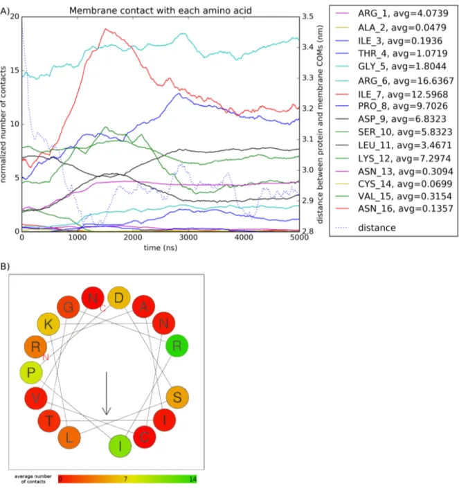
The system is built with the protein floating in water 1 nm away from the membrane. We can see in Figure 1 that it tends to stick to both membranes for the whole simulation. The average minimum distance computed with the GROMACS mindist function over the run is very close in both systems and close to the expected value of twice the Van der Waals radius of 0.235 nm used in the MARTINI force-field [24].
Electostatic forces drive helix-membrane interaction
Using the GROMACS mindist function with a radius of 8 ˚A, we plotted the number of con-tacts between the protein and the membrane, and between each amino acid residue and each lipid type in the membrane. We also computed the distance between Centers of Mass of the protein and membrane z-coordinate using the GROMACS traj function.
We find that some residues in the peptide have more contacts with the membrane than others (Figure 2.A). When applying the average number of contact for each amino acid onto the puta-tive secondary structure of the α-helix [19], we find there is no obvious repartition of contacts on one side of the helix that would be lipophilic (Figure 2.B).
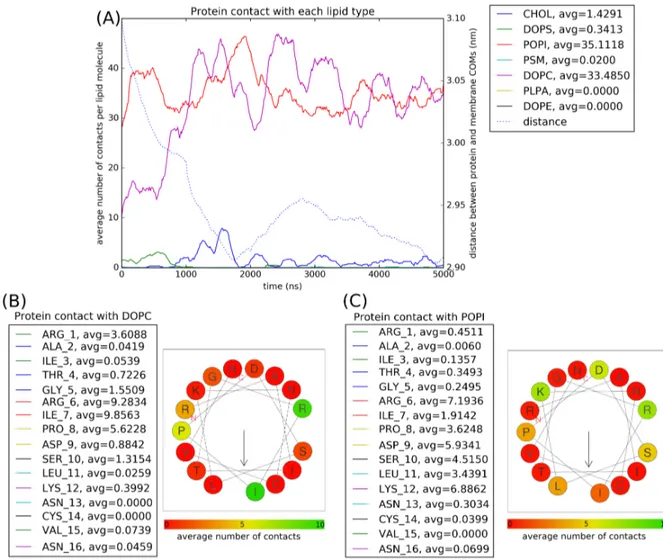
When plotting contacts between the protein and each lipid type, we can see that the protein interacts almost exlusively with POPI and DOPC molecules (Figure 3.A). These interactions can be explained by the fact that POPI has a negatively charged headgroup and DOPC is the most common in the membrane composition. We then plotted contacts between each residue and both lipid types (Figure 3.B & 3.C).
There does not appear to be any favored side of contact with DOPC molecules, probably be-cause of how overwhelming the molecule is in the membrane composition (49%, Table 1). However, the amino acids that interact more with POPI are Arginine and Lysine which are both positively charged at pH = 7.
Finally to inspect which parts of the lipids interact with positive residues, we measured contacts between positive residues and each bead in the head of DOPC and POPI. Positive residues inter-act more with the choline than the phopsphate of DOPC (Figure 4.A & 4.B), probably because cholines are the most protruding groups. However, we observe in Figures 4.C & 4.D that this is no the case for POPI. Positive residues actually have more contacts with the C1 inositol bead although it is not the most protruding one. Its number of contacts with PO4 is also on average equal to C2 & C3 beads, which are the most protruding.
Preferential contacts between negative headgroups and positive residues suggest that the inter-action between the peptide and membranes may be driven by electrostatic forces.
All-Atom secondary structure analysis
Structure prediction tools
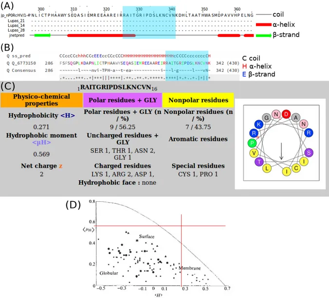
Figures 5.A & 5.B show that both tools predict the 16-residue sequence we are interested in to be mostly random coil. HELIQUEST predicts an average mean hydrophobicity (hHi = 0.271 on a scale from -1.01 to 2.25 [40]) with a low mean helical hydrophobic moment (hµHi = 0.569 on a scale from 0 to 3.26 [41]). These values do not allow us to place the protein onto the graph in Figure 5.D, which could have been another prediction of the peptide’s amphipathicity. These values mean that the 16-residue sequence has both hydrophobic and hydrophilic residues but that the helix has a low amphipathicity because there is no hydrophobic and hydrophilic face. However, the secondary structure prediction tools are designed for and most accurate on solu-ble proteins and they should not be considered very reliasolu-ble on membrane proteins, including peripheral ones.
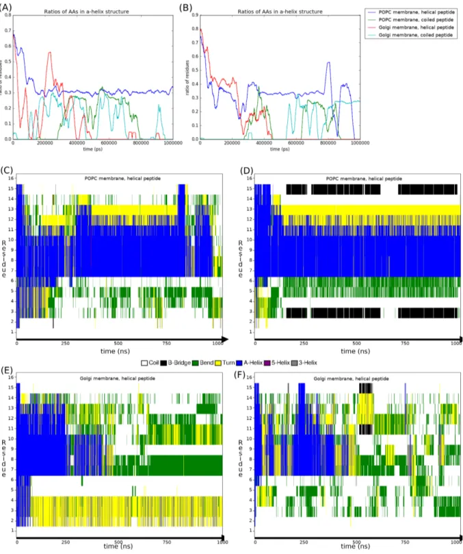
All-atom simulations also exhibit electrostatic attraction
When plotting the Minimum distance between peptides and membranes in both 1 μs All-atom runs, we can see that the peptide tends to spend more time in contact with the Golgi membranes (Figure 6). This could be explained by the fact that the protein has an overall charge of +2. In POPC membranes, all lipid headgroups are zwitterionic phosphatidylcholines while Golgi
membranes are comparatively more negatively charged, and is coherent with Coarse-grain ob-servations.
Secondary structure of the peptide is not stable in water
Considering the uncertainty of secondary structure predictions, we decided to use All-atom simulations since they have already been used to predict secondary structure [42]. For both 1 μs atomistic runs we plotted the DSSP results over time. Figures 7.A & 7.B show the ratio of amino acids from the peptide in α-helix conformation.
Peptides that are initially helical start unfolding during equilibration and tend towards a random coil conformation as the run progresses. There seems to be a recurring value of 0.3, meaning 5 amino acids assigned as α-helix. To know whether this is a full helix turn or disconnected bends, we plotted the secondary structure of each individual amino acid over time for the folded peptide simulations. Figures 7.C to 7.F show that residues from I7 to K12 are the ones which tend to be in the α-helical conformation. These residues are also the ones that had the most contacts with membranes in the CG simulations in Figure 2, meaning they could either be stabilized into helix by membrane contact or their helical conformation allows them to interact the membrane more.
Figure 1: Minimum distance between protein and membranes in both Coarse-Grain sim-ulations.
Figure 2: Contacts between the membrane and protein residues in Coarse-Grain models. (A) Contacts < 8 ˚A between each residue and the membrane. (B) Average number of con-tacts of the putative 16-residue α-helix (adapted from [19]). The arrow indicates the predicted hydrophobic face.
Figure 3:Contacts between the peptide and lipid types in Coarse-Grain models. (A) Con-tacts < 8 ˚A between each residue and the membrane. (B) Average number of conCon-tacts of the putative residue α-helix with DOPC. (C) Average number of contacts of the putative 16-residue α-helix with POPI.
Figure 4: Preferential contacts between positive residues and lipids in Coarse-Grain mod-els. (A) Contacts < 8 ˚A between DOPC headgroup beads and positive residues. (B) MARTINI mapping of DOPC molecules. (C) Contacts < 8 ˚A between POPI headgroup beads and positive residues. (D) MARTINI mapping of POPI molecules. (E) MARTINI bead types (adapted from the MARTINI website).
Figure 5: Algorithmic prediction of the peptide’s properties. (A,B) Secondary Structure prediction plots for CtB1/BARS using Jpred4 or HHPred, resp. The blue window indicates the putative 16-residue helix. Capitalization in (B) is proportional to amino acid volume. (C) HELIQUEST prediction of amphipathicity. (D) Placement of the 16-residue peptide onto an empiric clustering plot of proteins as a function of hHi and hµHi (adapted from [41]).
Figure 6:Minimum distance between peptides and membranes in both 1 μs All-atom runs (left vs. right). (A,B) Pure POPC membranes. (C,D) Golgi-like membranes.
Figure 7: Assignment of secondary structure by the DSSP algorithm over the two 1μs All-Atom runs (left vs. right). (A,B) Ratio of residues in the peptide assigned as α-helix. (C,D) DSSP plot for each residue in the POPC membrane-helical peptide runs. (E,F) DSSP plot for each residue in the Golgi membrane-helical peptide runs.
Discussion
Molecular Dynamics simulations validate the hypothesis that the peptide and the membrane in-teract. The study of contacts between amino acids and lipids suggests that the interaction might be driven by electrostatic forces between negatively charged lipids and basic residues bearing a positive charge at pH = 7. From predictive tools and the study of secondary structure in All-Atom simulations, we cannot confirm that the 16-residue sequence is helical when separated from the rest of the protein.
There are more physical properties of membranes we could study on longer simulations and with increasing peptide concentration : lipid diffusion, area per lipid and membrane thickness can all indicate conducting to the rearrangement of lipids. Compressibility modulus and lateral pressure profiles are also useful for measuring membrane curvature [43], another mandatory step for membrane fission. All of these measurements have been planned and will be carried out in the future, using the present work as a starting point.
Acknowledgments
This work was granted access to the HPC resources of CINES under the allocation DARI n ˚ A0040710138, project imp6353 attributed by GENCI (Grand Equipement National de Cal-cul Intensif).
This report will be shared with Dr. Luini’s team at the Institute of Protein Biochemistry in Naples, Italy.
Thanks
Many thanks to Luca for taking the time and trusting me to work on this project. It has been a really enriching experience and I met a lot of great people at IBCP. I’ll really miss coffee and lunch with the team!
I would also like to thank my teachers Jean-Marc and Sandrine for always being available for us students.
Last but certainely not least, huge props to all the wonderful people I met over the 3 years between spent in Rennes, Chicago and Lyon and who participated in making me un g´enie avant d’ˆetre ing´enieur.
References
[1] D. Corda, C. Hidalgo Carcedo, M. Bonazzi, A. Luini, and S. Span`o, “Molecular aspects of membrane fission in the secretory pathway,” Cellular and molecular life sciences : CMLS, vol. 59, pp. 1819–32, 12 2002.
[2] Y. Kozlovsky and M. M. Kozlov, “Membrane fission: model for intermediate structures,” Biophysical journal, vol. 85, no. 1, pp. 85–96, 2003.
[3] L. V. Chernomordik and M. M. Kozlov, “Protein-lipid interplay in fusion and fission of biological membranes,” Annual review of biochemistry, vol. 72, no. 1, pp. 175–207, 2003. [4] G. Praefcke and H. T. McMahon, “The dynamin superfamily: universal membrane tubu-lation and fission molecules?,” Nature reviews. Molecular cell biology, vol. 5, pp. 133–47, 03 2004.
[5] L. Pelkmans and A. Helenius, “Insider information: what viruses tell us about endocyto-sis,” Current Opinion in Cell Biology, vol. 15, no. 4, pp. 414 – 422, 2003.
[6] E. Harsay and R. Schekman, “A subset of yeast vacuolar protein sorting mutants is blocked in one branch of the exocytic pathway,” The Journal of Cell Biology, vol. 156, no. 2, pp. 271–286, 2002.
[7] S. Span`o, M. G. Silletta, A. Colanzi, S. Alberti, G. Fiucci, C. Valente, A. Fusella, M. Salmona, A. Mironov, A. Luini, et al., “Molecular Cloning and Functional Charac-terization of Brefeldin A-ADP-Ribosylated Substrate,” Journal of Biological Chemistry, vol. 274, no. 25, pp. 17705–17710, 1999.
[8] M. Nardini, S. Span`o, C. Cericola, A. Pesce, A. Massaro, E. Millo, A. Luini, D. Corda, and M. Bolognesi, “CtBP/BARS: a dual-function protein involved in transcription co-repression and golgi membrane fission,” The EMBO Journal, vol. 22, no. 12, pp. 3122– 3130, 2003.
[9] M. Bonazzi, S. Span`o, G. Turacchio, C. Cericola, C. Valente, A. Colanzi, H. S. Kweon, V. W. Hsu, E. V. Polishchuck, R. S. Polishchuck, et al., “CtBP3/BARS drives membrane fission in dynamin-independent transport pathways,” Nature cell biology, vol. 7, no. 6, p. 570, 2005.
[10] C. H. Carcedo, M. Bonazzi, S. Span`o, G. Turacchio, A. Colanzi, A. Luini, and D. Corda, “Mitotic Golgi partitioning is driven by the membrane-fissioning protein CtBP3/BARS,” Science, vol. 305, no. 5680, pp. 93–96, 2004.
[11] R. Weigert, M. G. Silletta, S. Span`o, G. Turacchio, C. Cericola, A. Colanzi, S. Senatore, R. Mancini, E. V. Polishchuk, M. Salmona, et al., “CtBP/BARS induces fission of Golgi membranes by acylating lysophosphatidic acid,” Nature, vol. 402, no. 6760, p. 429, 1999. [12] J. L. Gallop, P. J. G. Butler, and H. T. McMahon, “Endophilin and CtBP/BARS are not acyl transferases in endocytosis or Golgi fission,” Nature, vol. 438, no. 7068, p. 675, 2005. [13] A. Pagliuso, C. Valente, L. L. Giordano, A. Filograna, G. Li, D. Circolo, G. Turacchio, V. M. Marzullo, L. Mandrich, M. A. Zhukovsky, et al., “Golgi membrane fission requires the CtBP1-S/BARS-induced activation of lysophosphatidic acid acyltransferase δ,” Nature communications, vol. 7, p. 12148, 2016.
[14] J.-S. Yang, H. Gad, S. Y. Lee, A. Mironov, L. Zhang, G. V. Beznoussenko, C. Valente, G. Turacchio, A. N. Bonsra, G. Du, et al., “A role for phosphatidic acid in COPI vesi-cle fission yields insights into Golgi maintenance,” Nature cell biology, vol. 10, no. 10, p. 1146, 2008.
[15] A. G. Bellesis, A. M. Jecrois, J. A. Hayes, C. A. Schiffer, and W. E. Royer, “Assembly of human C-terminal binding protein (CtBP) into tetramers,” Journal of Biological Chem-istry, vol. 293, no. 23, pp. 9101–9112, 2018.
[16] E. Boucrot, A. Pick, G. Camdere, N. Liska, E. Evergren, H. T. McMahon, and M. M. Ko-zlov, “Membrane fission is promoted by insertion of amphipathic helices and is restricted by crescent BAR domains,” Cell, vol. 149, no. 1, pp. 124–136, 2012.
[17] A. Drozdetskiy, C. Cole, J. Procter, and G. J. Barton, “JPred4: a protein secondary struc-ture prediction server,” Nucleic Acids Research, vol. 43, pp. W389–W394, 04 2015. [18] L. Zimmermann, A. Stephens, S.-Z. Nam, D. Rau, J. K¨ubler, M. Lozajic, F. Gabler,
J. S¨oding, A. N. Lupas, and V. Alva, “A completely reimplemented MPI bioinformat-ics toolkit with a new HHpred server at its core,” Journal of molecular biology, vol. 430, no. 15, pp. 2237–2243, 2018.
[19] R. Gautier, D. Douguet, B. Antonny, and G. Drin, “HELIQUEST: a web server to screen sequences with specific α-helical properties,” Bioinformatics, vol. 24, no. 18, pp. 2101– 2102, 2008.
[20] S. Jo, T. Kim, V. G. Iyer, and W. Im, “CHARMM-GUI: A web-based graphical user interface for CHARMM,” Journal of Computational Chemistry, vol. 29, no. 11, pp. 1859– 1865, 2008.
[21] G. van Meer, “Lipids of the Golgi membrane,” Trends in cell biology, vol. 8, no. 1, pp. 29– 33, 1998.
[22] “The PyMOL Molecular Graphics System, Version 1.2r3pre, Schr¨odinger, LLC..”
[23] T. A. Wassenaar, H. I. Ing´olfsson, R. A. B¨ockmann, D. P. Tieleman, and S. J. Marrink, “Computational lipidomics with insane: A versatile tool for generating custom mem-branes for molecular simulations,” Journal of Chemical Theory and Computation, vol. 11, no. 5, pp. 2144–2155, 2015. PMID: 26574417.
[24] D. H. de Jong, G. Singh, W. F. D. Bennett, C. Arnarez, T. A. Wassenaar, L. V. Sch¨afer, X. Periole, D. P. Tieleman, and S. J. Marrink, “Improved parameters for the Martini Coarse-Grained protein force field,” Journal of Chemical Theory and Computation, vol. 9, no. 1, pp. 687–697, 2013. PMID: 26589065.
[25] J. B. Klauda, R. M. Venable, J. A. Freites, J. W. O’Connor, D. J. Tobias, C. Mondragon-Ramirez, I. Vorobyov, A. D. MacKerell, and R. W. Pastor, “Update of the CHARMM All-atom additive force field for lipids: Validation on six lipid types,” The Journal of Physical Chemistry B, vol. 114, no. 23, pp. 7830–7843, 2010. PMID: 20496934.
[26] R. B. Best, X. Zhu, J. Shim, P. E. M. Lopes, J. Mittal, M. Feig, and A. D. MacKerell, “Optimization of the Additive CHARMM All-atom protein force field targeting improved sampling of the backbone φ, ψ and side-chain χ1 and χ2 dihedral angles,” Journal of Chemical Theory and Computation, vol. 8, no. 9, pp. 3257–3273, 2012. PMID: 23341755.
[27] W. Jorgensen, J. Chandrasekhar, J. Madura, R. Impey, and M. Klein, “Comparison of sim-ple potential functions for simulating liquid water.,” Journal of Chemical Physics, pp. 926– 935, 1983.
[28] B. Hess, H. Bekker, H. J. Berendsen, and J. G. Fraaije, “LINCS: a linear constraint solver for molecular simulations,” Journal of computational chemistry, vol. 18, no. 12, pp. 1463– 1472, 1997.
[29] T. Darden, D. York, and L. Pedersen, “Particle mesh Ewald: An Nlog(N) method for Ewald sums in large systems,” The Journal of chemical physics, vol. 98, no. 12, pp. 10089– 10092, 1993.
[30] S. P´all and B. Hess, “A flexible algorithm for calculating pair interactions on SIMD archi-tectures,” Computer Physics Communications, vol. 184, no. 12, pp. 2641–2650, 2013. [31] G. Bussi, D. Donadio, and M. Parrinello, “Canonical sampling through velocity rescaling,”
The Journal of chemical physics, vol. 126, no. 1, p. 014101, 2007.
[32] M. Parrinello and A. Rahman, “Polymorphic transitions in single crystals: A new molec-ular dynamics method,” Journal of Applied physics, vol. 52, no. 12, pp. 7182–7190, 1981. [33] M. Abraham, D. van der Spoel, E. Lindahl, B. Hess, and the GROMACS development
team, GROMACS User Manual version 2016.4.
[34] W. G. Touw, C. Baakman, J. Black, T. A. te Beek, E. Krieger, R. P. Joosten, and G. Vriend, “A series of PDB-related databanks for everyday needs,” Nucleic acids research, vol. 43, no. D1, pp. D364–D368, 2014.
[35] W. Kabsch and C. Sander, “Dictionary of protein secondary structure: pattern recognition of hydrogen-bonded and geometrical features,” Biopolymers, vol. 22, no. 12, pp. 2577– 2637, 1983.
[36] J. D. Hunter, “Matplotlib: A 2D graphics environment,” Computing in Science & Engi-neering, vol. 9, no. 3, pp. 90–95, 2007.
[37] L. Monticelli, S. K. Kandasamy, X. Periole, R. G. Larson, D. P. Tieleman, and S.-J. Mar-rink, “The MARTINI Coarse-Grained Force Field: Extension to Proteins,” Journal of Chemical Theory and Computation, vol. 4, no. 5, pp. 819–834, 2008. PMID: 26621095. [38] I. G. Tironi, R. Sperb, P. E. Smith, and W. F. van Gunsteren, “A generalized reaction field
method for molecular dynamics simulations,” The Journal of chemical physics, vol. 102, no. 13, pp. 5451–5459, 1995.
[39] D. H. De Jong, S. Baoukina, H. I. Ing´olfsson, and S. J. Marrink, “Martini straight: Boost-ing performance usBoost-ing a shorter cutoff and GPUs,” Computer Physics Communications, vol. 199, pp. 1–7, 2016.
[40] J.-L. Fauchere and V. Pliska, “Hydrophobic parameters π of amino-acid side chains from the partitioning of N-acetyl-amino-acid amides,” Eur. J. Med. Chem, vol. 18, no. 3, pp. 369–375, 1983.
[41] D. Eisenberg, R. M. Weiss, and T. C. Terwilliger, “The helical hydrophobic moment: a measure of the amphiphilicity of a helix,” Nature, vol. 299, no. 5881, p. 371, 1982.
[42] L. Monticelli, D. P. Tieleman, and G. Colombo, “Mechanism of Helix Nucleation and Propagation : Microscopic View from Microsecond Time Scale MD Simulations,” The Journal of Physical Chemistry B, vol. 109, no. 43, pp. 20064–20067, 2005. PMID: 16853593.
[43] F. Campelo, C. Arnarez, S. J. Marrink, and M. M. Kozlov, “Helfrich model of membrane bending: from Gibbs theory of liquid interfaces to membranes as thick anisotropic elastic layers,” Advances in colloid and interface science, vol. 208, pp. 25–33, 2014.
Diplôme : Master 2 / Ingénieur Agronome Spécialité : Cursus Ingenieur Agronome
Spécialisation / option : Master BMC – Biologie Moleculaire et Cellulaire Enseignant référent : Dr. Jean-Marc FRASLIN
Auteur(s) : Thomas LE MÉTAYER
Date de naissance* : 17/02/1997
Organisme d'accueil :
Equipe Modeling Biological Macromolecules MMSB, UMR 5086 CNRS & U. Lyon
Adresse : 7 Passage du Vercors, 69637 Lyon, FRANCE
Maître de stage : Dr. Luca MONTICELLI
Nb pages : 23 Annexe(s) :
-Année de soutenance : 2018-2019
Titre français : Simulations Moléculaires des fissions membranaires mediées par la protéine BARS Titre anglais : Molecular Simulations of membrane fission mediated by BARS protein
Résumé : Il a été montré que la protéine CtBP1/BARS cause la fission de membranes golgiennes et de liposomes sans consommation d’ATP. Dans ce mémoire, nous analysons comment un fragment de 16 résidus supposément α-hélicoïdal issu du domaine C-terminal de BARS interagit avec les membranes en utilisant la simulation moléculaire.
Pour observer ces interactions, nous avons utilisé des simulations de Dynamique Moléculaire à échelle atomistique et gros-grain. Plusieurs modèles membranaires variant de simples bicouches lipidiques homogènes à des modèles simulant la composition des membranes golgiennes ont été considérés et confrontés à des modèles du fragment de BARS dans adoptant différentes structures secondaires.
Nos résultats montrent que la structure du fragment protéique varie fréquemment. Ce fragment interagit avec les modèles membranaires en se plaquant à leur surface via des interactions électrostatiques entre les résidus positifs et les lipides membranaires négatifs. Il n’est pas possible de confirmer que sa structure ou ses propriétés lorsqu’il est séparé du reste de la protéine sont celles observées in vivo et in vitro.
Produire plus de simulations avec de plus grands domaines de la protéine ou avec des concentrations croissantes du peptide pourrait permettre d’observer plus en détail comment les propriétés physiques de la membrane sont influencées par la protéine et aider à comprendre le mécanisme de fission engendré par BARS.
Abstract : The CtBP1/BARS protein has been shown to cause ATP-hydrolysis-independent membrane fission both in Golgi membranes and liposomes. This memoir analyzes how one putative amphipathic 16-residue α-helical fragment from the C-teminal domain of BARS interacts with membranes using in silico Molecular Simulations.
To observe these interactions, we used Molecular Dynamics simulations at atomistic and coarse-grained scales. We used several membrane models ranging from very simple homogeneous lipid bilayers to ones mimicking the composition of the Golgi membranes. These membranes were confronted to several models of the protein fragment varying in secondary structure.
Our results show that the protein fragment undergoes a lot of structural change. It interacts with model membranes by sticking to them, mostly via electrostatic interactions between positive residues and negative lipids. We cannot confirm that its structure or properties when detached from the rest of the protein are the same as observed in vivo or in vitro.
Running more simulations with bigger domaines of the protein or with an increasing concentration of the peptide in the future could provide more insight on how physical properties of membranes are influenced by the protein and help unveil the mechanism behind membrane fission.
Mots-clés : Molecular Dynamics, fission membranaire, interactions proteines-membranes Key Words: Molecular Dynamics, membrane fission, protein-membrane interactions