Title: Febrile seizures and increased stress sensitivity in children: how it relates to seizure characteristics
Authors: Fanny Thébault-Dagher a,b,c, Marc-Philippe Lafontaine a,b,c, Inga Sophia Knoth c, Florence Deguire a,b,c, Emilie Sheppard a,b,c, Ramona Cook c, Maryse Lagacé c, Jocelyn Gravel c, Sonia Lupien d,e, Sarah Lippé a,b,c
Affiliations:
a Department of Psychology, Université de Montréal, Marie Victorin Building, 90 Vincent-D’Indy Avenue, Montreal, Quebec, Canada
b Centre de recherche en neuropsychologie et cognition, Université de Montréal, Marie Victorin Building, 90 Vincent-D’Indy Avenue, Montreal, Quebec, Canada
c CHU Sainte-Justine Research Center, Université de Montréal, 3175 Chemin de la Côte-Sainte-Catherine, Montreal, Quebec, Canada
d Psychiatry Department, Université de Montréal, Roger-Gaudry Building, 2900 Edouard-Montpetit Boulevard, Montreal, Quebec, Canada
e Center for Studies on Human Stress, Institut universitaire en santé mentale de Montréal, 7331 Hochelaga Street, Montreal, Quebec, Canada
Authors e-mail adresses: fanny.thebault-dagher@umontreal.ca, marc.philippe.lafontaine@umontreal.ca, ingasophia.knoth@gmail.com,
florence.deguire@umontreal.ca, emilie.sheppard@umontreal.ca, ramona.cook@recherche-ste-justine.qc.ca, maryse.lagace@recherche-ste-ramona.cook@recherche-ste-justine.qc.ca, graveljocelyn@hotmail.com, sonia.lupien@umontreal.ca, sarah.lippe@umontreal.ca.
Contact information for the corresponding author: Fanny Thébault-Dagher, Université de Montréal, Marie-Victorin building, Office F-457, 90 Vincent-D’Indy Avenue, Montreal (Quebec), Canada 514-343-6111, ext 26905, fanny.thebault-dagher@umontreal.ca
ABSTRACT
Background: Studies suggest the relationship between seizures and stress starts early in life. However, evidence of long-term altered stress reactivity following early life seizures is lacking. Our objectives were to assess alterations in stress hormone reactivity in children with past febrile seizures (FS) and investigate how these alterations relate to clinical characteristics.
Method: This case-control study compared a convenience sample of children with simple FS (n=24), complex FS (n=18), and matched healthy controls (n = 42). Stress was induced by electrode placement for an electroencephalography exam. Salivary cortisol to stress, using three samples collected before and after the stressor, was compared between groups and sex. The relationship between stress reactivity and clinical characteristics (i.e. FS duration, age at first FS, time since the last FS) was investigated.
Results: Cortisol reactivity to stress was significantly different depending on study groups, F(1, 78) = 6.415, p = 0.003, η2
p = 0.141, but not sex nor was there a significant interaction between
group and sex (p ≥ 0.581). Simple FS participants showed higher cortisol reactivity to stress (M = 14.936, SD = 26.852) compared to complex FS (M = -4.663, SD = 18.649, p = 0.015) and controls (M = -3.817, SD = 18.907, p = 0.003). There was no significant difference between participants with complex FS and controls (p > 0.999). Stress reactivity was not linked to clinical characteristics.
Conclusions: Children with past simple FS showed greater changes in salivary cortisol following stress, suggesting enhanced stress sensitivity. As similar results were not found in a population with complex FS, our study shows stress alterations are not caused by seizure severity. Future
studies are needed to investigate whether stress sensitivity may be premorbid to simple FS and may contribute to simple FS incidence.
ARTICLE 1. Introduction1
Febrile Seizures (FS) are associated with fever episodes in young children without a history of spontaneous seizures or neurological insults [1]. They are the most common pediatric seizures, affecting 2 to 5% of children before age 5 [2, 3]. Although FS are common
neuropediatric emergencies, they are seen as benign, and seizure frequency is low, with only 30% of patients showing overall seizure recurrence [2]. Nevertheless, FS with complex features (i.e., >1 seizure during the fever episode, and / or focal, and / or ≥ 10 minutes seizure duration) have been associated with poorer outcome, including lasting increased anxiety and depressive symptoms [4-6]. Anxiety and depressive symptoms are amongst the most frequent seizure precipitants reported by people with epilepsy [7, 8], consequently a relationship between anxiety or depressive symptoms and seizures, which would be mediated through physiological stress, has received some support [9]. Incidentally, common underlying mechanisms between seizures and stress have been suggested through familial clustering research [10]. In that context, changes in basal stress hormone and stress reactivity axis found in people with epilepsy [11, 12] may be related to higher prevalence of psychiatric symptoms in this population [11, 13].
Clear evidences from animal studies suggest the seizure-stress relationship begins during early infancy, hence this relationship in early childhood has been suggested in humans as well [14]. More precisely, early life stress in animals and humans induces alterations in stress hormones secretion, regulation of the stress reactivity axis, and neuroanatomical changes predisposing the brain to seizures [15-17]. Complementarily, animal models show early life seizures are associated with changes in stress hormones levels [17-20]. Despite strong evidence
1Abbreviations: Analysis of variance (ANOVA), Area under the curve with respect to increase (AUCi), Electroencephalography (EEG), Febrile Seizures (FS), Socio-economical status (SES)
in late childhood and adult human studies of a seizure-stress relationship [21, 22], we do not know if early life seizures are associated with altered physiological reactivity to stress in human infants and toddlers. Moreover, it is unknown whether this association is present following seizures of low severity and frequency such as FS.
We aimed to investigate stress reactivity, as measured through salivary cortisol to
experimental stress, in children with past FS. Additionally, we assessed how FS characteristics relate to stress reactivity. We hypothesized that stress reactivity differs between children with past simple FS, complex FS, and healthy controls, and may vary depending on clinical characteristics (i.e. FS duration, age at first FS, time since the last FS). Alterations in
physiological stress following early-life benign seizures would increase our understanding of the seizure-stress relationship, providing insights into the mechanisms underlying FS prognosis.
2. Material and methods 2.1 Participants
This case-control study compared children with past simple FS, complex FS, and healthy controls. Cases were otherwise healthy children who were admitted to the emergency department of a tertiary care, university affiliated pediatric Hospital (CHU Sainte-Justine), between 10 to 24 months of age, following a simple or complex FS episode meeting the American Academy of Pediatrics criteria [1] from August 2015 to May 2018. A number of control equivalent to the total number of children with FS evaluated was recruited through social media. Children with simple FS, complex FS, and controls had similar age, sex, and socio-economical status (SES) distribution when tested. Children born before 34 weeks of gestational age, with a history of health problems that could affect neurological development or suspicions of developmental delay were excluded.
The study was approved by the CHU Sainte-Justine Research Ethics Board. Legal
guardians gave verbal consent when contacted and gave written informed consent when arriving at the laboratory. They were free to withdraw at any time in the study. Families were given a 30$ compensation for their participation.
2.2 Descriptive measures
Sociodemographic characteristics and personal history were assessed using an in-house questionnaire. Cognitive skills were evaluated using the Bayley Scales of Infant and Toddler Development - Third Edition [23], and adaptive behaviors were assessed using the Adaptive Behavior Assessment System – Second Edition [24] completed by the parent. Finally, as FS may lead to parental stress, parents of the FS groups were asked to complete the 4-item short form of the Perceived Stress Scale [25], measuring the degree to which they appraise their current situation as stressful. Developmental assessment and questionnaires were scored by a Doctoral student in Neuropsychology.
FS characteristics were extracted from medical records by a Doctoral student in Neuropsychology specialized in seizure disorders using a standardized report form. Dates, duration and type of each FS episode were noted. Results of neurological exams, if applicable, were also obtained. According to FS characteristics, children with FS were divided in two study groups: simple (isolated, <10 minutes, and generalized) and complex FS. For descriptive
purposes, complex FS were further classified as multiple (>1 during the fever episode), focal, prolonged (FS duration ≥ 10 minutes), and/or Febrile Status Epilepticus (FS duration ≥ 30 minutes, or repeated FS ≥ 30 minutes without regaining consciousness) [5].
Acute stress was induced using reduced parental support and mild arm restraint during an electroencephalography (EEG) net placement, as this evaluation was required to meet other objectives of the study. Studies in school-aged children show exposure to novel medical procedures leads to increased stress reactivity, as assessed by significant increase in cortisol levels [26]. However, stressor paradigms in children aged 10-24 months, including EEG net placement and evaluation, have a low success rate [27]. Reviews suggest supportiveness of adults could be buffering increased cortisol to stress [27]. In that context, threats to parental support may help induce cortisol reactivity to stress in infants and toddlers. Thus, during installation, participants were seated on their parent’s lap and, as children tried to pull on the EEG net, parents were asked to immobilize their children’s arms and maintain restraint despite fussiness and distress. Saliva sampling for assessment of cortisol levels were taken to measure stress reactivity to the EEG procedure. The first saliva sample was taken before entering the exam room. The second and third saliva samples were collected approximately 20 and 45 minutes following the stressor, providing cortisol measures during acute stress reactivity [28].
Salivary samples were obtained using sterile synthetic swabs (SalivaBio Children’s Swab; Salimetrics LLC, Carlsbad, CA). Cortisol concentration was determined with a high sensitivity enzyme immune assay kit (Salimetrics State College, PA, catalog No. 1-3102). Samples with a reading exceeding the upper limit for detection or significant intra-assay coefficient of variation were rerun at a dilution. All samples for a participant were analysed in the same batch, and each batch consisted of males and females of all study groups. Comprehensive information regarding cortisol assessment is shown in Supplementary material 1.
Experimental stress was induced during a morning or early-afternoon appointment at CHU Sainte-Justine. Morning and afternoon evaluations were evenly balanced across groups to control
for impact of the circadian rhythm on cortisol. Parents were instructed to postpone the evaluation if participants were sick, teething, or had an unusual bad night of sleep. No solid food was given during the testing and the mouth was thoroughly rinsed prior to saliva samples if participants had liquid food or put an object (e.g. pacifier) in their mouth to avoid contamination.
2.4 Statistical analyses
Children with cortisol readings exceeding the upper limit after dilution, significant outlying (± 3 Z score) timing characteristics (i.e. time at first sample, total evaluation duration) or missing samples were excluded. Outlying cortisol values were replaced by plausible values (Z score = 3) [29] to reduce possible bias. To obtain a single value representing stress reactivity, an area under the curve index with respect to increase (AUCi) [30] was calculated using cortisol levels for each sample and time between samples. The AUCi is commonly used to study stress reactivity
through changes in cortisol levels. Positive AUCi suggests increased cortisol and higher absolute values suggest larger changes. Supplemental information on cortisol analysis is shown in
Supplementary material 1.
To test our hypothesis, a two-way ANOVA with Bonferroni post-hoc tests was conducted to compare study groups (i.e. simple FS VS complex FS VS controls), sex, and the sex-group interaction on the AUCi. Sex was added to the analysis as animal testing on rodents show the seizure-stress relationship may depend on sex hormones [19]. To explore how clinical
characteristics relate to stress reactivity, the relationships between AUCi and duration of the longest FS, age at first FS and the time since the last FS of all FS participants were examined using Pearson’s bivariate correlations.
Analyses were carried out using IBM SPSS 25.0 (IBM, Armonk, NY). Normality was examined through visual inspection of histograms and quantile-quantile plots; logarithmic transformations were applied to non-normally distributed variables. Homogeneity of variance was assessed using Levene’s Test. Analyses were performed on complete cases, statistical significance was defined as a two-sided alpha level ≤ 0.05. One-way analyses of variance (ANOVA) and Chi-Square tests of independence were computed to examine differences in participants’ characteristics and study protocol to ensure group comparability. Pearson’s bivariate correlations were conducted between AUCi and participants’ characteristics to verify these variables would not confound our results. Assumptions necessary to conduct all analyses were verified and satisfied.
3. Results
3.1 Descriptive and control results
We identified 108 children with past FS, 54 families declined to participate, and seven children did not meet inclusion criteria, thus 47 children with FS were recruited and evaluated. The most common reason for refusal was lack of interest. Following exclusion (n = 5; 1 out-of-curve reading, 1 missing samples, 3 outlying timing), 42 children with FS were included in our analyses. Prior to analyses, they were classified into two study groups: those with past simple (n = 24; Median age = 19.07 months, IQR = 15.76 – 21.99) and complex FS (n = 18; Median age = 16.52 months, IQR = 13.43 – 20.75). They were compared to 42 healthy controls (Median age = 15.03 months, IQR = 10.93 – 25.80), leading to a total sample size of 84 children (Median age = 16.65 months, IQR = 12.21 – 21.26).
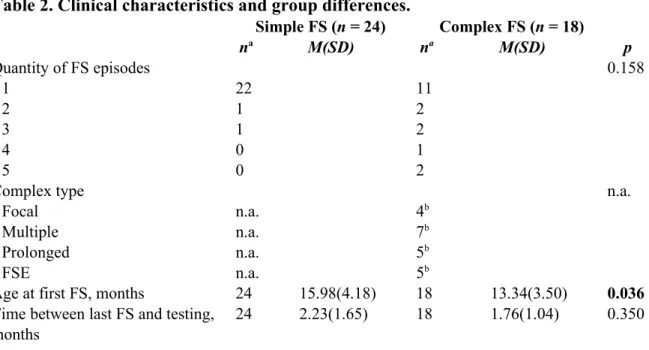
FS types and quantity are shown in Table 2. Children in the complex FS group had their first FS on average 2 months younger than the simple FS group. Overall median seizure duration
was 2 minutes (IQR = 1 - 5), with a bias for shorter duration. Finally, time since the last FS episode and perceived parental stress was similar between FS groups (p > 0.350). Parental stress was low, indicating parents “almost-never” experienced feelings of distress in the last month.
Descriptive statistics and comparisons are shown in Table 1. No significant group differences were found regarding descriptive variables, suggesting group comparability. Furthermore, there were no significant differences in study protocol, indicating data collection was comparable across study groups. Cortisol levels varied from 0.0135 to 1.1731 μg/dl. Comprehensive cortisol and evaluation results are shown in Supplementary material 1. Visual inspection of variability in cortisol trajectories (Figure 1a) shows stress was not successfully induced in all study participants, despite reduced parental supportiveness. More precisely, increased cortisol reactivity to stress, as defined by an untransformed AUCi > 0 was successfully achieved in 49% of study participants. AUCi was not associated with participants’ characteristics (p ≥ 0.080), thus these characteristics should not confound our main analysis.
3.2 Stress reactivity and febrile seizures type and clinical characteristics
A two-way ANOVA revealed cortisol reactivity to stress was significantly different depending on study group, F(1, 78) = 6.415, p = 0.003 (Figure 2). The effect size for group differences was large (η2
p = 0.141) [31]. Bonferroni post-hoc analyses revealed the simple FS
group showed higher cortisol reactivity to stress (M = 14.936, SD = 26.852) compared to complex FS (M = -4.663, SD = 18.649, p = 0.015, 95% CI = 3.06 to 36.49) and controls (M = -3.948, SD = 18.907, p = 0.003, 95% CI = 4.87 to 32.35). There was no significant difference between participants with complex FS and controls (p > 0.999). Visual inspection of
participants’ distribution (Figure 2) and variability in cortisol trajectory (Figure 1b, c, d) suggests children with past simple FS showed increased cortisol to stress more consistently than complex
FS and controls, although the study design was comparable across study groups. Coherently, untransformed AUCi data shows increased cortisol reactivity to stress was achieved in 71% of the children with simple FS group, as opposed to 33% and 43% of the complex FS and healthy controls groups, respectively. Significant differences were not found for sex (p = 0.581) or the sex-group interaction (p = 0.661).
Finally, Pearson’s bivariate correlations conducted on participants with past FS only (n = 42) suggest neither duration of the longest FS episode, age at first FS, nor time since the last FS were significantly associated with AUCi (p ≥ 0.150).
4. Discussion
We found that children with a history of simple FS show changes in salivary cortisol to experimental stress that are significantly different from children with complex FS and controls. Meanwhile, no significant difference was found between children with complex FS and controls. While average AUCi was positive for the simple FS group, it was negative for those with complex FS and controls. Furthermore, variability in cortisol trajectories shows participants in the simple FS group display increased cortisol to stress more consistently, while complex FS and control participants exhibited more diverse patterns of response in cortisol reactivity to
experimental stress. Thus, our results indicate that only our simple FS group was, on average, stressed during our experiment, suggesting children with simple FS may have a lower threshold for stress reactivity than their peers. Importantly, this difference cannot be explained by group differences in developmental characteristics, parental stress or study protocol. Our results did not support sex as a contributive factor to the FS-stress model and did not show stress reactivity to be associated with parental stress, duration of the longest FS, age at first FS or time since the incident.
While previous studies have shown increased cortisol immediately following FS in young children [32, 33], which may reflect an acute post-ictal stress response, this is the first study supporting long-term alterations in physiological stress sensitivity in young children with common and harmless seizures. These results may help to improve our understanding of the relationship between early-life seizures and stress in humans. Specifically, animal studies show that seizures may lower the stress threshold and increase stress reactivity [11, 12]. Our study cannot fully support this hypothesis in FS populations, as the threshold for stress reactivity in children with complex FS did not significantly differ from healthy controls’. However, it has been widely suggested through animal studies that early life stress can lead to reduced seizure threshold, increased seizure severity, and reduced therapeutic response to antiepileptic drugs [16-18]. Furthermore, while human studies of physiological stress in relation to seizures during early childhood are lacking, longitudinal human studies show perinatal maternal self-reported stress is linked to early life seizure severity and younger age at first simple FS [34, 35]. Thus, although our data does not support changes in stress sensitivity in all children affected by early life seizures, they do suggest that enhanced stress sensitivity can be associated with seizure vulnerability in some children, notably those with simple FS.
Although various animal studies show stress may be associated with increased seizure severity [36-41], children in our study with arguably more severe seizures, as a group, did not show altered stress reactivity. These results are coherent with multiple human studies suggesting stress may impact seizure genesis [35, 42-44] and increase seizure occurrence [7, 8], but not necessarily seizure severity. Notably, one study showed children with a first seizure provoked by stressful events tended to have more controlled epilepsies than those with a first seizure
than prognosis [42]. As children with complex FS in our study did not significantly differ from healthy controls, our study may further support altered early-life stress reactivity as being mostly associated with a low seizure threshold, and not enhanced seizure severity. Thus, seizure
occurrence in children with complex FS as a group may signal abnormal network connectivity in the developing brain or premorbid brain insults [10, 45], rather than reduced seizure threshold that induced by stress.
As a result, an important, novel aspect of our study is the disparity in stress sensitivity according to seizure type, suggesting that differences between simple and complex FS may go beyond clinical features. More precisely, we argue that the distinction between children with past simple and complex FS may include differences in environmental exposure, notably to stress, and genetic predisposition to stress sensitivity. Divergences in environmental and genetic origins between FS types have already received some support [46]. Additionally, familial clustering of epilepsy and behavioral disorders in humans suggest a greater role of genetics and environmental influences in uncomplicated epilepsies, whereas more severe seizures would be influenced by pre-existing neurological insults [10]. With regards to environmental factors, acute exposure to psychological stress in humans leads to changes in gene expression of polymorphisms [47] associated with increased incidence of simple but not complex FS in humans [48], and lower FS resistancy in rodents [49]. Thus, we suggest that in the context of simple FS fever could be enhancing neuronal excitability in already hyperexcitable children. Whether hyperexcitability was present in these children, and was due to premorbid stress, genetic predisposition, or a combination of both is beyond the scope of this study and should be the subject of future research. Studies of children and teenagers with epilepsy have shown altered stress reactivity
only in patients with stress sensitive epilepsy [22], further supporting the seizure-stress relationship may depend on patients’ stress sensitivity.
Descriptive analyses did not show group differences regarding cognition, nor could they support a relationship between cognition and stress reactivity. These results might be explained by the unspecific nature of instruments available to evaluate cognition during early childhood. Still, as stress may exacerbate seizures’ consequences on cognition [50], future longitudinal studies are needed to show how enhanced stress reactivity may relate to prognosis following simple FS.
4.1 Study design and limitations
Our study was conducted on a total sample size large enough to find a large effect size given our main hypothesis. Still, sample size for the complex FS group was small, and unequal sample size could lead to loss of statistical power. Furthermore, small sample size and large variance in cortisol data led to large confidence interval. Nevertheless, assumptions necessary to conduct our analyses were satisfied and our study design led to pertinent, statistically significant findings. Comparisons to healthy controls only could have biased interpretation of the results, as it is possible that increased stress sensitivity had been induced by minor illnesses or hospital visits. However, this seems unlikely as children were seen on average months following FS, and neither minor illnesses, nor repeated hospital visits for mild childhood illnesses are associated with long-term enhanced stress in children [51, 52]. Furthermore, significant differences between simple and complex FS participants provided interesting results on stress sensitivity in children with simple FS, while controlling for these variables.
While FS may be associated with neurodevelopmental disorders such as autism, our exclusion criteria may limit the generalisability of our results to neurotypical children. High SES in our studied families and recruitment in a large metropolitan area may limit generalisability as well. Personality and environmental factors may have led some families to decline to participate or fail to complete the questionnaires. These factors cannot be assessed, may have an impact on data interpretation and led to missing data in descriptive statistics, but not studied variables.
The use of salivary cortisol as our stress measure provides multiple advantages in early childhood studies as a non-invasive, easy to use method for assessing cortisol levels [28]. The efficiency of stressor paradigms in early childhood has been a concern of developmental researchers [27, 53], yielding only a 20% success rate in children aged 10-24 months [27]. As school-aged children show increased cortisol reactivity to medical procedures and novelty [26], it has been suggested that adult supportiveness may act as a buffer to increased cortisol to experimental stress in infants and toddlers. Thus, precautions were systematically taken while evaluating all participants in order to increase chances of inducing a physiological stress reaction (i.e. arm restraint, reduced parental support). Still, increased cortisol reactivity to stress was not successfully induced in all participants of the current study. Hence, mild arm restraint and parental unresponsiveness might be insufficient to induce cortisol reactivity to experimental stress in developmental research. In addition, studies have shown that parents preventing their children to interact with novel stimulus leads to enhanced cortisol reactivity to novelty [54]. In that context, children of parents who agreed to participate in a research-EEG, which is a novel situation, might show greater stress regulation to novelty. Nevertheless, our study design led to significant results regarding stress in children with FS. More precisely, as controls and complex FS did not show a significant increase in mean cortisol levels following the stressor, increased
cortisol reactivity to stress in children with past simple FS revealed increased sensitivity to experimental stress in this population. Moreover, developmental studies have previously suggested that increased cortisol reactivity to stress in single groups or individuals, while the overall study sample does not show such responses, may reveal individual characteristics, such as unsecure attachment or anxious predisposition, that could affect development [55, 56]. Still, future studies aiming to investigate stress reactivity in children with FS should incorporate multiple stressor paradigms that may represent a threat to parental supportiveness, as well as multiple measures of stress reactivity and stress regulation. For instance, autonomic nervous system responses may be less likely to be buffered by parental support behavior [53].
5. Conclusion
Stress is the most frequent seizure precipitant for people with epilepsy and is considered a burden for patients and their caretakers [7]. A growing body of literature suggests that the seizure-stress relationship starts in early infancy [14]. Our study supports the existence of
neuroendocrine alterations in children with common, benign seizures, but shows the relationship between early-life seizures and stress may depend on seizure type. These results add to previous studies suggesting that the relationship between stress and seizures, in humans, acts mainly on seizure genesis not severity, and may depend on patients’ stress sensitivity. While results from our study clarify this relationship, they raise questions about the link between stress and seizure severity in early childhood. Furthermore, although simple FS are considered benign, our study supports a reduced stress sensitivity threshold in this population. As animal studies suggest stress in the context of seizure disorders may have an impact on neurological and behavioral outcome [50, 57, 58], the relationship between stress and development in children with simple FS should be the subject of future studies. Thus, the next steps would involve studying early-life stress,
seizures, and cognitive or neurological outcome longitudinally, preferably combining multiple measures of stress reactivity and regulation. Findings of such studies would result in further clarifying the relationship between early-life stress and seizure, potentially leading to non-invasive interventions targeting stress, which are still rare in seizure disorders.
Declaration of interests: None. Funding sources
This work was supported by grants from the Fonds de Recherche du Québec Santé (FRQ-S) [22296] and a donation from the Jean-Pierre Hogue Legacy Foundation to Lippé. Thébault-Dagher is supported by scholarships from the Canadian Institutes of Health Research and the FRQ-S. Funding sources were not involved in study design, collection, analysis and
interpretation of data, writing and the decision to submit the article for publication.
Acknowledgements
The authors would like to thank the funding sources and the participating families. They would also like to gratefully acknowledge the whole team working at the Neuroscience of Early Development Laboratory for their contribution to data collection and processing, and the team at the Center for Studies on Human Stress for their help with the salivary cortisol analyses. Finally, they would like to acknowledge the neurological department at CHU Sainte-Justine, notably Doctor Lionel Carmant, for their support regarding clinical neurology.
References
[1] American Academy of Pediatrics. Neurodiagnostic evaluation of the child with a simple febrile seizure. Pediatrics 2011;127: 389.
[2] Offringa M, Bossuyt PM, Lubsen J, Ellenberg JH, Nelson KB, Knudsen FU, Annegers JF, El-Radhi ASM, Habbema JDF, Derksen-Lubsen G. Risk factors for seizure recurrence in children with febrile seizures: a pooled analysis of individual patient data from five studies. Journal of Pediatrics 1994;124: 574-584.
[3] International League Against Epilepsy. Guidelines for epidemiologic studies on epilepsy. Commission on Epidemiology and Prognosis, International League Against Epilepsy. Epilepsia 1993;34: 592-6.
[4] Sheppard E, Thébault-Dagher F, Knoth IS, Carmant L, Gravel J, Lippé S. The impact of complex febrile seizures on cognitive development in school-age children. Unpublished, manuscript in preparation. [5] Berg AT, Shinnar S. Complex febrile seizures. Epilepsia 1996;37: 126-133.
[6] Hesdorffer D, Benn E, Bagiella E, Nordli D, Pellock J, Hinton V, Shinnar S. Distribution of febrile seizure duration and associations with development. Annals of Neurology 2011;70: 93-100. [7] Frucht MM, Quigg M, Schwaner C, Fountain NB. Distribution of seizure precipitants among epilepsy syndromes. Epilepsia 2000;41: 1534-1539.
[8] Haut SR, Hall CB, Masur J, Lipton RB. Seizure occurrence: Precipitants and prediction. Neurology 2007;69: 1905-1910.
[9] Joëls M. Stress, the hippocampus, and epilepsy. Epilepsia 2009;50: 586-597.
[10] Hesdorffer D, Caplan R, Berg A. Familial clustering of epilepsy and behavioral disorders: evidence for a shared genetic basis. Epilepsia 2012;53: 301-307.
[11] Mazarati A, Shin D, Kwon YS, Bragin A, Pineda E, Tio D, Taylor AN, Sankar R. Elevated plasma corticosterone level and depressive behavior in experimental temporal lobe epilepsy. Neurobiology of disease 2009;34: 457-461.
[12] Zobel A, Wellmer J, Schulze-Rauschenbach S, Pfeiffer U, Schnell S, Elger C, Maier W. Impairment of inhibitory control of the hypothalamic pituitary adrenocortical system in epilepsy. European archives of psychiatry and clinical neuroscience 2004;254: 303-311.
[13] Tellez Zenteno JF, Patten SB, Jetté N, Williams J, Wiebe S. Psychiatric comorbidity in epilepsy:‐ a population based analysis. Epilepsia 2007;48: 2336-2344.‐
[14] van Campen JS, Jansen FE, de Graan PNE, Braun KPJ, Joels M. Early life stress in epilepsy: A seizure precipitant and risk factor for epileptogenesis. Epilepsy & Behavior 2014;38: 160-171.
[15] Lupien SJ, McEwen BS, Gunnar MR, Heim C. Effects of stress throughout the lifespan on the brain, behaviour and cognition. Nature Reviews: Neuroscience 2009;10: 434-445.
[16] Koe AS, Salzberg MR, Morris MJ, O'Brien TJ, Jones NC. Early life maternal separation stress augmentation of limbic epileptogenesis: The role of corticosterone and HPA axis programming. Psychoneuroendocrinology 2014;42: 124-133.
[17] Salzberg M, Kumar G, Supit L, Jones NC, Morris MJ, Rees S, O'Brien TJ. Early postnatal stress confers enduring vulnerability to limbic epileptogenesis. Epilepsia 2007;48: 2079-2085.
[18] Baram TZ, Schultz L. Corticotropin-releasing hormone is a rapid and potent convulsant in the infant rat. Developmental Brain Research 1991;61: 97-101.
[19] Desgent S, Duss S, Sanon NT, Lema P, Lévesque M, Hébert D, Rébillard R-M, Bibeau K, Brochu M, Carmant L. Early-life stress is associated with gender-based vulnerability to epileptogenesis in rat pups. PloS one 2012;7: e42622.
[20] Lai M-C, Yang S-N, Huang L-T. Neonatal isolation enhances anxiety-like behavior following early-life seizure in rats. Pediatrics & Neonatology 2008;49: 19-25.
[21] Allendorfer JB, Heyse H, Mendoza L, Nelson EB, Eliassen JC, Storrs JM, Szaflarski JP. Physiologic and cortical response to acute psychosocial stress in left temporal lobe epilepsy—A pilot cross-sectional fMRI study. Epilepsy & Behavior 2014;36: 115-123.
[22] van Campen JS, Jansen FE, Pet MA, Otte WM, Hillegers MHJ, Joels M, Braun KPJ. Relation between stress-precipitated seizures and the stress response in childhood epilepsy. Brain: A Journal of Neurology 2015;138: 2234-2248.
[23] Bayley N, Reuner G. Bayley scales of infant and toddler development: Bayley-III: Harcourt Assessment, Psych. Corporation; 2006.
[24] Harrison P, Oakland T. Adaptive behavior assessment system (ABAS-II). San Antonio, TX: The Psychological Corporation 2003.
[25] Cohen S, Kamarck T, Mermelstein R. A global measure of perceived stress. Journal of Health and Social Behavior 1983: 385-396.
[26] Lupien SJ, Parent S, Evans AC, Tremblay RE, Zelazo PD, Corbo V, Pruessner JC, Séguin JR. Larger amygdala but no change in hippocampal volume in 10-year-old children exposed to maternal depressive symptomatology since birth. Proceedings of the National Academy of Sciences 2011;108: 14324-14329.
[27] Gunnar MR, Talge NM, Herrera A. Stressor paradigms in developmental studies: What does and does not work to produce mean increases in salivary cortisol. Psychoneuroendocrinology 2009;34: 953-967.
[28] Gunnar MR, White BP. Salivary cortisol measures in infant and child assessment. . In: Singer LTE, Zeskind PSE, editors. Biobehavioral assessment of the infant. New York, NY, US; 2001, p. 167-189.
[29] Ghosh D, Vogt A. Outliers: An evaluation of methodologies. In: Joint Statistical Meetings: American Statistical Association San Diego, CA; 2012. p. 3455-3460.
[30] Pruessner JC, Kirschbaum C, Meinlschmid G, Hellhammer DH. Two formulas for computation of the area under the curve represent measures of total hormone concentration versus time-dependent change. Psychoneuroendocrinology 2003;28: 916-931.
[31] Cohen J. Statistical power analysis for the behavioral sciences. 2nd ed. New Jersey: Lawrence Erlbaum; 1988.
[32] Zelnik N, Kahana L, Rafael A, Besner I. Prolactin and cortisol levels in various paroxysmal disorders in childhood. Pediatrics 1991;88: 486-489.
[33] Dirik E, Sen A, Anal Ö, Cevik N. Serum cortisol and prolactin levels in childhood paroxysmal disorders. Pediatrics International 1996;38: 118-120.
[34] Gholipoor P, Saboory E, Ghazavi A, Kiyani A, Roshan-Milani S, Mohammadi S, Javanmardi E, Rasmi Y. Prenatal stress potentiates febrile seizure and leads to long-lasting increase in cortisol blood levels in children under 2years old. Epilepsy & Behavior 2017;72: 22-27.
[35] Thébault-Dagher F, Herba CM, Séguin JR, Muckle G, Lupien SJ, Carmant L, Simard M-N, Shapiro GD, Fraser WD, Lippé S. Age at first Febrile Seizure correlates with perinatal maternal emotional symptoms. Epilepsy Research 2017;135: 95 - 101.
[36] Frye CA, Bayon LE. Prenatal stress reduces the effectiveness of the neurosteroid 3α, 5α THP to‐ block kainic acid induced seizures. Developmental psychobiology 1999;34: 227-234.‐ ‐
[37] Lai M-C, Holmes GL, Lee K-H, Yang S-N, Wang A, Wu L, Tiao M-M, Hsieh S, Lee C-H, Huang L-T. Effect of neonatal isolation on outcome following neonatal seizures in rats—the role of corticosterone. Epilepsy research 2006;68: 123-136.
[38] Lee I, Strawn JR, Dwivedi AK, Walters M, Fleck A, Schwieterman D, Haut SR, Polak E, Privitera M. Childhood trauma in patients with self-reported stress-precipitated seizures. Epilepsy & Behavior 2015;51: 210-214.
[39] Ehlers CL, Killam EK. The influence of cortisone on EEG and seizure activity in the baboon Papio papio. Electroencephalography and clinical neurophysiology 1979;47: 404-410.
[40] Schridde U, van Luijtelaar G. Corticosterone increases spike-wave discharges in a dose-and time-dependent manner in WAG/Rij rats. Pharmacology Biochemistry and Behavior 2004;78: 369-375.
[41] Saboory E, Ahmadzadeh R, Roshan-Milani S. Prenatal exposure to restraint or predator stresses attenuates field excitatory postsynaptic potentials in infant rats. International Journal of Developmental Neuroscience 2011;29: 827-831.
[42] Bosnjak J, Vukovic-Bobic M, Mejaski-Bosnjak V. Effect of war on the occurrence of epileptic seizures in children. Epilepsy & Behavior 2002;3: 502-509.
[43] Moshe S, Shilo M, Chodick G, Yagev Y, Blatt I, Korczyn AD, Neufeld MY. Occurrence of seizures in association with work-related stress in young male army recruits. Epilepsia 2008;49: 1451-1456.
[44] Christensen J, Li J, Vestergaard M, Olsen J. Stress and epilepsy: a population-based cohort study of epilepsy in parents who lost a child. Epilepsy & Behavior 2007;11: 324-328.
[45] Dubé CM, Brewster AL, Richichi C, Zha Q, Baram TZ. Fever, febrile seizures and epilepsy. Trends in neurosciences 2007;30: 490-496.
[46] Rich S, Annegers J, Hauser W, Anderson V. Complex segregation analysis of febrile convulsions. American journal of human genetics 1987;41: 249.
[47] Brydon L, Edwards S, Jia H, Mohamed-Ali V, Zachary I, Martin JF, Steptoe A. Psychological stress activates interleukin-1β gene expression in human mononuclear cells. Brain, behavior, and immunity 2005;19: 540-546.
[48] Kira R, Torisu H, Takemoto M, Nomura A, Sakai Y, Sanefuji M, Sakamoto K, Matsumoto S, Gondo K, Hara T. Genetic susceptibility to simple febrile seizures: interleukin-1β promoter polymorphisms are associated with sporadic cases. Neuroscience letters 2005;384: 239-244.
[49] Dubé C, Vezzani A, Behrens M, Bartfai T, Baram TZ. Interleukin 1β contributes to the‐ generation of experimental febrile seizures. Annals of Neurology: Official Journal of the American Neurological Association and the Child Neurology Society 2005;57: 152-155.
[50] Huang LT, Holmes GL, Lai MC, Hung PL, Wang CL, Wang TJ, Yang CH, Liou CW, Yang SN. Maternal deprivation stress exacerbates cognitive deficits in immature rats with recurrent seizures. Epilepsia 2002;43: 1141-1148.
[51] Kolak AM, Frey TJ, Brown CA, Vernon-Feagans L. Minor illnesses, temperament, and toddler social functioning. Early Education and Development 2013;24: 1232-1244.
[52] Tiedeman ME. Anxiety responses of parents during and after the hospitalization of their 5-to 11-year-old children. Journal of pediatric nursing 1997;12: 110-119.
[53] Jansen J, Beijers R, Riksen-Walraven M, de Weerth C. Cortisol reactivity in young infants. Psychoneuroendocrinology 2010;35: 329-338.
[54] Hutt RL, Buss KA, Kiel EJ. Caregiver protective behavior, toddler fear and sadness, and toddler cortisol reactivity in novel contexts. Infancy 2013;18: 708-728.
[55] Nachmias M, Gunnar M, Mangelsdorf S, Parritz RH, Buss K. Behavioral inhibition and stress reactivity: The moderating role of attachment security. Child Development 1996;67: 508-522.
[56] Spinrad TL, Eisenberg N, Granger DA, Eggum ND, Sallquist J, Haugen R, Kupfer A, Hofer C. Individual differences in preschoolers' salivary cortisol and alpha-amylase reactivity: Relations to temperament and maladjustment. Hormones and behavior 2009;56: 133-139.
[57] Kazl C, Foote LT, Kim M-J, Koh S. Early-life experience alters response of developing brain to seizures. Brain Research 2009;1285: 174-181.
[58] Calabrese VP, Gruemer HD, Tripathi HL, Dewey W, Fortner CA, DeLorenzo RJ. Serum cortisol and cerebrospinal fluid β-endorphins in status epilepticus: their possible relation to prognosis. Archives of neurology 1993;50: 689-693.
TABLES Table 1. Descriptive statistics and group differences.
Simple FS Complex FS Control
(n = 24) (n = 18) (n = 42)
na M(SD) na M(SD) na M(SD) p
Participants’ characteristics
Age at testing, months 24 18.80(4.29) 18 16.75(4.25) 42 15.96(5.89) 0.103
Sex 0.543
Male 10 8 23
Female 14 10 19
Gestational age at birth 0.987
Term 17 14 34
Late preterm 1 1 2
Parental civil status 0.474
Together 19 15 31
Separated 0 0 3
Single-parent 1 0 1
Family income 0.311
<40 000$CA per year 2 0 5
≥40 000$CA per year 15 14 28
Maternal education, years 19 16.05(2.68) 15 16.67(2.41) 36 17.86(3.62) 0.116 Paternal education, years 18 15.44(3.58) 15 16.07(1.94) 34 16.79(3.86) 0.399 Cognitive skills, PR 24 57.21(20.43) 18 69.06(22.04) 42 62.12(23.88) 0.248 Adaptive behaviors, PR 17 43.65(23.75) 14 54.29(25.66) 32 47.91(28.68) 0.548 Perceived stress, sum 19 4.16(2.79) 13 4.00(2.80) n.a. n.a. 0.876b
Study protocol
Time at 1st sample, hh:mm 24 10:46(1:34) 18 10:49(1:41) 42 10:45(1:49) 0.987 Testing duration, min 24 54.88(5.00) 18 52.17(4.93) 42 53.00(4.13) 0.133 Cortisol 1st sample, μg/dl 24 0.15(0.13) 18 0.23(0.25) 42 0.19(0.18) 0.345 Cortisol 2nd sample, μg/dl 24 0.19(0.13) 18 0.20(0.18) 42 0.17(0.11) 0.703 Cortisol 3rd sample, μg/dl 24 0.19(0.12) 18 0.22(0.21) 42 0.16(0.15) 0.370
Note. Descriptive statistics (Mean, M; standard-deviation, SD) for participants’ characteristics (excluding clinical features) and study protocol. Values in the table are following winsorization, prior to transformation. Group differences were computed with one-way ANOVAs and Chi-square tests of independence, p-values are included in the table. FS = Febrile seizures; PR = Percentile rank. a Missing values account for n inconsistency, b Controls were not included in this analysis.
Table 2. Clinical characteristics and group differences. Simple FS (n = 24) Complex FS (n = 18) na M(SD) na M(SD) p Quantity of FS episodes 0.158 1 22 11 2 1 2 3 1 2 4 0 1 5 0 2
Complex type n.a.
Focal n.a. 4b
Multiple n.a. 7b
Prolonged n.a. 5b
FSE n.a. 5b
Age at first FS, months 24 15.98(4.18) 18 13.34(3.50) 0.036
Time between last FS and testing, 24 2.23(1.65) 18 1.76(1.04) 0.350 months
Note. Descriptive statistics (Mean, M; standard-deviation, SD) for clinical features. Values in the table are prior transformation. Group differences were computed with one-way ANOVAs and Chi-square tests of independence, p-values are included in the table. FS = Febrile seizures. a Missing values account for n inconsistency, b Total number adds over 18 because of overlap (one children had both focal and multiple seizures, one both focal and prolonged seizures and one both multiple and prolonged seizures).
FIGURE CAPTIONS
Figure 1.
Title: Changes in cortisol levels throughout the experiment.
Description: Dotted black line represents mean cortisol trajectory per group; shaded area represent overall variation in cortisol trajectory throughout the experiment. Models were baseline-adjusted and show changes in cortisol levels between measurement times (X1 = 0; X2 = Sample2 – Sample1; X3 = Sample3 – Sample1). Models built using untransformed and winsorized
cortisol values. A. All participants; B. Simple FS group; C. Complex FS group; D. Control group. Shows the Simple FS group has less variation in cortisol trajectory than the other study groups and overall study sample.
Figure 2.
Title: Differences in cortisol levels between study groups.
Description: A. Main difference between study groups in mean AUCi (“x”). Second quartile is represented by the boxes, whiskers indicate outside variability, and group outliers are plotted. * = p < 0.05; ** = p < 0.01. B. Mean cortisol trajectory separated per study groups. Trajectories are baseline-adjusted and show changes in cortisol levels between measurement times (X1 = 0; X2 = Sample2 – Sample1; X3 = Sample3 – Sample1). Model built using untransformed and winsorized
cortisol values. Suggests only the simple FS group show increased cortisol reactivity to experimental stress.