Université de Montréal
Epigenetic regulation of innate immune responses to infection
par Alain Pacis
Département de biochimie et médecine moléculaire Faculté de médecine
Thèse présentée à la Faculté des études supérieures en vue de l’obtention du grade de Ph. D.
en Bio-informatique
Faculté des études supérieures
Cette thèse intitulée :
Epigenetic regulation of innate immune responses to infection
présentée par : Alain Pacis
a été évaluée par un jury composé des personnes suivantes : Adrian Serohijos, président-rapporteur
Luis Barreiro, directeur de recherche Serge McGraw, membre du jury Guillaume Bourque, examinateur externe Claude Perreault, représentant de la doyenne de la FES
Résumé
L’importance des modifications épigénétiques sur le contrôle de l’expression génique est clairement établie dans la littérature. Il demeure cependant incertain si les marques épigénétiques modulent l’activité transcriptionnelle ou si ce sont plutôt des conséquences découlant de facteurs régulateurs qui modulent préalablement cette activité. Pour ma thèse, j’ai investigué le rôle de la méthylation de l’ADN dans le contexte de l’activation du système immunitaire inné. Plus précisément, j’ai conduit une analyse intégrant des données de méthylation à l’échelle génomique, de modifications d’histones, d’accessibilité à la chromatine et d’expression génique sur des cellules dendritiques avant et après une infection provoquée par
Mycobacterium tuberculosis (MTB). Dans le cadre du projet, je montre que la réponse
immunitaire à l’infection est associée à la perte de méthylation sur des milliers de sites CpG, indépendamment de la prolifération cellulaire. Les déméthylations actives se trouvent principalement sur des éléments amplificateurs éloignés des sites d’initiation de la transcription et sont fortement associées à l’induction de gènes situés dans leur voisinage. Cependant, une analyse longitudinale indique que la plupart des changements d’expression se produisent avant les changements perceptibles de méthylation. Une analyse de footprint de l’ADN a révélé que le recrutement de facteurs de transcriptions impliqués dans la réponse immunitaire, tel que NF-κB/Rel, précède les pertes de méthylation observées. Il est intéressant de noter que les niveaux de méthylation dans les régions déméthylées ne sont pas rétablis durant l'infection, même pour des gènes dont l’expression retourne à l’état basal. Ces résultats suggèrent que la déméthylation de l’ADN n’est probablement pas cruciale à la mise en place du programme de régulation central enclenché par les cellules du système immunitaire en réponse aux pathogènes. Celle-ci pourrait cependant jouer un rôle dans la mémoire épigénétique et pourrait permettre une réponse plus rapide à une seconde infection. De manière générale, les résultats ouvrent la porte à l’utilisation des régions de méthylation de l’ADN comme bio-marqueur prédictifs d’infections passées et présentes.
Abstract
The importance of epigenetic modifications in the control of gene expression is widely accepted. Yet, it often remains unclear whether altered epigenetic patterns themselves invoke transcriptional modulation or are instead downstream consequences of regulatory factors. During my thesis, I investigated the role of DNA methylation in the regulation of innate immune responses. Specifically, I performed an integrated analysis of data on genome-wide DNA methylation, histone modifications, chromatin accessibility, and gene expression, in dendritic cells (DCs), before and after infection with Mycobacterium tuberculosis (MTB). I demonstrate that the immune response to infection is associated with loss of methylation at thousands of CpG sites, independent of cell proliferation. Active demethylation was specifically targeted to distal enhancer elements and was strongly associated with induction of nearby genes. However, time course analysis further indicates that most changes in gene expression in response to infection occur prior to detectable changes in DNA methylation. Footprinting analysis revealed that the recruitment of immune-related transcription factors, such as NF-κB/Rel, to these regions preceded the observed loss in methylation. Interestingly, levels of methylation at differentially methylated CpG sites never reverted back to higher levels during the course of infection, even among genes for which expression levels return to basal state. Collectively, these results show that DNA demethylation is likely not crucial for the establishment of the core regulatory program engaged by innate immune cells in response to a pathogen. Instead, it might play a role in the establishment of epigenetic memory, which allows for a faster response to a secondary infection. More generally, the results from this thesis opens the door for using DNA methylation marks as a predictive biomarker of past or present infection.
Table of contents
Résumé ... iv
Abstract ... v
Table of contents ... vi
List of figures ... ix
List of tables ... xii
List of abbreviations ... xiii
Acknowledgments ... xv
1 Introduction ... 1
1.1 Epigenetics and chromatin ... 1
1.2 Histone modifications ... 2
1.3 DNA methylation ... 3
1.3.1 Writers ... 3
1.3.2 Erasers ... 6
1.3.3 Readers ... 8
1.4 Mapping the epigenome ... 10
1.4.1 ChIP-seq ... 10
1.4.2 BS-seq ... 10
1.4.3 ATAC-seq ... 12
1.5 Epigenetic regulation and innate immunity ... 13
1.5.1 Innate immunity ... 13
1.5.2 Dendritic cells: Linking innate and adaptive immunity ... 13
1.6 Research objectives ... 19
2 Article I ... 20
Abstract ... 22
Introduction ... 23
Results ... 25
MTB infection induces active changes in DNA methylation in human DCs ... 25
Active changes in methylation occur in regions enriched for 5-hydroxymethylcytosine . 29 MTB-DMRs overlap with enhancer elements that gain activation marks upon infection 31 MTB-DMRs are bound by signal-dependent transcription factors ... 35
MTB-DMRs are associated with genes differentially expressed in response to MTB infection ... 39 Discussion ... 43 Methods ... 46 Acknowledgments ... 50 Supplementary Methods ... 51 Supplementary Figures ... 65 3 Article II ... 84 Abstract ... 86 Introduction ... 87 Results ... 89 Bacterial infection induces stable loss of DNA methylation at enhancers of dendritic cells
Acknowledgments ... 112
Supplementary Figures ... 113
4 Discussion and Perspectives ... 121
4.1 Infection of human dendritic cells involves active, proliferation-independent DNA demethylation ... 121
4.2 Temporal hierarchy of transcriptional and epigenetic changes in response to infection ... 122
4.3 Methylation-sensitive transcription factors ... 126
4.4 Trained immunity ... 128
4.5 DNA methylation as a biomarker ... 131
5 References ... 133
List of figures
1 Introduction
Figure 1. Epigenetic mechanisms of gene regulation ... 1 Figure 2. Establishment of DNA methylation patterns in mammals ... 5 Figure 3. A complete pathway for dynamic cytosine modifications ... 7 Figure 4. Readers of methylation signal and their potential mechanism in gene repression .... 9 Figure 5. Detection of methylated DNA by bisulfite conversion ... 12 Figure 6. Dendritic cell maturation upon antigen encounter ... 15 Figure 7. Pioneer transcription factors organize the enhancer landscape required for stimulus-induced transcription in innate immune cells... 18
2 Article I
Figure 1. MTB-induced changes in methylation in post-mitotic human DCs ... 27 Figure 2. 5hmC is enriched in MTB-DMRs prior to infection ... 30 Figure 3. MTB-DMRs overlap with enhancer elements that become active upon infection in hypomethylated regions ... 33 Figure 4. MTB-DMRs are bound by signal-dependent transcription factors ... 37 Figure 5. Differential methylation is coupled to differential gene expression ... 41 Supplementary Figure 1. Correlation between DNA methylation levels among replicates ... 65
Supplementary Figure 5. Chromatin state annotation of infected and non-infected DC genomes…. ... 70 Supplementary Figure 6. Representative examples of a predefined and de novo enhancer at regions exhibiting loss in methylation ... 71 Supplementary Figure 7. Relationship between eRNA expression at hypomethylated regions and deposition of histone marks ... 72 Supplementary Figure 8. MTB-DMRs are enriched for signal-dependent TF footprints ... 73 Supplementary Figure 9. Correlation between changes in DNA methylation and gene expression, and other epigenetic marks ... 74 Supplementary Figure 10. Expression profiles of TET family of enzymes ... 75 Supplementary Figure 11. The enrichment for overlap with enhancer elements and DE genes is robust to the cutoffs used to define hypomethylated regions ... 76 Supplementary Figure 12. TF binding alone is not sufficient to induce loss in methylation levels.. ... 77 Supplementary Figure 13. Transcriptional responses using live MTB and heat-killed bacteria at different ratios ... 79 Supplementary Figure 14. Correlation of ChIP-seq signals for each histone mark between biological replicates ... 80 Supplementary Figure 15. Chromatin state emission probabilities and characterization of dynamic changes in histone marks in MTB-DMRs, based on a unified ChromHMM model 81 Supplementary Figure 16. Proliferation assays in non-infected cells or cells infected for 48 hours with MTB ... 83
3 Article II
Figure 1. DNA methylation dynamics in DCs during MTB infection ... 91 Figure 2. Time course analysis of changes in DNA methylation and gene expression in DCs in response MTB infection ... 94 Figure 3. Relationship between changes in DNA methylation and gene expression in macrophages in response to Salmonella infection ... 95 Figure 4. Relationship between changes in DNA methylation and transcription factor binding in DCs in response to MTB infection ... 98
Supplementary Figure 1. Characteristics of SeqCap target regions ... 113 Supplementary Figure 2. DNA methylation dynamics in DCs in response to infection with heat-inactivated MTB ... 114 Supplementary Figure 3. Expression of TET genes in non-infected DCs ... 115 Supplementary Figure 4. Time course analysis of gene expression in DCs in response to infection with heat-inactivated MTB ... 116 Supplementary Figure 5. Example of genes for which DNA demethylation occurred prior to gene activation ... 117 Supplementary Figure 6. Binding profiles of immune-related transcription factors within hypomethylated regions ... 118 Supplementary Figure 7. TET2 expression profile in MTB-infected DCs ... 119 Supplementary Figure 8. 5hmC enrichment in differentially methylated CpG sites ... 120
4 Discussion
Figure 1. Systematic assessment of the effects of epigenetic perturbations on the innate immune response ... 125 Figure 2. Emerging scenarios showing the effect of DNA methylation on transcription factor binding ... 128 Figure 3. Proposed model for the role of DNA methylation in innate immune responses to infection ... 130
List of tables
3 Article II
Table 1. List of motif IDs aggregated to their respective immune-related transcription factor family ... 109 Table 2. List of motif IDs categorized as methylation-sensitive (“methyl-minus”) transcription factors ... 110
List of abbreviations
5caC 5-carboxylcytosine 5fC 5-formylcytosine 5hmC 5-hydroxymethylcytosine 5mC 5-methylcytosine AM Active modification AR Active restorationATAC-seq Assay for transposase-accessible chromatin using sequencing BER Base excision repair
bp Base pair
ChIP-seq Chromatin immunoprecipitation sequencing cDNA Complimentary DNA
CGI CpG island
CpG Cytosine-phosphate-guanine dinucleotide
CRISPR Clustered regularly interspaced short palindromic repeats DC Dendritic cell
DM Differentially methylated
DMR Differentially methylated regions DNA Deoxyribonucleic acid
GO Gene Ontology
H3K4me1 Histone 3 lysine 4 monomethylation H3K4me3 Histone 3 lysine 4 trimethylation H3K9me3 Histone 3 lysine 9 trimethylation H3K27ac Histone 3 lysine 27 acetylation H3K27me3 Histone 3 Lysine 27 trimethylation H3K36me3 Histone 3 Lysine 36 trimethylation HDAC Histone deacetylase
kb Kilobase
mRNA Messenger RNA
MethylC-seq Whole genome shotgun bisulfite sequencing MTB Mycobacterium tuberculosis
PD Passive dilution
PCR Polymerase chain reaction Pyro-seq Pyrosequencing
RNA Ribonucleic acid RNA-seq RNA sequencing
scRNA-seq single-cell RNA sequencing SeqCap Epi Capture-based bisulfite sequencing TAB-seq TET-assisted bisulfite sequencing
TB Tuberculosis
TET TET methylcytosine dioxygenase tRNA Transfer ribonucleic acid
TF Transcription factor TSS Transcription start site
Acknowledgments
I would like to thank my supervisor, Luis Barreiro, for giving me the opportunity to work on an exciting project and for his invaluable assistance, support and guidance throughout the thesis. I also wish to acknowledge my collaborators for their excellent experimental contribution and scientific interest.
I am thankful to all past and present members of the Barreiro lab for their help in different parts of the work and for creating a scientifically motivating environment. I am fortunate to be a part of an amazing group who made this period as enjoyable as possible.
I gratefully acknowledge the financial support from le Fonds de recherche du Québec – Santé (FRQS), le Réseau de médecine génétique appliquée (RMGA), and la Faculté des études supérieures et postdoctorale de l’Université de Montréal (FESP).
1 Introduction
1.1 Epigenetics and chromatin
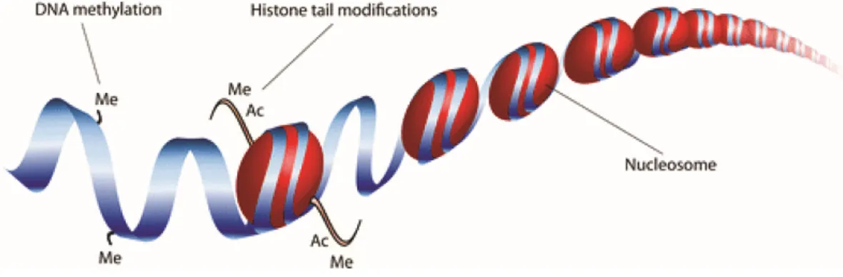
The term ‘epigenetics’ was coined by Waddington in 1942 to refer to ‘heritable changes in genome function that occur without changes in the DNA sequence’ (Waddington 2012). Despite the fact that every cell in a given multicellular organism contains the same genetic information, each cell exhibits different functions and morphologies. Knowing the nucleotide sequence alone is only a small part of the puzzle and the answer lies in the epigenetic regulation of genes. To understand epigenetics requires an understanding of chromatin structure. In eukaryotes, chromatin comprises of DNA wrapped ~147 bp around histone octamers (H2A, H2B, H3 and H4), which constitutes the nucleosome. Chromatin structure can either be loosely packed into euchromatin (open chromatin) or more densely packed into heterochromatin (closed chromatin) (Bell et al. 2011). Epigenetic mechanisms, such as DNA methylation and post-translational modifications of core histone tails, cooperatively determine chromatin configuration and the accessibility of the DNA to the transcription machinery and thus, govern the transcriptional regulation of the expression of genes (Berger 2007; Bernstein et al. 2007) (Figure 1).
Figure 1. Epigenetic mechanisms of gene regulation. DNA is wrapped around histones and
the combined loop of DNA and histone proteins is called a nucleosome. Epigenetic modifications can occur at the histone tails (including acetylation ‘Ac’ and methylation ‘Me’) or directly at the DNA (methylation). Image taken from (Hoeksema and de Winther 2016).
1.2 Histone modifications
There is a wide range of different modifications in the carboxy- and amino-terminal tails of histone proteins that can alter the structure and function of chromatin, including acetylation, methylation, phosphorylation, ubiquitylation, and SUMOylation (Berger 2007; Kouzarides 2007). The recent development of the chromatin immunoprecipitation technique (ChIP) using modification-specific antibodies and its adaptation sequencing (ChIP-seq) has revolutionized mapping of DNA-protein interactions (Furey 2012). It has provided new insights into the genome-wide distribution of histone modifications in many normal and disease-related processes (Barski et al. 2007; Consortium 2012; Roadmap Epigenomics et al. 2015). Although an in-depth review of histone modifications is outside the scope of this thesis, a few relevant modifications (i.e., H3K4me1, H3K4me3, H3K27me3, H3K36me3, H3K9me3, and H3K27ac) are described in more details below.
Histone methylation is associated with either transcriptional activation or inactivation (Kouzarides 2007). The effect of histone methylation on chromatin state is dependent not only on the specific lysine residue modified, but also on its degree of methylation, with the potential addition of one (me1), two (me2), or three methyl groups (me3). For example, histone H3 lysine 4 trimethylation (H3K4me3) shows increased signals in promoters of genes whereas H3K4me1 are associated with enhancer elements located far away from their target genes. It is also well-documented that H3K9me3 and H3K27me3 are associated with transcriptional repression or heterochromatin formation. In some cases, H3K4me3 and H3K27me3 co-exist as "bivalent domains" in genes that regulate development of stem cells, keeping these key genes in poised states for later activation (Bernstein et al. 2006). In the majority of cases, methylation at the aminoterminal domains of H3 and H4 is catalyzed by members of the SET-domain protein methyltransferase family (Dillon et al. 2005), and its removal is carried out by JmjC-containing
acetylation (H3K27ac), is positively correlated with activation in general. Acetylation removes positive charges and reduces the affinity between histones and DNA leading to an open chromatin structure, which is accessible to transcription factors. Co-occurrence H3K27ac and H3K4me1 marks has been widely used to classify active from inactive/primed (H3K4me1 only) enhancers (Creyghton et al. 2010; Calo and Wysocka 2013).
1.3 DNA methylation
In mammals, DNA methylation occurs primarily at the fifth carbon of cytosine residues that are followed by a guanine (CpG dinucleotides). Overall, mammalian genomes are very rich in DNA methylation with the exception of regions called CpG islands – CpG-rich regions found at promoters or near transcription start sites (TSS) of genes (Meissner et al. 2008; Lister et al. 2009). More recently, genome-wide DNA methylation analysis have identified lowly-methylated regions beyond CpG islands, that correspond to distal regulatory elements (enhancers, silencers and insulators) (Stadler et al. 2011). DNA methylation is often associated with transcriptional repression – for example during differentiation, X chromosome inactivation, and imprinting (Suzuki and Bird 2008). Aberrant patterns of DNA methylation can also have striking effects on individual health, including well-known links to cancer susceptibility and autoimmune disorders (Robertson 2005). Thanks to decades of research, many of the proteins and mechanisms involved in DNA methylation have already been identified. Processes that regulate DNA methylation are essentially broken down into three classes: “writers” are the enzymes that catalyze the addition of methyl groups onto cytosine residues, “erasers” modify and remove methyl groups, and “readers” recognize and bind to methyl groups to ultimately mediate changes in gene expression.
1.3.1 Writers
DNA methylation occurs by the addition of a methyl group from S-adenosylmethionine to cytosine with the help of DNA methyltransferases (DNMTs) (Goll and Bestor 2005;
Schermelleh et al. 2005). There are two types of DNA methyltransferase activities in mammals:
de novo and maintenance methylation, which are achieved by DNMT3 and DNMT1
respectively (Figure 2). DNMT1 was the first eukaryotic DNA methyltransferase to be discovered (Bestor et al. 1988). DNMT1 seemed to be responsible only for maintaining methylation after each round DNA replication, which led to the assignment of DNMT1 as a maintenance DNA methyltransferase (Bestor 2000; Goyal et al. 2006). This is supported by findings showing that that DNMT1 co-localizes with the replication machinery (Leonhardt et al. 1992; Schermelleh et al. 2007). At replication sites, hemimethylated DNA is formed when the newly synthesized unmethylated strand pairs with the methylated template strand. Strikingly, while virtually all of methylated CpG sites are methylated in both strands, 98% of methylated cytosines in non-CpG context are highly asymmetrical with only one of strands being methylated (Lister et al. 2009). This suggests that DNMT1 recognizes its substrate cytosine residue only if a guanine residue is beside it.
DNMT3A and DNMT3B are de novo methyltransferases that are responsible for establishing cytosine methylation patterns at unmethylated DNA (Okano et al. 1999). Although DNMT3A and DNMT3B show considerable functional redundancy in early developmental stages, they have different expression profiles in distinct cell types. Moreover, DNMT3B appears to be specialized in particular parts of the chromosome as it engages methylation only at the centromeric region (Xu et al. 1999). It has also been proposed that there is a possible cooperation between the de novo and the maintenance DNMTs (Siedlecki and Zielenkiewicz 2006). DNMT3A and DNMT3B may also participate in the maintenance of methylation by restoring methylation at cytosine residues which have been overlooked by DNMT1 during replication. There is a third homolog in the DNMT3 family found only in germ cells, called DNMT3L (DNA methyltransferase 3-like). Although this protein has been shown to not possess methyltransferase activity, it is essential as a regulatory cofactor of DNMT3A and DNMT3B
does not seem to act as a regulatory factor like DNMT3L – mice, flies and plants deficient of DNMT2 do not display any overt phenotype (Goll et al. 2006; Jeltsch et al. 2006). Interestingly, DNMT2 acts as a transfer RNA (tRNA) methyltransferase that specifically catalyzes the methylation of position 38 in tRNAAsp, tRNAGly and tRNAVal (Goll et al. 2006; Tuorto et al.
2012). Several reports have further demonstrated that DNMT2-mediated methylation contributes to the secondary structure of tRNAs and differential codon usage (Goll et al. 2006; Tuorto et al. 2012; Tuorto et al. 2015; Jeltsch et al. 2017; Zhang et al. 2018), suggesting its role as a modulator of protein translation.
Figure 2. Establishment of DNA methylation patterns in mammals. De novo
methyltransferases DNMT3A and DNMT3B introduce methyl groups (red circles) to the cytosine of previously unmethylated CpG dinucleotides on both strands. Replication of methylated DNA results in hemimethylated DNA in which the parent strand is methylated while the daughter strand is unmethylated. DNMT1 functions as maintenance methyltransferase by methylating the hemimethylated form of CpG sites. Image adapted from (Yu et al. 2011).
1.3.2 Erasers
DNA methylation is relatively stable compared with other epigenetic marks such as histone modifications. Nevertheless, loss of DNA methylation, or DNA demethylation, has been observed in different biological contexts. DNA demethylation is the process of removal of a methyl group from nucleotides in DNA and it may take place in a passive or active fashion. Passive DNA demethylation takes place in dividing cells. As DNMT1 maintains DNA methylation during cell replication, its absence allows newly synthesized DNA strands to be devoid of methylation. Active DNA demethylation occurs via direct removal of a methyl group independently of DNA replication and therefore can take place in both dividing and non-dividing cells. So far, there is no known mechanism in mammalian cells that can cleave the strong covalent carbon-to-carbon bond that connects cytosine to a methyl group. Instead, active demethylation occurs through a series of chemical reactions that revert 5mC back to C. A series of recent discoveries has brought clarity on our understanding of active DNA demethylation. Until recently, the only known covalent epigenetic modification on DNA was methylation at position 5’ of cytosine. A landmark discovery by Tahiliani et al. was made showing that 5mC is oxidized to 5-hydroxymethylcytosine (5hmC) by the enzyme ten-eleven translocation (TET) family proteins (Tahiliani et al. 2009). More importantly, work from the same group have shown that TET proteins and 5hmC may be involved in DNA demethylation – overexpression of TET1 leads to a decrease in 5mC levels. Since the discovery of TET, 5hmC has taken on a new central role in DNA demethylation. Given our current understanding, active demethylation involving TET fall into two groups, which both initially involve active modification (AM) of 5mC to generate 5hmC (Kohli and Zhang 2013) (Figure 3). In the process of passive dilution (PD), unmodified C is regenerated through DNA replication since DNMT1 does not recognize 5hmC and therefore cannot maintain it (Inoue et al. 2011; Inoue and Zhang 2011). Alternatively, active
rapid conversion of 5mC to unmodified C and therefore seems particularly well suited to locus-specific demethylation processes that require a rapid response to environmental stimuli.
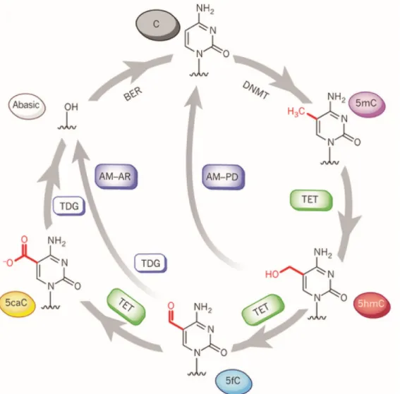
Figure 3. A complete pathway for dynamic cytosine modifications. 5mC bases are
introduced by DNA methyltransferase (DNMT) enzymes. 5mC is further oxidized to 5-hydroxymethylcytosine (5hmC), 5-formylcytosine (5fC) and 5-carboxylcytosine (5caC) during active DNA demethylation by TET family proteins. In the pathway of active modification followed by passive dilution (AM-PD), 5hmC is diluted in a replication-dependent manner to regenerate unmodified C. In the pathway of AM followed by active restoration (AM-AR), 5hmC is further oxidized by TET proteins to 5fC and then 5caC, which is then excised by TDG and repaired by the BER pathway into an unmodified cytosine, generating an abasic site. Image
1.3.3 Readers
There are potentially multiple ways in which DNA methylation can decrease transcription levels or completely turn off genes. One simple way this is accomplished is by directly preventing the binding of the transcriptional machinery or transcription factors to their respective regions by the methyl groups themselves (Hark et al. 2000; Jaenisch and Bird 2003). Alternatively, repression can be achieved via proteins that specifically recognize and bind 5mC. The identification of proteins that can read the methylation signal has shed light on how DNA methylation play a repressive role in gene expression. The first characterized methyl-binding proteins are the methyl-CpG binding domain (MBD) family which consists of five members, namely MBD1, MBD2, MBD3, MBD4 and MeCP2 (methyl-CpG-binding protein 2). These proteins each contain a conserved MBD domain that confers a high affinity for methylated CpG sites (Zhang et al. 1990; Nan et al. 1993; Hendrich and Bird 1998; Wade 2001; Hendrich and Tweedie 2003; Jaenisch and Bird 2003). Three of these proteins (MeCP2, MBD1 and MBD2) also contain a transcriptional repression domain (TRD) that allows these proteins to interact with a variety of repressor complexes and participate in methylation-dependent repression of transcription (Meehan et al. 1989; Nan et al. 1993) (Figure 4). The second family of methyl-binding proteins binds to methylated DNA via a zinc-finger domain and is composed of Kaiso, ZBTB4, and ZBTB38 (Prokhortchouk et al. 2001; Filion et al. 2006). Finally, there is the family of UHRF (ubiquitin-like, containing PHD and RING finger domain) proteins that includes, UHRF1 and UHRF2. These are multidomain proteins that bind methylated cytosines via a SET- and RING-associated DNA-binding domain (Hashimoto et al. 2008; Hashimoto et al. 2009).
Figure 4. Readers of methylation signal and their potential mechanism in gene repression.
(A) (Left to right) Transcription factor (TF) is able to bind to unmethylated DNA sequence (white circles); methylated CpG sites (black circles) or methyl-CpG-binding domain (MBD) proteins interfere with binding of TF. Image adapted from (Trzyna et al. 2012). (B) Graphical representation of the domains in the proteins that recognize and bind specifically methylcytosines. Image taken from (Bogdanovic and Veenstra 2009).
1.4 Mapping the epigenome
1.4.1 ChIP-seq
The increasing interest in the role of epigenetic processes in development and disease has coincided with the advancement of new methods to conduct large-scale and high-resolution epigenomic profiling. The most commonly used experimental approach to profile histone posttranslational modifications is chromatin immunoprecipitation sequencing (ChIP-seq) (Furey 2012). This method uses an antibody against a specific histone modification to immunoprecipitate chromatin regions bearing the corresponding modification. The key bioinformatics challenge in the analysis of ChIP-seq data is to accurately map thousands to millions of reads, corresponding to these regions, to the reference genome (Park 2009). Many sequence aligners for solving the problems of mapping sequence reads have been developed, such as Bowtie (Langmead et al. 2009) and BWA (Li and Durbin 2009). ChIP-seq is also a widely used approach to selectively enrich for DNA sequences bound by specific transcription factors, allowing for the generation of genome-wide binding site maps.
1.4.2 BS-seq
Although there are many DNA methylation analysis methods, bisulfite sequencing (BS-seq) is considered to be the gold standard method in DNA methylation studies (Harris et al. 2010; Laird 2010; Bock 2012). The direct examination of DNA methylation is hindered by the fact, that DNA methylation cannot be analyzed by standard DNA sequencing methods, since they are unable to distinguish 5-methylcytosine from unmodified cytosine. To overcome this, genomic
uracil with thymine (Figure 5). Initially, bisulfite sequencing was used to assay individual loci with locus-specific PCR followed by Sanger sequencing. Recently, reduced representation bisulfite sequencing has extended the genomic coverage of bisulfite sequencing by using high-throughput sequencing technology. Reduced representation bisulfite sequencing (RRBS) combines restriction digestion with BS for analysis of high CpG density regions. Finally, whole-genome bisulfite sequencing (WGBS or MethylC-seq) provides single-base resolution and quantitative rates of methylation for all of the ~29 million CpG sites in the human genome. Mapping of BS-seq reads is performed using as a specific alignment algorithm that implements a bisulfite converted reference genome (C-to-T and a G-to-A) (Krueger and Andrews 2011). Bisulfite treatment in combination with specially designed genotyping microarrays makes it possible to measure DNA methylation levels at a preselected fraction of CpG sites throughout the genome. The Illumina 450k Infinium methylation microarray, which contains over 450,000 CpG sites covering the majority of CpG islands, gene promoters and some enhancer regions, is the most commonly used array in human methylation research (Bibikova et al. 2011; Sandoval et al. 2011). Moreover, the recently developed MethylationEPIC 850k Infinium methylation microarray includes an additional 413,745 CpG positions that are enriched in human enhancer regions (Moran et al. 2016) provided by the ENCODE (Consortium 2012) and FANTOM5 (Lizio et al. 2015) consortia.
A potential issue with current bisulfite conversion-based methodologies is that they depend on the complete conversion of unmethylated cytosines (Wreczycka et al. 2017). Another limitation is that bisulfite conversion does not distinguish between methylcytosine (5mC) and 5-hydroxymethylcytosine (5hmC), the first derivative in the active DNA demethylation pathway (Huang et al. 2010; Jin et al. 2010) (Figure 5). Tet-assisted bisulfite sequencing (TAB-seq) overcomes this limitation and allows single-CpG resolution mapping of 5hmC (Yu et al. 2012).
Figure 5. Detection of methylated DNA by bisulfite conversion. The methyl group covalently
attached to the 5’position of cytosine protects against bisulfite conversion. Unmethylated cystosine (C) is converted to uracil (U; read as thymine (T) when sequenced), but not 5-methylcytosine (5mC). Bisulfite treatment converts 5-hydroxy5-methylcytosine (5hmC) to cytosine-5-methylenesulfonate (CMS), leaving both 5mC and 5hmC to be detected as C. Image
adapted from (Yu et al. 2012).
1.4.3 ATAC-seq
The chromatin accessibility of genomic regions can be profiled with methodologies such as DNase I hypersensitive site sequencing (DNase-seq) (Song and Crawford 2010), formaldehyde-assisted isolation of regulatory elements followed by sequencing (FAIRE-seq) (Giresi et al. 2007), and assay for transposase-accessible chromatin sequencing (ATAC-seq) (Buenrostro et al. 2013). ATAC-seq is now becoming increasingly popular owing to its simple workflow involving substantially fewer cells as starting material. The procedure relies on a hyperactive
1.5 Epigenetic regulation and innate immunity
1.5.1 Innate immunity
Classically, innate immunity is characterized by a rapid, nonspecific response to an invading pathogen (Medzhitov and Janeway 1997). Conversely, an adaptive immune response requires more time to mount, is highly specific to the invading pathogen, and forms memory cells that will respond faster and more robustly to a secondary challenge against an identical immune assault (Medzhitov and Janeway 1998). Understanding innate immune responses is fundamental as it provides the first line of defense against immune challenges (Medzhitov and Janeway 2000) and plays an important role in the activation of the adaptive system (Iwasaki and Medzhitov 2015). Innate immune cells, such as neutrophils, monocytes, macrophages and dendritic cells (DCs), are equipped with various pattern recognition receptors (PRRs), which recognize a wide array of conserved pathogen-associated molecular patterns (PAMPs) and discriminate between self and non-self molecules. Recognition of immune stimuli activates downstream molecular signaling pathways that culminate in the induction of sophisticated transcriptional programs involving the regulation of thousands of genes, which are coordinated with the help of signal-dependent transcription factors including NF-κB/Rel, AP-1, and interferon regulatory factors (IRFs) (Medzhitov 2001; Medzhitov and Horng 2009; Smale 2010; Smale 2011). Upon activation, these transcription factors bind to gene regulatory regions – promoters, enhancers, or silencers – where they function to initiate recruitment of various co-factors required for the activation of inflammatory and/or antiviral response signals. This cascade starts the process of pathogen clearance and the subsequent initiation of appropriate adaptive immune responses.
1.5.2 Dendritic cells: Linking innate and adaptive immunity
Dendritic cells (DCs) are professional antigen-presenting cells that have a central role in T cell activation and in initiation of adaptive immune responses. DCs express a number of pattern
recognition receptors, including Toll-like receptors (TLRs), Nod-like receptors (NLR) and RIG-I-like receptors (RLR), which recognize a wide array of pathogen-associated molecular patterns (Kapsenberg 2003). As immature cells specialized in antigen uptake and processing, DCs reside in and traffic through non-lymphoid peripheral tissues, continuously surveying the environment for invading microorganisms. As immature DCs capture antigens by endocytosis/phagocytosis, they undergo major changes in gene expression programs, evolving from immature, antigen-capturing cells to mature, antigen-presenting, T cell-priming cells. This process of DC maturation, in general, involves down-regulation of antigen internalization, a redistribution of major histocompatibility complex (MHC) molecules from intracellular endocytic compartments to the DC surface, an increase in the surface expression of costimulatory molecules, secretion of chemokines and cytokines, and surface expression of adhesion molecules and chemokine receptors (Tan and O'Neill 2005) (Figure 6).
To present foreign antigens to naïve T cells, DCs must migrate from inflamed or injured peripheral tissues to the closest draining lymph nodes through afferent lymphatic vessels. Migration of maturing DCs from the periphery into lymphoid tissues are coordinated by chemokines that interact with corresponding receptors on DCs (Alvarez et al. 2008). For example, immature DCs express CC-chemokine receptor 1 (CCR1), CCR2, CCR5 and CXC-chemokine receptor 1 (CXCR1) and are attracted to non-lymphoid tissues by their respective ligands, which are expressed constitutively or at inflammatory sites. DC maturation results in the downregulation of expression of these chemokine receptors and the upregulation of CCR7 expression. Expression of CCR7 switches DC responsiveness to its ligands, CC-chemokine ligand 19 (CCL19) and CCL21, that guide migration to secondary lymphoid organs. Maturation of DCs also induces the production of CCL22, CCL17 (i.e., chemokines that attract CCR4-expressing T cells), and CCL18. DC production of the chemokine CXCL16, in T cell-rich areas of lymphoid organs, may also function in promoting interaction between DCs and cytotoxic T
peptide-loaded MHC complexes to the cell surface. Antigen transport to the cell surface coincides with increased expression of costimulatory molecules, such as CD40, CD80 and CD86 (Tan and O'Neill 2005). These molecules amplify T cell receptor signaling and promote T cell activation. Moreover, the soluble cytokine profile secreted by DCs varies with the different stages of DC development and maturation thus influencing the different effector functions characteristic of immature vs. mature DCs (de Saint-Vis et al. 1998). A wide variety of cytokines may be expressed by mature DCs and the exact cytokine repertoire expressed will depend on the nature of the stimulus, maturation stage of the DC and the existing cytokine microenvironment. The distinct cytokine patterns released by mature DCs contribute to the commitment of naïve T cells into more specialized T cell subsets. For example, antigens that prime DCs to secrete IL-12 will typically induce Th1 differentiation (Heufler et al. 1996; Kalinski et al. 1999). Another example is the production of IL-10 and TGF-β, which leads to the generation of regulatory T (Treg) cells (Kushwah and Hu 2011).
Figure 6. Dendritic cell maturation upon antigen encounter. Maturation of dendritic cells,
complexes from within the endocytic system to the cell surface, the production of several cytokines and membrane associated T cell stimulatory molecules, and the remodeling of expressed chemokine receptors. These changes allow dendritic cells to migrate to draining lymph nodes and induce antigen-specific immune response by activating T cells. Image adapted
from (Hackstein and Thomson 2004).
1.5.3 Epigenetic control of the innate immune response
To elaborate an appropriate response to the threat, innate immune cells also undergo important epigenetic regulation. In response to immune stimuli, the most labile epigenetic changes involve the post-translational modifications of histone tails at promoter and enhancer regions (Hazzalin and Mahadevan 2005; Monticelli and Natoli 2013). At promoter regions, histone acetylation has been shown to be essential for the activation of many pro-inflammatory genes (Schmeck et al. 2005; Qiao et al. 2013). Genome-wide analysis, using ChIP-seq, have revealed that enhancer elements (marked by H3K4me1), also contribute to modulation of immune responses (Heintzman et al. 2007; Heintzman et al. 2009; Barish et al. 2010; Ghisletti et al. 2010; Garber et al. 2012; Calo and Wysocka 2013; Kaikkonen et al. 2013; Ostuni et al. 2013; Rogatsky and Adelman 2014). The canonical model is that binding of signal-dependent transcription factors (TFs) in response to stimuli occurs within cell type-specific repertoires of enhancers, that were already established by lineage-determining or “pioneer” TFs during cell differentiation (Barish et al. 2010; Ghisletti et al. 2010; Heinz et al. 2010; John et al. 2011; Mullen et al. 2011; Natoli et al. 2011; Trompouki et al. 2011; Garber et al. 2012; Kaikkonen et al. 2013; Ostuni et al. 2013; Rogatsky and Adelman 2014) (Figure 7). Interestingly, Ostuni et al. have reported a new class of distal regulatory elements, coined “latent enhancers” that appear after stimulation of mouse macrophages with different stimuli (Ostuni et al. 2013). These
In contrast to histone modifications, little is known about the regulatory implications of DNA methylation in innate immune responses. DNA methylation has historically been considered to be a relatively stable epigenetic mark (Bernstein et al. 2007), and thus unlikely to respond to environmental perturbations on a short time scale. Despite its thermodynamic stability, there is increasing evidence that DNA methylation can rapidly respond to environmental perturbations, as exemplified in post-mitotic cells in the brain during neuronal activation (Weaver et al. 2004; Guo et al. 2011); or during monocyte differentiation into macrophages or dendritic cells (Klug et al. 2010). Likewise, studies on dividing cells or cell lines also argue for the involvement of an active enzymatic mechanism, as the kinetics of the demethylation procedure are too fast to be dependent on cell proliferation (Bruniquel and Schwartz 2003; Murayama et al. 2006; Niehrs and Schafer 2012). Indeed, the recent description of transitions from 5-methylcytosine (5mC) into more labile oxidized intermediates – such as hydroxymethylcytosine (5hmC), 5-formylcytosine (5fC) and 5-carboxylcytosine (5caC) – by TET enzymes (Tahiliani et al. 2009) could provide a suitable mechanism to support rapid response genes.
Recent studies have reported altered DNA methylation patterns associated with activation of innate immune cells.(Marr et al. 2014; Zhang et al. 2014; Cizmeci et al. 2016; Wiencke et al. 2016). For instance, Marr et al. assessed epigenetic changes in macrophage DNA methylation in response to infection with an intracellular protozoa Leishmania donovani (Marr et al. 2014). Using 450K methylation array, they identified a set of 443 CpG sites with changes in methylation following live L. donovani infection. These epigenetic changes are linked to genes that play a critical role in host defense such as the JAK/STAT and Notch signaling pathway. Similarly, Sinclair et al. investigated DNA methylation dynamics in Anaplasma
phagocytophilum-infected human neutrophils using methylated DNA binding domain (MBD)
enrichment and next generation sequencing approach (MBD-seq) (Sinclair et al. 2015). Within 24 hours post-infection, marked increases in DNA methylation were observed genome-wide as compared with mock-infected controls. These studies, however, have focused exclusively on DNA methylation changes in promoter regions or at relatively few CpG sites in the genome at low-resolution, and that such changes are poorly predictive of changes in gene expression levels.
Figure 7. Pioneer transcription factors (TFs) organize the enhancer landscape required for stimulus-induced transcription in innate immune cells. During differentiation of
hematopoietic stem cells to macrophages or DCs, lineage-determining or pioneer TFs, such as PU.1, open condensed chromatin and promote the deposition of H3K4me1 at enhancers. Immune stimuli (e.g., infection) trigger the recruitment of signal-dependent TFs, such as NF-κB within the cell type-specific enhancer repertoires already established by PU.1 prior to immune stimulation (i.e., primed enhancers). NF-κB binding leads to the deposition of the H3K27ac activating mark and the subsequent upregulation of stimulus-responsive genes. Image
1.6 Research objectives
The main goal of this thesis was to elucidate the role of DNA methylation in the regulation of innate immune responses to infection. This work is divided into two chapters addressing different questions pertaining to the overarching goal.
Article I. Bacterial infection remodels the DNA methylation landscape of human dendritic cells
The first aim was to characterize changes in DNA methylation in innate immune cells during infection. Using high-throughput sequencing methods, I performed comprehensive transcriptional and epigenetic profiling of human dendritic cells, before and after infection with a pathogenic strain of Mycobacterium tuberculosis (MTB). A Carboxyfluorescein Diacetate Succinimidyl Ester (CFSE) proliferation assay was also performed to determine whether changes in methylation were independent of cell division.
Article II. DNA demethylation plays a limited role in the regulation of innate immune responses to infection
I next sought to assess the causal relationship between changes in DNA methylation and gene expression in response to infection. I generated paired data on DNA methylation, gene expression, and chromatin accessibility in non-infected and MTB-infected DCs at multiple time-points. These time-series datasets allowed the dissection of the relative order of regulatory events during infection.
2 Article I
Bacterial infection remodels the DNA methylation landscape of human
dendritic cells
Pacis A, Tailleux L, Morin AM, Lambourne J, MacIsaac JL, Yotova V, Dumaine A, Danckaert
A, Luca F, Grenier JC, Hansen KD, Gicquel B, Yu M, Pai A, He C, Tung J, Pastinen T, Kobor MS, Pique-Regi R, Gilad Y, Barreiro LB.
Genome Research 2015 Dec;25(12):1801-11. Epub 2015 Sep 21.
Bacterial infection remodels the DNA methylation landscape of
human dendritic cells
Alain Pacis1,2, Ludovic Tailleux3, Alexander M Morin4, John Lambourne5, Julia L Maclsaac4,
Vania Yotova1, Anne Dumaine1, Anne Danckaert6, Francesca Luca7, Jean-Christophe Grenier1,
Kasper D Hansen8, Brigitte Gicquel3, Miao Yu9, Athma Pai10, Chuan He9, Jenny Tung11, Tomi
Pastinen5, Michael S Kobor4, Roger Pique-Regi7, Yoav Gilad12,*, Luis B Barreiro1,13,*
1CHU Sainte-Justine Research Center, Department of Genetics, Montreal, H3T1C5, Canada; 2University of Montreal, Department of Biochemistry, Montreal, H3T1J4, Canada; 3Institut
Pasteur, Mycobacterial Genetics Unit, Paris, 75015, France; 4Centre for Molecular Medicine
and Therapeutics, Child and Family Research Institute, Department of Medical Genetics, University of British Columbia; 5Génome Québec Innovation Centre, Department of Human
Genetics, McGill University, Montréal, H3A0G1, Canada; 6Institut Pasteur, Imagopole, Paris,
75015, France; 7Wayne State University, Center for Molecular Medicine and Genetics and
Department of Obstetrics and Gynecology, Detroit, MI, 48202; 8Johns Hopkins Bloomberg
School of Public Health, Department of Biostatistics and McKusick-Nathans Institute for Genetic Medicine, Baltimore, MD, 21205; 9University of Chicago, Department of Chemistry
and Institute for Biophysical Dynamics, Chicago, IL, 60637; 10Department of Biology,
Massachusetts Institute of Technology, United States; 11Duke University, Departments of
Evolutionary Anthropology and Biology and Duke Population Research Institute, Durham, NC, USA 27708; 12University of Chicago, Department of Human Genetics, Chicago, IL, 60637; 13University of Montreal, Department of Pediatrics, Montreal, H3T1J4, Canada.
Abstract
DNA methylation is an epigenetic mark thought to be robust to environmental perturbations on a short time scale. Here, we challenge that view by demonstrating that the infection of human dendritic cells (DCs) with live pathogenic bacteria is associated with rapid and active demethylation at thousands of loci, independent of cell division. We performed an integrated analysis of data on genome-wide DNA methylation, histone mark patterns, chromatin accessibility, and gene expression, before and after infection. We found that infection-induced demethylation rarely occurs at promoter regions and instead localizes to distal enhancer elements, including those that regulate the activation of key immune transcription factors. Active demethylation is associated with extensive epigenetic remodeling, including the gain of histone activation marks and increased chromatin accessibility, and is strongly predictive of changes in the expression levels of nearby genes. Collectively, our observations show that active, rapid changes in DNA methylation in enhancers play a previously unappreciated role in regulating the transcriptional response to infection, even in non-proliferating cells.
Introduction
The first immune mechanisms recruited to defend against invading pathogens are those associated with innate immune cells, such as dendritic cells (DCs) or macrophages. Once they sense an intruder, these cells induce sophisticated transcriptional programs involving the regulation of thousands of genes, which are coordinated with the help of signal-dependent transcription factors, including NF-κB/Rel, AP-1, and interferon regulatory factors (IRFs) (Medzhitov 2001; Smale 2010). The regulation of this program is achieved through a series of epigenetic changes, which are thought to modulate the access of transcription factors to specific DNA regulatory elements (Bierne et al. 2012).
The most well-studied epigenetic responses to immune stimuli involve the post-translational modification of histone tails at promoter and enhancer regions (Bierne et al. 2012; Monticelli and Natoli 2013). Histone acetylation has been shown to be essential for the activation of many pro-inflammatory genes (Ghisletti et al. 2010; Qiao et al. 2013), whereas increased activity of histone deacetylases is often associated with gene repression in the context of inflammation (Villagra et al. 2009). Moreover, recent studies suggest that the response of innate cells to different immune challenges can result in the appearance of histone marks associated with de
novo enhancer elements (or latent enhancers) (Kaikkonen et al. 2013; Ostuni et al. 2013). These de novo enhancers have been postulated to contribute to a faster and stronger transcriptional
response to a secondary stimulus (Ostuni et al. 2013).
In contrast, we still know remarkably little about the role of other epigenetic changes in controlling responses to infection. DNA methylation has been particularly understudied, as a consequence of the belief that methylation marks are highly stable, and unlikely to respond to environmental perturbations on a short time scale (Bierne et al. 2012; Monticelli and Natoli 2013). Recent work, however, suggests that DNA methylation patterns can rapidly change in response to certain environmental cues (Klug et al. 2010; Guo et al. 2011; Dowen et al. 2012; Marr et al. 2014), raising the possibility that rapid changes in DNA methylation might play a role in innate immune responses. To date, no studies have comprehensively investigated the contribution of rapid, active changes in methylation (in contrast to passive changes during cell
replication) to the regulatory programs induced by innate immune cells in response to an infectious agent. More broadly, the few studies in mammalian cells that demonstrate cell division-independent changes in DNA methylation have only focused on a small number of CpG sites and, surprisingly, have suggested that such changes are poorly predictive of changes in gene expression levels (Bruniquel and Schwartz 2003; Klug et al. 2010; Guo et al. 2011; Marr et al. 2014). Here, we report the first comprehensive epigenome and transcriptome analysis of monocyte-derived DCs – professional antigen presenting cells that play a central role in bridging innate and adaptive immunity – before and after in vitro infection with live pathogenic bacteria. All the data generated in this study are freely accessible via a custom web-based browser that enables easy querying and visualization of epigenetic profiles at any genomic region of interest (http://luis-barreirolab.org/EpigenomeBrowser).
Results
MTB infection induces active changes in DNA methylation in human DCs
We infected monocyte-derived DCs from six healthy donors with a live virulent strain of
Mycobacterium tuberculosis (MTB), the causative agent of tuberculosis (TB) in humans.
Monocyte-derived DCs are ideally suited to study active changes in methylation because they are post-mitotic and not expected to proliferate in response to infection (Pickl et al. 1996; Ardeshna et al. 2000). To experimentally confirm this assumption, we performed a Carboxyfluorescein Diacetate Succinimidyl Ester (CFSE) proliferation assay. This method relies on the ability of the highly fluorescent dye carboxyfluorescein to incorporate within cells. Following each cell division, the equal distribution of these fluorescent molecules to progeny cells results in a halving of cell fluorescence levels. We did not detect any decrease in per-cell fluorescence at 18 hours post-infection, which confirms that DCs do not proliferate after MTB infection (Figure 1A). In contrast, we observed high rates of proliferation in our positive control, human monocytic THP-1 cells (Figure 1A).
At 18 hours after infection, we obtained paired data on single base-pair resolution DNA methylation levels (using whole genome shotgun bisulfite sequencing: i.e., MethylC-seq) and genome-wide gene expression data (using mRNA sequencing: i.e., mRNA-seq) in non-infected and MTB-infected DCs. For MethylC-seq data, we generated 8.6 billion single-end reads (mean of 648 ± 110 SD million reads per sample; Supplementary Table 1) resulting in an average coverage per CpG site of ~9X for each sample. We detected an average of 24 million CpG sites in each sample, corresponding to over 80% of CpG sites in the human genome. Genome-wide methylation data between biological replicates were strongly correlated, attesting to the high quality of the data (Supplementary Figure 1; mean r across all samples = 0.86).
As expected for mammalian cells, most CpG sites were highly methylated throughout the genome except near transcription start sites (TSSs), CpG islands, and putative enhancer elements (Supplementary Figure 2A,B). We found a significant negative correlation between gene expression levels and methylation levels around TSSs (r = -0.39; P < 1 × 10-16;
in the stable silencing of gene expression. Principal component analysis of our data along with MethylC-seq data from 21 other purified cell types and tissues revealed that the DC methylome is closely related to that of other blood-derived cells, particularly cells that share a common myeloid progenitor with DCs, such as neutrophils (Supplementary Figure 2E).
We next assessed the occurrence and the extent to which the response of DCs to a bacterial infection is accompanied by active changes in DNA methylation, using the BSmooth algorithm (Hansen et al. 2012). We defined MTB-induced differentially methylated regions (MTB-DMRs) as regions of 3 or more consecutive CpG sites exhibiting a significant difference in methylation between the two groups (P < 0.01) and an absolute mean methylation difference above 0.1 (Hansen et al. 2014). Using these criteria, we identified 3,271 MTB-DMRs, corresponding to both hypermethylated regions (48%) and hypomethylated regions (52%) (Figure 1B; Supplementary Table 2). To independently validate these changes, we generated methylation-sensitive pyrosequencing data on control versus MTB-infected DCs from 5 completely new individuals. We targeted 21 CpG sites that were differentially methylated in the MethylC-seq analysis, distributed across 4 hypermethylated (11 CpG sites) and 6 hypomethylated MTB-DMRs (10 CpG sites; Supplementary Table 3). We were able to validate 100% of the hypomethylated CpG sites, with effect sizes similar to or greater than those identified in the original bisulfite sequencing analysis (Figure 1B,C; Supplementary Figure 3A). In contrast, we were not able to validate any of the hypermethylated CpG sites (Supplementary Figure
3B), which indicates that most (if not all) active changes in methylation observed in response to
infection are losses rather than gains in methylation, in accordance with previous findings (Klug et al. 2010).
We found that only 6% of hypomethylated regions overlapped with a promoter (Figure 1D) and that the vast majority of hypomethylated regions were located distal to TSSs (median distance
including the regulation of transcription, signal transduction, and cell apoptosis (Figure 1F;
Supplementary Table 4). The set of genes near hypo-DMRs included virtually all of the
“master-regulators” of innate immune responses, including CREB5, REL, NFKB1, IRF2, and
IRF4. It also included key genes involved in DC-mediated activation of B and T cells (e.g., CD83) and the regulation of cell death (e.g., BCL2).
Figure 1. MTB-induced changes in methylation in post-mitotic human DCs. (A)
CFSE-labeled THP-1 (left) and CFSE-CFSE-labeled DCs (right). Proliferation was assayed in either non-infected cells or cells non-infected for 18 hours with MTB. Similar results were observed 48 hours post-infection (Supplementary Figure 16). (B) Example of a region showing active loss of DNA methylation in response to MTB infection (gray shading). The plot shows smoothed methylation values (y-axis) for six non-infected (blue) and six MTB-infected samples (red). Thick blue and red lines show average methylation levels for non-infected and infected cells, respectively. The inset on the right shows methylation levels at two individual CpG sites within the
hypomethylated region using bisulfite pyrosequencing as a validation method. (C) Scatter plot showing the correlation between MethylC-seq (x-axis; smoothed data) and pyrosequencing data (y-axis) for mean differences in methylation between infected and non-infected cells, at 10 CpG sites within hypomethylated DMRs. Data are represented as mean ± s.e.m., n = 6 for MethylC-seq and n = 5 for Pyro-MethylC-seq. (D) Pie charts showing the distribution of hypomethylated regions in different genomic regions. Each MTB-DMR is counted only once: the overlap of a genomic region excludes all previously overlapped MTB-DMRs, starting clockwise from promoters (TSS ± 500 bp; red). (E) Distribution of distances of MTB-DMRs to the nearest TSS. (F) Representative gene ontology (GO) terms enriched among genes associated with hypomethylated regions. To demonstrate that the enriched biological processes are largely robust to the cutoff used to define MTB-DMRs, we show how these results differ depending on the number of differentially methylated CpG sites (P < 0.01) required to call an MTB-DMR (from at least three to at least 5 consecutive sites).
Active changes in methylation occur in regions enriched for
5-hydroxymethylcytosine
The TET family proteins catalyze the conversion of methylated cytosine (5mC) to 5-hydroxy-methylcytosine (5hmC), and are thus key players in the process of active demethylation. To evaluate if 5hmC levels dynamically change in response to MTB infection (as expected if 5mC sites must pass through the 5hmC state before demethylation), we generated single base-pair resolution maps of 5hmC across the genome using TET-assisted bisulfite sequencing (TAB-seq) (Yu et al. 2012) in one of the 5 original donor. As previously described for other cell populations (Song et al. 2011; Lister et al. 2013), we found markedly higher levels of 5hmC in gene bodies of highly expressed genes, consistent with a role for 5hmC in maintaining and/or promoting gene expression (Figure 2A) (Hahn et al. 2013; Hon et al. 2014).
Next, we evaluated if 5hmC marks were enriched within hypomethylated MTB-DMRs. We found that regions that became hypomethylated post-infection were already associated with significantly higher levels of 5hmC prior to infection (3.6-fold enrichment; Wilcoxon test; P < 1 × 10-16). Upon infection, 5hmC levels increased even further (Wilcoxon test; P = 1.57 × 10⁻¹¹;
Figure 2B,C), suggesting that 5hmC plays an important role in the cascade of events leading to
active demethylation. The increase in 5hmC appears to be specific to hypomethylated regions since no enrichment was observed genome-wide, a result supported by quantitative immunocytochemistry data (Figure 2D,E). The striking enrichment of 5hmC within MTB-DMRs prior to infection strongly suggests that, in addition to its role as a transitory demethylation intermediate, 5hmC might also contribute to coordinating the gene expression program induced in response to a microbial stimulus.
Figure 2. 5hmC is enriched in MTB-DMRs prior to infection. (A) Metagene profiles of
5hmC levels relative to Ensembl transcripts expressed at different levels in human DCs. We grouped genes in four quantiles based on their expression levels on non-infected DCs. (B) Bar plots showing mean 5hmC/C ratios within hypomethylated regions, before (blue) and after infection (red). (C) Composite plots of patterns of 5hmC before and after MTB infection ±3 kb around the midpoint of hypomethylated regions. (D) 5hmC staining in non-infected (top panel) and MTB-infected DCs (bottom panel). 5hmC levels are given by the levels of Alexa 488 (green: middle panel). Cells counterstained with DAPI to localize the nucleus are shown in the first panel. (E) Boxplots showing the distribution of 5hmC staining intensity. No significant differences were observed between the two groups.
MTB-DMRs overlap with enhancer elements that gain activation marks upon
infection
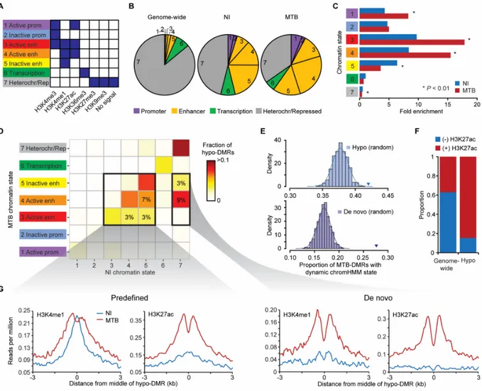
Given that MTB-DMRs are primarily found distal to TSSs, we predicted that MTB-DMRs would overlap with enhancer regions. To test this hypothesis and evaluate how the chromatin states associated with MTB-DMRs dynamically change in response to infection, we collected ChIP-seq data for six histone marks (H3K4me1, H3K4me3, H3K27ac, H3K27me3, H3K36me3 and H3K9me3) in non-infected and infected DCs (Supplementary Table 1) from two additional donors. Using these data, we generated genome-wide, gene regulatory annotation maps for non-infected and MTB-non-infected DCs using the ChromHMM chromatin segmentation program (Figure 3A; Supplementary Figure 5) (Ernst and Kellis 2012). We found that 41% of hypomethylated regions overlapped with a ChromHMM-annotated enhancer region (defined by the presence of H3K4me1) already present in non-infected DCs, a 7.4-fold enrichment compared to genome-wide expectations (χ2-test; P < 1 × 10-16; Figure 3B,C; Supplementary
Table 2). Slightly higher enrichments (8.1-fold; P < 1 × 10-16) were observed when defining
chromatin states in MTB-infected DCs. Given the high-resolution of our histone maps, we could further distinguish between active and inactive/poised enhancer elements based on the presence or absence of the H3K27ac mark, respectively, in addition to H3K4me1 (Heintzman et al. 2007; Creyghton et al. 2010; Rada-Iglesias et al. 2011). Overall, we found that MTB infection leads to a significant increase of active enhancer elements (and decrease of inactive/poised enhancers) colocalizing with MTB-DMRs (Figure 3B,C).
We next extended our analysis by examining chromatin transition states at hypomethylated regions in response to MTB-infection. We found that 42% of hypomethylated regions occurred in regions that exhibited infection-dependent changes in chromatin state, a significantly higher proportion than expected compared to the rest of the genome (Presampling < 0.001; Figure 3E). The
chromatin state transitions observed within hypomethylated regions were primarily explained by the acquisition of histone activating marks (e.g., H3K27ac) in MTB-infected cells. For example, among hypomethylated regions that overlapped with predefined enhancers (i.e., enhancers observable in non-infected cells), 85% of those that exhibit a change in chromatin state gained an activation mark (H3K27ac or H3K27ac+H3K4me3; Figure 3F,G;
Supplementary Figure 6A). This proportion was markedly larger than that observed
genome-wide (37%) (χ2-test; P = 1.1 × 10-59; Figure 3F). Notably, we also found a large number of
hypomethylated regions (n = 218; 12.7% of all hypomethylated regions) that overlapped with heterochromatin/repressed regions before infection but gained de novo enhancer marks upon MTB infection (H3K4me1 (+ H3K27ac + H3K4me3)). The number of de novo enhancers we observed among hypomethylated regions was significantly higher than expected by chance (Presampling < 0.001; Figure 3D,E,G; Supplementary Figure 6A). The identification of
enhancers only present in infected DCs resembles recent findings showing that, in response to different immune stimuli, mouse macrophages can gain de novo putative enhancer regions that were absent in naive cells (Kaikkonen et al. 2013; Ostuni et al. 2013). Interestingly, we observed that 5hmC was significantly enriched among de novo hypo-DMRs prior to infection (Wilcoxon test; P = 5.27 × 10-149), suggesting that 5hmC might be an early “pre-marking” mechanism of
enhancer activation, even before the deposition of H3K4me1 marks (Supplementary Figure
6A,B).
Finally, we found that MTB-induced activation or de novo gain of enhancer elements at hypomethylated regions was associated with the induction of putative enhancer RNAs (eRNAs) (Wang et al. 2011) in these intergenic regions (as measured by whole-transcriptome RNA-seq) as well as with increased levels of histone marks associated with transcriptional activity (Supplementary Figure 7). Moreover, changes in eRNA levels in response to MTB infection show a striking positive correlation with changes in gene expression levels of nearby genes (r = 0.49, P = 7.6 × 10⁻¹³;Supplementary Figure 7), in support of a mechanistic link between
demethylation, eRNA production and the regulation of proximal protein-coding genes (Lam et al. 2014).
Figure 3. MTB-DMRs overlap with enhancer elements that become active upon infection in hypomethylated regions. (A) Combination of histone patterns used to define the 7 chromatin
states. The precise relative contribution of each chromatin mark to each of the chromHMM-defined states can be found in Supplementary Figure 3. Note that state 7 was chromHMM-defined by either no signal or the presence of either H3K27me3/H3K9me3. (B) Pie charts showing the distribution of chromatin state annotations genome-wide (on non-infected cells) and within all MTB-DMRs in either non-infected (blue) or MTB-infected cells. The chromatin state codes are as defined in (A). (C) Fold enrichments of the different chromatin states within hypomethylated regions as compared to genome-wide expectations in non-infected (blue) and MTB-infected cells (red). (D) Heatmap of the proportion of hypomethylated regions by chromatin transition state. The x-axis represents the chromatin states defined in non-infected DCs and the y-axis the chromatin state of the same region in MTB-infected DCs. The two inner boxes indicate two