Université de Montréal
Binder of SPerm protein interference in sperm-egg
interaction
Par
Hamed Heidari Vala
Département de pharmacologie et physiologie
Faculté de Médecine
Mémoire présenté à la Faculté de Médecine en vue de l’obtention du grade de Maîtrise ès sciences (M.Sc.) en physiologie moléculaire, cellulaire et intégrative
Février 2018
Université de Montréal Faculté de Médecine
Ce mémoire intitulé :
Binder of SPerm protein interference in sperm-egg interaction
L’interférence de la protéine Binder SPerm (BSP) dans
l'interaction spermatozoïde-ovocyte
Présenté par Hamed Heidari Vala
A été évalué par un jury composé des personnes suivantes :
Réjean Couture Ph.D., président-rapporteur Puttaswamy Manjunath, Ph.D., directeur de recherche
Abstract
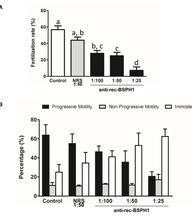
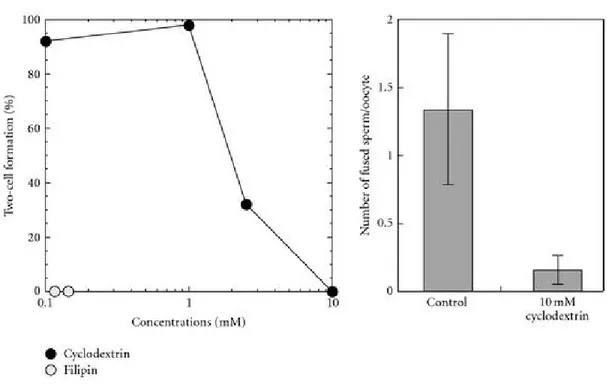
Binder of SPerm (BSP) proteins were first characterized in the laboratory of Dr. P. Manjunath, where the biochemistry of seminal plasma proteins and their interactions with sperm are being studied. These proteins were shown to bind to choline phospholipids on the ejaculated sperm membrane. In mice and humans, the Binder of SPerm homolog 1 (BSPH1) protein is exclusively expressed in the epididymis. BSPH1 proteins have been shown to be involved in the sperm membrane changes underlying capacitation. Findings from experiments with the recombinant mouse BSP homolog (rec-BSPH1) suggest that the protein initially resides on the surface of the sperm and then relocalizes over the head and mid-piece during capacitation and sperm-egg interaction, suggesting a potential role for BSPH1 in sperm-egg interaction. In the current study, we examined the role of the mouse recombinant BSP homolog 1 (rec-BSPH1) in sperm-egg interaction using an in vitro fertilization (IVF) assay. Oocytes were pre-treated with rec-BSPH1, control proteins or media alone, and inseminated with capacitated sperm. In addition to IVF assays, the potential binding of rec-BSPH1 to the oocyte surface was investigated using immunofluorescence. Finally, sperm-bound native BSPH1 was immuno-neutralized by anti-rec-BSPH1 antibodies in order to indirectly demonstrate the importance of BSPH1 in sperm-egg interaction and fertilization. Our results showed that eggs pre-incubated with rec-BSPH1 protein exhibited a dose-dependent decrease in fertilization rate compared to those exposed to control proteins or media alone. Since BSPH1 binding sites were not identified on the egg, the observed inhibition in fertilization rate when eggs were pre-incubated with rec-BSPH1 suggested that an alternate mechanism was at play. We hypothesize that bovine serum albumin contained in the media synergizes with rec-BSPH1 to
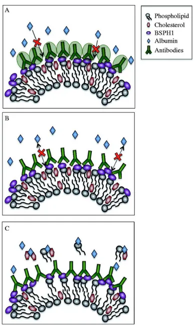
provoke oocyte membrane lipid raft disorganization, which interferes with fertilization. In addition, anti-rec-BSPH1 antibodies could effectively immuno-neutralize native protein on sperm, which led to dramatic motility suppression and failed hyperactivation, followed by compromised fertilization. Taken together with previously published research, our findings suggest that BSPH1 would mostly be involved in the late events of sperm capacitation.
Keywords: Binder of SPerm Homolog, BSPH1, in vitro fertilization, sperm-egg interaction, capacitation, cholesterol efflux
Résumé
Les protéines de la famille Binder of SPerm (BSP) ont été initialement caractérisées dans le laboratoire du Dr. P. Manjunath, où la biochimie des protéines du plasma séminal et leurs interactions avec les spermatozoïdes sont à l’étude. Il a été démontré que ces protéines lient les phospholipides portant un groupement choline de la membrane des spermatozoïdes éjaculés. Chez la souris et l’humain, la protéine « Binder of SPerm homolog 1 » (BSPH1) est exprimée exclusivement dans l’épididyme. Il a été démontré que les protéines BSPH1 sont impliquées dans les modifications membranaires qui sous-tendent la capacitation chez les spermatozoïdes. Les résultats d’expériences avec la protéine recombinante de l’homologue murin des BSP (rec-BSPH1) suggèrent que la protéine est initialement localisée sur toute la surface du spermatozoïde, pour ensuite se relocaliser sur la tête et sur la partie intermédiaire de la cellule pendant la capacitation et l’interaction entre le spermatozoïde et l’ovocyte, suggérant un rôle potentiel dans cette interaction. Dans cette étude, nous avons examiné le rôle de la protéine recombinante murine « Binder of SPerm homolog 1» (rec-BSPH1) dans l’interaction spermatozoïde-ovocyte par le biais d’un essai de fécondation in vitro (FIV).
Des ovocytes ont été pré-incubés avec soit des protéines rec-BSPH1, soit des protéines contrôles ou avec le milieu de culture cellulaire seul, et ensuite inséminés par des spermatozoïdes capacités. Outre les essais FIV, la liaison potentielle de rec-BSPH1 sur la surface de l’ovocyte a été investiguée par le biais d’expériences d’immunofluorescence. Enfin, la protéine BSPH1 native, liée au spermatozoïde, a été immuno-neutralisée à l’aide d’anticorps dirigés contre la protéine rec-BSPH1 (anticorps anti-rec-BSPH1) afin de démontrer indirectement l’importance de BSPH1 dans l’interaction spermatozoïde-ovocyte et la
fécondation. Nos résultats montrent que les ovocytes pré-incubés avec la protéine rec-BSPH1 ont connu une diminution dose-dépendante du taux de fécondation lorsque comparés à des ovocytes pré-exposés à des protéines contrôles ou au milieu de culture cellulaire seul. Puisque des sites de liaison à la protéine BSPH1 n’ont pas été identifiés sur la surface de l’ovocyte, la diminution observée dans le taux de fécondation lorsque les ovocytes étaient pré-incubés avec la protéine rec-BSPH1 suggère qu’un mécanisme alternatif était en jeu. Nous émettons l’hypothèse que l’albumine de sérum bovin contenue dans le milieu de culture cellulaire agit en synergie avec rec-BSPH1 afin de provoquer la désorganisation des radeaux lipidiques de la membrane des ovocytes, ce qui interfère avec la fécondation. De plus, les anticorps anti-rec-BSPH1 ont efficacement immuno-neutralisé la protéine native sur la surface des spermatozoïdes, ce qui a mené à une suppression dramatique de la motilité et un échec de l’hyperactivation, accompagné d’une diminution du taux de fécondation.
Combinés aux résultats de recherche publiés précédemment, les résultats de cette étude suggèrent que la protéine BSPH1 serait principalement impliquée dans les évènements tardifs de la capacitation des spermatozoïdes.
Mots-clés : Protéine « Binder of SPerm Homolog » (BSPH1), fécondation in vitro, interaction spermatozoïde-ovocyte, capacitation, efflux de cholestérol
Table of content
Abstract... i
Résumé ... iii
Table of content ... v
List of figures ... vii
List of acronymes ... viii
List of abreviations ...ix
Acknowledgments ... xiii
Introduction... 15
1. Epididymal milieu and sperm maturation ... 15
1.1. Epididymal microenvironment and secretome ... 15
1.2. Physiology of sperm maturation ... 17
1.2.1. Protein traffic between sperm and the epididymal milieu ... 19
1.2.2. Changes to the sperm membrane during maturation ... 20
1.2.3. Progressive movement competency ... 21
2. The Binder of SPerm protein family ... 21
2.1. Background ... 21 2.2. Structure ... 23 2.3. Binding properties ... 24 2.3.1. Gelatin ... 25 2.3.2. Glycosaminoglycans (GAGs) ... 25 2.3.3. Phospholipids ... 25 2.3.4. Cholesterol ... 26
2.4. Physiological function of BSP proteins ... 27
2.4.1. BSPs effects on sperm in the male reproductive tract... 27
2.4.2. BSPs effects on sperm in a cryopreservation environment ... 29
2.4.3.1. The oviductal sperm reservoir ... 29
2.4.3.2. Sperm capacitation ... 30
2.4.3.3. Implication of BSP proteins in sperm-egg interaction ... 33
3. Sperm-egg interaction... 33 3.1. Acrosome reaction ... 34 3.2. Sperm-egg fusion ... 35 4. Thesis Objectives ... 37 Article ... 39 Discussion ... 73
Conclusions and perspectives ... 85
List of figures
Figure 1 : Different mechanisms of epididymal secretion ... 17
Figure 2 : Schematic drawing of a mouse sperm cell ... 18
Figure 3a : Illustration of the structure of the bovine BSP1 protein ... 24
Figure 3b : 3D structure of bovine BSP1 ... 24
Figure 4 : Proposed mechanism for the involvement of BSPH1 in HDL-induced mouse sperm capacitation ... 32
Figure 5 : Physiological and morphological changes occurring in sperm during capacitation and acrosome reaction ... 35
Figure 6 : Fertilization scenario ... 36
Figure 7 : Cyclodextrin, a lipid-raft disruptor ... 76
Figure 8 : Proposed models for the inhibition of BSA-induced capacitation by anti-BSPH1 antibodies ... 80
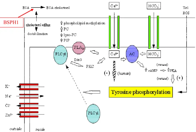
Figure 9 : Schematic illustration of the molecular pathways underlying BSA-induced sperm capacitation in vitro ... 83
List of acronymes
~ ... approximately % ... percent
e.g. ... for example i.e. ... for example µl ... microliter µg ... microgram µM ... micromolar °C ... degrees Celsius
List of abreviations
ABC ... ATP binding cassette AC ... adenylyl cyclase
ADAM ... a disintegrin and metalloprotease Akt ... protein kinase B
apoA-1 ... apolipoprotein 1 AR ... acrosome reaction ATP ... adenosine triphosphate
B-PER ... bacterial protein extraction reagent BSA ... bovine serum albumin
BSP ... Binder of SPerm
BSPH ... Binder of Sperm Homolog Ca2+... Calcium ion
cAMP ... cyclic adenosine monophosphate COC ... cumulus-oocyte complex
CRISP ... Cysteine-rich secretory protein DAPI... 4',6-diamidino-2-phenylindole DNA ... deoxyribonucleic acid
ERK ... extracellular signal regulated kinase FITC ... Fluorescein isothiocyanate
Fn2... fibronectin type II domain FSH ... follicle-stimulating hormone GAG ... Glycosaminoglycan
GPI ... Glycosylphosphatidylinositol h ... hour
hCG ... human chorionic gonadotropin HCO3- ... bicarbonate
HDL ... high-density lipoproteins His ... histidine
HTF ... human tubular fluid
ICSI ... intra-cytoplasmic sperm injection IgG... Immunoglobulin G
IMAC ... immobilized metal ion affinity chromatography IS ... initial segment
IVF ... in vitro fertilization K+ ... potassium ion kDa ... kilodalton KO ... knock-out LDL ... low-density lipoprotein LH ... luteinizing hormone M ... molar
MII... methaphase 2 (oocyte) mg ... milligram
min... minute ml ... milliliter mM ... millimolar
MβCD ... methyl beta cyclodextrine Na+ ... sodium ion
Ni ... nickel
NRS ... normal (pre-immune) rabbit serum OD ... optic density
PBS ... phosphate-buffered saline PC ... phosphatidylcholine PCR ... polymerase chain reaction PE ... phosphatidylethanolamine pH ... potential of hydrogen PI ... phosphatidylinositol PI ... propidium iodide
PKC ... protein kinase C PLA2 ... phospholipase A2
PMSG ... pregnant mare's serum gonadotropin PVDF ... polyvinylidene fluoride
rec-BSPH ... recombinant Binder of SPerm Homolog ROS ... reactive oxygen species
rpm ... rotation per minute S ... second
sAC ... soluble adenylyl cyclase
SDS-PAGE ... sodium dodecyl sulfate polyacrylamide gel electrophoresis SEM ... standard error of the mean
SM ... sphingomyelin
SPF ... specific pathogenic free SCA ... sperm class analyser Tris-HCL ... tris Hydrochloride Trx ... thioredoxin
WHO ... world health organization ZP ... zona pellucida
Acknowledgments
I would first like to thank my research director Dr. Manjunath, whose office door was always open whenever I encountered any trouble or had a question about experiments or writing. He consistently encouraged me to apply new ideas and more efforts; because he believes that a successful future career starts with high-quality research training. I would also like to thank Bruno Prud’homme who was involved in facilitating this research project through his teaching and training on the use of the laboratory instrumentation required for my project. I also had the opportunity to benefit from my colleagues’ experience, which greatly benefited my training; as such, I would like to thank Abdullah, Marzieh and Samin.
I would also like to acknowledge the Director of the Molecular, Cellular and Integrative Physiology program in which I was enrolled at the Université de Montréal, Dr. Réjean Couture. I often consulted Dr. Couture when I came across troubles in my coursework, and he provided me with sound advice. Moreover, he is a member of my thesis jury. I was always given timely notifications and reminders from kind administrators; thank you to Joanne Payette and Nicole Allard.
Special thanks go to the Hôpital Maisonneuve-Rosemont Foundation and donors who supported my research and life expenses. I also thank the Canadian Institutes of Health Research (CIHR) for the funding to Dr. Manjunath’s research program, part of which formed the basis of my master’s project and is presented in this thesis. I would thank the International office of the Université de Montréal, as well as the Department of Pharmacology and Physiology, which provided me with an additional tuition waiver.
Finally, I must express my very profound gratitude to my darling wife, Saeideh, who tolerated difficulties encountered over the last two years and bestowed me with the energy needed to succeed. My family abroad is also appreciated for their continuous encouragement throughout my years of study.
Introduction
1. Epididymal milieu and sperm maturation
1.1.
Epididymal microenvironment and secretome
The epididymis is a long and convoluted post-testicular tubule directing semen into the vas deferens. This duct is constructed of highly specified segments including the initial segment (IS), the caput, the corpus and the cauda epididymidis, with slight species-specific variations. These epididymal segments are further partitioned into sub-segment regions through connective tissue. Segment-specific functions are evidenced by the expression of distinct sets of genes in the different subdivisions [1-3].
In mouse, the initial epididymal segment with its typical cuboidal epithelium is thought to be the most active segment contributing to sperm maturation [4]. Epithelial cells from the innermost layer of the epididymis exhibit specific expression profiles and morphological characteristics. This specialized structure provides the blood-epididymis barrier and forms inside a luminal milieu [5, 6]. Almost all changes taking place inside the epididymis are made possible by the continuously modified epididymal fluid, whose composition corresponds to the different gene expressions profiles of the various specified segments. Epididymal gene expression and differentiation can be compromised by factors secreted from testes, namely lumicrine factors [7].
For sperm to acquire fertilizing ability, they must pass through the epididymis and undergo epididymal maturation. Therein, sperm come in contact with a specialized milieu, which contains a number of molecules that mediate a series of modifications that allow the
maturation of the male gamete. This set of changes empowers sperm to move through female reproductive tract and fertilize the egg. In addition, the sperm membrane gains several molecules originating from the epididymal lumen whilst migrating through the epididymis. Along with the acquisition of motility, sperm that have completed epididymal maturation should be efficiently organized so as to be capable of penetrating the egg’s surrounding layers. During their epididymal transit, sperm undergo a set of changes in lipid [8, 9] and glycoprotein [10] composition on their surface, as well as membrane antigen relocalization [11, 12], which collectively facilitate further maturation to allow capacitation and fertilization. These essential modifications are believed to be made possible via interactions between the epididymal secretome and sperm [13, 14].
The epididymal epithelium displays a blebbing behavior and releases blebbing vesicles, within which epididymally synthesized proteins are transferred to the epididymal lumen and then to the sperm membrane during sperm passage through the epididymis. It has been proposed that microparticles of epithelial origin work as vehicles for transferring specific proteins to the sperm membrane [15]. Such particles, named epididymosomes, are thought to contribute to sperm functionalization in most species, including human [15]. Currently, there exist various hypotheses to explain the mechanisms involved in altering the sperm surface in both the male (Figure 1) and female genital tract [15].
Figure 1 : Different mechanisms of epididymal secretion. Adapted from Brewis and Gadella
[15]1.2.
Physiology of sperm maturation
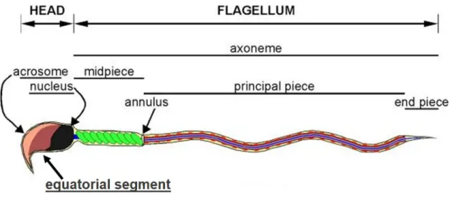
The sperm head is composed of two compartments: the nucleus containing a condensed haploid genome and a secretory apical structure named the acrosome [15-17]. The mid-piece is a segment that contains the sperm mitochondria, and is followed by the flagellum (Figure 2). Sperm surface biochemical structures differ from one compartment to the next, and this heterogeneous composition [18, 19] corresponds to the organelle distribution throughout the cell. In general, sperm cells have lost many somatic cell characteristics and do not activate gene expression; as such, both transcription and translation machinery are completely silenced [20]. In the absence of de novo protein synthesis, the endocytosis/exocytosis capacity of sperm is compromised and affects the sperm surface.
Figure 2 : Schematic drawing of a mouse sperm cell, illustrating the compartments (head
and flagellum) and their respective sub-compartments. Adapted from Buffone et al (2012) [21] with slight modifications.The molecular changes leading to surface specialization in the different segments of the sperm membrane, which take place during spermatogenesis, are yet to be fully understood. Once released into the seminiferous tubules, sperm encounter different milieus during their transit through the male and female reproductive tracts, on their voyage to meet the egg. These different milieus have varying molecular compositions, and thus sperm surface remodeling happens through interactions with different modulators encountered inside the various portions of the male and female tracts. The first major changes in the protein profile of the sperm surface occur within the epididymis [22-25]. At that time, sperm are coated by accessory fluids, which protect them against lipid raft disruptors and potential damage that may be encountered while they enter the female reproductive tract. Within the female tract, de-capacitation factors are removed from the sperm surface before they undergo de-capacitation [26-31]. Afterwards, sperm also interact with the oocyte cumulus cells, before penetrating the zona pellucida [32, 33], and pass through the perivitelline space. In this space, they interact with
components of its fluid before binding the oolemma [34, 35]. All these steps affect sperm surface remodeling, leading to either stimulatory or inhibitory effects on sperm fertilizing ability [36].
The majority of research groups believe that sperm acquire both their fertilizing ability and forward motion ability during epididymal transit [4, 37-40]. Sperm transit across the human epididymis for almost 6 days, while this transit can last 14 days in some species. To study the fertilizing ability of sperm, it is ideal to use cells that have completed their epididymal transit (and thus epididymal maturation). Following epididymal transit and prior to ejaculation, mature sperm are stored in the cauda epididymis. This sperm has acquired progressive motility, undergone an alteration of membrane charge as well as a reorganization of membrane proteins [4]. Although human in vitro fertilization (IVF) results show that epididymal sperm can be used for fertilization [41-43], fewer epididymal sperm can fertilize oocytes compared to ejaculated sperm that have passed through the epididymis completely. These results reveal that transit through all segments of the epididymis is required for optimal fertilizing capacity [40].
1.2.1. Protein traffic between sperm and the epididymal milieu
Epididymal proteins are secreted through both merocrine and apocrine pathways [44]. Merocrine pathways involve the rough endoplasmic reticulum and Golgi apparatus, which produce vesicles or vacuoles that shed into the lumen. Proteins lacking a signal peptide are secreted through apocrine pathways [45], in which proteins are delivered in small cytoplasmic protrusions at the apex of the epithelial cells. These highly hydrophobic proteins without signal peptides are likely associated with luminal hydrophobic complexes [46] and/or released as epididymosomes [47].
Epididymal proteins are implicated in the acquisition of sperm functions such as motility [48, 49], capacitation [50], ability to undergo the acrosome reaction [51], sperm–zona pellucida interaction [52] and fertilization [53-55]. Other epididymal proteins have been proposed to tag damaged or abnormal spermatozoa, as observed in sperm ubiquitination [56].
1.2.2. Changes to the sperm membrane during maturation
In addition to the acquisition of new proteins, spermatozoa undergo molecular changes on their surface during epididymal maturation. These alterations include the addition, removal, and/or modification of external sugars and lipids of the sperm plasma membrane. Glycoproteins and polysaccharides form an interface between the spermatozoa and its external environment, known as the glycocalyx [57, 58]. The sperm glycocalyx plays a role in recognizing the egg. Enzymes in the epididymal luminal fluid drive alterations of sperm surface glycoconjugates [10], leading to a net negative charge over the sperm surface [59, 60]. This negative surface charge increases as sperm advance through the epididymis; thus, cauda epididymal sperm are the mostly negatively charged [59]. This net negative surface charge is important for preventing the sperm from aggregating and from binding non-specifically to the female reproductive tract [61].
Epididymal sperm maturation is also associated with modifications of sperm membrane lipid composition. In most species, the cholesterol:phospholipid ratio decreases in the sperm membrane as they transit from the caput to the cauda epididymis [62]. This leads to changes in membrane fluidity that may be essential for later membrane events required for fertilization, such as the acrosome reaction and the ability to fuse with the egg oolemma.
1.2.3. Progressive movement competency
Cauda epididymal spermatozoa suspended in appropriate saline buffers display progressive motility, but testicular and caput epididymal spermatozoa do not [63]. However, if immature spermatozoa are demembranated in the presence of low concentrations of the non-ionic detergent Triton X-100, and then reactivated with ATP and cAMP, they become motile [63]. Following membrane changes induced by exposure to detergents, cholesterol efflux can trigger Ca2+ ion mobilization, with leads to the acquisition of motility and later to hyperactivation. Although the motility achieved by caput spermatozoa in these conditions is similar in intensity to that of cauda spermatozoa, they exhibit differences in flagellar bending [64].
2. The Binder of SPerm protein family
2.1. Background
After exiting the testes, sperm undergo maturation during epididymal transit. Upon ejaculation, sperm are mixed with fluids secreted by accessory glands (seminal vesicles, prostate and cowpers’ glands). This fluid is thought to play substantial role in sperm transit and fertilization events [65].
Amongst the proteins found in seminal plasma, the Binder of sperm (BSP) family of proteins is highly conserved in mammals. They were first discovered in the bovine species; the bovine BSP proteins were thus the first to be structurally characterized. Three BSP proteins (BSP1, BSP3 and BSP5) account for approximately 60% of the total protein fraction of bovine seminal plasma [66-69].
Following the first publication reporting possible epididymal BSP proteins in bovine [70], epididymal BSP genes and proteins were characterized in boar, ram, rabbit, mouse and human epididymides [71-74]. Now, genes and proteins from the BSP family have been identified in multiple mammalian species, where they originate from the same sources: the seminal vesicles and/or epididymis. BSP proteins bind to the sperm membrane through an interaction with choline phospholipids and are implicated in sperm fertilizing ability. This has been shown to be due to their role in modulating membrane remodeling, leading to capacitation. They are also involved in the formation of the oviductal sperm reservoir, the regulation of sperm cell volume and possibly in sperm-egg interaction [75].
Two BSP-homologous genes were identified in the mouse genome (Bsph1 and Bsph2). In mice and humans, the BSP proteins are exclusively expressed in the epididymis, whereas these proteins are expressed in the seminal vesicles and epididymis in ungulates. In general, phylogenetic analysis of the BSP superfamily designated different members into three subfamilies including BSP, BSPH1, and BSPH2. These studies strongly showed, BSP proteins secreted by seminal vesicles share commen characteristics through which are included in the BSP subfamily. However, the epididymal proteins are distributed in all three subfamilies. This distribution may justify the differences between the BSP proteins expressed in the epididymis and seminal vesicles. Proteins of the BSP subfamily expressed by seminal vesicles account for 1 to 50% of total seminal plasma proteins depending on the species (predominant in bovine) [76]. In ungulates, BSP proteins represent the major protein fraction in semen, whereas in murine and human, BSPs account for only 0.01% of total proteins in mouse and human semen [69, 77].
Nonetheless, functional analysis of BSPH1 proteins from mice and human revealed that these proteins, regardless of very low concentrations in seminal plasma, are already bound to sperm surface in the epididymis and function as efficiently as proteins expressed by seminal vesicles [78, 79].
As opposed to mouse and human, unbound free BSP proteins in bovine, owing to the high expression by seminal vesicles, act as acceptors and induce a first cholesterol efflux which proceeds till sperm pass through the cervical mucus. Then sperm bound BSP proteins can stabilize membrane until sperm reach the oviduct, where HDL interacts with BSP proteins. This induces a second cholesterol and phospholipid efflux, leading to the initiation of sperm capacitation [77].
2.2. Structure
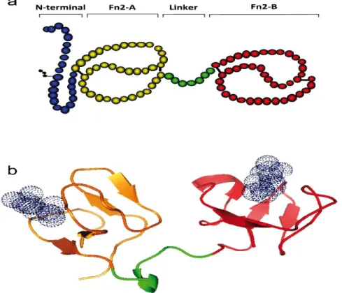
BSP proteins are mostly acidic and rather small, with molecular weights ranging from 15 to 30 kDa. Depending on the species, between one and six forms of the protein are expressed. They are composed of an N-terminal domain, two fibronectin type II domains (Fn2-A and Fn2-B) and a 7-amino acid linker that is shared among all BSP family members (Figure 3). Some BSPs also have a short, variable C-terminal domain [69]. The Fn2 domains are thought to be responsible for the functional roles of BSP proteins. Two disulfide bonds are found in each domain; thus, each BSP protein contains eight cysteine residues in its primary structure [80]. In the Fn2 domains, two anti-parallel β-sheets form a hydrophobic pocket [81-85]. Sequence analysis of BSPs revealed many conserved motifs, mostly around the cysteine residues [72]. As opposed to the conserved Fn2 domains, the N-terminal domain of BSP proteins varies in length from 15 to 71 amino acid residues, and exceptionally 380 amino acid residues in rabbit
BSP1 [86]. BSP proteins bind to the sperm surface; this enhances the binding of additional BSP proteins to those already bound to the sperm surface [87, 88].
Figure 3 :
a. Illustration of the structure of the bovine BSP1 protein. b. 3D structure of bovine BSP1; each Fn2 domain is composed of two anti-parallel β-sheets connected by a α-helix. Adapted from Plante et al. (2016) [75]2.3. Binding properties
Members of the BSP superfamily have many binding properties through which they interact with various ligands. Different biological functions of BSP proteins have been attributed to their interaction with known partners such as high-density lipoproteins (HDL), apolipoprotein A-I (apoA-I), phospholipids and glycosaminoglycans (GAGs)
[89-95]
. For example, the interaction of BSP proteins with milk proteins including casein micelles, α-lactalbumin and β-lactoglobulin have been shown to protect sperm during cryopreservation, thus justifying theaddition of milk to extender solutions
[96, 97]
. To find the interplay between binding properties and BSP’s funaction, structure essential for different interactions should be taken into consideration. A number of binding properties and the significance in vitro and in vivo application are further described below.2.3.1. Gelatin
Gelatin (a denatured derivative of type-I collagen) was the first macromolecule discovered to interact with BSP proteins [68], after which interaction with many other types of collagen (II– V) was also demonstrated [98]. Hydrophobic interactions are known to be responsible for gelatin binding by BSP proteins, and urea has been shown to disrupt this interaction [68, 99].
2.3.2. Glycosaminoglycans (GAGs)
The interaction of BSP proteins with GAGs such as heparin is due to the ionic charges. Findings in the bovine species showed that B-B-X-B and B-B-B-X-X-B (B represents a basic amino acid) consensus sequences play fundamental role in BSP binding to GAGs [89, 100, 101].
2.3.3. Phospholipids
The binding of BSP family proteins to phospholipids has been extensively studied in bovine. Most BSPs, including bovine BSP1 and BSP3 proteins, exhibit specific affinity for the phosphocholine motif, suggesting that this interaction is the main cause of BSP protein incorporation into phospholipid membranes [102]. In addition to binding to the phosphorylcholine moiety, bovine BSP5 also interacts with cardiolipin (exclusive phospholipid of mitochondrial membranes), phosphatidylethanolamine, phosphatidylserine, phosphatidylinositol and phosphatidic acid [90].
The Fn2 domains of BSPs are believed to form a binding site for the phosphocholine group [85, 90, 103, 104]. The crystal structure of BSP1 protein with bound phosphocholine showed that the two Fn2 binding sites were occupied [85, 105]. It has been shown that the phosphocholine binds to the Fn2 domains through cation–π interactions between the quaternary ammonium group of the choline group and a tryptophan residue of the BSP protein. In addition, a hydrogen bond between the hydroxyl group of the tyrosine residue and the phosphate group of the phospholipid increases affinity [85]. Hydrophobic interactions can contribute to the binding of BSP1 proteins to bilayer membranes. In this case, the hydrophobic pockets in the two Fn2 domains enable the protein to insert into a polar area of the bilayer membrane [106]. All of these features render the BSP1 protein capable of considerably binding to phospholipid bilayers. In fact, the saturation ratio for binding was suggested to be about 1 protein for 10–16 phospholipids [106-110].
2.3.4. Cholesterol
Previous studies indicated that cholesterol loaded into phospholipid bilayers can promote the interaction between BSPs and phospholipids [111, 112]. Though BSP proteins could not interact with immobilized cholesterol [90, 113, 114], they interacted with cholesterol residing in the phospholipid bilayer structure. While an exact mechanism for direct binding to cholesterol is not yet known, it might correspond to a cholesterol recognition domain in BSP proteins [115].
Beside binding to lipids, BSPs were also shown to modulate the efflux of phospholipids and cholesterol from the membrane bilayer of epididymal sperm [116, 117], from fibroblasts [113], and even from artificial membranes. However, Moreau and Manjunath (2000) showed
membrane and the amount of lipid efflux [118]. They also revealed that BSP-mediated cholesterol efflux is unidirectional and differs from the effect of ApoA-1 on cholesterol efflux. The different BSP proteins exhibit varying capacities to promote cholesterol efflux, with BSP1 showing higher capacity to promote efflux than BSP3 in bovine [117].
2.4. Physiological function of BSP proteins
Though BSP proteins are mostly implicated in sperm capacitation, they may also play a role in other steps of the fertilization process. BSPs have been hypothesized to function as chaperone-like molecules, cell death markers, mediators of sperm motility and viability, as well as to play a role in the oviductal sperm reservoir (will be discussed later) and in sperm-egg interaction.
2.4.1. BSPs effects on sperm in the male reproductive tract
Following spermiogenesis, sperm are subjected to maturation events while transiting through the epididymis, where they acquire fertilizing ability and are stored prior to ejaculation. During epididymal transit, maturation events occur; these molecular events allow sperm to acquire motility and lead to considerable modifications to the sperm plasma membrane. These changes render them competent to undergo capacitation and later fertilization events once they reach the female reproductive tract, as well as allow them to remain protected during the rest of their journey to the site of fertilization. These changes, which vary according to species, are mostly believed to result from interactions of the sperm with the epididymal milieu.
In the bovine species, soon after ejaculation sperm are mixed with accessory gland secretions, which contain BSP proteins. These BSP proteins bind (half time < 1 s) to the sperm membrane due to their affinity for choline phospholipids [66] and promote a first cholesterol
efflux from the sperm membrane (priming) [117]. Simultaneously, BSP proteins bound to sperm may also serve as decapacitating factors, allowing stabilization of the membrane lipid structure [109, 111] while entering the female genital tract, thus protecting ejaculated sperm by preventing premature capacitation [86].
Based on the proposed mechanism, BSP proteins saturate all binding sites on the sperm membrane upon ejaculation [118] and considerable amounts of BSPs remain free in seminal plasma. These free BSPs could act as cholesterol acceptors and contribute to the induction of the first cholesterol efflux, which occurs during sperm migration through the cervical mucus inside the female genital tract. BSP proteins bound to ejaculated sperm can also keep the membrane stabilized in order to prevent untimely capacitation (second cholesterol efflux). In mice, unlike ungulates, BSPH1 is expressed in the caput epididymis, so is thought to be implicated in the sperm membrane changes occurring during epididymal maturation [72]. BSPH1 binds to the sperm surface through affinity binding to the choline phospholipids and then stabilizes membrane lipid structures to avoid premature capacitation [66].
During epididymal transit, sperm not only undergo significant membrane remodeling, but also acquire sufficient motility before being stored in the cauda while awaiting ejaculation. Consistent with the sperm membrane localization of most proteins implicated in motility, BSPs also display localization over the sperm midpiece, suggesting a potential involvement in sperm motility [66, 73, 95, 119-121]. Sanchez-Luengo et al. showed that bovine BSP1 bound to the sperm midpiece may increase sperm motility through a mechanism involving the increased enzymatic activity of Ca2+-ATPase [122].
2.4.2. BSPs effects on sperm in a cryopreservation environment
There are also detrimental effects attributable to the long-term exposure of sperm to seminal plasma, and the removal of seminal plasma prior to cryopreservation can enable sperm to tolerate potential cryo-damages [123]. Some data from cryopreservation experiments suggest that the quantity of BSP1 proteins bound to the acrosome of ejaculated sperm is an indicator of the sensitivity of bovine sperm to cryostorage [124]. Results relating to the cryopreservation of sperm from farm animals suggest that BSP proteins influence sperm sensitivity to cold shock during cryopreservation and hence affect the fertilizing ability of sperm [116]. It has been shown that continuous exposure of sperm to BSP proteins found in seminal plasma may induce excessive lipid removal from the sperm membrane. During exposure to seminal plasma, sperm can lose 30–35 % of their phosphatidylcholine (PC) and cholesterol, which leads to cryosensitivity. To avoid cryo-damage, sperm extenders contain cryoprotectants such as egg yolk or milk along with conventional penetrating cryoprotectants [125]. Such a combination can sequestrate BSP proteins from seminal plasma and thus attenuate this excessive removal of lipids from the sperm membrane, leading to improved sperm cryosurvival [88].
2.4.3. BSPs effects on sperm in the female reproductive tract
2.4.3.1. The oviductal sperm reservoir
Ejaculated sperm form a reservoir within the female reproductive tract when they arrive in the oviduct and establish the contact with the oviductal epithelium. There, different mediators favor sperm storage in a reservoir in order to extend their window of viability, and for sperm energy to be saved so it can be directed towards capacitation and hyperactivation. This
biological phenomenon regulates the synchronization of sperm release with ovulation [126, 127]. The interaction of sperm with the oviductal epithelium to form a sperm reservoir prevents sperm from undergoing premature capacitation. This is possibly achieved via inhibition of a Ca++ efflux signaling pathway [128].
Upon ovulation, follicular fluid-driven mediators such as GAGs enter into the oviduct and interact with sperm-bound BSP proteins within the sperm reservoir. Following this interaction, sperm disperse from the reservoir and are then chemotactically attracted to the egg. During this time, sperm should ideally undergo capacitation, the acrosome reaction in order to be able to fertilize the oocyte [92, 129].
2.4.3.2. Sperm capacitation
Capacitation is a late maturation event that prepares sperm to fertilize an oocyte. It must occur in the proper time and place, within the female genital tract, in order to enable sperm to fertilize [130, 131]. Remodeling of the sperm membrane leads to changes that trigger capacitation. Although most of the proteins required for cell signaling, the acrosome reaction and zona binding are found within sperm membrane lipid raft domains along with cholesterol, gangliosides, and sphingolipids [132-134], non-raft membrane domains also contain proteins that contribute to the cholesterol/phospholipid efflux that takes place during capacitation. Moreover, certain studies have revealed that BSA-induced cholesterol removal during sperm capacitation is mediated by non-raft membrane fractions [135, 136]. In agreement with these findings, Plante and Manjunath (2015) showed that BSPH1 protein resides in non-raft membrane fractions and mediates the cholesterol/phospholipid efflux occurring during mouse sperm capacitation (Figure 4) [137].
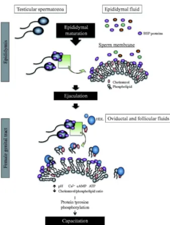
Once sperm reach the oviduct, they are stored in the oviductal sperm reservoir until ovulation. At ovulation, HDLs from oviductal and follicular fluids interact with the sperm membrane, triggering lipid efflux and cell signalling pathways that induce capacitation. Furthermore, HDL is thought to interact with BSPs to accept phospholipids and cholesterol from the destabilized membrane, thus disturbing the sperm membrane cholesterol/phospholipid ratio [137]. This remodeling results in, consecutively, an increase in intracellular pH, calcium influx, and an increase in cAMP. Together, these events activate a cascade of intracellular signalling pathways including the protein kinase A (PKA), protein kinase C (PKC), extracellular signal-regulated kinase (ERK) and phosphatidyl-inositol-3-kinase (PI3K)/Akt, pathways, which leads to protein tyrosine phosphorylation and capacitation [138-144].
In ungulates, heparin and heparin-like GAGs also play role in sperm capacitation, as do HDL and albumin. BSP proteins have been shown to bind heparin-like GAGs from the female reproductive tract and to potentiate GAG-induced capacitation. Upon ejaculation and entering the female genital tract, sperm-bound BSP proteins protect sperm from premature capacitation, until they come into contact with heparin-like GAGs near the site of fertilization. This exposure to heparin-like GAGs induces the release of BSPs from the sperm membrane. Following this release of decapacitation factors from the sperm membrane, calcium influx and protein tyrosine phosphorylation drive sperm into capacitation [145]. It seems that the interrupted interaction between BSPs and other proteins residing on the sperm membrane, such as calmodulin and phospholipase A2 (PLA2), promotes the activation of protein kinases, resulting in protein tyrosine phosphorylation and eventually the acrosome reaction.
Calmodulin is a modulator of calcium influx and is involved in capacitation and the acrosome reaction, both of which are Ca2+-dependent processes, at a point upstream of cAMP. Since
bovine BSP proteins bind to calmodulin, this interaction could also be implicated in capacitation through an effect on Ca2+ signaling [94, 146-149].
Although sperm membrane associated PLA2 is regulated by calmodulin, BSP proteins can also affect the regulation of this enzyme in a dose-dependent manner [150-152]. Following activation by zona-pellucida or progesterone, the lipolytic PLA2 enzyme can cleave phospholipids of the sperm membrane into free fatty acids such as arachidonic acid and lysophospholipids, which trigger acrosomal exocytosis and the acrosome reaction [153]. Interestingly, PLA2 activity showed a 100-fold increase in the presence of low concentrations of BSP proteins [151].
Figure 4 : Proposed mechanism for the involvement of BSPH1 in HDL-induced mouse
sperm capacitation. Unlike ungulates, there is no cholesterol efflux taking place inside the male reproductive tract. To be capacitated, sperm must undergo cholesterol efflux induced by the HDL content of follicular and/or oviduct fluids. Adapted from Plante and Manjunath2.4.3.3. Implication of BSP proteins in sperm-egg interaction
In bovine, zona glycoproteins that contain mannose-enriched saccharides possess binding affinity to BSP proteins [154]. It is thought that BSPs may mediate the interaction between sperm and the ZP. Interestingly, experiments conducted by Turmo et al. showed that the bovine BSPs have binding affinity for isolated ZP glycoproteins, and that anti-BSP antibodies could affect sperm-egg interaction [155]. Adding BSP1 to bovine IVF media can promote blastocyst rates in vitro, suggesting that BSP1 triggered the capacitation cascade in epididymal sperm, which eventually allowed them to undergo the acrosome reaction and successfully fertilize [156-158].
Moreover, Nixon et al. showed that the incubation of rabbit sperm with anti-BSP1 antibodies can inhibit fertilization rates in a dose-dependent manner [73]. On the other hand, immunolocalization results show that human and mouse capacitated sperm retain a certain amount of bound BSP proteins, suggesting a contribution of BSP proteins beyond capacitation [98, 159].
3. Sperm-egg interaction
Following capacitation, sperm are potentiated to fertilize an oocyte. Inside the oviduct, sperm are chemotactically attracted to the oocyte due to chemoattractant molecules secreted from cumulus cells (progesterone is the main if not the sole) [160]. Once a sperm reaches the oocyte, it must pass through the cumulus oophorous and then bind to the ZP. This interaction and the subsequent intracellular molecular signaling cascade induce acrosomal exocytosis and the acrosome reaction, allowing sperm to penetrate the zona pellucida. Finally, acrosome reacted sperm that have penetrated the ZP bind directly to the egg plasma membrane and the
sperm and egg membranes fuse together, resulting in pronucleus fusion and zygote formation [16]. Obviously, these steps could be compromised in IVF, while other stages can be bypassed and or facilitated.
3.1. Acrosome reaction
Upon binding to the ZP, sperm undergo the acrosome reaction, which is an irreversible step of fertilization. During the acrosome reaction, the outer membrane of the acrosome and the plasma membrane merge and form vesicles, shedding the enzymatic content of the acrosome [161]. This release of proteolytic enzymes from the acrosome allows sperm to penetrate across the loosened structures of the zona pellucida [162].
In vivo, acrosomal exocytosis and the acrosome reaction are triggered by a synergistic cooperation between zona pellucida glycoprotein 3 (ZP3) and progesterone released from cumulus cells. ZP3 binds to a specific receptor at the anterior segment of the sperm head, and progesterone also acts via a different sperm receptor [163]. Both ZP3 and progesterone activate intracellular signaling transduction pathways, causing an increased influx in Ca2+, which then stimulates PKC, adenylyl cyclase (AC) and PLA2 activation and production of cAMP [164]. The mobilization of internal calcium reserves, as well as Ca2+ influx through various channels can give rise such an increase in intracellular Ca2+ (Figure 5).
In vitro, the acrosome reaction can be induced in capacitated sperm in the presence of calcium ionophores, progesterone or soluble zona pellucida glycoproteins. Calcium ionophores are small molecules that can penetrate through the cytoplasmic membrane and co-transport calcium into intact cells [163].
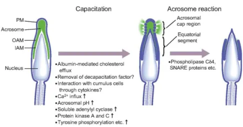
Figure 5 : Physiological and morphological changes occurring in sperm during capacitation
and acrosome reaction. These changes include the phosphorylation of various proteins, activation of PKA and PKC, removal of cholesterol from the membrane and elevation of intracellular Ca2+ levels. Adapted from Okabe (2013) [165].3.2. Sperm-egg fusion
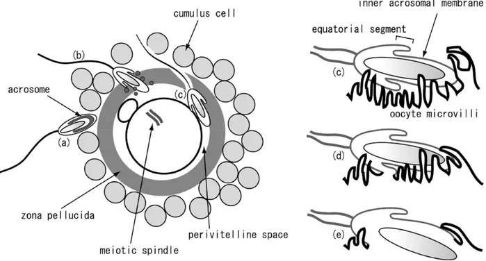
Fertilization should be terminated by the fusion between the sperm and egg membranes. Immediately before fusion, the head of the acrosome-reacted sperm is surrounded by oolemma microvilli (Figure 6) [166]. In sperm, fusion of its membrane to that of the oocyte begins from the equatorial segment. The sperm surface IZUMO protein forms a complex with the JUNO protein, which resides on the oocyte surface. The IZUMO1 sperm membrane protein and its oocyte counterpart JUNO have been shown to be modulating factors for sperm-egg interaction and fusion [167]. This complex allows the formation of fusion pores in the oolemma microvilli surrounding the sperm head and triggers the fusion of the two plasma membranes [162]. The oocyte CD9 protein and sperm IZUMO protein are essential for sperm to adhere to the oocyte. Other sperm proteins such as ADAM (A disintegrin and metalloproteinase) proteins, cysteine-rich secretory proteins (CRISP1 and CRISP2) also assist in oocyte adhesion
but are not as essential as IZUMO. Consequently, mice lacking IZUMO or CD9 are infertile, whereas the loss of ADAM proteins, CRISP1 or CRISP2 does not cause infertility [166, 168].
Figure 6 : Fertilization scenario. a. After passing through the layer of cumulus cells, a sperm
cell interacts with the zona pellucida b. once recognized, the sperm undergoes the acrosome reaction and penetrates the ZP. c. Sperm passes through the perivitelline space to make contact with oocyte microvilli. d. Sperm-egg fusion initiates from the merging of the sperm equatorial segment and the microvilli membrane. Adapted from Kaji and Kudo (2004) [166].Once the membranes are fused, the male and female pronuclei combine to form the nucleus of the zygote. To avoid penetration of the egg by multiple sperm (polyspermy), immediately after the fusion process a signal triggers a signalling cascade that mediates Ca2+ oscillation and causes changes in the rigidity of the ZP and the oocyte plasma membrane. Cortical granules residing beneath the oocyte membrane are activated and fuse with the egg cytoplasmic membrane, after which a hyaline layer (fertilization envelope) is formed. Membrane
biosynthesis, the formation of fertilization envelope and other parallel mechanisms render the fertilized egg membrane impermeable to additional sperm [162].
4. Thesis Objectives
Based on a WHO report, “One in every four couples in developed countries has been found to be affected by infertility”. Even though reproductive medicine is quickly expanding, the general burden of infertility and subfertility has not shown any decrease over the last 20 years. The molecular events underlying fertilization have recently begun to be addressed, which will help elucidate the reasons for certain unexplained difficulties in fertilization, occurring both naturally and in vitro. The mutual interaction between sperm and egg is a determining event, involving multiple players that can influence the fertilization process. Deficiencies in any of the requisite interactions may cause an infertility phenotype.
Numerous studies have proven that specific secretory proteins released into the epididymis associate to sperm passing through the organ. These proteins are believed to play a substantial role in mammalian sperm maturation, which includes capacitation and fertilization. There is reasonable evidence showing the participation of epididymal proteins in the events that prepare sperm for fertilization, such as the sperm surface changes that occur due to the lipid efflux from membrane raft domains. In addition, certain epididymal proteins may directly influence interaction between gametes. These bound proteins appear to mediate sperm-egg interplay at different levels. Binder of SPerm (BSP) proteins are exclusively secreted by the epididymis in mice and humans. Earlier studies on bovine BSP proteins suggested that they play a crucial role in fertilization. As such, further investigations were undertaken to functionally characterize the homologous murine and human proteins. In line with this and
previous findings, we hypothesized that BSP homologs may play a modulatory role in sperm-egg interaction, thereby affect fertilization. Therefore, in this project, we aimed to evaluate the potential role of Binder of SPerm homolog 1 in mouse sperm-egg interaction and fertilization. Specifically, the following objectives were pursued.
l) To examine the role of BSPH1 in sperm-egg interaction. We pre-incubated eggs retrieved from super-ovulated mice with rec-BSPH1 protein. Treated eggs were inseminated, and monitored for fertility.
2) To characterize potential BSPH1 binding sites on the egg surface. We performed immunodetection experiments with eggs that were pre-treated with rec-BSPH1 protein.
3) To further confirm the role of the BSPH1 protein in egg interaction. Native sperm-bound BSPH1 was immuno-neutralized with anti-BSPH1 antibodies and then this sperm was used to fertilize eggs.
Article
Role of Binder of SPerm homolog 1 (BSPH1) protein in mouse
sperm-egg interaction and fertilization
Hamed Heidari-Vala
1,2, Samin Sabouhi-Zarafshan
1,3, Bruno Prud’homme
1Abdullah Alnoman
1,2and Puttaswamy Manjunath
1,2,3,41Maisonneuve-Rosemont Hospital Research Centre, Montreal, Quebec, Canada, H1T 2M4
2Department of Pharmacology and Physiology, Faculty of Medicine, University of Montreal,
Montreal, Quebec, Canada, H3C 3J7
3Department of Biochemistry and Molecular Medicine, Faculty of Medicine, University of
Montreal, Montreal, Quebec, Canada, H3C 3J7
4Department of Medicine, Faculty of Medicine, University of Montreal, Montreal, Quebec,
Canada, H3C 3J7
4To whom correspondence should be addressed at: Centre de Recherche de l’Hôpital Maisonneuve-Rosemont, 5415 boulevard de l’Assomption, Montreal, Quebec, H1T 2M4
Canada. Tel.: +1 514 252 3562. Fax: +1 514 252 3569. E-mail: [email protected]
ABSTRACT
In mice, the Binder of Sperm Homolog 1 (BSPH1) protein is exclusively expressed in the epididymis, similarly to its human counterpart. Previous studies with mouse and human BSPH1 revealed that BSP proteins play a role in the membrane modification events that occur during sperm capacitation. In the current study, we investigated the role of mouse recombinant BSP homolog 1 (rec-BSPH1) in sperm-egg interaction. Mouse rec-BSPH1 was produced by transforming E. coli with a pET32a vector carrying BSPH1 cDNA and purified using immobilized metal (Ni2+) affinity chromatography. Mouse oocytes were co-incubated with different concentrations of rec-BSPH1 or control proteins and then inseminated with sperm. In order to establish whether rec-BSPH1 interfered with IVF of mouse oocytes, rec-BSPH1 binding to egg and sperm was first tested using an immunodetection assay. In separate experiments, sperm were immuno-neutralized by anti-rec-BSPH1 antibodies to indirectly verify the implication of BSPH1 in sperm-egg interaction and fertilization. The study revealed a dose-dependent inhibition of fertilization when oocytes were pre-incubated with rec-BSPH1. Moreover, sperm immuno-neutralization with anti-rec-BSPH1 antibodies led to dramatic motility changes, followed by compromised fertilization. In view of these results, we conclude that BSPH1 could be a marker of sperm fertility and thus an eventual target for male contraceptive development.
Keywords: Binder of SPerm (BSP) protein, sperm-egg interaction, in vitro fertilization, capacitation and mouse
INTRODUCTION
Sperm are specialized cells whose primary function is to carry paternal genetic material to an egg. As such, these haploid cells have lost many of the properties that are typically implicated in gene expression in somatic, diploid cells. Due to this unique state, the protein expression machinery of sperm is compromised upon spermiogenesis (Brewis & Gadella, 2010; Gur & Breitbart, 2006; Hosken & Hodgson, 2014; Pitnick, Hosken, & Birkhead, 2009). Once the post-testicular journey has started, sperm surface modifications begin to occur that prepare the sperm for fertilization (Skerget, Rosenow, Petritis, & Karr, 2015). The sperm surface is first modified during epididymal transit, and further membrane modifications occur during their passage through the female reproductive tract. These steps correspond to epididymal maturation and capacitation, respectively. Such membrane remodeling events are triggered by the epididymal secretome, accessory gland fluids as well as fluids secreted in the oviduct, and they collectively enable sperm to fertilize the oocyte (Cornwall, 2009; Sullivan & Mieusset, 2016).
Modifications to the sperm membrane during epididymal maturation mostly involve lipid and protein composition, and are localized to sperm membrane microdomains called lipid rafts. In addition to proteins localized in lipid rafts, other proteins on the sperm membrane can also participate in sperm surface remodeling (Boerke et al., 2013). From the latter, Binder of SPerm (BSP) proteins are secreted in the epididymis and interact with phosphocholine moieties of the sperm membrane, triggering cholesterol/phospholipid efflux. After these alterations in membrane fluidity, sperm capacitation occurs while sperm are exposed to capacitating factors in the female reproductive tract (Plante & Manjunath, 2015a; Plante, Thérien, & Manjunath, 2012). Besides their role in capacitation, BSP proteins have been
suggested to have a chaperone-like activity, to be a cell viability marker as well as to be involved in the formation of the oviductal sperm reservoir and, more recently, in sperm-egg interaction.
Ever since the BSP family of proteins was first discovered in bovine seminal plasma (Manjunath, 1984), several other mammals have also been shown to express homologs of these proteins with molecular weights varying from 15 to 30 kDa (Plante et al. 2016). BSP family members contain a variable N-terminal domain followed by two tandemly arranged fibronectin type II (Fn2) domains separated by a short linker sequence, and in some cases a short C-terminal domain (Manjunath, Lefebvre, Jois, Fan, & Wright, 2009). Sequencing analysis showed considerable conserved motifs throughout the members of the BSP family, most commonly in the region of the Fn2 domains (Fn2-A and Fn2-B). This particular structure is implicated in the biological functions of BSP proteins (reviewed in Manjunath, et al., 2009; Plante, Prud'homme, Fan, Lafleur, & Manjunath, 2016).
Besides ungulate BSP proteins, a role for murine Binder of SPerm Homolog 1 (BSPH1) in sperm membrane stabilization (decapacitation) and destabilization (capacitation) was recently proposed. BSP proteins are secreted by the epididymis and they bind to sperm membrane phospholipids, thereby preventing the free movement of lipids. This stabilization of the sperm membrane structure, or decapacitation, protects the membrane from untimely destabilization, in order to prevent premature capacitation. Upon ejaculation, sperm pass through the oviduct where high density lipoproteins (HDL) and glycosaminoglycans (GAGs) interact with sperm-bound BSP proteins, which destabilizes the membrane by removing BSPs as well as phospholipids and cholesterol (Plante & Manjunath, 2015b; Plante, et al., 2012).
The multistep capacitation process includes several biochemical changes, including an increase in ion permeability and intracellular pH, which initiates an intracellular molecular signaling cascade resulting in protein kinase activation and protein tyrosine phosphorylation and, ultimately, leads to the acrosome reaction. Only capacitated sperm can undergo the acrosome reaction (Beltran et al., 2016; Naz & Rajesh, 2004). In addition to undergoing their own maturation, sperm must additionally travel through the cumulus cells surrounding the oocyte, through the zona pellucida and the oolemma, where they come in contact with many different molecules, before fertilization takes place. Many sperm and egg molecules such as Izumo/Juno and ADAM proteins have been proposed to be involved in the interactions leading to eventual fertilization (Mou & Xie, 2017).
Until now, BSP proteins were best known for their interaction with molecules that modulate capacitation; however, a potential role for BSP proteins during the fertilization process has never been investigated. Our studies indicate that BSPH1 protein retains its localization on the sperm surface beyond capacitation, ascribing a possible role in sperm-egg interaction (Plante, Fan, & Manjunath, 2014; Plante & Manjunath, 2015a, 2015b). The aim of the present study was to elucidate the contribution of BSPH1 to sperm-egg interaction in the mouse model.
MATERIALS AND METHODS
Animals
Health-certified CD-1 (ICR) mice (male: ≥8 weeks; female: 5-8 weeks) were purchased from Charles River Laboratories (Kingston, NY, USA) and were accommodated in the animal care facility of the Maisonneuve-Rosemont Hospital research center. Animals had ad libitum access to filtered water and food, under temperature-controlled (22 ± 1°C), light-controlled (a light cycle of 12 h light: 12 h dark) and specific pathogen-free (SPF) environment at least 3-5 days before experimentation. Animal protocols were approved by the Maisonneuve-Rosemont Hospital ethics committee and experimental work was carried out according to the guidelines of the Canadian Council of Animal Care.
Recombinant protein expression and purification
The recombinant BSPH1 protein was expressed as previously described (Lefebvre, Boileau, & Manjunath, 2009; Plante, et al., 2012). In brief, E.coli OrigamiB (DE3) pLysS transformed with the pET32a vector carrying the His-tagged BSPH1 cDNA construct were grown under IPTG-induction to reach O.D600nm ~ 0.5-0.8. Cell suspensions were then centrifuged at 6,000 ×g for 10 min at 4°C, resuspended in 4X binding buffer (2 M NaCl, 80 mM Tris-HCl, 20 mM imidazole, pH 7.4) and stored at -20°C for downstream analysis. Once the cell suspension was thawed, an equal volume of bacterial protein extraction reagent (B-PER) was added, the mixture was incubated with rotation for 20 minutes and then cells were subjected to sonication with an amplitude of 50% (five cycles of 10 sec on ice and 1 min off) (Branson Digital 450 Sonifier). Urea was added to achieve a final concentration of 6 M, the solution was kept mixing for an hour, and cell extracts were finally centrifuged at 20,000 ×g for 30 min at 4°C.
Cell lysate supernatant was filtered through Acrodisc® (1.2 μm) and then subjected to chromatography on a Ni2+ charged His-Bind® resin (Novagen, MilliporeSigma, La Jolla, CA) column, where gradual on-column refolding was performed using a urea gradient (6 M to 0 M). The column was then washed with 20 mM imidazole to remove unspecific interactions and BSPH1 proteins were eluted using elution buffer (500 mM NaCl, 20 mM Tris-HCl and 200 mM imidazole, pH 7.4).The absorbance of eluted fractions was monitored at 280 nm, after which selected protein fractions from the chromatography were pooled and concentrated to 2 ml using an ultrafiltration technique. Protein was desalted on a gel filtration column (Sephadex G-25, 1.5 x 24 cm), which was pre-equilibrated with 0.05 mM ammonium bicarbonate. The desalted protein fractions were pooled, lyophilized and stored at 4°C.
Electrophoresis and Western blotting
SDS-PAGE was performed according to the Laemmli method (Laemmli, 1970) on 15% polyacrylamide gels using the Mini-Protean apparatus (Bio-Rad; Mississauga, ON, Canada). Two gels were run for each sample under the same conditions; the first was stained with Coomassie Brilliant Blue R-250 (Bio-Rad), and proteins in the other gel were transferred electrophoretically to Immobilon-P PVDF membranes (Bio-Rad) overnight in a cold room. Immunodetection was performed using either His-Probe monoclonal antibody (Santa Cruz Biotechnology, Santa Cruz, CA) or affinity-purified antibody against (His)6-tagged recombinant BSPH1 (anti-rec-BSPH1) (produced by Medimabs, Montréal, QC, Canada), at a concentration of 1:1000 and 1:500, respectively. Goat anti-mouse IgG-HRP (1:3000) or goat anti-rabbit IgG-HRP (1:10 000) were used as secondary antibodies. The bands were revealed using a chemiluminescence reagent (Perkin-Elmer, Boston, MA) and a Fuji LAS-3000 image
analyzer (Fujifilm; Stamford, CT). To verify protein loading, membranes were stained with a solution of 0.5% Amido Black 10B (Bio-Rad).
Superovulation and egg retrieval
In order to get MII oocytes, 5-8 week-old females were subjected to superovulation via intraperitoneal injection of 9 IU PMSG (≥1,000 IU/mg; Sigma-Aldrich) and 9 IU hCG (Sigma-Aldrich) 48 h apart. Mice were then euthanized 16 h post-hCG, and both oviducts were cut out and placed in M2 medium (Millipore). Oviducts were transferred to a new dish, where swollen ampullae were nicked with dissection forceps under a stereo microscope. The released cumulus-oocyte complexes (COCs) were then treated with 3-5 µl hyaluronidase (10 mg/ml) (from bovine testes; Sigma-Aldrich) in M2 medium for 2-3 min. Upon cumulus cell removal, eggs were washed sequentially 2 times with M2 medium and 3 times with HTF medium, which were previously equilibrated in 5% CO2 and humidified atmosphere at 37°C. To obtain zona-free oocytes, eggs were treated with Tyrode’s solution (Sigma-Aldrich) for 30 seconds to digest the zona pellucida (ZP).
Immunolocalization of rec-BSPH1 on oocytes
The immunolocalization method was adapted from published protocols (Ellerman et al., 2006; Herrero et al., 2005; Huang et al., 2017). Ten to 20 oocytes were treated with 300 μg/mL rec-BSPH1 protein in PBS containing 3% bovine serum albumin (BSA), or with PBS-3% BSA alone, for 1 h at room temperature. Eggs were then washed five times in PBS containing 3% BSA, fixed in 4% paraformaldehyde for 30 min at room temperature then washed another five times. Afterwards, eggs were incubated with either anti-rec-BSPH1 polyclonal antibody
(1:100), anti-BSPH1 15-mer (1:100), corresponding to the 15 C-terminal amino acids of BSPH1, monoclonal His-probe (1:50), or normal rabbit serum IgG (NRS-IgG) as a control (1:100) in PBS containing 3% BSA for 1 h at 37°C with 5% CO2. To remove excess antibodies, eggs were washed five times with PBS containing 3% BSA and then incubated with fluorescein isothiocyanate (FITC)-labeled goat rabbit IgG or FITC-labeled goat anti-mouse IgG at a dilution of 1:200, in PBS containing 3% BSA for 1 h at 37°C with 5% CO2. To counterstain the nucleus, eggs were soaked for 5-10 minutes at room temperature in 10 µg/ml DAPI in PBS containing 3% BSA and then washed five times before mounting with slow fade media on the stage of an Olympus IX73P1F (Olympus Corp., Japan) inverted microscope. Images were acquired using a monochromatic Peltier cooled 1.4 megapixel CCD Olympus XM10 (Olympus Corp., Japan) camera controlled by the Olympus cellSens software.
Sperm retrieval and preparation
CD-1 male mice (≥8 weeks) were sacrificed by cervical dislocation and then dissected to remove both the cauda epididymis and vas deferens while trying to avoid adipose tissue and vascular debris. Tissues were minced (four to five times) in 300 μl of equilibrated (5% CO2 and humidified atmosphere, 37°C) HTF medium and left at 37°C for 10 min to allow motile sperm to swim out. Sperm suspensions were gently removed and placed into tubes (45º-inclined) containing 1 ml equilibrated HTF at 37°C for 90 min, where motile sperm were allowed to swim up and capacitate. From the upper layer, 10 μl were removed to measure sperm concentration.
Immunolocalization of recombinant and native BSPH1 on sperm
This protocol was mostly adapted from Plante et al (Plante, et al., 2014; Plante, et al., 2012). 2×10⁶ sperm were incubated for 1 h with 30 μg/ml rec-BSPH1 or without protein (control), in PBS or HTF medium, respectively, for uncapacitated and capacitated sperm treatment. To induce the acrosome reaction, 200 μl of capacitated sperm were incubated for an additional 30 min with 5μM calcium ionophore A23187 (Sigma-Aldrich). Sperm suspensions were fixed with 4% paraformaldehyde for 30 min at room temperature and washed 3 times (2 min; 8,000 ×g) with PBS containing 1% BSA. 15 μl were smeared on Poly-L-lysine coated slides (FisherScientific, Ottawa, ON, Canada) and then allowed to dry. Sperm fixed on the poly-L-lysine slides were permeabilized for 10 minutes with PBS containing 0.1% TritonX-100 and 0.2% paraformaldehyde, washed three times with PBS, and incubated for 1 h at room temperature in PBS-1% BSA (or 30 min in PBS-3% BSA) to avoid nonspecific binding. Slides were then incubated for 1 h at room temperature with anti-rec-BSPH1 antibody (1:50) or NRS-IgG (1:50) in PBS containing 0.1% BSA. Excess antibodies were removed by washing three times with PBS containing 1% BSA, and then slides were incubated for 1 h at room temperature with FITC-conjugated goat anti-rabbit IgG (1:100) in PBS-0.1% BSA. After washing thoroughly, slides were counterstained with Propidium Iodide (Thermo Fisher Scientific) for 5-10 min. Finally, slides were washed, air dried and mounted with Permount (Fisher Chemical) or slowfade mounting media (Thermo Fisher Scientific). Observations were made using a fluorescence microscope and images were captured as mentioned previously. To immunolocalize native BSPH1 on sperm, an experiment was adapted from Herrero et al. and Plante et al. (Herrero, et al., 2005; Plante, et al., 2012). Approximately 30,000 capacitated or uncapacitated sperm were smeared on Poly-L-lysine slides and air-dried. Sperm were then
![Figure 1 : Different mechanisms of epididymal secretion. Adapted from Brewis and Gadella [15]](https://thumb-eu.123doks.com/thumbv2/123doknet/1962290.154/20.918.285.656.121.530/figure-different-mechanisms-epididymal-secretion-adapted-brewis-gadella.webp)