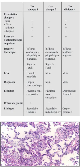
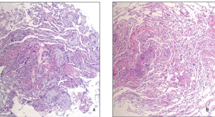
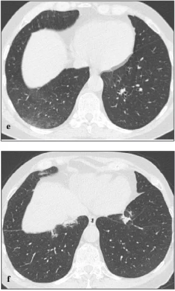
Pneumopathies organisées: à propos de 3 cas
Texte intégral
Figure


Documents relatifs
In [Silva et al., 2012] we ad- dressed concept drift detection using a different type of neu- ral network, namely Adaptive Resonance Theory (ART) net- works. Figure 2 illustrates
– Marco Antonio SOBREVILLA CABEZUDO, University of S˜ao Paulo, ICMC, BRAZIL. – F´elix Arturo ONCEVAY MARCOS, Pontificia Universidad Cat´olica del Per´ u,
Francisco Edgar Castillo Barrera , UASLP,Mexico. Technical Program
• Jorge Carlos VALVERDE REBAZA, University of S˜ao Paulo, ICMC, BRAZIL. • Alneu de Andrade LOPES, University of S˜ao Paulo,
• Ricardo Bastos CAVALCANTE PRUDENCIO, Federal University of Per- nambuco, Brazil. • Estevam HRUSCHKA, Federal University of S˜ao
Naomi Myake, Center for Research and Development of Higher Education, University of Tokyo Giulio Sandini, Robotics, Brain and Cognitive Sciences, Italian Institute of
• Juan Antonio LOSSIO VENTURA, Montpellier 2 University, LIRMM, Montpellier, France. • Hugo ALATRISTA SALAS, UMR TETIS,
Patient concerns: We report the case of a 38-year-old male smoker with COP presenting in the form of diffuse micronodules on computed tomography (CT) scan and describe the