Discrimination between Bifidobacterium Species from Human
and Animal Origin by PCR-Restriction Fragment
Length Polymorphism
V. DELCENSERIE, N. BECHOUX, T. LEONARD, B. CHINA, AND G. DAUBE
University of Liege, Faculty of Veterinary Medicine, Food Sciences Department, Sart Tilman, B43b 4000 Liege, Belgium
ABSTRACT
Bifidobacteria are normal intestinal flora in humans and animals. The genus Bifidobacterium includes 31 species of significant host specificity. Taking into account their properties, we proposed to use bifidobacteria as fecal contamination indicators. PCR-restriction fragment length polymorphism on the 16S rDNA gene was used to distinguish the different Bifidobacterium species. Sixty-four strains belonging to 13 different species were differentiated from animal or human origin using one or two restriction enzymes. Moreover, the primers used were specifics of the Bifidobacterium genus. Therefore, this method made it possible to determine both the presence of bifidobacteria in a sample and its origin of contamination.
Members of the genus Bifidobacterium are generally nonpathogenic, gram-positive, anaerobic, nonmotile, and non-spore-forming bacteria (20). They possess fructose-6-phosphate phosphoketolase and produce acetic acid and lactic acid as end products of glucose metabolism (18). The habitats of bifidobacteria range from sewage to the intestines of humans, animals, and honeybees (4, 21). Although 31 species are currently known in the genus Bifidobacterium, the taxonomy is sometimes confusing because of existing conflicts between genetic and phenotypic characteristics (11). A phylogenetic analysis of the genus Bifidobacterium and related genera based on 16S rDNA sequences has been done (17). The Bifidobacterium species form an independent phylogenic cluster that can be divided into two subclusters: subcluster 1, which is composed of most Bifidobacterium species, and subcluster 2, which consists of two species, B. denticolens and B. inopinatum. Both of these were isolated from human dental caries. Because of the limitations of 16S rDNA analysis in the phylogenetic study of closely related bacterial taxa, a more recent phylogenic analysis of the genus Bifidobacterium was based on Hsp60 gene sequences (9).
Bifldobacteria are well known for their probiotic effects (5) and are incorporated in many food products (23). Nevertheless, because bifldobacteria are also isolated from the feces of humans and many animals-such as ruminants, pigs, poultry, rodents, and rabbits (18)-they represent a potential indicator of the fecal contamination of food products. Moreover, these bacteria are strictly anaerobic (2), and, in the presence of oxygen, they stop growing but remain cultivable (2). It is therefore possible to estimate the initial amount of bifldobacteria present in the food product. Finally, another advantage of bifldobacteria over other fecal contamination indicators, such as Escherichia coli, is the host specificity of the Bifidobacterium species. For example, B. pseudolongum subsp. globosum, B. thermophilum, and B. bourn are present in ruminant feces (10); B. suis in swine; B. cuniculi and B. magnum in rabbit; B. pullorum in chicken; B. adolescentis, B. dentium, B. bifidum, B. breve, B. catenulatum, B. infantis, and B. longum are present in the human intestine (13). Thus, by determining the Bifidobacterium species, one can also determine the origin of the contamination (human and/or animal). The identification of Bifidobacterium by molecular methods was performed using several strategies: the 16S rDNA is a common target for the identification of bifidobacteria species by PCR. The amplicon is generated either by using species-specific primers (15, 22) or by using genus-species-specific primers followed by either sequencing (17) or hybridization with species-specific probes (12) or a restriction analysis (13, 14, 19, 22). This last method, PCR-restriction fragment length polymorphism (RFLP) based on 16S rDNA, although already described, was applied using a new set of enzymes, to distinguish human- and animalborne strains.
MATERIALS AND METHODS
Bacterial strains. The Bifidobacterium strains used in the present study are listed on Table 1. Before testing, the strains were withdrawn from frozen storage on Rosenow medium (Sanofi-Syn-thelabo, Marnes-la-Coquette, France) and grown on brain heart infusion (BHI; Bio-Rad, Marnes-la-Coquette, France) at 37°C for 48 to 72 h under anaerobic conditions, using an anaerobic cabinet (Ruskinn Technology Limited, Leeds, UK).
Other bacteria tested were five E. coli strains, one Salmonella Typhimurium strain, one Campylobacterjejuni strain, one Yersin- ia enterocolitica strain, one Listeria monocytogenes strain, five Clostridium prefringens strains, five enterobacteria strains, six Lactobacillus strains, five Staphylococcus strains, five Bacillus ce-reus strains, and five Pseudomonas strains.
Target DNA preparation. Bacterial cultures in BHI broth were centrifuged at 12,000 X g for 2 min using a bench-top centrifuge. The pellets were resuspendedin sterile, demineralized water, and the DNA was extracted using a Wizard Genomic DNA purification kit (Promega, Madison, Wis.). The purity and concentration of DNA were spectrophotometrically estimated (Ge-nequant-plus, Amersham Pharmacia Biotech, Amersham, UK). PCR. To detect Bifidobacterium, we used the following primers, chosen using Oligo 6 software (Molecular Biology Insights, Cascade, Colo.): 16S direct, 5'-aat agc tcc tgg aaa cgg gt-3' and 16S reverse, 5'-cgt aag ggg cat gat gat ct-3' (Eurogentec, Seraing, Belgium), which corresponds to a fragment of ~1,050 bp from 16S rDNA sequence. The PCR mix was 0.2 mM dNTPs, 400 pmoles 1-1 each primer, 4 U of Dap Goldstar (Eurogentec), 1X
buffer (20 mM Tris-HCl [pH 8.0], 100 mM KC1, 0.1 mM EDTA, 1 mM dithiothreitol, 50% glycerol, 0.5% Nonidet P-40, and 0.5% Tween 20; Eurogentec), 5% dimethylsulfoxide (1.1 kg/L; Merck Eurolab, Leuven, Belgium), 20 µl DNA (40 to 200 ng), and H2O in a total volume of 80 µl. The following cycles were applied:
95°C for 5 min, followed by 50 cycles of 95°C for 30 s, 58°C for 30 s, and 72°C for 1 min 30 s. Amplified PCR products were then analyzed by gel electrophoresis using 1% gel agarose (20 by 10 cm) and 1 X Tris, acetic acid, and EDTA buffer (TAE; Bio-Rad). The voltage used was 10 V CM-1for 1 h. After electrophoresis, gels were
stained with ethidium bromide (1 mg ml-1) and photographed under UV light (302 nm).
Restriction enzyme analysis. For restriction analysis of the PCR products, we used two enzymes, Alul (MBI Fermentas, St. Leon-Rot, Germany) and TaqI (Roche, Basel, Switzerland), according to the recommendations of the manufacturer Twenty mi-croliters of the PCR product was restricted by 1 U of enzyme in 1X buffer at 37°C for 3 h with AluI and at 65°C for 3 h with Taql in a total volume of 30 µl. After digestion, the products were analyzed by gel electrophoresis using 2.5% agarose gel in 1X TAE buffer under 10 V cm-1 constant voltage.
After electrophoresis, gels were stained with ethidium bromide (1 mg ml-1) and photographed under UV light
(302 nm). The size of the bands was determined using Kodak 1D software (Thermolabsystems, Brussels, Belgium).
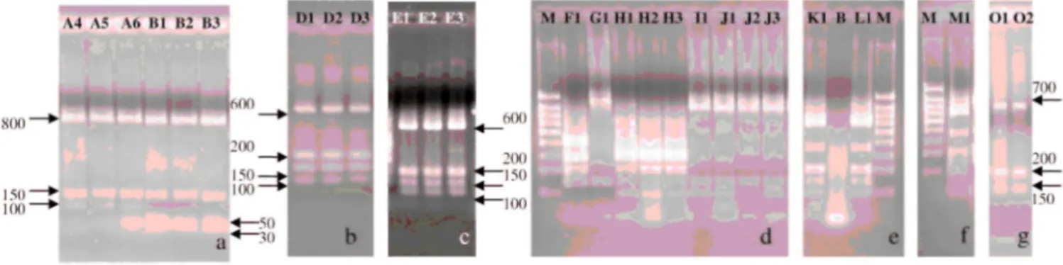
FIGURE 1. Alu/ restriction digest patterns. B, blanc; M, 5 µl molecular-weight marker
(1,000-800-700-600-500-400-300-200-100 bp). (a) Strains A4, A5, and A6 (B. animalis) show pattern I (800-150-100 bp), and strains B1, B2, and B3 (B. thermophilum) show pattern VII (800-150-50-30 bp). (b) Strains Dl, D2, and D3 (B.
pseudolongum subsp. globosum) show pattern II (600-200-150-100 bp). (c) Strains El, E2, and E3 (B. pseudolongum subsp. pseudolongum) show pattern II (600-200-150-100 bp). (d) Strain F1 (B. merycicum) shows pattern III (400-300-200-150 bp), strain G1 (B. ruminantium) shows pattern IV (900-150 bp), strains H1, H2, and H3 (B. minimum) show pattern V (310-290-200-150-100 bp), and strain II (B. cuniculi) and strains J1, J2, and J3 (B. adolescentis) show pattern I (800-150-100 bp). (e) Strains Kl (B. bifidum) and LI (B. breve) show pattern II (600-200-150-100 bp). (f) Strain Ml (B. dentium) shows pattern II (600-200-150-100 bp), and strains Ol, 02, and 03 (B. pseudocatenulatum) show pattern VI (700-200-150 bp).
TABLE 1. Bacterial strains used in the study International or INRA internal reference Laboratory's strains number Species Origin
ATCC 27672 A1 B. animalis Animal
Biavati PI6 A2 B. animalis Animal
Biavati F434 A3 B. animalis Animal
Biavati RA16 A4 B. animalis Animal
Biavati RA20 A5 B. animalis Animal
NCFB 2242T A6 B. animalis Animal
Cheval 1/1 Bl B. thermophilum Animal
Pigeon 1/2 B2 B. thermophilum Animal
LC294/2 B3 B. thermophilum Animal
LC103/1 B4 B. thermophilum Animal
B39/3 B5 B. thermophilum Animal
B105/5 B6 B. thermophilum Animal
LC288/1 B7 B. thermophilum Animal
Porc 3/1 B8 B. thermophilum Animal
B42/1 B9 B. thermophilum Animal
LCI 10/1 BIO B. thermophilum Animal
T585/1/2 Bll B. thermophilum Animal
Pigeon 1/1 B12 B. thermophilum Animal
Cheval 5/1 B13 B. thermophilum Animal
T528/4 B14 B. thermophilum Animal
B79/3 B15 B. thermophilum Animal
Internal Dl B. pseudolongum subsp. globosum Animal
Internal D2 B. pseudolongum subsp. globosum Animal
Biavati RU224 D3 B. pseudolongum subsp. globosum Animal
Internal El B. pseudolongum subsp. pseudolongum Animal
Biavati MB7 E2 B. pseudolongum subsp. pseudolongum Animal
LC287/2 E3 B. pseudolongum subsp. pseudolongum Animal
LC289/2 E4 B. pseudolongum subsp. pseudolongum Animal
LC302/2 E5 B. pseudolongum subsp. pseudolongum Animal
LC407/1/1 E6 B. pseudolongum subsp. pseudolongum Animal
B81/1 E7 B. pseudolongum subsp. pseudolongum Animal
LC312/2 E8 B. pseudolongum subsp. pseudolongum Animal
LC317/2 E9 B. pseudolongum subsp. pseudolongum Animal
LC405/3 E10 B. pseudolongum subsp. pseudolongum Animal
LC290/1 Ell B. pseudolongum subsp. pseudolongum Animal
LC464/3 E12 B. pseudolongum subsp. pseudolongum Animal
LC287/1 E13 B. pseudolongum subsp. pseudolongum Animal
LC305/2 E14 B. pseudolongum subsp. pseudolongum Animal
B81/1 E15 B. pseudolongum subsp. pseudolongum Animal
LC304/1 E16 B. pseudolongum subsp. pseudolongum Animal
LC323/1 E17 B. pseudolongum subsp. pseudolongum Animal
LC324/2 E18 B. pseudolongum subsp. pseudolongum Animal
LC340/3 E19 B. pseudolongum subsp. pseudolongum Animal
LC306/1 E20 B. pseudolongum subsp. pseudolongum Animal
Bs82 E21 B. pseudolongum subsp. pseudolongum Animal
RU915BT Fl B. merycicum Animal
RU687T Gl B. ruminantium Animal
DSM20102T HI B. minimum Animal
LC396/4 H2 B. minimum Animal
LC300/1 H3 B. minimum Animal
Internal 11 B. cuniculi Animal
Internal Jl B. adolescentis Human
Bs3 J2 B. adolescentis Human
CCUG18363T J3 B. adolescentis Human
5031e J5 B. adolescentis Human
DSM20082 Kl B. bifidum Human
NCFB2257T LI B. breve Human
CCUG18363T Ml B. dentium Human
DSM20438T Ol B. pseudocatenulatun Human
B2B 02 B. pseudocatenulatun Human
BS40 03 B. pseudocatenulatun Human
C19I 04 B. pseudocatenulatun Human
C20B 05 B. pseudocatenulatun Human
FIGURE 2. Taq/ restriction digest patterns. B, blanc; M, 5 µl molecular-weight marker
(1,000-800-700-600-500-400-300-200-100 bp). (a) Strains Al, A2, and A3 (B. animalis) show pattern VIII (470-330-250 bp). (b) Strain II (B. cuniculi) shows pattern VIII (470-330-250 bp), and strains Jl, J2, and J3 (B. adolescentis) show pattern IX (470-250-210-120 bp). (c) Strains Kl, L1, and Ml (B. bifidum, B. breve, and B. dentium) show pattern IX (470-250-210-120 bp), and strains Ol and 02 (B. pseudocatenulaturn) show pattern VIII (470-330-250 bp). (d) Strains Dl, D2, and D3 show pattern VIII (470-330-250 bp), and strains E18, E19, and E20 show pattern VIII (470-330-250 bp).
TABLE 2. Restriction patterns obtained with Alu/ and Taq/ on 13 Bifidobacterium species or subspecies
Species Tested strains Origin First digestion:
AluI pattern
Second digestion: TaqI pattern
B. animal is 6 Animal I VIII
B. cuniculi 1 Animal I VIII
B. adolescentis 5 Human I IX
B. pseudolongum subsp. globosum 3 Animal II VIII
B. pseudolongum subsp. pseudolongum 21 Animal II VIII
B. bifidum Human II IX
B. breve Human II IX
B. dentium Human II IX
B. merycicum Animal III
B. ruminantium Animal IV
B. minimum 3 Animal V
B. pseudocatenulatun 5 Human VI
B. thermophilum 15 Animal VII
RESULTS
The selected primers were used on DNA isolated from different bacteria: E. coli, Salmonella Typhimurium, C. je-juni, Y. enterocolitica, L. monocytogenes, C. perfringens, enterobacteria, Lactobacillus, Staphylococcus, B. cereus, Pseudomonas, and Bifidobacterium species. The results indicated that an ~l,050-bp product was only detected in Bifidobacterium species.
The obtained PCR products were first digested by Alul. Seven different patterns were observed: pattern I (800-150-100 bp) included B. animalis (A), B. cuniculi (I), and B. adolescentis (J) (Fig. 1a and 1d); pattern II (600-200-150-100 bp) included B. pseudolongum subsp. globosum (D), B. pseudolongum subsp. pseudolongum (E), B. bifidum (K), B. breve (L), and B. dentium (M) (Fig. 1b, 1c, 1e, and 1f); pattern III (400-300-200-150 bp) included B. mery-cicum (F) (Fig. 1d); pattern IV (900-150 bp) included B. ruminantium (G) (Fig. 1d); pattern V (310-290-200-150-100 bp) included B. minimum (H) (Fig. 1d); pattern VI (700-200-150 bp) included B. pseudocatenulatum (O) (Fig. 1f); and pattern VII (800-150-50-30) included B. thermophilum (B) (Fig. la). Because the aim of the present study was to be able to distinguish bifidobacteria of animal origin from those of human origin as well as to identify the species, we had to use a second restriction enzyme. Indeed, group I and group II were heterogeneous. In group I, B. adolescentis was of human origin, and B. animalis and B. cuniculi were of animal origin. We used then the enzyme TaqI to differentiate the different species. Species of human origin can be distinguished from species of animal origin (Fig. 2a and 2b). In group II, B. bifidum, B. dentium, and B. breve were of human origin, and B. pseudolongum subsp. globosum and B. pseudolongum subsp. pseudolongum were of animal origin. It was also possible to distinguish animal from human strains by using TaqI (Fig. 2c through 2e). The other groups were homogeneous: groups III and IV regrouped strains ofanimal origin, groups V and VII contained strains of animal origin, and group VI contained strains of human origin. Table 2 shows the different restriction patterns obtained with the two enzymes on 13 Bifidobacterium species or subspecies.
DISCUSSION
Food quality is a priority in our modern society, because the food chain is frequently the subject of periodic crises. Therefore, it is important to have objective tests to control the hygienic quality of food. Microbiological control is of major importance. It is important to be able to detect pathogens (16) and to detect fecal
contamination (6-8). Indeed, that kind of contamination is the signature of a hygiene problem during foodstuff preparation. Classically, the bacterium used for this purpose is E. coli (6). However, because this species is present both in animal and humans, it is difficult to determine the origin of the contamination. Moreover, these bacteria continue to multiply after contamination, giving a false idea of the initial contamination level. To bypass these drawbacks, the use of Bifidobacterium species was proposed (1). Indeed, the Bifidobacterium species have good host specificity; therefore, it seems possible to identify the origin of the contamination. Moreover,
bifidobacteria are strictly anaerobic, giving a better idea of the contamination level because there is no bacterial multiplication after the initial contamination in aerobic conditions. Beerens (1) compared E. coli and
Bifidobacterium as indicators of fecal contamination in meat and meat products by classical microbiological methods and found that there was good correlation in the presence or absence of both bacteria. Moreover, most of the isolated bifidobacteria were of animal origin. The sequence of the gene encoding 16S rRNA (16S rDNA) is a common taxonomic tool to identify bacterial species. This gene was sequenced in 21 Bifidobacterium species, but the deduced dendrogram did not allow discrimination of the species regarding their host (human or animal) (17). Therefore, identifying the Bifidobacterium species origin was challenging. To distinguish the Bifidobacterium species, we chose the PCR-RFLP technique (13, 14, 19, 22). In previous studies, the 16 rDNA amplicon was digested by HaeIII or TaqI, which allowed the distinction between human and cow strains (3), but this approach was incomplete, because only contamination by cows was investigated. The most extensive PCR-RFLP study on Bifidobacterium species using 16 rDNA was done by Ventura et al. (22). Sixteen species were investigated using the enzymes Sau3AI and BamHI. However, some species were not investigated (B.
merycicum, B. ruminan-tium, B. minimum, and B. thermophilum). In our study, PCR primers were selected in the 16S rDNA region, because this sequence is available for most Bifidobacterium species. The specificity of the chosen primers was good-the other bacteria tested were negative in amplification. The strategy used was as follows: the Alul enzyme was used to perform a first classification, which allowed us to obtain seven different groups. Because some of these groups contained both animal and human strains, the TaqI enzyme was then used to distinguish strains of human or animal origin. Finally, a fast and simple strategy to determine both the
presence and origin (human or animal) of bifidobacteria was obtained. The next step will be to apply this method to artificial and natural contaminated food samples. Among possible PCR templates, Hsp60 also seems to be a good candidate (9), because it was sequenced in most Bifidobacterium species and it is more variable between species than the 16S rDNA sequence. For the quantification of Bifidobacterium in feces by real-time PCR, the hsp60 gene can be used opposite to 16SrDNA, because it is present in a single copy.
ACKNOWLEDGMENTS
This work was supported European Community grant QLK1-CT-2000-00805. We thank Dr. Gavini for providing the Bifidobacterium strains.
REFERENCES
1. Beerens, H. 1998. Bifidobacteria as indicators of faecal contamination in meat and meat products: detection, determination of origin and comparison with Escherichia coli. Int. J. Food. Microbiol. 40: 203-207.
2. Beerens, H., F. Gavini, and C. Neut. 2000. Effect of exposure to air on 84 strains of Bifidobacteria. Anaerobe 6:65-67.
3. Bernhard, A. E., and K. G. Field. 2000. Identification of nonpoint sources of fecal pollution in coastal waters by using host-specific 16S ribosomal DNA genetic markers from fecal anaerobes. Appl. Environ. Microbiol. 66:1587-1594.
4. Biavati, B., V. Scardovi, and W. Moore. 1982. Electrophoretic pattern of proteins in the genus Bifidobacterium and proposal of four new species. Int. J. Syst. Bacterial. 32:358-373.
5. Brigidi, P., B. Vitali, E. Swennen, L. Altomare, M. Rossi, and D. Matteuzzi. 2000. Specific detection of Bifidobacterium strains in a pharmaceutical probiotic product and in human feces by polymerase chain reaction. Syst. Appl. Microbiol. 23:391-399.
6. Catsaras, M. V. 1991. Les indices de contamination fécale, p. 247-259. In C. M. Bourgeois and J. Y. Leveau (ed.), Techniques d’analyses et de contrôle dans les industries agro-alimentaires. Lavoisier-Tec et Doc, Paris.
7. Council of the European Communities. Council directive 92/46/EEC of 16 June 1992 laying down the health rules for the production and placing on the market of raw milk, heat-treated milk and milk-based products. Official Journal of the European Communities, Brussels.
8. Council of the European Communities. Council directive 94/65/EC of 14 December 1994 laying down the requirements for the production and placing on the market of minced meat and meat preparations. Official Journal of the European Communities, Brussels.
9. Jian, W., L. Zhu, and X. Dong. 2001. New approach to phylogenetic analysis of the genus Bifidobacterium based on partial hsp60 gene sequences. Int. J. Syst. Evol. Microbiol. 51:1633-1638.
10. Klein, G., A. Pack, C. Bonaparte, and G. Reuter. 1998. Taxonomy and physiology of probiotic lactic acid bacteria. Int. J. Food. Microbiol. 43:103-125.
11. Lauer, E., and O. Kandler. 1983. DNA-DNA homology, murein types and enzymes patterns in the type strain of the genus Bifidobacterium. Syst. Appl. Microbiol. 4:42-64.
12. Lynch, P. A., B. J. Gilpin, L. W. Sinton, and M. G. Savill. 2002. The detection of Bifidobacterium adolescentis by colony hybridization as an indicator of human faecal pollution. J. Appl. Microbiol. 92:526-533. 13. Mangin, I., Y. Bouhnik, N. Bisetti, and B. Descaris. 1999. Molecular monitoring of human intestinal Bifidobacterium strain diversity. Res. Microbiol. 150:343-350.
14. Mangin, I., N. Bourget, and B. Decaris. 1996. Ribosomal DNA polymorphism in the genus Bifidobacterium. Res. Microbiol. 147:183-192.
15. Matsuki, T, K. Watanabe, J. Fujimoto, Y Miyamoto, T. Takada, K. Matsumoto, H. Oyaizu, and R. Tanaka. 2002. Development of 16S rRNA-gene-targeted group-specific primers for the detection and identification of predominant bacteria in human feces. Appl. Environ. Microbiol. 68:5445-5451.
16. Mescle, J. E, and J. Zucca. 1996. Origine et comportement des mi-croorganismes des aliments, p. 4-11. In C. M. Bourgeois (ed.), Mi-crobiologie alimentaire: aspect microbiologique de la sécurité et de la qualité des aliments. Lavoisier Tec & Doc, Paris.
17. Miyake, T, K. Watanabe, T. Watanabe, and H. Oyaizu. 1998. Phy-logenic analysis of the genus Bifidobacterium and related genera based on 16S rDNA sequences. Microbiol. Immunol. 42:661-667.
18. Prevot, A.R., A. Turpin, and P. Kaiser. 1967. Sous-genre: Bifidobacterium, p. 1840-1878. In A. R. Prevot, A. Turpin, and P. Kaiser (ed.), Les bactéries anaérobies. Editions Dunod, Paris.
19. Roy, D., and S. Sirois. 2000. Molecular differentiation of Bifidobacterium species with amplified ribosomal DNA restriction analysis and alignment of short regions of the Idh gene. FEMS Microbiol. Lett. 191:17-24.
20. Scardovi, V. 1986. Genus Bifidobacterium, p. 1418-1434. In P. H. Sneath, N. S. Mair, M. E. Sharpe, and J. G. Holt (ed.), Bergey's manual of systematic bacteriology. Williams & Wilkins, Baltimore, Md.
21. Scardovi, V., and L. D. Trovatelli. 1969. New species of bifidobacteria from Apis mellifica L. and Apis indica F: a contribution to the taxonomy and biochemistry of the genus Bifidobacterium. Zent. Bak-teriol. Parasitenkd. Infektionkr. Hyg. Abt. 123:64-88.
22. Ventura, M., M. Elli, R. Reniero, and R. Zink. 2001. Molecular mi-crobial analysis of Bifidobacterium isolates from different environments by the species-species amplified ribosomal DNA restriction analysis (ARDRA). FEMS Microbiol. Ecol. 36:113-121.
23. Vinderola, C. G., N. Bailo, and J. A. Reinheimer. 2000. Survival of probiotic microflora in Argentinian yoghurts during refrigerated storage. Food Res. Int. 33:97-102.