GUIDELINES FOR CLINICAL PRACTICE
Donation after cardio-circulatory death liver transplantation
Hieu Le Dinh, Arnaud de Roover, Abdour Kaba, Séverine Lauwick, Jean Joris, Jean Delwaide, Pierre Honoré, Michel Meurisse, Olivier DetryHieu Le Dinh, Arnaud de Roover, Pierre Honoré, Michel Meurisse, Olivier Detry, Department of Abdominal Surgery and Transplantation, University Hospital of Liège, University of Liège, 4000 Liège, Belgium
Abdour Kaba, Séverine Lauwick, Jean Joris, Department of Anesthesia and Intensive Care Medicine, University Hospital of Liège, University of Liège, 4000 Liège, Belgium
Jean Delwaide, Department of Hepatology and Gastroenterol-ogy, University Hospital of Liège, University of Liège, 4000 Liège, Belgium
Author contributions: Le Dinh H performed the literature re-view and wrote the manuscript; de Roover A, Kaba A, Lauwick S, Joris J, Delwaide J, Honoré P and Meurisse M constitute the team involved in the care of the liver transplant patients and they reviewed and commented the manuscript; Detry O super-vised the review.
Correspondence to: Olivier Detry, Professor, Department of Abdominal Surgery and Transplantation, University Hospital of Liège, University of Liège, Sart Tilman B35, 4000 Liège, Belgium. oli.detry@chu.ulg.ac.be
Telephone: +32-4-3667645 Fax: +32-4-3667069 Received: December 9, 2011 Received: March 27, 2012 Accepted: March 29, 2012
Published online: September 7, 2012
Abstract
The renewed interest in donation after cardio-circu-latory death (DCD) started in the 1990s following the limited success of the transplant community to expand the donation after brain-death (DBD) organ supply and following the request of potential DCD families. Since then, DCD organ procurement and transplantation activities have rapidly expanded, particularly for non-vital organs, like kidneys. In liver transplantation (LT), DCD donors are a valuable organ source that helps to decrease the mortality rate on the waiting lists and to increase the availability of organs for transplantation despite a higher risk of early graft dysfunction, more frequent vascular and ischemia-type biliary lesions, higher rates of re-listing and re-transplantation and lower graft survival, which are obviously due to the
inevitable warm ischemia occurring during the declara-tion of death and organ retrieval process. Experimental strategies intervening in both donors and recipients at different phases of the transplantation process have focused on the attenuation of ischemia-reperfusion in-jury and already gained encouraging results, and some of them have found their way from pre-clinical success into clinical reality. The future of DCD-LT is promising. Concerted efforts should concentrate on the identifica-tion of suitable donors (probably Maastricht category Ⅲ DCD donors), better donor and recipient matching (high risk donors to low risk recipients), use of ad-vanced organ preservation techniques (oxygenated hy-pothermic machine perfusion, normothermic machine perfusion, venous systemic oxygen persufflation), and pharmacological modulation (probably a multi-factorial biologic modulation strategy) so that DCD liver al-lografts could be safely utilized and attain equivalent results as DBD-LT.
© 2012 Baishideng. All rights reserved.
Key words: Non-heart-beating donation; Complication;
Bile duct; Allocation; Ischemia; Ischemia-reperfusion injury; Liver disease
Peer reviewers: Bijan Eghtesad, Associate Professor, Department of General Surgery, Cleveland Clinic Foundation, 9500 Euclid Avenue, Cleveland, OH 44195, United States; Tokihiko Sawada, Associate Professor, Second Department of Surgery, Dokkyo University School of Medicine, Kitakobayashi 880, Mibu, Shimotsuga, Tochigi 321-0293, Japan; Philip Rosenthal, Professor, Pediatrics, UCSF, 500 parnassus Avenue, San Francisco, CA 94143-0136, United States
Le Dinh H, de Roover A, Kaba A, Lauwick S, Joris J, Delwaide J, Honoré P, Meurisse M, Detry O. Donation after cardio-circu-latory death liver transplantation. World J Gastroenterol 2012; 18(33): 4491-4506 Available from: URL: http://www.wjgnet. com/1007-9327/full/v18/i33/4491.htm DOI: http://dx.doi. org/10.3748/wjg.v18.i33.4491
© 2012 Baishideng. All rights reserved.
INTRODUCTION
The first human liver transplantations (LT) were per-formed from donation after cardio-circulatory death
(DCD) in the 1960s[1-4]. DCD-LT was nonetheless almost
universally abandoned in the following two decades, given the well-recognized Harvard brain-dead concept in 1968 and given the better results of LT originating from
donation after brain death (DBD)[5]. In 1983, LT was
ap-proved as a therapeutic modality for end-stage liver dis-eases after a long period considered as an experimental procedure. The renewed interest in DCD donors started in the 1990s following the limited success of the trans-plant community to expand the DBD organ supply and following the request of potential DCD families.
If DCD kidneys are increasingly accepted around the
world[6], the use of DCD livers remains limited in
expe-rienced transplant centers due to higher risks of primary graft dysfunction and biliary complications as well as a lack of a reliable viability testing prior to liver implanta-tion. However the number of DCD-LT increased rap-idly over the past decade. In the United States, 276 DCD liver transplants were performed in 2008 compared to only 23 cases in 1999, making up 5% of the deceased
donor (DD) liver transplants[7-10]. The same trend was
observed in the United Kingdom[11-13], Spain[14],
Neth-erlands[15] and Belgium[15,16]. Netherlands had the
high-est rate of DCD- over DD-LT in the world (22.5% in
2008)[15]. France has just initiated its DCD-LT program
since 2010[17]. In Japan, although DCD donors were the
essential DD source, its use was reserved mainly for
kidney, pancreas and islet transplantation[18]. Using a
mathematical model to analyze the potential impact of a DCD policy on LT programs, Chaib reported if 1%, 5% and 10% of deceased individuals became DCD donors, there would be 8%, 27% and 37% relative reductions in
the size of waiting list, respectively[19]. The use of DCD
livers could increase the supply of transplants by 53%[20].
Centers with active DCD-LT programs usually reported
4%-10% rates of LT from the DCD source[21]. The
po-tential impact of DCD use on the DBD availability is also a controversial issue. Controlled DCD programs might negatively influenced DBD activity in Belgium, Netherlands and United Kingdom while uncontrolled DCD donors seemed to be a clear additional source of
organs for transplantation in France and Spain[22].
Most countries use Maastricht-category-3 DCD do-nors for LT, except France and Spain, where categories 1 and 2 are exclusively used due to legal interdiction of discontinuation of therapy in irreversibly brain-injured
individuals[17,23,24]. German law prohibits any DCD organ
procurement and transplant activity. In Italy, death of a human being must be declared 20 min after cardiac arrest using continuous electrocardiography. The pro-cedure therefore will enable, at best, retrieval of only a few marginal kidneys and some tissues, and will not be
helpful for patients on LT waiting lists[25]. This article is
aimed at reviewing mono- and multi-centric DCD-LT outcomes, experimental strategies on animal models to
optimize the utilization of this donor source and its fu-ture development.
DIFFERENCES BETWEEN DCD AND DBD
DONORS PERTINENT TO LT OUTCOMES
Generally results of DCD-LT are inferior to those from DBD-LT with regard to both short-and long-term graft and patient survival as well as post-transplant morbid-ity. Expected DCD-LT outcomes could be explained by inherent differences between DCD and DBD donors in circumstances of death, warm ischemia time (WIT) and donor cause of death. Consequently, a different strategy of DCD use in terms of logistics of organ retrieval and preservation, allocation and recipient selection appears necessary to guarantee acceptable results. These differ-ences will be briefly discussed prior to considering re-sults of DCD-LT in detail.
Circumstances of death and consequent warm ischemia time
In DCD, donor death is diagnosed on the basis of irre-versible cessation of cardio-pulmonary function instead of conventional neurologic criteria. As a result, organs from DCD donors are subjected to a period of hypoten-sion, hypoxia and acirculation prior to organ procure-ment and this WIT adversely affects tissue viability and
graft function after transplantation[26]. An international
classification of DCD donors into 4 categories was first
proposed in 1995 and widely accepted up to now[27].
New DCD categories have been recently suggested in
Spain[28,29], Italy[30] and Belgium[31]. The length of WIT
varies greatly according to the type of DCD process. It is longest among uncontrolled category -1 and -2 (usually 90-120 min) and shorter among controlled category -3 and -4 DCD donors (usually 20-30 min). In brain death, issues related to donor warm ischemia are eliminated be-cause DBD donors have an effective natural organ per-fusion and a potentially well-preserved organ function and WIT is thus nearly equal to zero.
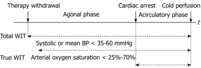
However, WIT is heterogeneously defined among
authors[32]. In the controlled DCD context, the
common-est definition is the time interval between withdrawal of both ventilator and cardiac support to start of cold
flushing of the organ[33,34]. This definition includes the
no-touch period and the time of death declaration and is proposed to have two phases (withdrawal and acircula-tory phases). Other authors used a blood pressure (BP) or oxygen saturation threshold below which would be defined as the beginning of true WIT (systolic or mean BP < 35-60 mmHg, oxygen saturation < 25%-70% or
unreadable)[35-42]. de Vera et al[34] did not use a BP
thresh-old to define the start of WIT because tissues are still hypoxic in a DCD donor who maintains a BP but has ceased to ventilate. It is unknown at what BP or oxygen saturation the liver parenchyma and biliary system
under-go irrecoverable injury[43]. The first international
suggested WIT should be calculated from the moment of cardiac arrest until the start of hypothermic
flush-out[44]. This definition may be useful for consistency but
is inaccurate at the cellular level. Hypoxia starts when the blood flow or oxygenation no longer meets cellular
metabolic needs[37]. The start of WIT may be chosen
prior to asystole, and the end of WIT may be at or after
aortic flushing[45]. Apparently a well-accepted definition
of donor organ ischemic times is needed to standardize nomenclature and allow accurate comparisons of
indi-vidual DCD studies[46,47] (Figure 1).
In transplant practice, WIT should be minimized as much as possible. For controlled DCD donors, the pos-sibility to predict whether a potential donor will or will not expire in a time frame consistent with donation is extremely important, because prolonged time to asystole, likely resulting in suboptimal organ perfusion, is a
com-mon reason for non procurement of DCD grafts[48,49].
Time between therapy withdrawal and cardiac arrest usu-ally does not exceed 1 h in most DCD donors. However, if a DCD donor has a period of relatively hemodynamic stability after life-support withdrawal, this period may be extended beyond 1 h without additional warm injury to
the organs[50]. Some authors emphasized during the
with-drawal phase, time to a systolic BP < 50 mmHg should
be < 30 min[20] and the hypotensive period (mean BP
< 50 mmHg) < 15 min[51]. Manara et al[52] proposed the
so-called functional WIT, which is measured from the donor’s systolic BP < 50 mmHg, the arterial oxygen sat-uration < 70%, or both, to the start of cold perfusion, should not exceed 30 min and may be limited to 20 min in suboptimal donors. Several factors have been identi-fied as predictors of rapid death following treatment withdrawal and include the DCD tool of University of
Wisconsin[53], donor Glasgow coma scale, inotropic use,
BP at treatment discontinuation, high FiO2 and mode of
ventilation[54,55]. Withdrawal of therapy is preferably
oc-curred in the operating room with a donor surgical team immediately available. Prior to cessation of the ventilator and organ perfusion support, the donor may be already prepared and draped, and the surgical instruments, pres-ervation solution and tubing are set up to facilitate rapid organ recovery. The super rapid recovery technique is
preferable and organs may be removed en bloc[39,50]. For
uncontrolled donors, in vivo organ preservation
tech-niques, like in-situ intravascular cooling using a double balloon and triple lumen catheter or hypo- and normo-thermic cardiopulmonary bypass with extracorporeal membrane oxygenation (ECMO), should be employed. With regard to the logistic organization, two frequently mentioned initiatives are the “Maastricht’s box” and the
“Madrid’s rapid identification and response system”[6].
Donor cause of death
DCD donors do not experience the brain dead process. Brain death provokes a cascade of changes in hemo-dynamics, hormones, and immune response, which negatively affect donor organ viability and transplant
outcomes[56,57]. Hemodynamic instability may have
del-eterious effects on liver function, although the liver has a high tolerance to marked hypotension and a large physi-ological reserve. Only a few histphysi-ological changes were observed in the liver both on light and electron
micro-scopic examination during the brain dead process[58,59].
The most important changes are the increased liver immunogenicity with subsequent increased host allo-responsiveness and the occurrence of apoptosis of
he-patocytes[60]. Clinical findings in livers from DBD donors
revealed significantly higher leukocyte infiltrates, up-regulation of adhesion molecules [intercellular adhesion molecule (ICAM), vascular cell adhesion molecule] and pro-inflammatory cytokines [interleukin-6 (IL-6), IL-10, IL-1β, interferon γ and tumor necrosis factor-alpha (TNF-α)], along with an increased expression of major
histo-compatibility complex-Ⅱ relative to livers from
living donors[61,62]. The peak time of cytokine
expres-sion and cell infiltration is during brain death and organ
procurement but not after reperfusion[61]. These changes
may amplify ischemia-reperfusion injury (IRI) during the transplant procedure and accelerate graft rejection after
transplant[63]. In reality, donor brain-death mechanisms
are quite varied and large differences may exist in the de-gree of impaired organ quality and transplant outcomes. The impact of donor cause of death on transplant out-comes has been recently confirmed in a United Network for Organ Sharing (UNOS) registry analysis, in which the cerebro-vascular accident presented as a predictor of worse graft survival across all organs relative to other
donor modes of death[64].
Uncontrolled DCD donors whose cause of death is usually other than neurologic do not undergo the pro-cess of brain death, while most controlled DCD donors have sustained irreversible cerebral injuries. As a result, organs from controlled DCD donors are likely to suffer more from the harmful immunologic and inflammatory effects of acute brain injury than those from
uncon-trolled DCD donors[65].
Allocation policy
It is reported that organs that have already subjected to warm ischemic injury have an increased susceptibility to
damage during cold storage[66]. The incidence of primary
non-function (PNF) was 2.5 times less in patients with
Therapy withdrawal Cardiac arrest Cold perfusion
Agonal phase Acirculatory phase
Total WIT True WIT
Systolic or mean BP < 35-60 mmHg Arterial oxygen saturation < 25%-70%
t
Figure 1 Different ways of warm ischemia time definition in the controlled donation after cardio-circulatory death setting (see text for more details).
True warm ischemia time (WIT) is also called complete or functional WIT; Total WIT is also called overall WIT; Agonal phase is also called withdrawal phase. BP: Blood pressure; t: Time.
cold ischemia time (CIT) ≤ 8 h vs those with CIT > 8 h
(5% vs 13%)[34]. The incidence of graft failure within 60 d
of transplantation was 10.8% if CIT < 8 h and substan-tially increased to 30.4% and 58.3% if CIT > 8 h and >
12 h, respectively[67]. Proper and rapid allocation of DCD
livers thus appears pivotal to minimize CIT. One-year graft survival of DCD livers shared regionally was less
good than those shared locally (67% vs 77%)[68] and the
relative risk of graft failure from nationally shared DCD livers was 31% higher than locally or regionally shared
ones[69]. Thus a policy to favor local use of DCD livers
seems reasonable[67,68]. However, parallel (backup) offers
should also be made to expedite organ placement[33]. The
exchange of DCD livers between transplant centers has been successfully done but requires a more efficient and rapid referral system due to a lower tolerance of these
al-lografts to cold storage[70].
Regarding recipient selection criteria, DCD livers could be routinely discussed and offered to all
recipi-ents on the waiting list[20,70,71] or selectively reserved to
uncomplicated cases to ensure short CIT (by avoiding cases with extensive history of abdominal surgery or
portal-vein thrombosis)[20,35]. An expected long
surgi-cal procedure exceeding 8 h of CIT, logistisurgi-cal reasons for an extended CIT, combined organ transplantation, recipients with high Model for End-Stage Liver Disease (MELD) scores or a large age difference between donors and recipients could all result in the refusal of a DCD
liver[51]. Patients with stable cholestatic liver disease or
re-transplantation were also excluded from DCD programs because of problems related to the quality of life in primary biliary cirrhosis and to the fear that pre-existent warm ischemic biliary damage could trigger the
recur-rence of primary sclerosing cholangitis[72]. Using DCD
livers in re-transplanted patients might increase the CIT associated with a difficult hepatectomy. Recently LaMat-tina has demonstrated the feasibility of simultaneous
liver and kidney (SLK) transplantation using DCD do-nors and shown short-term results comparable to those of SLK transplantation using DBD donors, making it a valid approach to safely expanding the donor organ pool
for patients with end-stage liver and kidney disease[73].
It is still controversial whether it is better to trans-plant such grafts into healthy or sicker recipients (i.e., according to the recipient liver disease severity). UNOS database reviews advocated utilizing DCD livers in
“low-risk” recipients[32,67,74]. de Vera et al[34] also observed better
graft survival when DCD livers were utilized in patients
with MELD scores ≤ 30, but simultaneously could
demonstrate that “sicker”, high-risk recipients (at MELD scores > 30 or on organ-perfusion support, like mechan-ic ventilation or hemodialysis) had a greater patient and graft survival benefit from the transplantation of DCD livers compared to patients who are not as critically ill. Risk classification for DCD donors and DCD-LT recipi-ents is summarized in Table 1. Other groups of patirecipi-ents that may have a true survival benefit from DCD-LT in-clude MELD “disadvantaged” patients (hepato-cellular carcinoma patients beyond the Milan criteria or who are listed in areas with long waiting times, patients with low MELD scores that do not adequately reflect their level
of illness and their critical need for a transplant)[34,72].
Studies about the effect of DCD liver grafts on hepatitis-C virus positive (HCV+) recipients transplant
outcomes were inconsistent. Nguyen et al[45] and recently
Hernandez-Alejandro et al[75] found a negative effect of
HCV on DCD livers, but a formal contraindication for the use of DCD liver allografts in HCV+ recipients was not justified except for older donors. In fact, while single-center series reported no significant difference in graft and patient survival rates of HCV+ recipients and graft loss from HCV recurrence between DCD and
DBD groups[20,34,76,77], as well as no deleterious effects of
DCD liver grafts on the disease progression (fibrosis)
Authors Donors Recipients
Mateo et al[74] Low risk Both WIT ≤ 30 min and CIT ≤ 10 h RCRR ≤ 1.5
High risk WIT > 30 min and/or CIT > 10 h RCRR > 1.5
Re-transplantation and/or On life-support and/or
A combination of ≥ 3 risk factors: Hospitalization or in an intensive care unit Serum creatinine > 2 mg/dL
On dialysis Age > 60 yr
Lee et al[32] Low risk Donors with no identified donor risk factors Recipients with no identified recipient risk factors
High risk Donors with at least one identified donor risk factor: Recipients with at least one identified recipient risk factor:
Donor age > 45 yr Previous transplantation
WIT > 15 min Life support at transplantation
CIT > 10 h
de Vera et al[34] Low risk - MELD scores ≤ 30
High risk - MELD scores > 30
On life support (mechanical ventilation, hemodialysis) Table 1 Risk classification for donation after cardio-circulatory death donors and donation after cardio-circulatory death-liver trans-plantations recipients
in comparison with DBD liver grafts in HCV+
recipi-ents[77], the most recent UNOS registry data showed
inferior graft survival but similar patient survival of HCV+ recipients with DCD donors compared to ones with DBD donors. Furthermore, DCD livers on HCV disease do not fare worse than DCD livers on non-HCV disease. DCD livers thus appeared to be important
source of LT for HCV patients[78]. Split livers from DCD
donors have also been reported in recent years with
ac-ceptable results[79,80].
TRANSPLANT OUTCOMES
Currently one-year patient survival after DBD-LT and to a certain extent after controlled DCD-LT is about 85%-90% in comparison to 60% in the early eighties and around 30% in the early d of LT and at 5 years post-transplant patient survival rate remains over 70%. Medi-cal progress over the past 40 years in the field of organ preservation, surgical techniques, immunosuppressive drugs, treatment of post-transplant complications and organ allocation has permitted DCD to become reality in the modern era. Although there are concerns about the quality of such organs, with evidence that a pro-longed WIT causes a raised incidence of PNF and bili-ary complications as well as suboptimal graft and patient survival when compared to DBD livers, DCD livers may be life-saving for those who would die waiting for
a DBD liver[68] and do increase the number of organs
available for LT. With careful donor/recipient selection and matching, minimization of ischemia and good post-operative care, acceptable results can be achieved. Es-sential results of most important publications in the last
decade in DCD-LT are presented in Tables 2-4.
Primary non-function
PNF is usually defined as unrecoverable hepato-cellular dysfunction leading to patient death or re-transplantation within the first week post-transplant after excluding other causes of graft failure such as vascular thrombosis,
bili-ary complications, rejection or recurrent disease[81-84].
Initial studies using uncontrolled DCD donors reported
a rate of PNF as high as 50%[85]. Currently only a few
transplant centers in the world (like Spain, France) used this kind of donors because of aforementioned reasons.
By using different in vivo organ preservation methods
to maintain DCD donors and by strictly applying
do-nor selection criteria, authors in Madrid[71], Barcelona[29]
and La Coruña[86-89] could obtain promising results from
Maastricht category Ⅰ and Ⅱ donors with a PNF rate
of 10%-25%. The discard rate nevertheless was high up
to 50%-75%[29,71]. In controlled DCD donors, the PNF
rates are 0% to 12%. Matched analysis[34,72] and registry
data[67,68] showed a higher rate of PNF in controlled DCD than DBD donors, although no difference was found in
most comparative studies[20,43,90,91] except one[92]. The
in-creased risk of PNF in DCD-LT recipients was also con-firmed in a recent meta-analysis (odds ratio = 3.6, 95%
CI: 2.1-6.4)[93]. Case-series reports of controlled DCD-LT
also had a rate of PNF between 0% and 10%[42,70,94-97].
PNF is the consequence of severe IRI with the initial period of warm ischemia playing a crucial role. Experi-mental evidence supported that donor WIT should be
less than 30 min to minimize PNF[98]. This warm ischemia
(WI) period increases graft susceptibility to damage dur-ing cold preservation and CIT was a main contributdur-ing
factor to PNF[34,67]; therefore, both periods of ischemia
must be kept to a minimum. Many laboratory tests have been developed both in animal models and in human to predict the probability of occurrence of PNF
post-transplant, but none is yet clinically efficient[99]. Recently
Dahaba proposed bispectral index monitoring as an early
intra-operative indicator of early graft dysfunction[100].
Biliary complications
Since the introduction of LT up to now, biliary compli-cations are always regarded as the “Achilles heel” and a major cause of morbidity and graft failure in patients
after LT[101]. The most common biliary complications
are bile leakage and bile duct stricture[102,103]. Strictures
involving the donor bile duct (> 1 cm above the biliary anastomosis) and requiring endoscopic or radiological dilatation/stenting or surgery in the face of a patent, non-stenotic hepatic artery was referred to as ischemic-type biliary lesions (ITBL), based on the radiologic resemblance of those occurring after hepatic artery
thrombosis (HAT)[51,91,103].
Abt et al[104] first mentioned the significantly higher
incidences of overall biliary complications as well as ITBL in DCD-LT recipients, the finding which was
later confirmed in both matched[34,72] and
compara-tive[20,43,51,89,91,104] studies except series of Fujita et al[105] and
Manzarbeitia et al[106]. The rates of overall biliary
com-plications and ITBL were 10.5%- 53% and 8.3%-38%, respectively in DCD-LT compared to 8.3%-22% and 0%-8%, respectively in DBD-LT. Especially Jimenez-Galanes reported only a 5% incidence of ITBL in their patients receiving livers from uncontrolled DCD donors
under normothermic ECMO[71]. A recent meta-analysis
revealed that DCD recipients had a 2.4 times increased odds of biliary complications (95% CI: 1.8-3.4) and a
10.8 times increased odds of ITBL (95% CI: 4.8-24.2) vs
DBD recipients. In average, biliary complications were present in 29% of DCD compared with 17% of DBD
recipients and ITBL in 16% of DCD vs 3% of DBD
re-cipients[93].
Furthermore DCD recipients who developed ITBL experienced a fairly rapid clinical deterioration, charac-terized by a relatively short mean time from transplant to first endoscopic retrograde percutaneous cholangio-pancreatography (ERCP), from first ERCP to relisting
and from relisting to re-transplantation (within 180 d)[36,69].
ITBL results in re-operation, multiple endoscopic and percutaneous biliary interventions, re-transplantation and even patient death with markedly increased medical
loss with ITBL for mation w as 3.02 (95% CI: 1.9-5.3) and graft sur vi val w as significantly decreased in patients with non-anastomotic strictures , compared to patients without it [89] . Up to 50% of all occur rences of
ITBL lead to death and/or re-transplantation
[108] . ITBL is usually a reflection of sev ere IRI in relation to various factors . In animal models , ir rev ersible biliar y tract damag e has been obser ved after 40 min of cardiac ar rest although he pato-cellular function could be preser ved [109] . Clinical obser vations sho w ed that total WIT > 30 min and chaotic donor ph ysiolog y before asytole ma y increase the risk of post-transplant biliar y stricture [33,110] . T he mec hanism could come from the stasis of blood and clot for mation in the peri-biliar y micro-circulation whose blood is solely supplied by the he patic ar ter y [96] . Many m ulti variate analysis recognized DCD liv er grafts as an inde pendent risk factor for the appearance of ITBL (RR = 47.1) [51,89] .
Authors study period
Transplant center Pu bl ic at io n year Pa ti en t nu m be r an d M aa st ri ch t ca te go ry WIT (min) CIT (min) Mean follow-up PNF % M aj or b ili ar y co m pl ic at io ns % ITBL % HAT % HAS % Rejection % R et ra ns pl an -tation % Graft survival % Patient survival % 1 yr 3 yr 5 yr 10 yr 1 yr 3 yr 5 yr 1 0 y r Casavilla et al [85] 1989-1993
Pittsburgh, United States
1995 6 DCD 4 37 10.6 h 50 1 16.6 83.3 17 67 6 DCDc 23.8 11 h 0 1 33.3 33.3 50 50 Otero et al [87] 1995-2000 Madrid, Spain 2003 20 DCD 2 10 647 > 2 yr 25 1 30 1 0 -27 25 80 80 40 DBD 8 405 3 1 8 1 34 5 55 83 Quintela et al [86] 1995-2004 Spain 2005 9 D C D 2 + 1 D C D 4 80 561 -10 -10 100 100 Suárez et al [89] 1994-2005 Spain 2008 27 DCD 2 13 635 > 3 mo 18 1 41.7 1 2 5. 0 1 3.6 17.4 49 1 62 471 DBD 7 3 1 16.8 1 2 .3 1 3.1 28.6 68 1 74 Fondevila et al [29] 2002-2006 Barcelona, Spain 2007 10 DCD 1 399 -10 10 - 10 -20 50 70 20 DBD 0 0 5 5 75 80 J im én ez -G al an es et al [7 1] 2006-2008 Madrid, Spain 2009 20 DCD 2 12 432 360 d 10 5 50 0 1 1 80 85.5 40 DBD 6 409 2.5 50 1 87.5 87.5 Pine et al [72] 2002-2008
St. James, London, United Kingdom
2009 39 DCDc 13.4 352 2.5 yr 5.1 33.3 1 2 0. 5 1 2.6 12 .8 1 20.5 7.6 1 79.5 1 6 3. 6 1 80 1 6 8. 2 1 39 DBD 593 6.6 yr 10.2 1 5.1 23.1 2.5 1 97.4 1 9 7. 4 1 100 1 1 1 de Vera et al [34] 1993-2007
Pittsburgh, United States
2009 141 DCDc 19.8 657 12 1 25 1 1 6. 3 1 66 18 1 69 1 56 1 44 1 79 70 57 282 DBD 636 3 1 13 1 < 1 1 7 1 82 1 73 1 63 1 85 76 64 Yamamoto et al [90] 1984-1988 Stoc kholm , Swed en 2010 24 DCDc 6 7 h > 20 yr 8.3 37.5 1 3 3. 3 1 70.8 54.2 - 37.5 37.5 61.9 42.9 42.9 16 DBD 6.8 h > 20 yr 18.7 6.3 1 56.2 43.8 37.5 37.5 63.6 54.5 Fujita et al [105] 1990-2006
Gainesville, Floria, United States
2007 24 DCDc 12.8 7.6 h 2.8 25 12.5 8.3 39.1 20.8 69.1 58.6 86.8 81.7 -1209 DBD 8.1 h 20.5 4.1 9.4 78.7 70.2 84 76 Foley et al [91] 1993-2002
Wisconsin, United States
2005 36 DCDc 17.8 8.2 h 3 yr 5.5 33 1 13.8 5.5 16.6 1 61 19.4 1 67 1 56 1 80 1 68 1 553 DBD 8.3 h 4.6 yr 1.3 10 1 8 11.8 5.4 1 56 7 1 86 1 80 1 91 1 84 1 Manzarbeitia et al [106] 1995-2002
Philadelphia, United States
2004 19 DCDc 19.6 574 1000 d 5.2 10.5 -- 10.5 89.5 311 DBD 557 13.8 8.7 84.2 Abt et al [104] 1996-2001
Pennsylvania, United States
2003 15 DCDc 20.4 366 819 d 6.7 33.3 1 2 6. 7 1 3.2 -20 6.6 1 71.8 71.8 79 79 221 DBD 464 690 d 3.6 9.5 1 2 .3 1 21.3 3.6 1 85.4 73.9 90.9 77.7 Nguyen et al [45] 1998-2001 M ay o C lin ic , F lo ri a, United States 2009 19 DCDc 16 6.7 h > 4.5 yr 5.3 26.3 10.5 0 5.3 5.3 1 15.8 73.7 68.4 63.2 89.5 89.5 89.5 234 ECD 7.1 h 4.7 22.6 33.3 1 8.5 1 85 78.6 72.3 214 SCD 7.5 h 1.7 15.9 33.2 1 19.6 1 84.3 80.7 76.5
Table 2 Results of donation after cardio-circulatory death-liver transplantations in single-center studies
DCDc: Controlled donors after cardiac death; DCD 1, DCD 2 and DCD 4: Maastricht category-1, category-2 and category-4 DCD donors; DBD: Donors after brain death; SCD: Standard criteria donors; ECD: Extended criteria donors; WIT: Warm ischemia time; CIT: Cold ischemia time; PNF: Primary non-function; ITBL: Ischemic-type biliary lesions; HAT: Early hepatic artery thrombosis; HAS: Early hepatic artery stenosis. Major symptomatic biliary complica
-tions include biliary leak, anastomotic and non-anastomotic stenosis.
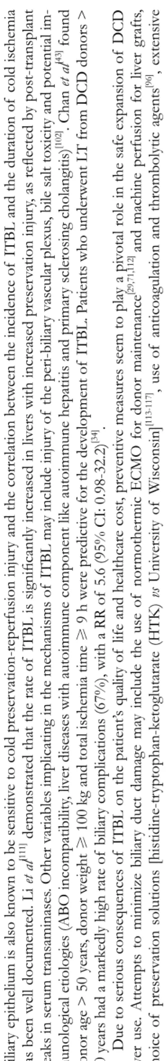
Biliar y epithelium is also kno wn to be sensiti ve to cold preser vation-re perfusion injur y and the cor relation betw een the incidence of ITBL and the duration of cold isc hemia has been w ell documented. Li et al [111] demonstrated that the rate of ITBL is significantly increased in liv ers with increased preser vation injur y, as reflected by post-transplant pe aks in se rum t ransaminase s. Ot he r v ariable s implic at ing in t he me chanisms of ITBL ma y inc lude injur y of the pe ri-biliar y v asc ular ple xus , bile salt to xic ity and pot ent ial im -m unological etiologies (ABO incompatibility , li ver diseases with autoimm une component lik e autoimm une he patitis and primar y sclerosing cholangitis) [102] . Chan et al [43] found donor ag e > 50 years , donor w eight ≥ 100 kg and total isc hemia time ≥ 9 h w ere predicti ve for the dev elopment of ITBL. P atients who underw ent LT from DCD donors >
60 years had a mark
edly high rate of
biliar y complications (67%), with a RR of 5.6 (95% CI: 0.98-32.2) [34] . D ue to s er io us c on se qu en ce s of IT BL o n th e pa tie nt ’s qu ality of life and healthcare cost, prev enti ve measures seem to pla y a pi votal role in the safe expansion of DCD liv er use . Attempts to minimize biliar y duct damag e ma y include the use of nor mother mic ECMO for donor maintenance [29,71,112] and mac hine perfusion for liv er grafts , choice of preser vation solutions [histidine-tr yptophan-k etoglutarate (HTK) vs Uni versity of Wisconsin] [113-117] , use of anticoagulation and thrombolytic ag ents [96] , extensi ve
Authors study period
Transplant center Publication year Pa tie nt n um be r an d M aa st ric ht c at eg or y WIT (min) CIT (min) Mean follow-up PNF % Major biliary complications % ITBL % HAT % HAS % Rejection % Retransplantation % Graft survival % Pa ti en t su rv iv al % 1 yr 3 yr 5 yr 1 yr 3 yr 5 yr Grewal et al [20] 1998-2006
Mayo Clinic, Floria,
United States 2009 108 DCDc 22.3 6.3 h 3.7 8.3 1 0.9 14.8 79.3 74.5 71 91.5 88.1 88.1 77.2 1328 DBD 7.1 h 1.4 1.9 1 1.7 9.3 81.6 74.7 69.1 87.3 81.1 Kaczmarek et al [41] 1999-2006 2007 11 DCDc 34.6 7.6 h > 14 m o 0 45.4 1 1 1 0 9.1 1 0 1 164 DBD 16.4 1 8.2 1 Dubbeld et al [51] 2001-2006 2010 55 DCDc 16.5 456 2 28 1 24 1 74.7 18 74 68 85 80 471 DBD 515 1.5 8.3 1 7.9 1 10.4 80.4 74.5 86.3 80.8 Chan et al [43] 2003-2006
Seattle, United States
2008 51 DCDc 3 yr 0 3.3 23.5 1 13.7 1 4.8 9.8 79 79 83 83 334 DBD 8.9 1 1.2 1 85 77 88 78 Skaro et al [36] 2003-2008
Chicago, United States
2009 32 DCDc 15.8 5.5 h 3 53 1 38 1 93 - 2 1 61 1 53 1 74 74 237 DBD 5.2 h 1 22 1 2 1 27 1 85 1 74 1 90 81 Jay et al [107] 2004-2008
Chicago, United States
2010 28 DCDc 16.5 5.7 h 1.8 yr 3.6 57.7 1 44 1 10.7 7.1 21.4 1 60 1 50 1 70 1 70 1 198 DBD 5.3 h 0.5 21 1 1.6 1 3 6.1 7.1 1 89 1 78 1 96 1 93 1 Dezza et al [92] 2003 -2006 Ghent, Belgium 2007 13 DCDc 10 6.16 h 163 d 8 1 23.1 1 31 1 54 1 62 1 98 DBD 9.14 h 603 d 1 1 12 1 79 1 86 1 Maheshwari et al [95] 1997-2006
Johns Hopkins Balti
-more, United States
20 DCDc 33 8.7 h 5 60 50 5 20 62 62 30 78 78 40 Muiesan et al [70] 2001-2004 London, United Kingdom 2005 31 DCDc 14.7 8.6 h 3.1 9.4 0 3.1 28.1 3.1 86.5 89.6 Abou Abbass et al [97] 2004-2008 United States 2010 26 DCDc 39 5.3 h 0 46 15.4 11.5 7.7 26.9 23 77 92 Detry et al [94] 2003-2007 2010 58 DCDc 25 451 3.4 38 32.7 3.4 3.4 13.8 72.4 48.8 83.3 66.9 H er na nd ez -A le ja nd ro et al [4 2] 2006-2007 London, United Kingdom 2010 10 DCDc 54.7 5.8 h 10 10 0 0 0 10 Hashimoto et al [96] 2005-2009 United States 2010 22 DCDc 21 422 4.5 27 9 0 0 9 81 81
-Table 3 Results of donation after cardio-circulatory death- liver transplantations in single-center studies
DCDc: Controlled donors after cardiac death; DBD: Donors after brain death; SCD: Standard criteria donors; ECD: Extended criteria donors; WIT: Warm ischemia time; CIT: Cold ischemia time; PNF: Primary non-function; ITBL: Ischemic-type biliary lesions; HAT: Early hepatic artery thrombosis; HAS: Early hepatic artery stenosis. Major symptomatic biliary complications include biliary leak, anastomotic and non-anastomotic stenosis. 1Numbers denote
Graft and patient survival
Graft survival is defined as the time from transplantation to either re-transplantation or patient death, with “early” and “late” graft failure occurring within and beyond 1
year post-transplant, respectively[34]. Few studies reported
experience with LT from uncontrolled DCD donors. Early results were poor with a PNF rate of 50% and
one-year graft survival rate of only 17%[85] leading to a
scarce usage of this donor category in the United States.
Subsequent series in Spain using advanced in vivo organ
preservation methods showed promising outcomes with one- and five-year graft survival rates of 50%-80% and 49%, and one- and five-year patient survival rates
of 70%-85.5% and 62%, respectively[29,71,89]. LT from
controlled DCD donors offered better results although they still appeared inferior to DBD-LT in matched
studies[34,72], registry data analysis[32,67-69,74,125] and in some
comparative studies[36,91,92]. One-, three-, five- and
ten-year graft survival rates were 54%-79.5%, 53%-74.5%, 37.5%-71% and 37.5%-44%, respectively. Patient surviv-al rates at corresponding time points were 61.9%-91.5%, 62.8%-89.5%, 42.9%-89.5% and 42.9%-57%, respective-ly. Transplant outcomes comparable to those obtained from DBD-LT have been sporadically reported in select centers through careful donor selection and optimization of CIT or through invasive techniques designed to
opti-mize recovery before declaration of death[20,43,51,104].
Significant risk factors for DCD liver graft loss have been identified by multivariate Cox regression technique in both single center studies and large data registry analy-sis[32,67-69,74,126,127]. Among donor risk factors, age > 50 years, total WIT > 30-35 min, CIT > 6 h, body weight > 100 kg and regional or national liver distribution had
dele-terious effects on graft survival[32,74,127]. There is a stepwise
increase in the relative risk of graft failure among donor
age, WIT and CIT[32,127]. Strong recipient determinants of
irrigation of the donor bile duct and pressure perfusion of the hepatic artery during organ retrieval and/or at
back table[113,118,119], early porto-caval shunt to reduce
por-tal hypertension in the recipient, choice of reperfusion
techniques (concomitant vs sequential reperfusion of
portal vein and hepatic artery)[120] and certainly the most
important thing is always minimizing warm and cold
ischemia period[121].
HAT and stenosis
HAT is a thrombo-embolic occlusion of the hepatic artery that can occur early or late after LT. Most authors used the first 30 d post-transplant as a time point to
distinguish between early and late HAT[122]. Early HAT
results in fulminant hepatic failure, bile duct necrosis and leaks, relapsing bacteremia and ultimately graft loss and recipient death. The frequencies of early HAT after DCD-LT varied from 0% to 16.6% and did not seem significantly higher than those after DBD-LT in most
studies[20,29,34,36,43,51,71,72,89,91,104,105] except Yamamoto et al[90]
(33.3% vs 0%). Risk factors for early HAT have been
well analyzed in a recent systemic review[123]. Few
de-tailed studies discussed late HAT.
The incidence of hepatic artery stenosis (HAS) was not consistently found higher in DCD than DBD grafts
(12.8%-16.6% vs 0%-5.4%)[72,91]. It is possible that
he-patic arteries are susceptible to WI during DCD organ retrieval, resulting in subsequent scar and stenosis. More-over the increased susceptibility of DCD livers to post-operative arterial ischemia might be responsible for more biliary strictures in DCD than DBD recipients with HAS
(83% vs 37%) as well as shorter time to the development
of biliary strictures after HAS in the DCD group[91].
In-adequate surgical technique, vascular trauma by clamps, graft rejection, recurrent hepatic disease might also play
a role in the mechanisms for HAS[72,124].
Authors and
study period Publication year Patient number and Maastricht category WIT (min) CIT (h) PNF (%) Retransplantation (%)
Graft survival % Patient survival %
1 yr 3 yr 5 yr 1 yr 3 yr 5 yr Abt et al[67] 1993-2001 2004 144 DCD 12.7 8.1 11.81 13.91 70.21 63.31 - 79.7 72.1 26 856 DBD 8.9 6.41 8.31 80.41 72.11 85 77.4 Mateo et al[74] 1996-2003 2006 367 DCD 15.6 8.3 - - 711 601 531 - - 33 111 DBD 8.4 801 721 651 Lee et al[32] 1996-2006 2006 874 DCD 15.4 7.9 - - 72.11 61.81 38.81 82.31 75.91 65.31 43 734 DBD 8.2 80.71 71.91 65.61 85.41 77.51 71.51 Doshi et al[68] 1998-2004 2007 345 DCD - 8.2 6.41 13.01 75 65 - 83 77 20 289 young-DBD 8.1 3.91 5.61 831 751 881 801 3604 old-DBD 8.2 5.31 76 64 83 73 Merion et al[125] 2000-2004 2006 472 DCD - 7.9 - - 70.11 60.51 - - - 23 598 DBD 8.1 831 751 Selck et al[69] 2002-2007 2008 855 DCD - - - 21.61 73.81 57.61 - - - 21 089 DBD 8.81 84.41 74.41 Mathur et al[127] 2001-2009 2010 1567 DCD 16.1 7.5 - 13.6 - - - 78 64.9
-Table 4 Results of donation after cardio-circulatory death-liver transplantations in United Network for Organ Sharing data base registry
DCD: Donors after cardiac death; DBD: Donors after brain death; WIT: Warm ischemia time; CIT: Cold ischemia time; PNF: Primary non-function. Major
symptomatic biliary complications include biliary leak, anastomotic and non-anastomotic stenosis. 1Numbers denote the statistically significant difference
graft failure include age > 55 years, history of previous transplantation, medical status at transplantation [intensive care unit (ICU) or non-ICU hospitalization, life support, dialysis, renal insufficiency], high MELD score (> 30) and
positive HCV serology[32,74,127]. In the DBD-LT model, it
has been shown that a single risk factor lessened outcome marginally, however, the additive effect of multiple risk
factors in a given donor-recipient pair were disastrous[83].
Grafts with ≥ 3 donor risk factors had significantly lower
1-year post-transplant survival than no or only 1 or 2 risk
factors (58.3% vs 72.6%, 69.2% and 73.9%, respectively).
No grafts with 4 risk factors survived within 1 year[128].
The relative risk of allograft failure from LT utilizing DCD donors was 31%-87% higher than LT utilizing
DBD donors[67-69,125,126]. Causes of early graft failure
in-cluded PNF, biliary complications, HAT and deaths from sepsis/multi-organ failure. Late graft failure was often secondary to chronic rejection and recipient death with a functioning graft.
Although DCD livers may not be as good as DBD ones with potential inferior transplant outcomes, there are subgroups of grafts and recipients that could give favorable results through appropriate graft and recipient matching. Low-risk DCD grafts which are transplanted in low-risk patients lead to comparable graft survival rates with DBD livers. Livers from DCD donors transplanted into high-risk recipients fared poorly independent of the
allograft quality[74]. Doshi et al[68] showed DCD liver grafts
were not inferior to DBD livers from older donors (≥ 60
years). Given the ever increasing demand for LT, DCD livers appear to be a reasonable alternative to increasing use of older or split livers and are a reasonable option
when death is imminent[68]. Even if graft or/and patient
survival is lower with a DCD liver, it is still better than dying because of turning down a DCD offer and con-tinuing to wait for a DBD liver on these d as the patient’ s choice is frequently not between marginal livers (includ-ing DCD) and standard livers but between marginal
liv-ers and no livliv-ers[105]. The benefit of earlier access to LT
provided by a DCD graft could outweigh the risks of
prolonged waiting for a standard graft[77].
Re-transplantation
DCD recipients more often require re-transplantation.
Re-spectively, 21.6%-42% vs 8.8%-16% of DCD and DBD
recipients were listed for re-transplantation[36,69]. The
re-transplantation rate ranged from 7.6% to 31% in DCD-LT
compared to 2.5%-12% in DBD-LT[20,29,34,36,51,67-69,71,72,91,92,106].
DCD livers exhibited a 2.1 times greater risk of graft fail-ure, a 2.5 times greater risk of re-listing, and a 3.2 times greater risk of re-transplantation compared with DBD
livers[36]. The majority of re-listing and re-transplantation
in the DCD group were a consequence of biliary com-plications, especially ischemic cholangiography, but not due to an increased incidence of PNF, HAT or technical
complications[36,69]. Particularly DCD livers had a
tempo-rally different failure pattern within the first year
post-transplant that limited access to re-post-transplantation[36,69]:
graft failure was more likely to occur within the first 180
d (18.1% vs 11.7%[67], 10.2% vs 2.5%[72] and 20.5% vs
11.5%[69] of DCD and DBD grafts failed within 60, 90
and 180 d, respectively); at transplantation, DCD re-cipients waited longer and received higher risk allografts; and more DCD recipients remained waiting for re-transplantation with fewer removed for death, clinical de-terioration, or improvement. Re-transplantation arouses controversy on medical, economic, and ethical grounds: patient and graft survival rates after a second LT are inferior to those after initial grafting, the procedure is more expensive and in the context of organ shortage, re-transplantation inevitably denies organs to first-time
recipients[129].
Utilization of DCD allografts for re-transplantation
was rare (2.5% of initial DCD vs 3.1% of initial DBD)
and outcomes from each group were comparable[69].
The general practice is to avoid re-transplantation with a
DCD graft[36]. The use of DCD donors in the setting of
re-transplantation resulted in an increased risk of
recipi-ent death (hazard ratio = 2.1, 95% CI: 1.2-3.6)[129].
Acute rejection
The acute rejection rate did not differ significantly be-tween DCD- and DBD-LT in most studies (1.9%-29%
vs 0.6%-34%)[20,72,87,89,104]. Foley et al[91] reported a one-year
rejection rate of 61% in the DCD group similar to that in the DBD group (56%). There were little data looking at the impact of DCD source on the risk of acute rejection.
EXPERIMENTAL STRATEGIES TO
IMPROVE DCD-LT OUTCOMES
The progressively increased DCD liver procurement to solve the shortage of DBD organs and to alleviate the waiting-list mortality has raised many challenges to the
transplant community and transplant policy makers[110].
A lot of experimental researches have been performed over the past decade, intervening in both donors and recipients at different phases of the transplantation pro-cess, at the aim of tackling some of these challenges and providing a deep insight into IRI mechanisms.
Donor pre-treatment
Various cyto-protective substances have been successful-ly administered into the donor prior to cardiac arrest for prevention of liver microcirculatory disturbance. Micro-circulatory disturbance was the main obstacle to success-ful DCD-LT, which was due to four major mechanisms: deterioration of sinusoidal endothelial cells (SEC) caused by activated Kupffer cells, sinusoidal narrowing caused by some vasoconstrictors and swollen hepatocytes,
leu-kocyte and platelet adhesion, and hyper-coagulability[130].
Up to now, only Heparin and phentolamin (an
anticoagu-lative substance and alpha-adrenergic antagonist) are
allowed in clinical DCD organ procurement[131], other
its powerful immunosuppression, enabled to prevent
liver normothermic IRI by multiple mechanisms[132].
Mil-rinone, a type 3 phosphodiesterase inhibitor, attenuated
graft injury caused by warm and cold ischemia via an
increase in intracellular cAMP levels, protection of SEC, relaxation of hepatic stellate cells, inhibition of platelet
aggregation and anti-inflammatory effect[133]. Lazaroids,
an antioxidant designed to inhibit iron-dependent lipid
peroxidation, ameliorated SEC viability via antioxidant
effects and membrane stabilization[134]. N-acetylcystein has
a direct effect on oxygen free radicals, but its usage had
no effect in both graft viability and lipid peroxidation[135].
Animal studies clearly showed the concept of phar-macological modulation of organ donors before pro-curement is feasible to improve the viability of marginal grafts. Nevertheless there are no definitive recommen-dations for the use of these drugs. Application of this method to clinical LT would require management of
some practical problems and possible ethical conflicts[136].
Organ preservation
Preservation of DCD livers by hypothermic machine perfusion (HMP) was shown superior to static cold
stor-age (SCS) in many experimental studies[137,138].
Nonetheless a putative drawback of HMP for liv-ers is to induce alterations at the vascular endothelial site, especially if HMP was performed for a long time
or under suboptimal conditions[139]. Endoplasmic stress
activation promoted cellular apoptosis via activation of
caspase-12[140,141]. The efficiency of HMP was markedly
increased by oxygenation of the perfusate[142]. The
con-cern that high oxygenation might favor the generation of oxygen free radicals, which in turn could impair tissue integrity, was not justified. Several investigators could demonstrate the beneficial effect of oxygenated HMP in reducing the liver expression of pro-inflammatory cyto-kines (TNF-α, IL-8), adhesion molecules (ICAM-1) and
major histocompatibility complex class Ⅱ antigens[143-145].
This benefit will likely be more pronounced in marginal
grafts such as elderly, steatotic and DCD livers[144].
Cyto-protective agents can be added into the machine perfu-sion (MP) solution to ameliorate the efficiency of HMP
organ preservation[146].
The positive effects of HMP on warm-ischemically pre-damaged livers were observed even after a brief pe-riod of MP, before (pre-conditioning) or after SCS
(post-conditioning)[143,147] and therefore it was not necessary to
require MP over a full preservation period and helped
avoid side-effects of HMP on vascular endothelium[141].
The use of HMP as the initial method for organ preser-vation followed by secondary SCS during transportation combined the advantage of aerobic resuscitation (i.e., restitution of cellular homeostasis) with an ease of SCS
for later surveillance and transportation[141]. Manekeller
showed a post-conditioning of 1 h after SCS can ame-liorate the viability of marginal livers. The extension or abbreviation of post-conditioning time seems to have no
further beneficial effects[148].
Schön et al[66] reported advantages of normothermic
machine perfusion (NMP) over SCS in pig DCD-LT models. Livers subjected to 1 h of WI and then cold-stored for 4-24 h were rendered completely nonviable while such livers under 4-24 h of oxygenated NMP
recovered function to a viable level[149]. Due to the
com-plexity of the logistics of clinical multi-organ recovery and of the NMP device, a period of cold preservation prior to warm perfusion of the liver is unescapable. A brief period of cold preservation (1 h) prior to NMP could maintain the synthetic and metabolic function but resulted in significant hepatocellular damage, sinusoidal
endothelial cell dysfunction and Kupffer cell injury[150].
Once this duration was prolonged to 4 h, NMP
com-pletely failed to resuscitate porcine livers[151].
Normother-mically perfusing DCD livers throughout the preserva-tion period not only replenished cellular substrate, ame-liorating the ischemic injury, but also provided a clear assessment of liver function and therefore could permit the use of severely injured organs with reassurance of
function[149,152].
Despite the aforementioned benefits of MP over SCS in liver preservation, only SCS is clinically approved up to now, MP is still in the pre-clinical stage and early
clinical studies[153]. Tojimbara showed the impact of
vis-cosity and temperature of initial flushing solutions on graft function. A low viscosity flushing solution was as-sociated with lower vascular resistance, whereas a warm flush solution prevented cold-induced vasospasm and therefore improved the washout effect of the
microcir-culation[154]. HTK solution possessing a low viscosity and
low potassium is more preferable in the DCD setting. The role of aeration of the cold-stored liver was also clarified. Oxygen provided either by surface diffusion (surface oxygenation) or intravascular diffusion (oxygen persufflation) helps improve the energy status of organs thus leading to earlier recovery. Surface oxygenation was not in use any more due to complicated technique,
limited efficiency and risk of oxygen intoxication[155].
Ve-nous systemic oxygen persufflation (VSOP) was shown to improve organ viability during hypothermic storage of the grafts and to be a feasible means for recondition-ing of warm-ischemically pre-injured livers from DCD
donors[155-158]. Experimentally even a short period of
VSOP prior to long-term preservation of the liver by SCS may be sufficient for a relevant improvement of
liv-er integrity upon repliv-erfusion[159]. Gaseous persufflation
with carbon monoxide was also tested in a DCD-LT rat
model with enhanced liver graft viability[160]. However no
additive or synergistic effect was noted when livers were persufflated with a mixture of gaseous oxygen and
car-bon monoxide[161].
Pharmaceutical interventions during SCS aimed at conditioning marginal organs also increasingly gained at-tention. Different cyto-protective drugs have been added into the flush and/or preservation solution, like
vaso-dilators (phentolamin, epoprosterol, dopamine)[162,163],
(strepto-kinase)[164], antioxydants (superoxide dismutase,
edara-vone)[165,166], antibiotics, hormones (glucagon, growth
factors)[167]. In the DCD setting, vasodilators,
anti-coag-ulants, thrombolytic agents and antibiotics seem particu-larly necessary because the organs tend to develop vaso-spasm, thrombus formation in the microcirculation and the risk of colonic bacterial contamination secondary to
translocation of organisms during the WI period[168,169].
Viability testing
Due to serious consequences of transplanting a DCD liver with potentially severe IRI (PNF, re-transplantation or even recipient death), it would be ideal if the viability of such livers could be predicted prior to rather than after transplantation. WIT is not always exactly known and thus cannot be a reliable parameter. Light microscopic examination of biopsy specimens was unable to
uniformly predict liver function after transplantation[170].
Monbaliu showed the extent of parenchyma vacuolation
predicted pig liver graft viability before LT[171]. Muiesan et
al[70] applied the mechanical digestion of liver biopsies with
collagenase and assessed the viability of hepatocytes by trypan blue exclusion method. However, the test was not helpful and the decision as to whether to use the liver was generally made on gross appearance, ease of perfusion,
degree of steatosis and donor characteristics[172].
Another approach is to evaluate the vascular resis-tance and enzyme release in the perfusate of HMP livers. Resistance index of the portal vein and hepatic artery
showed no utility[173]. Biomarkers of liver cell damage,
like transaminases, lactate dehydrogenase and liver fatty acid binding protein, correlated well with WI dura-tion and concomitant hepatocyte damage in pig
DCD-LT models[174]. Possible other parameters are the ATP
content and redox active iron status of the liver during HMP[175]. During NMP, the assessment of liver viability may be easier because the liver is in a normal metabolic state. Bile production was a good viability indicator be-sides the measurement of other liver functions
(detoxifi-cation, metabolism or synthesis)[176]. Recently Liu et al[177]
has tested the utility of magnetic resonance imaging and proton magnetic resonance spectroscopy to evaluate WI livers without success.
Recipient treatment
Pharmaceutical strategies aimed at modulating IRI mechanisms were also applied successfully in animal recipients and generally did not impose ethical problems as donor pre-treatment. Such protocols without donor pretreatment will be favorable in clinical application. Most studies tested a single agent for a specific target of the IRI process. A multi-factorial approach acting on different pathways of the IRI process have been advocated and
remarkably ameliorated transplant outcomes[162].
PERSPECTIVES
The future of DCD-LT is promising. Concerted efforts
should concentrate on the identification of suitable
donors (probably Maastricht category Ⅲ DCD donors),
better donor and recipient matching (high risk donors to low risk recipients), use of advanced organ preservation techniques (oxygenated HMP and NMP, VSOP), and pharmacological modulation (probably a multi-factorial biologic modulation strategy) so that liver procurement and transplantation from DCD donors could be widely expanded and attain equivalent results as DBD-LT.
REFERENCES
1 Starzl TE, Marchioro TL, Vonkaulla KN, Hermann G,
Brit-tain RS, Waddell WR. Homotransplantation of the liver in humans. Surg Gynecol Obstet 1963; 117: 659-676
2 Starzl TE, Groth CG, Brettschneider L, Penn I, Fulginiti VA,
Moon JB, Blanchard H, Martin AJ, Porter KA. Orthotopic homotransplantation of the human liver. Ann Surg 1968;
168: 392-415
3 Calne RY, Williams R. Liver transplantation in man. I.
Ob-servations on technique and organization in five cases. Br
Med J 1968; 4: 535-540
4 Calne RY. Early days of liver transplantation. Am J
Trans-plant 2008; 8: 1775-1778
5 Ascher HL. Liver transplantation--the first 25 years. West J
Med 1988; 149: 316-321
6 Ledinh H, Bonvoisin C, Weekers L, de Roover A, Honoré P,
Squifflet JP, Meurisse M, Detry O. Results of kidney trans-plantation from donors after cardiac death. Transplant Proc 2010; 42: 2407-2414
7 Thuluvath PJ, Guidinger MK, Fung JJ, Johnson LB, Rayhill
SC, Pelletier SJ. Liver transplantation in the United States, 1999-2008. Am J Transplant 2010; 10: 1003-1019
8 Klein AS, Messersmith EE, Ratner LE, Kochik R, Baliga
PK, Ojo AO. Organ donation and utilization in the United States, 1999-2008. Am J Transplant 2010; 10: 973-986
9 Kauffman HM, Rosendale JD, Taranto SE, McBride MA,
Marks WH. Non–heart-beating donors (then) and donation after cardiac death (now). Transplant Rev 2007; 21: 237-248 10 Wynn JJ, Alexander CE. Increasing organ donation and
transplantation: the U.S. experience over the past decade.
Transpl Int 2011; 24: 324-332
11 UK Transplant archive activity reports. Available from: URL: http: //www.uktransplant.org.uk/ukt/statistics/transplant _activity_report/archive_activity_reports/pdf/ukt/transpla nt_activity_uk_2008-2009.pdf. Access date: 15/1/2011 12 Devey L, Wigmore SJ. Non-heart-beating organ donation.
Br J Surg 2009; 96: 833-835
13 Roberts KJ, Bramhall S, Mayer D, Muiesan P. Uncontrolled organ donation following prehospital cardiac arrest: a potential solution to the shortage of organ donors in the United Kingdom? Transpl Int 2011; 24: 477-481
14 Newsletter Transplant. Available from: URL: http: //www. edqm.eu/medias/fichiers/Newsletter_Transplant_Vol_14_ No_1_Sept_2009.pdf. Access date: 15/1/2011
15 Euro Transplant annual reports. Available from: URL: http: //www.eurotransplant.org/files/annual_report/ar_2008. pdf. Access date: 15/1/2011
16 Squifflet JP. Why did it take so long to start a non-heart-beating donor program in Belgium? Acta Chir Belg 2006; 106: 485-488
17 Le rapport annuel de l’Agence de BioMedecine. Available from: URL: http: //www.agence-biomedecine.fr/uploads/ document/RA_Biomed_2009-B.pdf. Access date: 15/1/2011 18 Noguchi H, Hatanaka N, Matsumoto S. Renal and islet
transplantation from non-heart-beating donors in Japan. In: Talbot D, D’Alessandro AM, editors. Organ donation and