Prise en charge des syndromes de Fanconi secondaires à une immunoglobuline monoclonale
Texte intégral
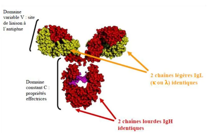
Figure


Documents relatifs
Il est aussi important de pouvoir évaluer l’évolu- tion clonale de ces tumeurs au cours du temps, puisque certains clones peuvent régresser et d’autres persister au cours
The first patient developed cast nephropathy in the graft, leading to ESRD, with no recovery of graft function despite BD treatment FIGURE 2: Survival of MM/SMM patients and
Demographic and clinical baseline characteristics of patients with monoclonal gammopathy MG and controls and a comparison of features in cases with or without chronic pruritus
By combining detailed information on the displacement structure with global measurements of pressure, saturation and the capillary number Ca, we obtain a scaling relation
FigUre 3 | Sialylation of IgGs as assessed by mass spectrometry: Purified pc IgGs from six healthy volunteers (HV) and pc IgGs and mc IgGs from eight patients [four
Recently updated analysis of the large southeastern Minnesota cohort [5] concerning 1384 patients with MGUS with a median follow-up of 34 years showed among patients with non-IgM
The natural history of MM has been clarified: MM occurs from obligatory precursor stages, monoclonal gammopathy of undetermined significance (MGUS), and smoldering MM (SMM), through
To model clusters of clonal and subclonal point mutations, allowing inference of the number of subclones and the fraction of cells within each subclone, we used a 2-D Bayesian
