PROPROTEIN CONVERTASE SUBTILISIN/KEXIN TYPE 9 IN HUMAN DISEASE Par Zuhier Awan Département de Biochimie Faculté de Médecine Février 2011
Mémoire présenté à la Faculté des études supérieures en vue de l’obtention du grade de Maître ès sciences (M.Sc.) en biochimie
Faculté des études supérieures
Ce mémoire est intitulé:
PROPROTEIN CONVERTASE SUBTILISIN/KEXIN TYPE 9 IN HUMAN DISEASE
Présenté par: Zuhier Awan
a été évalué par un jury composé des personnes suivantes :
. Dre Martine Raymond
président-rapporteur
. Dr Nabil Georges Seidah
directeur de recherche
. Dr Robert Scott Kiss
membre du jury
RÉSUMÉ
Les maladies cardiovasculaires (MCV) demeurent au tournant de ce siècle la principale cause de mortalité dans le monde. Parmi les facteurs de risque, l’hypercholestérolémie et l’obésité abdominale sont directement liées au développement précoce de l’athérosclérose. L’hypercholestérolémie familiale, communément associée à une déficience des récepteurs des lipoprotéines de basse densité (LDLR), est connue comme cause de maladie précoce d’athérosclérose et de calcification aortique chez l’humain. La subtilisine convertase proprotéine/kexine du type 9 (PCSK9), membre de la famille des proprotéines convertases, est trouvée indirectement associée aux MCV par son implication dans la dégradation du LDLR. Chez l'humain, des mutations du gène PCSK9 conduisent soit à une hypercholestérolémie familiale, soit à une hypocholestérolémie, selon que la mutation entraîne un gain ou une perte de fonction, respectivement. Il demeure incertain si les individus porteurs de mutations causant un gain de fonction de la PCSK9 développeront une calcification aortique ou si des mutations entraînant une perte de fonction provoqueront une obésité abdominale. Dans cette étude, nous avons examiné :
1) l’effet d’une surexpression de PCSK9 dans le foie de souris sur la calcification aortique ; 2) les
conséquences d’une déficience en PCSK9 (Pcsk9 KO), mimant une inhibition pharmacologique, sur le tissu graisseux.
Nous avons utilisé un modèle de souris transgénique (Tg) surexprimant le cDNA de PCSK9 de souris dans les hépatocytes de souris et démontrons par tomographie calculée qu’une calcification survient de façon moins étendue chez les souris PCSK9 Tg que chez les souris déficientes en LDLR. Alors que le PCSK9 Tg et la déficience en LDLR causaient tous deux une hypercholestérolémie familiale, les niveaux seuls de cholestérol circulant ne parvenaient pas à prédire le degré de calcification aortique. Dans une seconde étude, nous utilisions des souris génétiquement manipulées dépourvues de PSCK9 et démontrons que l’accumulation de graisses viscérales (adipogenèse) apparaît régulée par la PCSK9 circulante. Ainsi, en l’absence de PCSK9, l’adipogenèse viscérale augmente vraisemblablement par régulation post-traductionnelle des récepteurs à lipoprotéines de très basse densité (VLDLR) dans le tissu adipeux.
Ces deux modèles mettent en évidence un équilibre dynamique de la PCSK9 dans des voies métaboliques différentes, réalisant un élément clé dans la santé cardiovasculaire. Par conséquent, les essais d’investigations et d’altérations biologiques de la PCSK9 devraient être pris en compte dans un modèle animal valide utilisant une méthode sensible et en portant une attention prudente aux effets secondaires de toute intervention.
Mots clés:
Subtilisine convertase proprotéine/kexin type 9, récepteur des lipoprotéines de faible densité, hypercholestérolémie familiale, calcification aortique, métabolisme des graisses.
ABSTRACT
Cardiovascular disease (CVD) is the leading cause of death in the 21st century. Among risk factors, hypercholesterolemia and abdominal obesity are directly linked to premature development of atherosclerosis. Familial hypercholesterolemia, commonly due to low-density lipoprotein receptor (LDLR) deficiency, is known to cause premature atherosclerosis and aortic calcification in humans. Proprotein convertase subtilisin/kexin 9 (PCSK9), a member of the proprotein convertase family, is indirectly associated with CVD through enhanced LDLR degradation. Mutations in the human PCSK9 gene lead to either familial hypercholesterolemia or hypocholesterolemia, depending on whether the mutation causes a gain or a loss of function, respectively. It is uncertain if individuals carrying mutations causing a gain-of-function of PCSK9 will develop aortic calcification or whether loss-of-function mutations will lead to abdominal obesity. In this thesis, we investigated: 1) the effect of PCSK9 overexpression on aortic calcification; 2) the consequences of PSCK9 deficiency, mimicking pharmacological inhibition of PCSK9 on fat tissue.
We employed a transgenic (Tg) mouse model overexpressing mouse PCSK9 and illustrated by micro-computerized tomography that calcification occurs to a lesser extent in PCSK9 Tg mice than in LDLR-deficient mice. While both PCSK9 Tg and LDLR deficiency caused familial hypercholesterolemia, circulating cholesterol levels alone could not dictate the degree of aortic calcification. In another study, we used genetically modified mice lacking PCSK9 and demonstrated that visceral fat accumulation (adipogenesis) is regulated by circulating PCSK9. Thus in the absence of PCSK9, visceral adipogenesis increases likely via post-translational regulation of very-low-density lipoproteins receptors (VLDLR) in the adipose tissue.
In conclusion, these two studies highlight the dynamic balance of PCSK9 in different metabolic pathways, making it a key element in cardiovascular health. Consequently, attempts to survey and/or alter PCSK9 biology should be performed in a valid animal model using sensitive methods and with careful attention to side effects of any given intervention.
Key words:
Proprotein convertase subtilisin/kexin 9, low-density lipoprotein receptor, familial hypercholesterolemia, aortic calcification, fat metabolism.
TABLE OF CONTENTS
RÉSUMÉ... iii
ABSTRACT... iv
TABLE OF CONTENTS... v
LIST OF FIGURES... vii
LIST OF TABLES... viii
LIST OF ABBREVIATIONS... ix
DEDICATION... xi
ACKNOWLEDGEMENTS... xii
CHAPTER 1: LITERATURE REVIEW... 1
1. Pandemic of cardiovascular disease... 2
1.1. Risk factors... 2
1.1.1. Hypercholesterolemia... 3
1.1.1.1. Lipid metabolism and the role of LDLR... 4
1.1.1.2. The etiology of hypercholesterolemia... 9
1.1.1.3. Treatment of hypercholesterolemia... 11
1.1.2. Abdominal obesity ... 12
1.1.2.1. Fat metabolism and the role of VLDLR... 13
1.1.2.2. The etiology of obesity... 14
1.1.2.3. Measurement of obesity... 16
1.1.3. Atherosclerosis... 18
1.1.3.1. The pathogenesis of atherosclerosis... 19
1.1.3.2. The progression to arterial calcification... 19
1.1.3.3. Measurement of arterial calcification... 25
2. The proprotein convertases family... 26
2.1. Mammalian proprotein convertases in lipid metabolism... 27
2.1.1. PCSK9 domains and crystal structure... 28
2.1.2. PCSK9 site of synthesis and distribution... 30
2.1.3. PCSK9 natural substrate and activity ... 30
2.1.4. PCSK9 and lipid metabolism... 31
2.1.5. PCSK9 as a potential therapeutic target... 33
3. Hypotheses and objectives... 35
3.1. First hypothesis and objectives... 35
CHAPTER 2: PCSK9 AND AORTIC WALL CALCIFICATION... …….... 37
4. Foreword: Article 1... 38
4.1. The LDLR Deficient and PCSK9 Gain-of-function Mouse as Models for Aortic Calcification and Quantification by Micro-Computed Tomography… 39 4.1.1. Abstract... 40
4.1.2. Introduction... 41
4.1.3. Material and methods... 43
4.1.4. Results... 45 4.1.5. Discussion... 48 4.1.6. Clinical perspective ... 50 4.1.7. Acknowledgment... 51 4.1.8. References... 52 4.1.9. Tables... 57
4.1.10. Figures and legends... 58
CHAPTER 3: PCSK9 AND FAT METABOLISM... 62
5. Foreword: Article 2... 63
5.1. Circulating Proprotein Convertase Subtilisin/Kexin 9 (PCSK9) Regulates VLDLR Protein and Triglyceride Accumulation in Visceral Adipose Tissue 65 5.1.1. Abstract... 66 5.1.2. Introduction... 67 5.1.3. Methods... 69 5.1.4. Results... 69 5.1.5. Discussion... 74 5.1.6. Acknowledgment... 76 5.1.7. References... 77
5.1.8. Figures and legends... 81
5.1.9. Supplemental material... 87
CHAPTER 4: GENERAL DISCUSSION AND CONCLUSIONS... 98
6. General discussion... 99
7. Conclusions... 106
7.1. Conclusions: Article 1... 106
7.2. Conclusions: Article 2... 107
REFERENCES... 109
APPENDIX I... xiii
APPENDIX II... xiv
LIST OF FIGURES
CHAPTER 1: LITERATURE REVIEW
Figure 1. Lipoprotein classes and subclasses... 4
Figure 2. Exogenous, endogenous and reverse cholesterol transport pathways... 5
Figure 4. The LDL receptor superfamily... 8
Figure 3. LDLR recycling and associated genetic defects... 10
Figure 5. The VLDLR role in fat metabolism... 14
Figure 6. Etiology of obesity: genetic vs. cultural and non-transmissible factors... 15
Figure 7. Potential interplay of lipids and inflammation with genetics in the pathogenesis of calcification... 24
Figure 8. The proprotein convertases family... 26
Figure 9. SREBPs are master regulators of lipid homeostasis... 28
Figure 10. PCSK9 domains, biology and crystal structure... 29
Figure 11. The PCSK9 protein and mutations influencing plasma LDL-C... 32
Figure 12. Novel pharmacology approaches to treat hypercholesterolemia... 34
CHAPTER 2: PCSK9 AND AORTIC WALL CALCIFICATION Figure 1. Measurement of aortic calcification in mice by micro-CT... 58
Figure 2. Age-dependent aortic calcification in Ldlr-/-mice... 59
Figure 3. Aortic calcification in Ldlr-/-and PCSK9 Tg mice on a high fat diet... 60
Figure 4. The degree of AoCS in relation to circulating cholesterol... 61
CHAPTER 3: PCSK9 AND FAT METABILISM Figure 1. Pcsk9-/-mice accumulate fat in visceral adipose tissue... 81
Figure 2. PCSK9 deficiency leads to adipocyte hypertrophy... 82
Figure 3. In vivo dietary lipid uptake and ex vivo triglyceride synthesis are altered in Pcsk9-/-mice... 83
Figure 4. PCSK9 deficiency leads to adipocyte hypertrophy in a LDLR-deficient background... 84
Figure 5. VLDLR protein levels are higher in Pcsk9-/-perigonadal fat... 85
Figure 6. Circulating PCSK9 regulates VLDLR protein levels in adipose tissue... 86
CHAPTER 4: GENERAL DISCUSSION AND CONCLUSIONS Figure 1. Future experiments to identify the mechanism of aortic calcification…….... 107
LIST OF TABLES
CHAPTER 1: LITERATURE REVIEW
Table I. Risk Factors for CVD in the INTER-HEART study... 3
Table II. Medications that may lead to weight gain and obesity... 16
Table III. Arteriosclerosis vs. atherosclerosis... 19
Table IV. Mechanisms of calcification... 22
Table V. Factors that affect vascular calcification... 23
CHAPTER 2: PCSK9 AND AORTIC WALL CALCIFICATION Table I. Attributes of WT, Ldlr -/- and PCSK9 Tg mice on chow diet... 57
LIST OF ABBREVIATIONS
ABCA1 ATP-binding cassette, sub-family A1
AoCS aortic calcium score
apoER2 apolipoprotein E receptor 2
BMI body mass index
CETP cholesteryl ester transfer protein
CVD cardiovascular disease
DEXA dual-energy X-ray absorptiometry
EGF epidermal growth factor-like repeat
ER endoplasmic reticulum
FDB familial defective apolipoprotein B
FFA free fatty acid
FH familial hypercholesterolemia
HDL high-density lipoprotein
HDL-C high density lipoprotein cholesterol
HMG-CoA hydroxymethylglutaryl coenzyme A reductase HSPG heparin sulfate proteoglycans
IDL intermediate-density lipoprotein
KO knockout
LCAT lecithin:cholesterol acyl transferase
LDL low-density lipoprotein
LDL-C low-density lipoprotein cholesterol
ldlr –/– ldlr gene knockout in mice
LDLR low-density lipoprotein receptor
LNA locked nucleic acid
LPL lipoprotein lipase
LRP LDL-receptor-related protein
mAb monoclonal antibody
micro-CT micro computed tomography
mRNA messenger RNA
PC proprotein convertase
PCNA proliferating cell nuclear antigen
PCR polymerase chain reaction
PCSK PC subtilisin/kexin
Pcsk9 –/– Pcsk9 gene knockout in mice
PCSK9 proprotein convertase subtilisin/kexin type 9
PLTP phospholipid transfer protein
QPCR quantitative polymerase chain reaction
RAP receptor-associated protein
RCT reverse cholesterol transport
RGD arginine-glycine-aspartic acid
S1P site 1 protease
siRNA small interfering RNA
SKI-1 subtilisin kexin isozyme-1
SNP single nucleotide polymorphism
SR-BI scavenger receptor BI
SREBP sterol regulatory element binding protein
LIST OF ABBREVIATIONS (cnt’d) Tg transgenic
TG triglycerides
TGN trans-Golgi network
UWW underwater weighing
VLDL very-low-density lipoprotein
VLDLR VLDL receptor
WC waist-circumference
WD Western diet
WHO World Health Organization
WHR waist-hip ratio
I dedicate this thesis to my wife, kids, parents and siblings for their solid support and caring, without whom, none of this would have been possible.
I extend this dedication also to my academic family, mentors and colleagues from whom the source of my energy, continuity and search for knowledge has been acquired.
ACKNOWLEDGEMENTS
First and foremost, I would like to thank my supervisor Dr. Nabil G. Seidah for providing me with the opportunity to work in his laboratory, for his support and guidance during the course of my studies, for making these research projects possible and for his intellectual contribution. Truly he is an extraordinary scientist and a worldwide authority in his field and it was a real pleasure to learn from him. I have the equal pleasure of learning from Dr. Annik Prat who introduced me to the world of basic science, animal modeling and who vigorously reviewed all my manuscripts. She demonstrated a rich knowledge of the subject and literature.
Exceptional thanks and respect to my first mentor Dr. Jacques Genest for taking the time to help me in conducting biomedical research, design experiments and analyze data. I benefited from his insightful comments and critical thinking. I will never forget whenever I sought out his advice he would squeeze me in his busy schedule and help me overcome any obstacle. A similar gratitude and admiration goes to Dr David Goltzman.
I am also grateful to Anna Roubtsova and Maxime Denis, two very good colleagues and true friends, for allowing me to learn from their experiences. They taught me a lot of techniques and were ready to answer any question. Big thanks to Michel Marcil, Steve Poirier, Dana Bailey, Iulia Iatan, Rashid Essalmani, Suzanne Benjannet, Edwige Marcinkiewicz, Ann Chamberland, Josée Hamelin and Brigitte Mary for their companionship and for making my learning experience a pleasant one. Finally, I would like to express my thanks to everyone who gave me feedback on the first draft of this thesis and the committee members that reviewed the final version of my thesis.
CHAPTER 1
INTRODUCTION
CHAPTER 1
LITERATURE REVIEW
1. Pandemic of cardiovascular disease
Cardiovascular disease (CVD) is the number one killer disease in developed countries (Murray et al 1997, Yusuf et al 2001). According to the World Health Organization (WHO) report this has been also the case in developing countries during last year (WHO 2011). The rapid urbanization of the developing world has led to the pandemic of CVD with significant impact on individuals, societies and global well-being (Ruff et al 2010). It is estimated that around 80% of CVD deaths will strike low- to middle-income countries with limited medical resources. In less than 20 years close to 24 millions of individuals will die from CVD worldwide. The largest percentage will occur in the Middle East and the largest absolute number of deaths alone will take place in the Indian subcontinent and the Far East (WHO 2011).
This is in accord with epidemiological studies provided insight into the shifting etiology of CVD from the drastic infection-related rheumatic heart and syphilitic aortitis (Nakazone et al 2010) to an insidious and slowly progressing etiology related to physical inactivity and caloric excess. This shift has led to the blowup of secondary dyslipidemia that gradually emerged from affluent societies to less wealthier ones. The emphasis today is to tackle modifiable risk factors such as hypercholesterolemia and improve treatment strategies to lessen this burden (Ruff et al 2010).
In this chapter I will discuss hypercholesterolemia and abdominal obesity and the challenges facing disease detection and cure. Chapter 2 will be an application of an imaging method extensively used in human studies that has been applied to two hypercholesterolemic animal models to evaluate arterial calcification. Chapter 3 will explore the effect of a hypothetical pharmacological approach to treat hypercholesterolemia and the potential abdominal obesity as a side effect. Finally I will conclude and summarize the overall progress in Chapter 4.
1.1. Risk factors
Diabetes mellitus, abdominal obesity and hypercholesterolemia are among the top modifiable risk factors of CVD along with hypertension and smoking (Yusuf et al 2004). Individuals with features of the first three risk factors are diagnosed with the metabolic syndrome and therefore at a higher risk of atherosclerosis and vascular calcification
(Ellison et al 2005) (Table I). Other important behavioral risk factors of CVD are Western diet and inactivity.
Table I: Risk factors for cardiovascular disease †
Risk factor Population-attributable risk (%)
Diabetes mellitus * No exercise
No fruit and vegetable intake No alcohol intake Hypertension Psychosocial factors ‡ Abdominal obesity * Current smoking Hypercholesterolemia* (High Apo B:Apo A1 ratio)
12.3 12.5 12.9 13.9 23.4 28.8 33.7 36.4 54.4
†From the INTER-HEART study. * Risk factors for atherosclerosis and arterial
calcification. ‡Depression, perceived stress at home or work (general stress), low locus of control, and major life events. (Abbreviations: Apo, apolipoprotein; MI, myocardial infarction). Taken from Ruff et al. Nat. Rev. Cardiol; 2010.
Behavioral risk factors are responsible for about 80% of CVD and are usually modifiable, but genetic risk factors including gender and age, are not (Ruff et al 2010). Adherence to treatment guidelines to modify risk factors is a current priority. Although not always easy to treat, failing to succeed in reducing modifiable risk factors will aggravate chronic illness to a new unanticipated dimension. Along the same lines, advancement in biomedical research leading to our better understanding of complex disease should translate into higher and earlier detection rate that will most certainly enhanced treatment modalities and engender new pharmacological approaches.
1.1.1. Hypercholesterolemia
Hypercholesterolemia is caused by excessive circulating cholesterol levels above the 95th percentile of a given population (Bae et al 1993). Hypercholesterolemia is a well known risk factor to CVD (Goldstein et al 1987). In the INTER-HEART study (an international heart study designed to assess the importance of risk factors on CVD worldwide) it had the highest attributable risk (54%) for development of myocardial infarction (Ruff et al 2010) (Table I). In order to understand how excess cholesterol may leads to medical complications we have to study how cholesterol is carried in the blood
and the essential role of different players and mediators like the essential role of the low-density lipoprotein receptor (LDLR) in disease progression and treatment.
1.1.1.1. Lipid metabolism and the role of LDLR
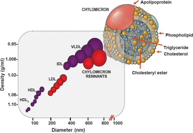
Lipoprotein particles including chylomicrons, very-low density lipoprotein (VLDL), intermediate density lipoprotein (IDL), low-density lipoprotein (LDL), and high-density lipoprotein (HDL) carry cholesterol in the blood. Lipoprotein particles also transfer and accept cholesterol, triglycerides (TG) and proteins from other lipoprotein particles and tissues. These lipoprotein particles are separated and classified according to their densities and diameters that reflect their load of cholesterol, TG and apolipoproteins (Figure 1) (Grundy et al 1990). A single lipoprotein particle is usually composed of apolipoproteins, phospholipids, TG, free cholesterol and cholesteryl esters.
Figure 1. Lipoprotein classes and subclasses. A lipoprotein particle is composed of apolipoprotein, phospholipids, triglycerides, cholesterol and cholesteryl esters. Taken from Grundy et al. Cholesterol and atherosclerosis; 1990.
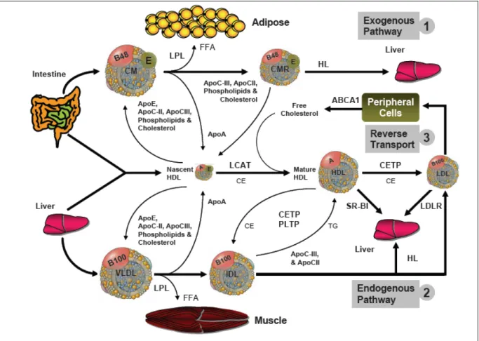
Lipoproteins are subjected to plasma and tissue enzyme actions and are removed from the circulation through receptor–mediated uptake by three pathways (Brown et al 1983, Von Eckardstein et al 2001) (Figure 2):
1. The exogenous pathway, which dictates the fate of dietary lipids.
2. The endogenous pathway, which dictates the fate of lipid synthesized in liver. 3. The reverse cholesterol transport pathway, which dictates the fate of peripheral
tissue lipids and the excess return back to the liver.
Figure 2. Lipid metabolism; exogenous, endogenous and reverse cholesterol transport pathways are depicted. VLDL, CM and HDL initial and mediate the exogenous, endogenous and reverse cholesterol transport processes respectively in the human body. (Abbreviation – ABCA1: ATP-binding cassette 1, CETP: Cholesteryl ester transfer protein, CM: chylomicron, FFA: free fatty acid, HDL: high-density lipoprotein, IDL: intermediate-density lipoprotein, LCAT: lecithin:cholesterol acyl transferase, LDL: low intermediate-density lipoprotein, LPL: lipoprotein lipase, PLTP: phospholipid transfer protein, VLDL: very low density lipoprotein). Taken from Marcil M et al. Hypoalphalipoprotéinémie familiale. Aspects cliniques, génétiques, cellulaires et moléculaires. Doctoral Thesis, Université de Montréal, 1999; p. 17.
The exogenous pathway. The intestinal epithelium assembles dietary TG and cholesterol, along with apolipoproteins (apo) B-48 and apoA and phospholipids into chylomicron (CM) particles. This facilitates its travel inside the aqueous plasma to deliver lipid to different tissue after having a meal. Following their secretion into the plasma, an exchange of components takes place between chylomicrons and HDL, wherein plasma the cholesterol, apoE, apoC-II, apoC-III and phospholipids are transferred from HDL to chylomicrons and apoA is transferred from chylomicrons to HDL. ApoC-II in the chylomicrons activates the lipoprotein lipases (LPL) found on the epithelium of the muscle and adipose tissue (Saxena et al 1991) and associated with heparan sulfate proteoglycans (HSPG). When a VLDL particle docks on a VLDL receptor (VLDLR), the TG in the core are hydrolyzed by LPL. Newly liberated free fatty acids (FFA) then enter either the muscle cells (metabolized for energy via β-oxidation) or the adipocytes synthesized back to TG for storage (Jensen et al 2003). As a result, the chylomicrons shrink in size and apoC-II, apoC-III, phospholipids and free cholesterol are transferred back to HDL particles. Chylomicron remnants containing apoB-48, apoE and cholesteryl esters are internalized and catabolized after binding the hepatic apoE receptor (Brown et al 1983, Gregory et al 2001).
The endogenous pathway. De novo synthesized lipids by the liver are incorporated in nascent VLDL particles. These VLDL, contain TG, cholesterol, apoB-100 and phospholipids assembled and secreted by the hepatocytes into the plasma when the body is in a fasting state. In the plasma, apoC-II, apoC-III and apoE are transferred to VLDL from HDL. Apo C-II activates LPL to hydrolyze TG from VLDL and yield a smaller and denser intermediate-density lipoprotein (IDL) particle. Some IDLs particles are removed by the liver via an apoE receptor (Brown et al 1983), while the remaining particles transfer CE to HDL in exchange for TG. Conversion of IDL to LDL is complete after the cholesteryl esters are transferred from HDL to IDL, while TG and apolipoproteins (except apo B-100) are removed from IDL. LDL delivers cholesterol to hepatocytes and peripheral cells (e.g. adrenal gland) via LDL receptor (LDLR) uptake. Scavenger cells (e.g. macrophages) in blood vessels may pick up oxidized LDL particles, in case of hypercholesterolemia, leading to atherosclerotic plaque formation (Brown et al 1983, Gregory et al 2001).
Reverse cholesterol transport. HDL particles mediate the return of cholesterol from peripheral cells to the liver to protect the vessels form the harmful effect of excess lipids. Nascent HDL particles containing apoA1, apoE are assembled and secreted by the liver and intestine. Free cholesterol from other lipoproteins or peripheral cells is transferred to HDL and esterified by LCAT. The cholesteryl esters can be transferred to other lipoproteins such as IDL and LDL to be derived back to the liver or transported directly by HDL particles to the liver through scavenger receptor class B type I (SR-BI) (Von Eckardstein et al 2001).
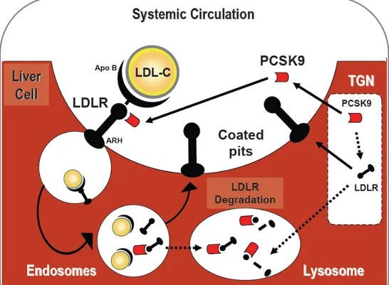
The LDLR is mainly a hepatic cell surface receptor (Brown et al 1983) that recognizes the apoB-100 ligand, but also apoE carried on VLDL remnant, IDL and some classes of HDL (Innerarity et al 1990, Kane et al 2001). Shortly after the LDLR discovery in 1979, Brown and Goldstein won the Nobel Prize for what was considered the key element in the cholesterol endogenous pathway (Brown et al 1983).The receptor-mediated endocytosis of the LDL particles by LDLR occurs in most, but not all, cells in a clathrin coated vesicles assisted by the adapter proteins ARH (Garcia et al 2001). Then when internalized the low pH in the vesicles facilitate the dissociation of the LDLR from its ligand. LDLR then recycles back to the surface to be associated with more LDL particle and eventually cholesterol levels fall in the circulation (Goldstein et al 1985). The liver then utilizes the cholesterol and TG to synthesize VLDL particles which are then exported once again to the circulation and/or excreted in the intestine as bile acids.
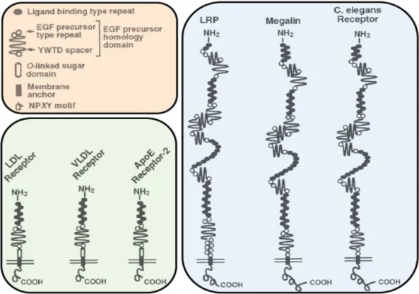
The LDLR is a 839-amino acids transmembrane glycoprotein that is encoded on chromosome 19 (Brown et al 1987) (Figure 3). Its N-terminal ligand-binding domain is 292-amino acids long that compose a cysteine-rich sequence. Its C-terminal cytoplasmic domain composes 50-amino acids long and serves to direct the receptor to the coated pits. In addition the extracellular portion of the receptor contains a 22-amino acid membrane-spanning region, a 58-amino acid region of 18 O-linked carbohydrate chains and a 400-amino acid region that is homologous to the precursor for epidermal growth factor (EGF) (Brown et al 1987).
Figure 3. The LDL receptor superfamily. The structural organization of some members of the LDL receptor gene family is depicted. The closest member to the LDLR is the VLDLR followed by the ApoER2. Taken from Schneider et al. J Am Soc Nephrol; 1999.
The EGF precursor homology domains consist of EGF repeats and YWTD spacer regions whish are involved in the pH-dependent release of the ligands from LDLR. The LDLR belongs to the LDLR superfamily that shares structural similarity with members of the LDL receptor gene family (Figure 3). Sequence alignment programs revealed that the closest members to the LDLR in structure were VLDLR (59% identity) and apolipoprotein E receptor 2 (ApoER2) (46% identity) followed by LRP, Megalin and C-elegans receptor (Poirier et al 2009).
There are over one thousand mutations identified in the LDLR gene to date (Guardamagna et al 2009) and they are classified into the 4 following groups (Goldstein et al 1987, Hobbs et al 1990):
1. Null mutations or LDLR defective synthesis (e.g. >15kb deletion in LDLR genes, a common French-Canadian mutation).
2. LDLR defective transport from the endoplasmic reticulum (ER) to the trans-Golgi network (TGN) (or gain-of-function mutation in PCSK9 gene which targets intra-cellular LDLR).
3. LDLR defective binding (or loss of function mutation in APOB gene encoding the apoB-100 ligand).
4. LDLR defective internalization where receptors do not cluster in the coated pits, thereby minimizing LDL internalization (e.g. mutations in autosomal recessive hypercholesterolemia “ARH” gene encoding the adaptor protein ARH).
1.1.1.2. The etiology of hypercholesterolemia
Monogenic hypercholesterolemia (or familial hypercholesterolemia - Fredrickson classification IIa) is the most common cause of markedly elevated serum cholesterol concentrations. Familial hypercholesterolemia (FH) is diagnosed by plasma low-density lipoprotein cholesterol (LDL-C) elevations 2-fold (heterozygote) or 3- to 5-fold (homozygote) above the average of a given population, age- and gender–matched, while serum TG concentrations remains within normal range (Rader et al 2003). Mixed hypercholesterolemia (familial combined dyslipidemia, Fredrickson classification IIb) has both LDL-C and TG elevations. This condition is caused by a susceptible genotype aggravated by one or more risk factors, including atherogenic diet (saturated fat, trans-fat, and cholesterol) (Hu et al 1997), obesity, and lack of exercise. Monogenic hypercholesterolemia is associated with xanthelasma and cutaneous xanthomas that can be sometimes extensive and disfiguring in homozygous FH. More importantly, affected FH patients are at increased risk for premature coronary artery disease and death at a younger age. Homozygous FH can experience their first cardiac event in childhood and only few survive to reach adulthood (Awan et al 2008).
The carrier frequency of mutations leading to FH in the general population is 1:500 (Brown et al 1983); however, this frequency increases in the French-Canadian descent who may suffer the “founder effect” of the first French settlers. This is due to many years of geographical isolation and a small genetic pool. The carrier frequency in some of the rural sites in Quebec can reach 1:80 (Scriver 2001). This phenomenon can be observed not only in the French-Canadian population but in similar culturally isolated environments in South-Africa and in some regions of Italy, Scandinavia and Lebanon (Khachadurian 1964).
From a practical point of view, human mutations in FH are of two major types depending on LDLR residual activity: < 2% (receptor-negative, poor response to statin) or between 2-25% (receptor-defective, good response to statin) (Rader et al 2003). Thus levels of residual LDLR activity correlates negatively with plasma levels of LDL-C. Etiologically speaking hypercholesterolemia can be a problem of cholesterol clearance (primary) or a problem of over production (secondary):
A. Primary or genetic etiologies are collectively called FH; classically a monogenic disorder caused by mutations in LDLR gene leading to either an absence or low activity of the LDLR. Since LDLR seminal discovery by Brown et al, three additional gene mutations have been discovered as primary cause of FH (Figure 4): APOB gene (FDB, familial defective apolipoprotein B) (Humphries et al 2006), ARH gene (encoding the adaptor protein, also known as autosomal recessive hypercholesterolemia) (Garcia et al 2001) and the recently described gain-of-function mutations in PCSK9 gene. PCSK9 will be discussed in greater details in Section 2.
Figure 4. LDLR recycling and associated genetic defect. Familial Hypercholesterolemia (FH) is caused by mutations in LDLR gene leading to either an absence or low activity of the LDLR, mutations in APOB gene (Familial defective apolipoprotein B), mutations ARH gene (Autosomal Recessive Hypercholesterolemia) or gain-of-function mutations in PCSK9 gene (encoding the LDLR regulatory protein).
B. Secondary or acquired etiologies of hypercholesterolemia may include metabolic disorders like liver disease (bile duct obstruction), renal failure (nephrotic syndrome) (Awan et al 2009) and hypothyroidism (Abrams et al 1981).
1.1.1.3. Treatment of hypercholesterolemia
Before the statin era, cholestyramine (bile acid binder) and niacin (vitamin B3 analog) (Goldstein et al 1987, Grundy et al 1981) were among the commonly used drugs to lower cholesterol, but were limited in usage due to their intolerability. Statins on the other hand represent a class of medication that inhibits the hydroxymethyl glutaryl coenzyme A (HMG-CoA) reductase (Goldstein et al 1987), a rate limiting enzyme in endogenous cholesterol synthesis. As a consequence statins, by reducing the endogenous levels of cholesterol synthesis, increase the uptake of circulating LDL particles, with significantly lower side effects leading to higher patient compliance. The mechanism of statins’ action is mediated by the upregulation of the levels of cell surface LDLR, which is found ubiquitously in all cells, but predominantly in hepatocytes (Goldstein et al 1987). Upregulation of the LDLR at the surface of hepatocyte increases LDL particles uptake in the liver and effectively lowers plasma cholesterol in blood (Goldstein et al 1987). The LDLR gene is under the influence of sterol regulatory element binding protein 2 (SREBP-2) which binds specific sterol regulatory elements in the promoter of the LDLR gene, thus up regulating the expression of LDLR in response to low sterol (Horton et al 2002). The transcription factor SREBP-2, in response to statin, also upregulates a molecule termed PCSK9 to counter regulate the LDLR effect (Dubuc et al 2004). PCSK9 binds to the LDLR via its catalytic domain, which interacts with the epidermal growth factor (EGF) A domain on the extracellular region of the LDLR (Zhang et al 2007) (Figure 3). The EGF precursor domain modulates LDL particle binding to the LDLR and mediates the acid-dependent dissociation of the ligand from the LDLR. Although the EGF-like repeats on LDLR are homologous to other LDLR family members (Christensen et al 1999) (Figure 3), there was no in vivo evidence (until our study) that PCSK9 binds other members of the LDL receptor gene family like the closely related VLDLR and the apoER2, although this was reported in cell lines (Poirier et al 2008).
Statins not only increase LDLR and decrease cholesterol levels, but also have been associated with a 20-25% reduction in CVD in over 14 randomized clinical trials (Baigent et al 2005). The additional protective role on CVD beyond cholesterol lowering can be
explained by the pleiotropic effects of statins (Davignon et al 2004). These pleiotropic properties influence various aspects of cell functions, inflammation, coagulation and vasomotor activity (De Lorenzo et al 2006). However, in patients lacking LDLR or having significantly lower LDLR activity, the statin class of medications is not efficient to lower cholesterol on their own, and ezetimibe (Rader et al 2003), a class of specific inhibitors of intestinal cholesterol absorption, are usually added. If all pharmacological attempts to lower cholesterol fail, a drastic invasive technique, LDL apheresis (Rader et al 2003), is then required. LDL apheresis, a type of dialysis to filtrate lipoproteins, has allowed homozygous FH patients to live beyond their life expectancy; however they are not free of disease as they experience arterial calcification as a late complication of the original disease (Awan et al 2008).
In summary lipoproteins and lipoprotein disorders play an important role in the onset and progression of CVD. Available treatments to date, including statins and LDL apheresis, are not sufficient to eliminate cardiovascular risk and the search for newer and safer cholesterol-lowering agents continues. Therefore, classes of drugs, such as the PCSK9 inhibitors are viable candidates, as will be discussed in section 2.
1.1.2. Abdominal obesity
A state of excess visceral fat in the abdomen is termed abdominal obesity. The WHO terms abdomen obesity a worldwide epidemic. Public health leaders marked obesity and inactivity as the second leading cause of preventable deaths in the West (Mokdad et al 2005). In the INTER-HEART study (an international heart study designed to assess the importance of risk factors on CVD worldwide) abdominal obesity carried a 33.7% attributable-risk of developing myocardial infarction (Table I). The WHO was also alarmed by the rise of childhood obesity (Koletzko et al 2002). Excess calories in children fuel the formation of new adipocytes and lead to hyperplastic obesity. This differs from adulthood caloric-excess that leads to hypertrophic obesity. Individuals with hyperplastic obesity are more likely to encounter difficulties in losing weight, since diet alone decreases the size but not the number of adipocytes. Childhood overweight and obesity is associated with higher CVD risk (Teixeira et al 2001) and the persistence of obesity is linked to the development of multiple risk factors (Srinivasan et al 1996), which in addition to developing CVD may lead to diabetes and cancer (Haslam et al 2005).
One billion adults and one tenth of the world’s children are now classified as overweight or obese and will experience diminished life expectancy (Haslam et al 2005). For instance obesity has been shown to decrease life expectancy by 7 years at the age of 40 (Peeters et al 2003).
1.1.2.1. Fat metabolism and the role of VLDLR
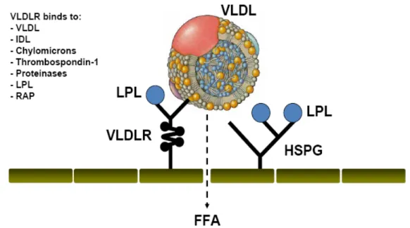
Similar to the LDLR in hepatocytes, the very-low-density lipoprotein receptor (VLDLR) plays a major role in adipocyte fat metabolism. VLDLR is highly expressed in adipose tissue (Takahashi et al 2004, Tacken et al 2001). In rabbits, the mRNA expression of VLDLR was significant in muscles and adipose tissues, but not detectable in the liver. This indicates a key role of VLDLR in the fatty acid metabolism of peripheral tissues (Oka et al 1994). Different from the LDLR, the VLDLR is not regulated by cellular sterols and does not bind efficiently LDL particles. However estradiol, thyroid hormone and active vitamin D modulate VLDLR expression (Tacken et al 2001). Feeding status per se did not influence mRNA levels of VLDLR, but atherogenic diets upregulate the expression of VLDLR in different animal models (Tiebel et al 1999).
Initially the involvement of VLDLR in lipoprotein metabolism could not be easily illustrated using a VLDLR knockout mouse. Apart from a 15-20% adipose mass reduction, lipid parameters were not significantly altered (Frkman et al 1995). It was only later, when VLDLR and LDLR double knockout mice became available, that the contribution to fatty acid delivery from TG-rich lipoprotein particles in peripheral tissues was elucidated (Tacken et al 2000). VLDLR-deficient mice were protected against diet-induced obesity and insulin resistance (Goudriaan et al 2000). VLDLR binds apoE-enriched chylomicrons and VLDLs, IDLs, as well as LPL that hydrolyzes TG into FFA (Figure 5). These interactions are facilitated by HSPG that binds LPL as well (Takahashi
et al 2004, Tacken et al 2001), whereas binding to ApoC-I, inhibits FFA hydrolysis (Jong et al 1996). Once the binding is facilitated by VLDLR (Figure 5), LPL starts to hydrolyze TG from TG-rich particles and FFAs diffuse into the cell were they become esterified or reform back to TG and contribute to the lipid core.
Figure 5. The VLDLR role in fat metabolism. The VLDLR and HSPG facilitate LPL proximity to VLDL leading to TG hydrolysis and FFA entry to the cell. Taken and modified from Tacken et al. Curr Opin Lipidol; 2001.
1.1.2.2. The etiology of obesity
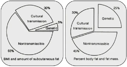
A simplified mechanism of obesity is energy disequilibrium leading to excessive calories stored in adipose tissue when energy expenditure is reduced (Rosenbaum et al 1997). However, the exact mechanism of obesity is not fully understood and is likely multifaceted (Bouchard et al 1991) (Figure 6). Evidence to support the fact that obesity is under genetic influences comes from identical twins studies (Bouchard et al 1990). Depending on the method used, genetic variation is estimated to account for up to 50% of the weight variability (Bluchard et al 1988). The total transmission effect to obesity represents 35% when estimated by body mass index (BMI), but only 5% are linked to a genetic defect (Bouchard et al 1991). Hence, in contrast to overall weight, BMI is more sensitive to lifestyle and environmental conditions. Genetics can explain up to 25% of the individual differences in percent body fat and mass (Bouchard et al 1991). Therefore genetic predisposition alone does not necessarily translate into obesity. Although the transmission of a way of life and some genetic factors influence how the body regulates appetite and energy expenditure (Grundy et al 1998), the total transmission effect alone could not contribute significantly to the endemic rise of obesity (Figure 6). Therefore classification of obesity by genetic means may require more studies (Pi-Sunyer 2000).
Figure 6. Etiology of obesity: genetic vs. non-genetic factors. Total transmissible variance and its genetic component for body mass index (BMI), subcutaneous fat (skin-fold thickness) and for total body fat from underwater weighing. Taken from Bouchard et al. Am J Clin Nutr; 1991.
The only fully studied gene that causes direct obesity is the gene leading to congenital leptin deficiency (Rosenbaum et al 1997). Mouse model simulating the human disease is designated ob/ob mice and becomes morbidly obese and insulin resistant on a regular diet (Ingalla et al 1950). Conversely polygenic mutation in over 15 genes has been linked to Bardet-Biedl syndrome that is characterized by obesity, mental retardation, and hypogonadism (Green et al 1989, Farooqi et al 2005). Interestingly, single-nucleotide polymorphism (SNP) in the gene termed proprotein convertase 1 gene (PCSK1) leads to hypoinsulinemia and elevated proinsulin and pro-opiomelanocortin (POMC). These SNPs were associated with severe childhood obesity (Heni et al 2010, Farooqi et al 2004).
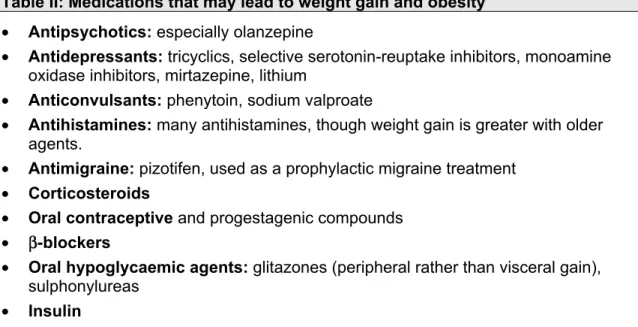
On the other hand, environmental factors such as eating habits and physical inactivity play a major role in weight gain in westernized communities. The type and quantity of refined carbohydrates and saturated fat consumed is a direct cause of the alarming incidence of obesity (Hu FB 2010). Obesity usually gives rise to many chronic diseases such as hypertension, diabetes, dyslipidemia and CVD (Haslam et al 2005). Obesity can also be a side effect of chronic diseases such as in Cushing's syndrome and hypothyroidism, but the cause of obesity is sometimes hard to distinguish (Longe et
al 2006). For instance, depression in patients may be a cause of obesity, or commonly depression may lead to overweight and obesity (Table II). Antipsychotic and antidepressant medications per se may increase weight likely by activating the SREBP transcription factors as seen in human primary hepatocytes, with subsequent up-regulation of downstream genes involved in cholesterol and fatty acid biosynthesis (Raeder et al 2006). A list of medications that may lead to weight gain and obesity are indicated in Table II. Therefore, whenever designing newer drugs that influence lipid metabolism, weight gain must be examined.
Table II: Medications that may lead to weight gain and obesity • Antipsychotics: especially olanzepine
• Antidepressants: tricyclics, selective serotonin-reuptake inhibitors, monoamine oxidase inhibitors, mirtazepine, lithium
• Anticonvulsants: phenytoin, sodium valproate
• Antihistamines: many antihistamines, though weight gain is greater with older agents.
• Antimigraine: pizotifen, used as a prophylactic migraine treatment • Corticosteroids
• Oral contraceptive and progestagenic compounds • β-blockers
• Oral hypoglycaemic agents: glitazones (peripheral rather than visceral gain), sulphonylureas
• Insulin
Taken from Haslam et al. Lancet; 2005.
1.1.2.3. Measurement of obesity
The absolute measurement of body fat is difficult and no single method is readily available for routine clinical use (Pi-Sunyer 2000). Chemical analysis of fat content in animals are commonly performed while in humans were only performed in cadavers during the 60’s (Widdowson 1965, Sheng et al 1979) and the outcome from these studies have shown a wide range of body fat content among different individuals. The underwater weighing (UWW) method or hydrodensitometry is based on the assumption that the body is divided into two compartments: adipose tissue and lean body mass (muscle and bone); as fat has a lower density under water therefore the body weighs less. Apart from being tedious and labor intensive, this method does not take into
account the high density of bones and muscles in athletes, which leads to body fat percentage underestimation, while the body fat of an osteoporotic patient may be overestimated (Haslam et al 2005). In contrast dual-energy X-ray absorptiometry (DEXA) is based on a three compartment measurement: bones, muscles and fat tissues. This technique is based on the assumption that bone content is directly proportional to the amount of X-ray absorbed by the bone being studied. In a comparative study, DEXA and UWW provided complementary information with DEXA values being lower. Thus, we used absolute measurement of body fat in mice for accurate results (Chapter 3). Nevertheless, DEXA is becoming the new gold standard method for human studies (Kennedy et al 2009).
Relative fat measurement assessing total body weight and body mass index (BMI) is also used in human studies. Obesity has conventionally been determined as a 20% increment in weight relative to subjects having the lowest death rate, matched for age and gender (Laquatra et al 2000, Longe et al 2006). This has been replaced completely by BMI, which is the ratio of weight (kg) to squared height (m2). WHO now adopts and accepts a BMI of 25 kg/m2 or more as overweight and obese when the BMI reaches 30 kg/m2 or more (WHO 2000). This is relevant as dyslipidemia gradually develop when BMI increases above 21 kg/m2, and when BMI reaches 30 kg/m2, small and dense LDL particle increases the risk of coronary heart disease by 3.6-fold (Willett et al 1995). In addition, associated low levels of HDL cholesterol and high concentrations of TG further and independently aggravate CVD risk (Wannamethee et al 1998).
More importantly the pattern of fat distribution will lower the threshold at which CVD of obesity starts. Obese patients with metabolic syndrome have an apple-like abdominal obesity and are at earlier risk for diabetes, than pear-shaped fat distributed around hips and thighs (Lebovitz et al 2003). The former is associated with cytokines release into the circulation, such as the tumor necrosis factor, leading to a proinflammatory state, insulin resistance and CVD (Lebovitz et al 2003). From the 52 countries participating in the INTER-HEART study (Yusuf et al 2004) the predictive ability of the waist/hip ratio (WHR) is superior compared to waist circumference (WC) or BMI, although WC measurements are more practical. In contrast, in another study BMI and WC were found to perform better over WHR for fatness assessments (Neovius et al 2005). A waist circumference of greater than 102 cm in men and 88 cm in women is an established risk factor for insulin
resistance, diabetes mellitus and CVD (Pi-Sunyer 2000). However the most accurate way to measure abdominal obesity is by magnetic resonance imaging (MRI) or computed tomography (CT) scanning, although both are expensive for routine clinical use (Pi-Sunyer 2000).
In summary, the surge in obesity rate continues to threaten our communities and economies. Health problem related to obesity does not depend on the absolute amount of fat but rather the distribution. Quantitatively and qualitatively ways to measure fat is required to prevent chronic disease and/or detect side effect of treatment modalities. The development of safe PCSK9 inhibitors must not lead to abdominal obesity as a side effect, as this would defeat their purpose, since abdominal obesity may lead to an increased incidence of atherosclerosis and CVD.
1.1.3. Atherosclerosis
The true incidence of atherosclerosis is impossible to compute, because it remains asymptomatic for years before a person starts to complain. Therefore the frequency of clinical manifestations of atherosclerosis is used instead. The process of atherosclerosis begins during childhood with the development of fatty streaks (McGill et al 2000). These lesions can be found in the aorta shortly after birth and appear in increasing numbers between 8 and 18 years (Guardamagna et al 2009). But serious lesions begin to develop in high risk groups when an individual reaches the fourth and fifth decades of life (Goldstein et al 1987).
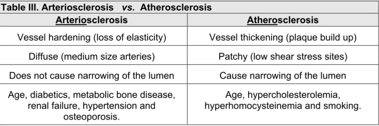
However, we must make a distinction between arteriosclerosis and atherosclerosis. Arteriosclerosis occurs in smooth muscle cell layer of the tunica media, while the atherosclerosis occurs in sub-endothelial layer of the tunica intima (Porter et al 2004). Arteriosclerosis refers to vessel hardening or loss of elasticity and occurs diffusely in medium size arteries. It does not cause narrowing of the lumen. Arteriosclerosis occurs as a consequence of aging, diabetes, metabolic bone diseases, renal failure, hypertension and osteoporosis (Table III). Atherosclerosis on the other hand refers to vessel thickening due to plaque build up. It occurs as patches at sites of low shear stress and causes narrowing of the lumen. Atherosclerosis occurs as a consequence of hypercholesterolemia, diabetes, hyperhomocysteinemia and excessive smoking Table III illustrates the main differences between arteriosclerosis and atherosclerosis.
Table III. Arteriosclerosis vs. Atherosclerosis
Arteriosclerosis Atherosclerosis
Vessel hardening (loss of elasticity) Vessel thickening (plaque build up) Diffuse (medium size arteries) Patchy (low shear stress sites) Does not cause narrowing of the lumen Cause narrowing of the lumen Age, diabetics, metabolic bone disease,
renal failure, hypertension and osteoporosis.
Age, hypercholesterolemia, hyperhomocysteinemia and smoking. Summarized and taken from wikipedia. 2011
1.1.3.1. The pathogenesis of atherosclerosis
Atherosclerosis is characterized by foam cells and plaque formation that potentially may erupt and cause thrombosis. This may lead to blood obstruction in vital organs, like the heart or the brain. Nevertheless, atherosclerotic plaques may also regress in response to statins and become less susceptible to eruption, but the associated calcific lesions may not regress (Chan KL et al 2010). The oxidized LDL particles that find their way into the sub-endothelium space as a result of endothelium dysfunction are engulfed by macrophages. Foam cell are then formed and subsequently die to give fatty streaks and plaques. The plaque formation process stimulates the cells of vascular bed to differentiate and produce substances that accumulate in the sub-endothelium. Notably, calcium deposition and calcified connective tissue start to form. The inner layer of the arterial wall thickens, the artery diameter shrinks and the blood flow starts to be compromised.
1.1.3.2. The progression of arterial calcification
Aortic calcification is complex and the exact mechanism is unknown, but damage to the endothelium may be the initial hit. Damage can be due to high cholesterol, blood glucose, blood pressure or cigarette smoking (Demer et al 2008). The role of triglycerides in aortic calcification is unclear (Awan et al 2008). High levels of triglycerides are often associated with diabetes, obesity, and low levels of high-density lipoprotein cholesterol, features of the metabolic syndrome and another cause of calcific atherosclerosis (Ellison et al 2005). The latter are all modifiable, while heredity, sex and age are not. Some people experience calcification in their 50’s or 60’s, others have rapid
progressive atherosclerosis and/or calcification during their 20’s, e.g., when they have a genetic predisposition as in FH (Alrasadi et al 2009).
From over 30 prospective clinical studies, arterial calcification was associated with most conventional cardiovascular risk factors and was deemed an independent risk factor for CVD (Rennenberg et al 2009). Arterial calcification is a cause of morbidity and mortality at any site: coronary arteries, peripheral arteries or aortic wall (Budoff et al 2007, Blacher et al 2001, Awan et al 2008). Plaque instability is a feature of calcified plaque, inducing mechanical failure leading to plaque rupture (Hoshino et al 2009). Aortic rigidity also leads to systolic hypertension, myocardial enlargement, myocardial infarction, heart failure, aortic aneurysm, ischemic limbs and death (Shao et al 2010, Awan et al 2008, London et al 2002, Budoff et al 2007). Calcification occurs most commonly in renal failure (Moe et al 2004), followed by uncontrolled diabetes, aggressive smoking and hypercholesterolemic disorders (Shao et al 2010). However, more extensive lethal calcification occurs in rare metabolic mutations as in homozygous deficiency of the enzyme nucleotide pyrophosphatase 1, a potent calcification inhibitor leading to infantile CVD (Rutsch et al 2008). Patients with fibrodysplasia ossificans progressiva, caused by a mutation in the BMP type-I receptors, are characterized by ectopic bone formation in muscles and soft-tissues that are mediated by cells of vascular origin (Hegyi et al 2003). FH patients develop premature severe calcific aortopathy and valvulopathy, which underscores the role of hyperlipidemia and/or the LDLR-deficiency. Correction of the hyperlipidemia does not alter the progression of calcification (Awan et al 2008). Furthermore a gene-dose effect of aortic calcification was observed in FH independent of cholesterol levels (Alrasadi et al 2009). Similarly loss of other genes like the NOTCH gene may lead to aortic valve calcification in susceptible individuals by favoring osteoblast differentiation (O’Brien 2006).
For over one hundred years, pathologists observed extra skeletal bone formation in a vascular bed (Bunting et al 1906, Virchow et al 1863). Later when the field of lipids expanded, calcified plaques were thought to occur passively in degenerative tissues. In the last few years, numerous mechanisms have been implicated in arterial calcification (Budoff et al 2007). Atheromatous calcification is currently considered a highly regulated process involving cellular components and regulatory factors (Wallin et al 2001, O'Brien 2006). Whether atherosclerosis activates the calcification machinery, or calcification
leads to the hardening of the vessels is still an enigma. Likely both occur in parallel, but one mechanism might be the initiating event (Table IV). In the case of kidney disease, mineral imbalance is an important contributor since the calcium, phosphate and calcification inhibitors and stimulators are dysregulated once kidney function becomes abnormal (Touyz et al 2009).
Arterial calcification is divided into two main histological categories: Mönckeberg and atheromatous calcification (Table IV):
Mönckeberg type of calcification is a cellular independent and dependent process. Amorphous mineral is believed to be deposited first, leading to the formation of amorphous calcified matrix that may be fueled by losing inhibitory calcification factors or acquiring pro-calcification factors (Sage et al 2010). Mineral imbalance is caused by excess calcium, phosphate and parathyroid hormone elevation. This induces a cellular differentiation process leading to chondro-osseous formation and eventual bone formation in the arterial wall (Cecilia et al 2004).
Atheromatous type of calcification is classically related to hypercholesterolemia (oxidized LDL), excess glucose, high homocysteine (Van Campenhout et al 2009) or oxidative stress (smoking) (Marangon et al 1998, Ker-Zabel et al 2003). Endothelial dysfunction arises from any of the previously mentioned insults. Scavenger cells infiltrate the site of insult and plaque is formed in a patchy manner, leading to inflammation that drives local cellular differentiation to an osteoblast-like phenotype, which later may form bone in the arterial wall.
Table IV: Associated calcification mechanisms*
Arteriosclerosis Atherosclerosis
Mönckeberg calciphylaxis Atheromatous calcification Smooth muscle layer (Tunica media) Sub-endothelium layer (Tunica intima) Diffuse (circumferential around elastic
laminae)
Patchy (within atheromatous plaques in Type I collagen)
Cellular independent and dependent
process Cellular dependent process
Amorphous matrix formation followed by chondro-osseous formation
Osteochondrogenesis followed by mineralization
Mineral imbalance related mechanism: • Calcium, phosphate and
pyrophosphate imbalance. • Losing inhibitory factors. • Acquiring promoting factors. • Cellular differentiation.
Metabolic excess related mechanism: • Endothelial dysfunction.
• Oxidative stress and inflammation. • Macrophage infiltration.
• Plaque formation. • Cellular differentiation. * Taken from Sage et al. Nature review; 2010.
Thus, in both types of calcification, differentiation of bone-like cells ultimately appears to occur, producing a calcified matrix. Although the initiating events may differ, ultimately the calcifications many mechanisms may overlap. Furthermore, both types of calcification can simultaneously occur in the same individual and/or the same vascular bed (Sage et al 2010).
Arterial calcification can also be re-classified in four categories based on their underlying mechanisms: 1) imbalance in mineral metabolism (Cecilia et al 2004, Dellegrottaglie et al 2006), 2) loss of calcification inhibitory factors (Weissen-Plenz et al 2008), 3) gain of calcification activator factors (Touyz et al 2008) (Table V), and 4) chondro-osteogenic cell differentiation (Shao et al 2006).
Table V: Factors that affects vascular calcification* Activators Inhibitors • Inorganic phosphates • TGFb1 • 25-hydroxycholesterol • cAMP • MAP kinase • Acetylated LDL • Homocysteine • Glucose • Endothelin-1
• Elastin degradation products • Pit-1
• Leptin
• Reactive oxygen species • BMP2-Msx2-Wnt • Wnt–β-catenin–lrP-5 • Vitamin D • Collagen type I • Alkaline phosphatase • Pyrophosphate • Statins • N-3 fatty acids • Tropoelastin • Bisphosphonates • Matrix Gla Protein • Osteopontin • Osteoprotegrin • NPP-1 via PPi • Fetuin-A (Ahsg) • Smad6 • Klotho • Carbonic anhydrase-2 • Calcimimetics • BMP-7 • CaR • Vitamin K • Estrogen * Taken from Touyz et al. Cardiovasc Res; 2009.
The pathophysiology of arterial calcification in FH is likely to be different from the vascular calcification observed in patients with renal disease treated with hemodialysis, where the imbalance in calcium and phosphate homeostasis, parathyroid hormone and vitamin D may contribute to the differentiation of local vascular cells to an osteoblast-like phenotype (Johnson et al 2006). Imbalance in mineral metabolism was not found in aortic calcification of FH patients, as addressed in our recent publication (Awan et al 2010).
Cells in the arterial walls of patients with either type of vascular calcification share many features with osteoblasts, including expression of bone morphogenic protein 2 (BMP2), core binding factor α1 (Cbfa1) and osterix, which have been isolated from calcified arterial wall lesions (Figure 7). This osteoblast-like phenotype of cells in the vessel wall is further supported by the prevalence of aortic valve calcification seen in individuals harboring a mutation in the NOTCH gene leading to the activation of the Runx2/Cbfa1 pathway (O’Brien 2006), one of two known pathways involved in osteoblast formation, with the other being the Wnt/Lrp5/β-catenin pathway (O’Brien 2006, Shao et al 2010).
Figure 7. Potential interplay of lipids and inflammation with genetics in the pathogenesis of calcification. Oxidized lipids, may induce osteoblastic differentiation of fibroblasts by upregulating expression of BMP2, which activates the Wnt/Lrp5/B-catenin pathway through upregulation of the transcription factor, Msx2. In addition, multiple cytokines, including TNF-, IL-1, and RANKL, may also promote calcification by activation of this pathway. Hypercholesterolemia is also associated with activation of the Runx2/Cbfa1 pathway in aortic valve disease. Increased phosphate (PO4) levels, as are seen in chronic kidney disease, might promote valve calcification through upregulation of Runx2/Cbfa1. Genetic factors may interact to further promote osteoblastic differentiation. NOTCH1 is shown as one example of how a genetic abnormality might contribute to both valvular morphological abnormalities and valvular calcification. Taken from O'Brien. Arterioscler Thromb Vasc Biol; 2006.
Similarly, other mutations associated with FH may affect these pathways (Figure 7). One possible mechanism is that similar signaling dysregulation in one pathway may be involved with LDLR−/− cells causing vascular calcification, as observed in Ldlr−/−, APOB100/100 mice. Controversy about the origin of these osteoblast-like cells, i.e. whether
they represent phenotypic changes in vascular smooth muscle cells, pericytes or differentiated mesenchymal stem cells is unresolved. Nevertheless, the pathophysiology of arterial calcifications in FH may well be caused by an interplayed between atherogenic factors and underlined genetic predisposition.
Furthermore, it would be of interest to test the remaining mechanisms of arterial calcification in an animal model. Before embarking on approaches to examine mechanisms, a sensitive examination method to measure calcification in soft tissue must be established in the same animal model that resembles human disease. This will validate any claims of cure or attenuation of arterial calcification.
1.1.3.3. Measurement of arterial calcification
For many years, physicians have made the diagnosis of atherosclerotic calcification during physical examination by simple means of palpation of hard arteries, auscultation by stethoscope for carotid bruits and more recently and accurately by Doppler examination. Vascular calcification in humans can be also detected on a standard x-ray or quantified more precisely by CT scan (Iijima et al 2011; Alrasadi et al 2009). Computed tomography or CT scan enabled physicians to measure the degree of calcified atherosclerotic plaques in the aorta and coronary arteries (APPENDIX I). This step is critical in patients at risk of aortic calcification undergoing major heart or aortic surgery to prevent intra-operative complications (Awan et al 2008).
Although conventional histology allows quantification of calcification, this occasionally leads to destruction of sample integrity and reduces in vivo structural preservation. In the proceeding chapter (CHAPTER 2), we applied micro-CT, a miniaturized version of CT, to detect and quantify aortic calcification in mice with a resolution of 5-20 µm. The micro-CT used (SkyScan) was initially developed to evaluate bone density in human teeth and skeletal parts of rodents. Therefore the apparatus imposed technical challenges to adopt a soft tissue mouse aorta to fit a small slot designed for holding small hard objects.
In summary, atherosclerosis and arterial calcification are complex processes. They require good novel tools to elucidate the cause of progression and evaluate potential therapeutic approaches. Similar to LDLR mutations, PCSK9 gain-of-function mutations carry at risk of atherosclerosis and arterial calcification. The understanding of PCSK9 biology is advancing each day and will benefit from innovative research tools.
2. The Proprotein Convertase Family
Proprotein convertases (PCs) are calcium-dependent serine endoproteases that play a key role in the post-translational processing of precursors of bioactive peptides. Many precursors of neuropeptides, hormones, enzymes, receptors or growth factors are synthesized as inactive precursors (prohormones) and are activated by proteolytic cleavage; this catalytic function is carried out by specific PCs (Seidah et al 1999, Fugère et al 2002).
Mammalian PCs belong to a family of 9 subtilisin/kexin proteases (Seidah et al 2008). The genes coding for PCs are designated by the letters PCSK and they are numbered from 1 to 9. PCSKs products are critically involved in various physiological processes depending on their protease activities resulting in activation/inactivation events, some of which could lead to cardiovascular homeostasis (Seidah et al 2007, Tall 2006) (Figure 8A).
Figure 8. The Proprotein Convertases Family. (A) Classification of proteases of the genome with emphasis on the PCs. (B) Schematic representation of the structure of bacterial subtilisin, yeast kexin and the mammalian PCs. The different domains and active site residues are shown. Taken and modified from Seidah et al. 2006 and 2007.
All PCSK products have an N-terminal signal peptide, a pro-segment domain, a catalytic domain and a P domain (Seidah et al 2008) (Figure 8B). The catalytic domains are highly conserved between many species. The 80-90 amino acids length pro-segment first is cleaved by an auto-catalytic mechanism in the endoplasmic reticulum (ER). The pro-segment acts both as an intramolecular chaperone to promote proper folding and as a competitive inhibitor of PCs function; this seems to prevent PCs from being accidentally activated during synthesis. The second round of cleavage occurs after a second set of basic amino acid residues in the pro-segment releasing it from the catalytic domain leading to activation of the PCs (Bergeron et al 2000, Seidah et al 2008).
2.1. Mammalian proprotein convertases in lipid metabolism
The first seven members of the mammalian PCs are similar in that they cleave protein precursors at basic amino acids. Out of these seven PCs, the fifth member, proprotein convertase subtilisin/kexin 5 (PCSK5), has relevance to lipid metabolism. Variation in the coding sequence of PCSK5 gene affects the HDL cholesterol levels as shown in a recent study (Iatan et al 2009). HDL cholesterol is inversely correlated to cardiovascular disease. This is achieved by the direct inactivation of endothelial lipase by PCSK5. PCSK5 also indirectly cleave and activate angiopoetin-like protein 3, a natural inhibitor of endothelial lipase and, consequently, affects atherosclerotic CVD risk (Iatan et al 2009) (Figure 8B).
The eighth member, PCSK8 also known as subtilisin kexin isozyme-1 (SKI-1) or site 1 protease (S1P), is known to cleave membrane-bound transcription factors, including sterol regulatory element binding proteins (SREBP-1 and -2), in the luminal domain of the Golgi apparatus, which when further cleaved by S2P, results in the release of their DNA-binding domain in absence of sterols. After cleavage, the cytosolic N-terminal domain becomes water soluble and translocates to the nucleus. This activated SREBP then binds to specific sterol regulatory element DNA sequences, thus upregulating the synthesis of enzymes involved in sterol/lipid biosynthesis. Sterols inhibit the cleavage of additional SREBP through a negative feedback loop, and thus synthesis of additional sterols is turned down (Horton et al 2002). Therefore, SREBPs are named master regulators of lipid homeostasis (Eberlé et al 2004) (Figure 9).