Université de Montréal
Effect of fatty acids on hyphal growth in the pathogenic
yeast Candida albicans
par Julie Shareck
Département de microbiologie et immunologie Faculté de Médecine
Thèse présentée à la Faculté de Médecine
en vue de l’obtention du grade de Philosophiae Doctor (Ph.D.) en microbiologie et immunologie
Septembre 2011
Faculté des études supérieures et postdoctorales
Cette thèse intitulée :
Effect of fatty acids on hyphal growth in the pathogenic yeast Candida albicans
Présentée par : Julie Shareck
a été évaluée par un jury composé des personnes suivantes :
Dr. Guy Lemay, président-rapporteur Dr. Pierre Belhumeur, directeur de recherche
Dr. Martine Raymond, membre du jury Dr. Malcolm Whiteway, examinateur externe Dr. Luis A. Rokeach, représentant du doyen de la FES
Résumé
Candida albicans est une levure pathogène qui, à l’état commensal, colonise les
muqueuses de la cavité orale et du tractus gastro-intestinal. De nature opportuniste, C.
albicans cause de nombreuses infections, allant des candidoses superficielles (muguet
buccal, vulvo-vaginite) aux candidoses systémiques sévères. C. albicans a la capacité de se développer sous diverses morphologies, telles que les formes levures, pseudohyphes et hyphes. Des stimuli environnementaux mimant les conditions retrouvées chez l’hôte (température de 37°C, pH neutre, présence de sérum) induisent la transition levure-à-hyphe (i.e. morphogenèse ou filamentation). Cette transition morphologique contribue à la pathogénicité de C. albicans, du fait que des souches présentant un défaut de filamentation sont avirulentes. Non seulement la morphogenèse est un facteur de virulence, mais elle constituerait aussi une cible pour le développement d’antifongiques. En effet, il a déjà été démontré que l’inhibition de la transition levure-à-hyphe atténuait la virulence de C.
albicans lors d’infections systémiques. Par ailleurs, des études ont démontré que de
nombreuses molécules pouvaient moduler la morphogenèse. Parmi ces molécules, certains acides gras, dont l’acide linoléique conjugué (CLA), inhibent la formation d’hyphes. Ainsi, le CLA posséderait des propriétés thérapeutiques, du fait qu’il interfère avec un déterminant de pathogénicité de C. albicans. Par contre, avant d’évaluer son potentiel thérapeutique dans un contexte clinique, il est essentiel d’étudier son mode d’action.
Ce projet vise à caractériser l’activité anti-filamentation des acides gras et du CLA et à déterminer le mécanisme par lequel ces molécules inhibent la morphogenèse chez C.
albicans. Des analyses transcriptomiques globales ont été effectuées afin d’obtenir le profil
transcriptionnel de la réponse de C. albicans au CLA. L’acide gras a entraîné une baisse des niveaux d’expression de gènes encodant des protéines hyphes-spécifiques et des régulateurs de morphogenèse, dont RAS1. Ce gène code pour la GTPase Ras1p, une protéine membranaire de signalisation qui joue un rôle important dans la transition levure-à-hyphe. Des analyses de PCR quantitatif ont confirmé que le CLA inhibait l’induction de
RAS1. De plus, le CLA a non seulement causé une baisse des niveaux cellulaires de Ras1p,
mais a aussi entraîné sa délocalisation de la membrane plasmique. En affectant les niveaux et la localisation cellulaire de Ras1p, le CLA nuit à l’activation de la voie de signalisation Ras1p-dépendante, inhibant ainsi la morphogenèse. Il est possible que le CLA altère la structure de la membrane plasmique et affecte indirectement la localisation membranaire de Ras1p. Ces travaux ont permis de mettre en évidence le mode d’action du CLA. Le potentiel thérapeutique du CLA pourrait maintenant être évalué dans un contexte d’infection, permettant ainsi de vérifier qu’une telle approche constitue véritablement une stratégie pour le traitement des candidoses.
Mots clés: Candida albicans, morphogenèse, transition levure-à-hyphe, acides gras, acide conjugué linoléique, Ras1p, voie de signalisation
Summary
The yeast Candida albicans is an inhabitant of the oral cavity, the gastrointestinal and genitourinary tracts of humans. Generally encountered as a commensal, it is also an opportunistic pathogen that causes a spectrum of infections, ranging from superficial mycoses (thrush, vulvovaginitis) to severe and life-threatening systemic infections. A striking feature of C. albicans is its ability to grow in different morphological forms, including budding yeasts, pseudohyphae, and hyphae. Environmental cues that mimic host conditions (elevated temperature, neutral or alkaline pH, and serum) induce the yeast-to-hypha transition. Morphogenesis is considered to be an attribute of pathogenesis, as mutants locked as yeasts or filamentous forms are avirulent. Given that the yeast-to-hypha transition is a virulence factor, it may also constitute a target for the development of antifungal drugs. Indeed, evidence has shown that impairing morphogenesis is a means to treat systemic candidiasis. Concurrently, a number of molecules have been reported to modulate morphogenesis in C. albicans. For instance, several fatty acids, including conjugated linoleic acid (CLA), inhibited the yeast-to-hypha transition. By interfering with an important attribute of C. albicans pathogenesis, CLA may harbor antifungal properties. However, before assessing its therapeutic potential in a clinical context, it is mandatory to address CLA’s mode of action.
The present study aims to further characterize the hypha-inhibiting properties of fatty acids and CLA and to elucidate the mechanism by which these molecules inhibit the yeast-to-hypha transition in C. albicans. Gene expression analyses were performed to gain insight into the transcriptional response of cells to CLA on a genome-wide scale and to probe the fatty acid’s mode of action. CLA downregulated the expression of hypha-specific genes and blocked the induction of genes encoding regulators of hyphal growth, including that of RAS1, which encodes the small GTPase Ras1p. A membrane-associated signaling protein, Ras1p plays a major role in morphogenesis. Quantitative PCR analyses showed that CLA prevented the increase in RAS1 mRNA levels which occurred at the onset of the
yeast-to-hypha transition. Unexpectedly, CLA reduced the steady-state levels of Ras1p. Additionally, CLA caused the delocalization of GFP-Ras1p from the plasma membrane. These findings indicate that CLA treatment results in suboptimal Ras1p cellular concentrations and localization, which impedes Ras1p signaling and inhibits the yeast-to-hypha transition. CLA may indirectly affect Ras1p localization by altering the structure of the plasma membrane. These studies have provided the mechanism underlying CLA’s hypha-inhibiting properties and may serve as the rationale to examine CLA’s therapeutic potential in the context of a Candida infection. There is a general lack of clinical evidence demonstrating that impairing morphogenesis is a sound approach to treat candidiasis. To remedy this situation, the therapeutic potential of molecules that modulate morphogenesis, such as CLA, should be clinically assessed.
Keywords: Candida albicans, yeast-to-hypha transition, morphogenesis, hyphal growth, fatty acids, conjugated linoleic acid, Ras1p signaling
Table of contents
RÉSUMÉ ... III SUMMARY ... V TABLE OF CONTENTS ... VII LIST OF TABLES ... XII LIST OF FIGURES ... XIII LIST OF ABBREVIATIONS ... XVI REMERCIEMENTS ... XIX
INTRODUCTION ... 1
1. CHAPTER 1. LITERATURE REVIEW ... 5
1.1 CELL MORPHOLOGIES OF C. ALBICANS ... 6
1.1.1 Pseudohyphae and true hyphae ... 6
1.1.2 Biofilms ... 10
1.1.3 Phenotypic switching ... 12
1.1.4 Chlamydospores ... 14
1.2 MORPHOGENETIC SIGNALS ... 14
1.3 MORPHOGENETIC SIGNALING PATHWAYS IN C. ALBICANS ... 17
1.3.1 The mitogen-activated protein kinase signaling pathway ... 21
1.3.2 The cAMP-PKA signaling pathway ... 25
1.3.3 The Czf1p signaling pathway ... 30
1.3.4 The Cph2p-Tec1p signaling pathway ... 31
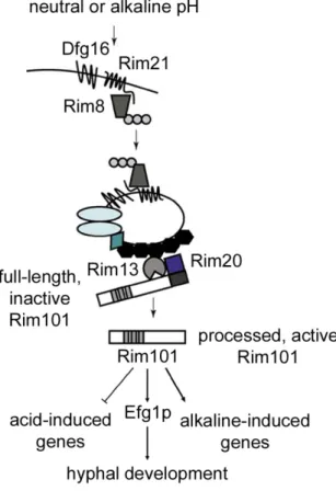
1.3.5 The Rim101p pH signaling pathway ... 33
1.3.6 The Tup1p signaling pathway ... 36
1.4 DOWNSTREAM TARGETS OF MORPHOGENETIC SIGNALING PATHWAYS ... 39
1.4.1 Virulence factors ... 40
1.4.1.1 Adhesins ... 40
1.4.1.2 Extracellular hydrolytic enzymes... 41
1.4.1.3 Morphogenesis ... 44
1.4.2 Hgc1p and the cyclin-dependent kinase Cdc28p ... 45
1.5 CANDIDA INFECTIONS ... 48
1.5.1 Skin and mucosal candidiasis ... 49
1.5.2 Invasive candidiasis ... 51
1.6 TREATMENT OF CANDIDIASIS ... 54
1.6.1 Classical antifungal drugs ... 55
1.6.1.1 Polyenes ... 55
1.6.1.2 5-Flucytosine ... 57
1.6.1.3 Ergosterol biosynthesis inhibitors ... 57
1.6.1.4 Echinocandins... 59
1.6.1.5 Griseofulvin ... 60
1.6.2 Targeting virulence: a new paradigm for antifungals ... 60
1.6.3 Morphogenesis, a target for the development of novel antifungals ... 61
1.7 RAS1P, A MEMBRANE-ASSOCIATED PROTEIN ... 62
1.7.1 Brief overview of plasma membrane constituents ... 62
1.7.2 Ras membrane attachment and subcellular trafficking ... 64
1.8 RATIONALE, HYPOTHESES, AND RESEARCH OBJECTIVES ... 68
2. CHAPTER 2. CONJUGATED LINOLEIC ACID INHIBITS HYPHAL GROWTH IN CANDIDA ALBICANS BY MODULATING RAS1P CELLULAR LEVELS AND DOWNREGULATING TEC1 EXPRESSION ... 70
2.1 ABSTRACT ... 74
2.2 INTRODUCTION ... 74
2.3.1 Strains and growth conditions ... 77
2.3.2 Hyphal growth assays in liquid and on solid media ... 79
2.3.3 Gene expression profiling ... 80
2.3.4 Northern blot analysis ... 81
2.3.5 Quantitative PCR analysis of C. albicans transcripts ... 81
2.3.6 Protein extraction and immunoblotting ... 83
2.3.7 Microarray data accession number ... 83
2.4 RESULTS ... 83
2.4.1 CLA inhibits hyphal growth in C. albicans in response to various hypha-inducing conditions ... 83
2.4.2 CLA impedes germ tube formation without affecting cellular growth ... 86
2.4.3 Gene expression analysis ... 88
2.4.4 UME6 and RFG1 are not required for CLA-mediated inhibition of hyphal growth ... 94
2.4.5 CLA downregulates TEC1 expression in a Ras1p-dependent manner .... 95
2.4.6 CLA reduces GFP-Ras1p protein levels and affects its localization ... 96
2.4.7 CLA affects the Tup1p-Nrg1p signaling pathway ... 100
2.5 DISCUSSION ... 102
2.6 ACKNOWLEDGMENTS ... 109
2.7 REFERENCES ... 110
3. CHAPTER 3. MODULATION OF MORPHOGENESIS IN CANDIDA ALBICANS BY VARIOUS SMALL MOLECULES ... 118
3.1 ABSTRACT ... 121
3.2 INTRODUCTION ... 121
3.3 FARNESOL ... 124
3.4 BACTERIAL AND FUNGAL AUTOREGULATORY MOLECULES ... 127
3.6 PEPTIDES AND PROTEINS ... 131
3.7 RAPAMYCIN ... 132
3.8 GELDANAMYCIN ... 133
3.9 HISTONE DEACETYLASE INHIBITORS ... 134
3.10CELL CYCLE INHIBITORS ... 135
3.11OTHER SMALL MOLECULES ... 136
3.12CONCLUSION ... 137
3.13ACKNOWLEDGMENTS ... 143
3.14REFERENCES ... 144
4. CHAPTER 4. DISCUSSION AND PERSPECTIVES ... 158
4.1 FATTY ACIDS INHIBIT HYPHAL GROWTH IN CANDIDA ALBICANS ... 159
4.2 USING TRANSCRIPTIONAL PROFILING TO ADDRESS THE MODE OF ACTION OF CLA .. ... 162
4.2.1 Hyphal growth program in Spider medium ... 163
4.2.2 Genes upregulated by CLA ... 166
4.2.3 Genes downregulated by CLA ... 168
4.3 CLA TARGETS RAS1P SIGNALING ... 170
4.3.1 CLA affects GFP-Ras1p levels and its subcellular localization ... 170
4.3.2 Causes of GFP-Ras1p delocalization ... 173
4.3.3 A link between plasma membrane properties and hyphal growth ... 175
4.3.4 Potential mechanism for the CLA-mediated delocalization of GFP-Ras1p ... 176
4.3.5 Downstream consequences of CLA-modulated Ras1p signaling ... 180
4.3.5.1 Activation of the MAP kinase pathway ... 180
4.3.5.2 Activation of the cAMP-PKA-Efg1p pathway ... 180
4.4 CLA AFFECTS THE TUP1P-NRG1P SIGNALING PATHWAY ... 187
4.6 THERAPEUTIC POTENTIAL OF MOLECULES THAT MODULATE MORPHOGENESIS IN C.
ALBICANS ... 192
4.6.1 Modulating morphogenesis as a means to treat candidiasis ... 192
4.6.2 Putative therapeutic applications of CLA and fatty acids ... 193
4.7 ECOLOGICAL ROLE OF FATTY ACIDS AND OTHER QUORUM SENSING MOLECULES IN C. ALBICANS MORPHOGENESIS ... 195
CONCLUSION ... 198
REFERENCES OF CHAPTERS 1 & 4 ... 200
List of tables
CHAPTER 1Table I Predisposing factors for candidiasisa ... 48
CHAPTER 2
Table II Candida albicans strains used in this study ... 78 Table III Primers used in this study ... 82 Table IV Selected genes upregulated during the yeast-to-hypha transition in Spider medium
... 90
CHAPTER 3
Table V Small molecules that modulate morphogenesis in C. albicans by affecting the Ras1p-cAMP-PKA signaling pathway ... 141
List of figures
CHAPTER 1Figure 1. 1 Phylogenetic tree of fungi pathogenic to humans and animals. ... 3
Figure 1. 2 Morphological forms of Candida albicans... 7
Figure 1. 3 Characteristic features of cell division for the yeast, pseudohyphal, and hyphal growth forms in C. albicans. ... 9
Figure 1. 4 Candida albicans biofilms. ... 11
Figure 1. 5 White-opaque switching in C. albicans. ... 13
Figure 1. 6 Environmental cues promote hyphal development via specific signaling pathways in C. albicans. ... 15
Figure 1. 7 Selected methods for gene disruption in C. albicans. ... 20
Figure 1. 8 Regulation of morphogenesis in C. albicans by multiple signaling pathways. . 23
Figure 1. 9 The Rim101p pH signaling pathway in C. albicans. ... 35
Figure 1. 10 Several virulence factors contribute to early events in the pathogenesis of candidiasis. ... 43
Figure 1. 11 Role of cyclin-dependent kinases in morphogenesis in C. albicans. ... 47
Figure 1. 12 Infection cycles of Candida albicans. ... 52
Figure 1. 13 Antifungal drug targets and resistance mechanisms. ... 56
Figure 1. 14 Membrane trafficking of mammalian Ras proteins. ... 65
Figure 1. 15 Depalmitoylation/palmitoylation cycle model of subcellular trafficking of H-Ras and N-H-Ras. ... 67
CHAPTER 2 Figure 2. 1 Conjugated linoleic acid (CLA) inhibits hyphal growth in Candida albicans. . 85
Figure 2. 2 CLA impedes germ tube formation of Candida albicans without affecting cellular growth. ... 87
Figure 2. 4 UME6 and RFG1 are not required for CLA-mediated hyphal growth inhibition.
... 94
Figure 2. 5 TEC1 downregulation by CLA is Ras1p-dependent. ... 97
Figure 2. 6 CLA reduces GFP-Ras1p protein levels and affects its localization. ... 100
Figure 2. 7 CLA affects the Tup1p-Nrg1p signaling pathway... 101
Figure 2. 8 Proposed model underlying the mechanism by which CLA inhibits hyphal growth in Candida albicans. ... 107
Figure S 1 Transcriptional profiles of CLA-treated cells... 108
CHAPTER 3 Figure 3. 1 Summary of the modes of action of selected small molecules which modulate morphogenesis in Candida albicans. ... 140
CHAPTER 4 Figure 4. 1 Unsaturated fatty acids inhibit hyphal growth in Candida albicans. ... 160
Figure 4. 2 Other Candida species respond to CLA. ... 162
Figure 4. 3 Differentially expressed transcription factors dispensable for hyphal growth. 165 Figure 4. 4 Inhibition of hyphal growth by CLA does not depend on its metabolism. ... 168
Figure 4. 5 CLA decreases RAS1 mRNA and protein levels. ... 172
Figure 4. 6 Effect of CLA on the subcellular localization of GFP-Ras1p in C. albicans. . 179
Figure 4. 7 CLA affects the activation of the MAP kinase pathway. ... 181
Figure 4. 8 CLA’s effect on cAMP signaling ... 183
Figure 4. 9 Morphology of PKA mutant strains. ... 184
Figure 4. 10 CLA does not modulate the Efg1p-dependent branch of Ras1p signaling. ... 186
Figure 4. 11 CLA affects the expression of Tup1p-Nrg1p-regulated genes. ... 188
À mes parents, parce qu’ils m’ont beaucoup appris, un peu à leur insu.
“All we have is now”
List of abbreviations
3OC12HSL: 3-oxo-C12-acyl homoserine lactone 5-FC: 5-flucytosine
5-FOA: 5-fluoroorotic acid 5-FU: 5-fluorouracil A: adenine
AIDS: Acquired immune deficiency syndrome AMP: adenosine monophosphate
ATP: adenosine triphosphate BEC: buccal epithelial cells bHLH: basic helix-loop-helix bp: base pairs
C.: Candida
C: cytosine
cAMP: cyclic adenosine monophosphate CDK: cyclin-dependent kinase
cDNA: complementary DNA CLA: conjugated linoleic acid COX: cyclooxygenase
CRIB: Cdc42/Rac interactive binding CSP: competence-stimulating peptide CT: threshold cycle
DAB: 1,4-diamino-2-butanone
DAPI: 4’-6’diamidino-2-phenyl-indole DHA: docosahexaenoic acid
DIC: differential interference contrast DNA: deoxyribonucleic acid
DOC: deoxycholate Dox: doxycycline
EBI: ergosterol biosynthesis inhibitors EC: esophageal candidiasis
ESCRT: endosomal-sorting complex required for trafficking FBS: fetal bovine serum
FLP: flippase
FRT: flippase recognition target FTS: S-farnesylthiosalicylic acid G: guanine
GAP: GTPase-activating protein GDP: guanosine diphosphate
GEF: guanine nucleotide exchange factor GFP: green fluorescent protein
GI: gastrointestinal
GlcNAc: N-acetylglucosamine GO: gene ontology
GPI: glycosylphosphatidylinositol
GRACE: gene replacement and conditional expression GTP: guanosine triphosphate
HA: hemagglutinin
HDAC: histone deacetylase
HIV: human immunodeficiency virus HSG: hypha-specific gene
HUVEC: human umbilical vein endothelial cells IL: interleukin
kDa: kilodalton
MAP: mitogen-activated protein MIC: minimal inhibitory concentration mRNA: messenger RNA
MTL: mating-type-like
NRE: Nrg1p response element OD: optical density
OPC: oropharyngeal candidiasis
PAGE: polyacrylamide gel electrophoresis PAK: p21-activated kinase
PBS: phosphate-buffered saline PCR: polymerase chain reaction PKA: protein kinase A
PKC: protein kinase C RA: Ras-association
rDNA: ribosomal deoxynucleic acid RIPA: radioimmunoprecipitation assay RNA: ribonucleic acid
S.: Saccharomyces
SAHA: suberoylanilide hydroxamic acid SAP: secreted aspartic protease
SDS: sodium dodecyl sulfate
SEM: scanning electron microscopy SLAD: synthetic low ammonium dextrose T: thymine
TBS-T: tris-buffered saline Tween 20 Tor: target of rapamycin
tRNA: transfer RNA TSA: trichostatin A TX: thromboxane
VVC: vulvovaginal candidiasis YNB: yeast nitrogen base YPD: yeast peptone dextrose μM: micromolar
Remerciements
Je tiens d’abord à remercier mon directeur de recherche, le Dr. Pierre Belhumeur, pour m’avoir accueillie dans son laboratoire et accompagnée dans mon cheminement scientifique, tout en me soutenant et en m’épaulant dans les moments difficiles qui ont pu joncher ce parcours. C’est grâce à la confiance qu’il a investie en moi que ce projet a pu être mené à terme.
Je me dois de souligner le soutien inégalé du Dr. Martin Clément, agent de recherche au laboratoire. Merci Martin pour avoir partagé avec moi tout ton savoir, pour avoir répondu à mes nombreuses questions ainsi que pour m’avoir conseillée et aidée pendant toutes ces années. Sans toi, ce projet n’aurait jamais vu le jour. Je remercie le Dr. André Nantel pour son aide dans le design expérimental et l’analyse et l’interprétation des résultats de biopuces ainsi que Christian Charbonneau pour son assistance en microscopie. Un merci tout particulier aux membres du laboratoire, passés et présents, ainsi qu’à ceux du département de microbiologie et immunologie de l’Université de Montréal. Je souligne aussi le soutien financier du conseil de recherches en sciences naturelles et en génie du Canada (CRSNG), le fonds québécois de recherche sur la nature et la technologie (FQRNT) et la Faculté des études supérieures (FES) de l’Université de Montréal.
Enfin, je tiens enfin à remercier les Drs. Guy Lemay, Martine Raymond, Malcolm Whiteway et Luis Rokeach pour avoir accepté de faire partie du jury de ma thèse et pour leurs commentaires qui seront certainement fort appréciés.
Introduction
The growing population of immunocompromised humans has allowed many fungi to become pathogens. The most common agents of fungal infections include Candida,
Aspergillus, and Cryptococcus (Pappas, 2010). In the genus Candida, almost 200 species of
ascomycetous, asexual yeasts are currently listed, yet only C. albicans, C. dubliniensis, C.
glabrata, C. krusei, C. tropicalis, C. parapsilosis, and C. lusitaniae are encountered as
opportunistic pathogens of humans (Odds et al., 2007). C. albicans is the most common cause of invasive fungal infections in hospital settings, in addition to being the causative agent of superficial mycoses such as oral thrush and vulvovaginitis (Horn et al., 2009; Miceli et al., 2011) (discussed in section 1.5).
The first description of (oral) thrush, an infection of the mucous membranes of the mouth and throat, dates back to 400 B. C. and was made by Hippocrates who mistook the infection for oral ulcers (Adams, 1939). It was not until 1846 that thrush was shown to be caused by a contagion rather than by abnormalities of the host (Berg, 1846). Using a “scientific approach”, Berg reproduced thrush in healthy newborns by infecting them with material from aphthous lesions. While Berg’s experiments demonstrated that thrush was caused by an organism (i.e. a fungus), it was not identified correctly. Confusion regarding the true identity of the fungus was perpetuated until 1923, when Berkhout proposed that the organism causing thrush should bear the generic name Candida (Berkhout, 1923). In 1954, the nomen conservandum (C. albicans) was officially approved (Calderone, 2002). The name Candida is derived from the Latin phrase “toga candida” which was used to describe the special white robe worn by candidates for the Roman Senate; albicans means “to whiten”. The name “Candida albicans” may refer to the oral lesions of aphthae or thrush or to the characteristic white colonies produced by C. albicans on agar (Calderone, 2002).
C. albicans is now ranked as the third or fourth most common agent of microbial
septicemia in hospitals, having surpassed many bacterial infections in terms of incidence and morbidity (Almirante et al., 2005; Beck-Sague & Jarvis, 1993; Tortorano et al., 2004).
These severe, often lethal Candida infections are on the rise due to a growing population of hospitalized patients with underlying immune deficiencies stemming from treatment for cancer or immunosuppression following a transplantation (Enoch et al., 2006). Treatment of such infections is complicated by the limited arsenal of antifungal drugs, the severe side effects in patients, the development of antifungal drug resistance, and the emergence of species refractory to conventionally used agents (Sanglard & Odds, 2002). There is a need for new targets or new strategies in antifungal therapy (discussed in section 1.6).
An Ascomycete, C. albicans belongs to the Saccharomycotina subphylum, to the Hemiascomycetes class, and to the Candida albicans clade (Figure 1.1) (Taylor, 2007). Baker’s yeast Saccharomyces cerevisiae also belongs to the Hemiascomycetes class, which makes it a close relative of C. albicans. Both yeasts diverged approximately 200 million to 800 million years ago (Hedges, 2002). Yet, about two-thirds of the ~6,500 genes in the C.
albicans genome have clear homologues in S. cerevisiae, allowing many gene functions to
be inferred and key aspects of various processes in the pathogenic to be understood (discussed in 1.3). Additionally, both yeasts are similar in their cellular morphologies, their ability to metabolize fermentable carbon sources, and their capacity to reproduce asexually by budding. A parasexual cycle has been identified in C. albicans, but differs from the complete sexual cycle described for S. cerevisiae because the pathogenic yeast is diploid, does not exist naturally in a haploid form, and does not undergo meiosis (discussed in 1.1.3).
A striking feature that differentiates C. albicans from S. cerevisiae and other
Candida species is its ability to grow in a variety of morphological forms, including as
budding yeasts, as pseudohyphae, and as true hyphae (Sudbery et al., 2004) (discussed in section 1.1). The yeast-to-hypha transition, also known as morphogenesis or filamentation, is induced by a variety of environmental cues (discussed in section 1.2) which activate a complex network of signaling pathways composed of transducers, kinases, and transcription factors (Biswas et al., 2007; Brown et al., 2007; Ernst, 2000) (discussed in
section 1.3). These signaling pathways are involved in sensing and transmitting inducing signals and in regulating the regulatory and structural elements essential for hyphal growth. Interestingly, the same pathways regulate the expression of virulence factors of C. albicans such as cell surface adhesins, secreted aspartic proteases, lipases, and phospholipases (discussed in section 1.4). Because it contributes to the overall pathogenesis of C. albicans, morphogenesis also constitutes one of its virulence factors (see section 1.4.3).
Figure 1. 1 Phylogenetic tree of fungi pathogenic to humans and animals.
A maximum-likelihood phylogeny based on small-subunit rDNA sequences of diverse fungi. Taxa of commonly encountered pathogenic fungi were added to the phylogeny. Adapted from Taylor (2007).
It has been suggested that virulence factors of C. albicans may be potential targets for the development of new antifungal drugs (see section 1.6.2). One such virulence factor that could be targeted by antifungal agents is morphogenesis. Interestingly, many small molecules have been reported to modulate the yeast-to-hypha transition. For instance, fatty acids, including conjugated linoleic acid (CLA), inhibited hyphal growth in C. albicans (Clement et al., 2007). Thus, CLA and fatty acids may be candidates for the development of novel antifungal agents. However, understanding how fatty acids inhibit morphogenesis is a prerequisite before undertaking studies to assess their therapeutic potential. In the present study, CLA is used to probe the mechanism by which fatty acids inhibit hyphal growth in C. albicans.
This thesis is divided into four major sections. In Chapter 1, morphogenesis in C.
albicans is described in depth, in terms of the different morphological growth forms,
inducing signals, and molecular events involved in the morphogenetic transition. The association between the yeast-to-hypha transition and virulence is described, demonstrating why morphogenesis may constitute a target for the development of antifungal drugs. In Chapter 2, findings pertaining to the hypha-inhibiting effects of CLA and to its mode of action are presented. These results were published in an article entitled “Conjugated linoleic acid inhibits hyphal growth in Candida albicans by modulating Ras1p cellular levels and downregulating TEC1 expression” in the scientific journal Eukaryotic Cell. In Chapter 3, a review on small molecules that modulate morphogenesis is presented. This minireview entitled “Modulation of morphogenesis in Candida albicans by various small molecules” was submitted to the scientific journal Eukaryotic Cell and is presently in revision. Finally, in Chapter 4, issues that are relevant to this work are discussed and perspectives are proposed.
In this chapter, the various aspects of morphogenesis, including cell morphologies, morphogenetic signals and signaling pathways, and downstream targets of these signaling pathways, are reviewed. The different types of Candida infections are described. Conventional antifungal drugs available to treat such infections as well as novel treatments are also discussed. Most of the notions constituting the backdrop of this study are provided in this chapter.
1.1 Cell morphologies of C. albicans
C. albicans was initially considered to be dimorphic, as it can grow in a budding
yeast form or as branching, filamentous forms such as pseudohyphae and true hyphae. However, the pathogenic yeast also exists in a mating-competent opaque form (Bennett & Johnson, 2005; Slutsky et al., 1987) and can be induced to form thick-walled spherical cells known as chlamydospores (Staib & Morschhauser, 2007). The term pleiomorphic appears to be better suited to describe C. albicans, as it fully reflects the spectrum of its morphologies (Figure 1.2).
1.1.1 Pseudohyphae and true hyphae
Even though pseudohyphae superficially resemble hyphae, both morphological forms have different cell shapes. A feature that characterizes hyphal cells is the absence of constrictions at the mother-daughter junction or at subsequent septal junctions, which results in cells with parallel sides along their entire length (Figure 1.2C). In contrast, pseudohyphae have constrictions at the mother-daughter junction and between each individual cellular compartment (Sudbery et al., 2004) (Figure 1.2B). Sides of pseudohyphal cells are not parallel, resulting in compartments being wider in the middle than at the two ends (Merson-Davies & Odds, 1989). Additionally, hyphae are narrower than pseudohyphae, with a width of ~2 μm compared to ~2.8 μm-5 μm, respectively (Sevilla & Odds, 1986).
Figure 1. 2 Morphological forms of Candida albicans.
Yeasts (A), pseudohyphae (B), and hyphae (C). Budding yeast cells are similar to diploid S.
cerevisiae cells. Pseudohyphal cells have constrictions at the mother-daughter junction and at the
positions of septa. Hyphae have parallel cell walls and no constrictions. (D) White-opaque phenotypic switching of the C. albicans WO-1 strain, grown on salt-dextrose at 23°C for three days. White (W) and opaque (O) colonies are seen. The cellular phenotype of white (E) and opaque cells (F). The white cell is round with a relatively smooth surface while the opaque cell is twice the size of the white cell and has unique wall pimples. (G) Chlamydospores are thick-walled spherical cells that are ~3 to 4 times larger than normal yeast cells. Adapted from Brown et al. (2007); Berman & Sudbery (2002); Staib & Morschhauser (2007).
C. albicans undergoes reversible morphological transitions between unicellular
budding yeast cells and filamentous growth forms such as pseudohyphae and true hyphae. A variety of environmental conditions induce the switch from yeast growth to filamentous growth (Sudbery et al., 2004) (see section 1.2). Pseudohyphae develop into elongated buds that remain attached to the mother cells, resulting in filaments of elongated cells with constrictions at the septa. In contrast, true hyphae grow by continuous apical extension followed by septation. Although morphological and cell cycle studies suggest that pseudohyphal and hyphal growth forms represent distinct developmental stages (Berman & Sudbery, 2002; Sudbery et al., 2004), recent work has demonstrated that the pseudohyphal morphology is not an alternate fate, but rather a true intermediate state between the yeast and hyphal morphologies (Carlisle et al., 2009).
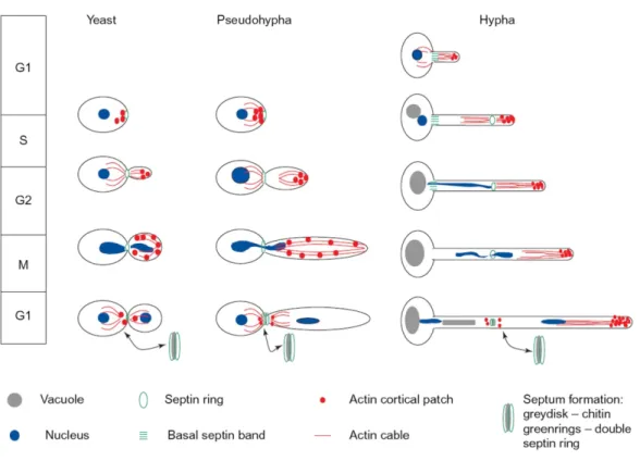
Several aspects of the cell cycle of hyphae and pseudohyphae also differ (Figure 1.3). In pseudohyphal cells, the daughter bud emerges from the mother cell at the start of the cell cycle. In hyphae, germ tube formation occurs before the start of the cell cycle, and is thus cell cycle-independent (Hazan et al., 2002). Polarized growth in yeast and pseudohyphal cells involves the polarisome, while apical extension of the germ tube is driven by the Spitzenkörper. Specific to hyphal cells, this membranous structure is usually found in strictly filamentous fungi and resembles the yeast polarisome (Whiteway & Bachewich, 2007). In pseudohyphal cells, the first septum is laid down at the mother-daughter junction, whereas in hyphae, it is formed within the growing germ tube (Sudbery
et al., 2004). The position of the septum affects the position of the first mitosis, which
occurs at the mother-daughter junction in pseudohyphae, but entirely within the germ tube in hyphae. The absence of cytokinesis is common to both pseudohyphal and hyphal growth modes, which results in cells remaining attached. However, while hyphae appear as long, septated filaments, pseudohyphae have constrictions at the septa.
Figure 1. 3 Characteristic features of cell division for the yeast, pseudohyphal, and hyphal growth forms in C. albicans.
In the yeast and pseudohyphal growth modes, a bud emerges from the mother cell, defining the position of the first septum. Changes in the pattern of actin polarization reflect a switch from polarized growth at the tip to isotropic growth throughout the growing bud. In pseudohyphal growth, daughter cells become increasingly elongated due to an extended period of polarized growth. Mitosis occurs across the plane of the septum at the mother-daughter junction. In yeast growth, cytokinesis yields two discrete cells while in pseudohyphal growth, cells remain attached, separated by constrictions. In hyphal development, a germ tubes emerges from the mother cell before the G1/S transition. The first septum is formed within the germ tube, where mitosis occurs. One nucleus migrates back into the mother cell, while the other moves further into the germ tube. Actin cortical patches continue to cluster at the growing tip, resulting in an elongated hypha. Hyphal cells remain attached, forming septated filaments. Adapted from Sudbery et al. (2004).
1.1.2 Biofilms
C. albicans biofilms are defined as surface-associated microbial communities,
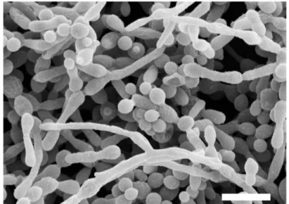
encased within a matrix of extracellular polymers (Costerton et al., 1995). Although not a distinct C. albicans morphology per se, hyphae and pseudohyphae play a determinant role in biofilm development. C. albicans biofilms show a complex three-dimensional architecture and display extensive spatial heterogeneity, consisting of a dense network of yeasts, hyphae, and pseudohyphae encased within a matrix of exopolymeric material (Figure 1.4A). Biofilm formation occurs in three distinct stages: (i) attachment and colonization of yeast cells to a surface, (ii) germ tube formation and proliferation of yeast and filamentous cells, which allows the formation of a basal layer of anchoring cells, and (iii) growth of pseudohyphae and hyphae and secretion of a carbohydrate- and protein-rich extracellular matrix (Chandra et al., 2001; Douglas, 2003; Ramage et al., 2001) (Figure 1.4B). While hyphal cells provide the scaffold to build the biofilm, they also express adhesin-encoding genes (discussed in section 1.4.1), which contribute to cell surface adherence properties required to maintain the structural integrity of the biofilm.
C. albicans biofilms have important clinical consequences. They are involved in
mucosal candidiasis (thrush, vulvovaginitis), form at the surface of implanted medical devices, such as prostheses, stents, shunts, implants, and various types of catheters, and can cause their failure (Kojic & Darouiche, 2004), have increased antifungal drug resistance, resist host immune defenses, and constitute a sustained reservoir of infecting cells (Lopez-Ribot, 2005).
Figure 1. 4 Candida albicans biofilms.
(A) Scanning electron microscopy (SEM) image of a mature (48 h) C. albicans biofilm, composed of yeast cells, pseudohyphae, and hyphae. Bar = 10 μm. Adapted from Ramage et al. (2005). (B) The SEM images show the stages of C. albicans biofilm formation, starting with the initial adhesion and attachment of C. albicans yeast cells to a surface, followed by the formation of germ tubes. Yeast and filamentous cells proliferate, forming a basal layer of anchoring cells. Further proliferation of microcolonies and filamentation contribute to biofilm development. As the biofilm matures, it becomes encased within a matrix of extracellular material. Adapted from Ramage et al. (2009).
1.1.3 Phenotypic switching
A change in cellular morphology also occurs during colony switching, which is seen only in specific strains (Pomes et al., 1985; Slutsky et al., 1985). At low frequency, C.
albicans strain 3153A can spontaneously and reversibly convert from smooth, white
dome-shaped colonies to variant colony shapes in which cells grow in yeast and filamentous forms (star, ring, irregular wrinkle, hat, stipple, and fuzzy) (Berman & Sudbery, 2002). In the C. albicans strain WO-1, the white-opaque switch involves the transition from white domed colonies containing white cells to opaque, flat colonies containing opaque cells (Figure 1.2D). White cells are round with a morphology resembling that of Baker’s yeast while opaque cells are oblong, twice the size of white cells, and have pimples on the surface of the cell wall (Anderson & Soll, 1987) (Figure 1.2E-F). White and opaque cells differ in virulence and in the capacity to colonize and infect different body locations. Indeed, white cells appear to be better suited for internal infections, while opaque cells thrive in skin infections (Kvaal et al., 1999).
The white-opaque switch has garnered more attention since it was shown to be required for mating (Bennett & Johnson, 2005; Miller & Johnson, 2002). In C. albicans, there are two mating-type-like (MTL) loci, MTLa and MTLα, which reside on chromosome 5 and include the MTL genes MTLa1, MTLα1, and MTLα2 (Hull & Johnson, 1999). For mating to occur, diploid cells have to undergo loss of heterozygosity at the MTL loci, resulting in a or α strains which express only MTLa or MTLα genes. Strains that are homozygous or hemizygous at the MTL have a tendency to undergo the white-opaque switch (Lan et al., 2002; Slutsky et al., 1987) (Figure 1.5A). Moreover, opaque cells mate more efficiently (Miller & Johnson, 2002). Diploid a and α opaque cells mate to create tetraploid a/a/α/α cells (Figure 1.5B). Although meiosis has not been observed in C.
albicans, tetraploid cells undergo chromosome loss under certain laboratory conditions,
Johnson, 2003). By making the white-opaque switch a prerequisite for mating, C. albicans ensures that mating only occurs under specific conditions.
Figure 1. 5 White-opaque switching in C. albicans.
(A) A sectored colony on an agar plate, in which most of the cells are in the white phase, but containing a minority of cells in the opaque phase. SEM images show white cells, which are round, and opaque cells, which appear oblong, twice the size of white cells and have pimples on the surface of the cell wall. The transition from the white phase to the opaque phase is blocked by the a1 and α2 proteins, ensuring that a/α strains are not permissive for white-opaque switching. Only a or α strains can switch to the opaque phase, the state in which they are primed to mate. (B) Mating of opaque cells yields a mononuclear tetraploid a/a/α/α cell. Reduction in ploidy can be achieved by chromosome loss, thereby regenerating a- and α–mating competent progeny and completing a parasexual cycle. Meiosis has not been observed in C. albicans. Adapted from Bennett & Johnson (2005).
1.1.4 Chlamydospores
Compared to other morphological growth forms, the chlamydospore growth mode remains elusive. Chlamydospores, thick-walled spherical cells that are ~3 to 4 times larger than normal yeast cells, appear to be a dormant growth form (Jansons & Nickerson, 1970) (Figure 1.2G). They have a high lipid and carbohydrate content, and are known to germinate under certain conditions (Fabry et al., 2003; Miller et al., 1974). Chlamydospore formation is induced at low temperature and in conditions of oxygen, light, and nutrient depletion (Staib & Morschhauser, 2007). Media consisting of rice or cornmeal agar supplemented with the detergent Tween 80 have been designed to induce chlamydospore production. This growth mode remains obscure as chlamydospores have rarely been observed during infection and do not appear to play a role in pathogenicity.
1.2 Morphogenetic signals
The yeast-to-hypha transition in C. albicans is triggered by a large number of different treatments which are presumed to reflect host conditions encountered by the pathogenic yeast in vivo (Odds, 1988a). Environmental cues that promote hyphal development are presented in Figure 1.6, and include a neutral or alkaline pH (6.5-8.0) and a growth temperature of 37°C (Buffo et al., 1984; Davis, 2003; Shapiro et al., 2009), nitrogen and/or carbon starvation (Csank et al., 1998), embedded growth and/or low oxygen concentration (Brown et al., 1999), and a wide range of chemicals (Odds, 1988a). The most commonly used chemicals that have morphogenetic potential are serum, amino acids (proline, methionine), the amino sugar N-acetylglucosamine, glucose, ammonium, and 5% CO2/bicarbonate (Biswas & Morschhauser, 2005; Ernst, 2000; Hudson et al., 2004;
Klengel et al., 2005; Land et al., 1975b; Maidan et al., 2005b; Mattia et al., 1982; Miwa et
al., 2004). Phosphate- or alkane-enriched media induce pseudohyphal growth (Hornby et al., 2004; Sorkhoh et al., 1990).
While serum remains the most potent inducer of hyphal growth, its chemical complexity has hampered the quest to identify its hypha-inducing factors. Initially, it was believed that serum induced hyphal growth by imposing nitrogen starvation, given that it is mainly composed of proteins that constitute an inaccessible source of nutrients until they are hydrolyzed (Brown & Gow, 1999). However, serum still stimulates hyphal growth when combined with a rich source of nutrients such as YPD, indicating that a factor other
Figure 1. 6 Environmental cues promote hyphal development via specific signaling pathways in C.
albicans.
Hyphal growth is induced by neutral or alkaline pH, serum, glucose, N-acetylglucosamine (GlcNAc), amino acids, nitrogen and carbon starvation, low oxygen concentration, and embedding in a physical matrix. A high temperature (37°C) is a common requirement for all hypha-inducing conditions, except for embedded growth. Specific signaling pathways mediate the responses to these signals. There appears to be some functional overlap between some of these pathways. Adapted from Brown et al. (2007).
than nitrogen starvation is responsible for morphogenesis. Low molecular weight compounds (< 1 kDa) were suggested to be the active factors in serum, as a serum filtrate induced hyphal growth (Feng et al., 1999). Accordingly, Hudson et al. (2004) demonstrated that glucose was responsible for ~80% of the serum activity. However, given that hyphal growth is induced using 5-20% serum in media containing glucose at concentrations 10- to 20-fold higher than those found in serum alone, it appears impossible that glucose is the only active agent in serum. Glucose does remain a morphogen, as low concentrations ranging between 0.01% and 0.3% were shown to stimulate hyphal development in liquid and solid media (Hudson et al., 2004; Maidan et al., 2005b). Recently, muramyl dipeptides have been identified as the hypha-inducing factors in serum (Wang & Xu, 2008; Xu et al., 2008).
Although a variety of environmental signals induce hyphal growth, neutral pH and a temperature of 37°C are the two factors that are common to most of the hypha-inducing regimes. A shift from acidic (pH 4.5) to alkaline (pH ≥ 6.5) conditions combined to an increase in temperature is known to promote hyphal growth (Buffo et al., 1984; Lee et al., 1975). Additionally, a distinct pH signaling pathway is responsible for mediating the effects of alkaline pH (Davis, 2009) (see section 1.3.5). In contrast, although it was known that temperature played a pivotal role in triggering morphogenesis, the missing link between high temperature and the onset of hyphal induction was uncovered recently. High temperatures are required to compromise the function of the heat shock protein Hsp90p, a repressor of hyphal growth (Shapiro et al., 2009) (discussed in section 3.8).
Different media are routinely used to induce hyphal growth in C. albicans. In these media, hypha-inducing cues are a combination of many individual factors, including a high temperature (37°C), a neutral pH, and a low cell density (OD600 < 0.1-0.5). Poor carbon
sources (e.g. mannitol, N-acetylglucosamine) or low glucose concentrations (e.g. in tissue culture media) impose a state of carbon starvation, which promotes hyphal development (Brown et al., 2007). Moreover, hypha-inducing media contain a complex mixture of amino
acids, including proline and methionine, which have morphogenetic properties (Lee et al., 1975; Maidan et al., 2005b; Odds, 1988a). Morphogenesis results from the synergistic effect of a variety of environmental cues (Brown et al., 2007; Ernst, 2000).
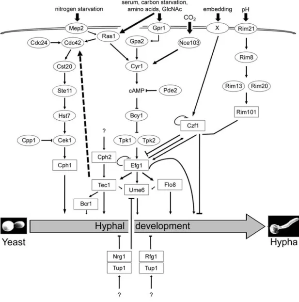
1.3 Morphogenetic signaling pathways in C. albicans
A complex network of signaling pathways regulate the yeast-to-hypha transition (Figure 1.8). The mitogen-activated protein (MAP) kinase, cAMP-protein kinase A (PKA), Czf1p, Cph2p-Tec1p, and Rim101p pathways stimulate hyphal growth, while the Tup1p signaling pathway represses the yeast-to-hypha transition. Although it was initially thought that each pathway made a somewhat independent contribution in regulating hyphal development, it is now clear that there is cross-talk between pathways. Most of the knowledge pertaining to morphogenetic signaling pathways in C. albicans has been gained by examining phenotypes of mutant strains lacking one (or more) specific gene(s). Thus, before describing the role of signaling pathways in morphogenesis, several aspects of C.
albicans genetics are discussed.
The study of the molecular mechanisms regulating the yeast-to-hypha transition in
C. albicans has been hindered by genetic features of the pathogenic yeast, including its
diploidy, its lack of an exploitable sexual cycle, its alternative codon usage, and the unavailability of good molecular tools and transformation protocols. One approach used to gain insight into biological aspects and gene functions is to analyze strains in which a specific gene is deleted or mutated. Given that C. albicans is an obligate diploid organism, a mutation has to become homozygous to observe a pronounced phenotype. In diploid organisms, homozygotes are generated by using genetic cross. However, although C.
albicans carries a mating-type-like locus (Hull & Johnson, 1999) and can be engineered to
sexual cycle is not exploitable, rendering genetic cross superfluous. Moreover, molecular tools to sequentially disrupt both alleles of a gene in C. albicans were initially unavailable.
For these reasons, C. albicans genes (involved in morphogenesis or in other biological processes) used to be identified/analyzed based on their ability to complement or suppress mutations in the corresponding S. cerevisiae genes (Liu et al., 1994) or to interfere with or constitutively activate the corresponding signaling pathways in S. cerevisiae (Whiteway et al., 1992). The sequencing and annotation of the C. albicans genome revolutionized research in C. albicans: gene functions could be inferred from those described for S. cerevisiae genes and functional analyses could be conducted using reverse genetics (Braun et al., 2005). However, comparative genome analyses showed that several
C. albicans genes, such as those encoding lipases, proteases, and cell wall proteins, had no
homologues in S. cerevisiae. In addition, functions of homologous genes had diverged. Moreover, it was becoming clear that while specific components of signaling pathways were conserved between both yeasts, molecular mechanisms and environmental cues had often diverged, perhaps due to the fact C. albicans had co-evolved with its human host (Biswas et al., 2007).
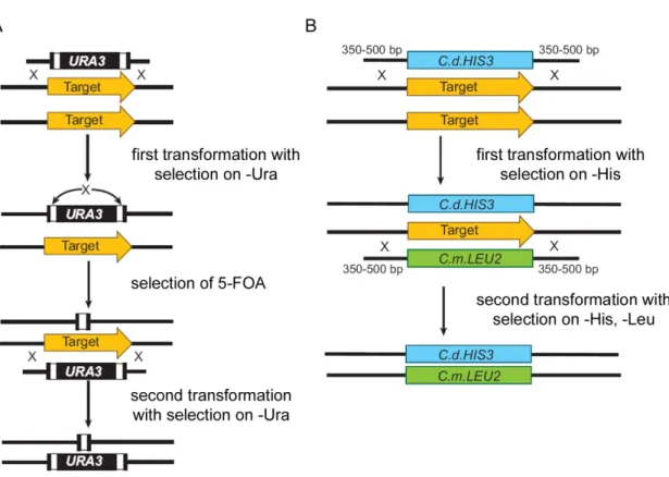
Advances in gene disruption technology have enabled to carry out studies directly in the pathogenic yeast. The Ura blaster method, the first to be developed, relies on homologous recombination to replace the chromosomal copy of a target gene by the disruption cassette whose 5’ and 3’ ends are homologous to DNA sequences that flank the target gene (Fonzi & Irwin, 1993) (Figure 1.7A). Direct repeats of the hisG sequence from
Salmonella thyphimurium flank the C. albicans URA3 gene, enabling intramolecular
recombination events. The disruption cassette is transformed into a strain of C. albicans auxotrophic for uracil; Ura+ heterozygous disruption mutants, in which one allele of the target gene is replaced by the URA3 cassette, are selected on medium lacking uridine. The
URA3 marker is “recycled” by passaging Ura+ transformants on medium containing 5-fluoroorotic acid (5-FOA) (Alani et al., 1987; Boeke et al., 1984). 5-FOA selects for strains
that delete the URA3 gene via intramolecular recombination between the hisG flanks. The resulting Ura- heterozygous strain is transformed again using the URA3 cassette to disrupt the second allele of the target gene.
Initially, the Ura blaster method was embraced by the C. albicans community. Many mutants were created in order to determine the role of genes in virulence. However, mutant strains displayed several problems. First, URA3 expression is subject to chromosomal positioning (Brand et al., 2004; Cheng et al., 2003b; Lay et al., 1998). Second, high URA3 expression levels are required for many processes, including morphogenesis, adhesion, and virulence (Brand et al., 2004; Cheng et al., 2003b; Sundstrom et al., 2002b). Thus, virulence defects may be linked either to the deletion of a target gene or to poor URA3 expression. Moreover, 5-FOA is a significant mutagen that can cause major chromosomal abnormalities (Wellington et al., 2006).
To circumvent these problems, several solutions have been proposed, including reintegrating a copy of URA3 at a standard locus from which it is consistently expressed (Murad et al., 2000; Sundstrom et al., 2002b), using dominant selectable markers conferring resistance to mycophenolic acid (Staib et al., 1999; Wirsching et al., 2000) and nourseothricin (Reuss et al., 2004; Roemer et al., 2003; Shen et al., 2005), and using auxotrophic markers that do not affect virulence, such as HIS1, LEU2, and ARG4 (Noble & Johnson, 2005). New and improved gene disruption tools have also been made available and have been reviewed elsewhere (Noble & Johnson, 2005) (Figure 1.7B).
Furthermore, regulatable promoters, including the glucose repressible PCK1 and
MAL2 (Backen et al., 2000; Brown et al., 1996; Leuker et al., 1997), the
methionine/cysteine-repressible MET3 promoter (Care et al., 1999), and the tetracycline-repressible promoter system (Nakayama et al., 2000) have been developed to generate conditional null mutants and to conduct functional studies of essential genes. A collection of gene replacement and conditional expression (GRACE) C. albicans mutants has also
been generated, in which one allele of a target gene is replaced by the HIS3 auxotrophic marker while the other allele is placed under the control of the tetracycline-repressible promoter (Roemer et al., 2003).
Figure 1. 7 Selected methods for gene disruption in C. albicans.
(A) The Ura blaster method consists in transforming a gene disruption cassette into a strain of C.
albicans auxotrophic for uracil (ura3/ura3). The cassette harbors the URA3 gene, flanked by direct
repeats of the hisG sequence from Salmonella typhimurium and has 5’ and 3’ ends homologous to sequences flanking the target gene. Mutants in which one allele of the target gene is replaced by the
URA3 cassette are selected on medium lacking uridine. Counterselection on 5-fluoroorotic acid
(5-FOA) identifies mutants that have lost the URA3 sequences through recombination between the hisG repeats. Transformation with the disruption cassette is repeated to disrupt the second copy of the target gene. (B) A linear gene disruption fragment with long regions of homology (300-500 bp) to sequences flanking the target gene is created using a fusion PCR technique. The first allele of a target gene is disrupted by transforming the first disruption fragment marked by the HIS1 gene from
C. dubliniensis, and selecting on medium lacking histidine. The second copy of the target gene is
disrupted by transforming the second disruption fragment, marked by the C. maltosa LEU2 gene, and selecting on medium lacking histidine and leucine.
A second issue that has hindered gene function analysis in C. albicans is that it displays a noncanonical codon usage: CTG is decoded as a serine instead of a leucine. Consequently, many heterologous markers such as Escherichia coli lacZ and jellyfish green fluorescent protein (GFP) cannot be functionally expressed in C. albicans unless they are modified (Berman & Sudbery, 2002). To overcome these impediments, a number of specialized reporter genes have been developed (reviewed in Berman & Sudbery [2002]). Convenient cassettes harboring these reporters have been generated, enabling gene expression to be monitored at the RNA or protein level and gene products to be localized in vivo (Gerami-Nejad et al., 2001; Gerami-Nejad et al., 2004; Gerami-Nejad et al., 2009). Thus, the development of molecular tools, combined to the availability of a sequenced genome and to improved transformation protocols, have made it fairly straightforward to conduct functional analyses of signaling genes in C. albicans.
1.3.1 The mitogen-activated protein kinase signaling pathway
The first morphogenetic signaling components to be identified in C. albicans were members of a MAP kinase pathway, analogous to the filamentous and invasive growth pathways in S. cerevisiae (Alonso-Monge et al., 2006; Gimeno et al., 1992; Palecek et al., 2002). In C. albicans, the MAP kinase pathway is comprised of three kinases, including Cst20p, homologous to the PAK Ste20p (Csank et al., 1997; Ushinsky et al., 2002), Hst7p homologous to the MAP kinase kinase Ste7p (Leberer et al., 1996), and Cek1p, the
Candida ERK-like kinase homologous to the MAP kinases Fus3p and Kss1p (Whiteway et al., 1992) (Figure 1.8). Although a homologue of the S. cerevisiae MAP kinase kinase
kinase STE11 has been annotated (Braun et al., 2005), the C. albicans STE11 gene has not been functionally dissected (Brown et al., 2007). These kinases function by sequential phosphorylation, and ultimately activate the transcription factor Cph1p, a homologue of Ste12p which lies downstream of Cek1p (Liu et al., 1994). The tyrosine phosphatase Cpp1p is a negative regulator of the MAP kinase, as constitutive hyphal development of a
The MAP kinase signaling cascade plays a minor role in hyphal growth. Strains lacking CST20, HST7, CEK1, and CPH1 were unable to form hyphae on solid Spider medium, containing the poor carbon source mannitol and on SLAD nitrogen starvation medium, but filamented in liquid Spider, Lee, and serum media (Csank et al., 1998; Kohler & Fink, 1996; Leberer et al., 1996; Liu et al., 1994). Thus, the MAP kinase induces morphogenesis in response to nutrient limitation on solid media only, while other pathways promote hyphal development in response to other inducing factors.
Activation of the MAP kinase pathway occurs through the GTPase module Cdc42p/Cdc24p (Bassilana et al., 2003) (Figure 1.8). Cdc42p, a Rho-type G protein (Bassilana et al., 2003; Hazan & Liu, 2002) and Cdc24p, its exchange factor, have critical roles in viability and in hyphal growth (Bassilana et al., 2003; Bassilana et al., 2005; Ushinsky et al., 2002; VandenBerg et al., 2004). In response to serum, CDC24 expression was transiently induced and Cdc24p was recruited to hyphal tips, where it maintained Cdc42p in its GTP-bound active state (Bassilana et al., 2005). Cdc42p was shown to bind with high affinity to a Cst20p motif in vitro and to Cla4p, another PAK kinase (Leberer et
al., 1997; Su et al., 2005). Given that Ras1p regulates the MAP kinase pathway (Leberer et al., 2001), Bassilana et al. (2003) proposed that the Cdc42p/Cdc24p GTPase module
formed a complex with Ras1p to activate Cst20p and the MAP kinase pathway. Additionally, in response to nitrogen, the transmembrane ammonium permease Mep2p was shown to be involved in activating the MAP kinase and cAMP-PKA pathways in a Ras1p-dependent manner and in inducing hyphal growth (Biswas & Morschhauser, 2005).
Figure 1. 8 Regulation of morphogenesis in C. albicans by multiple signaling pathways.
The MAP kinase, cAMP-PKA, and pH signaling pathways activate morphogenesis, while the yeast-to-hypha transition is repressed by Tup1p, Nrg1p, and Rfg1p. A variety of environmental signals trigger simultaneously different signaling pathways, resulting in the activation of specific transcription factors (boxed) which induce or repress hyphal development. An important regulator of hyphal growth, the small GTPase Ras1p, functions upstream of the MAP kinase and the cAMP-PKA signaling pathways, which operate through the transcription factors Cph1p and Efg1p, respectively. Efg1p appears to act as a hub, as it induces hyphal growth in response to different environmental cues, including serum, carbon starvation, CO2, and neutral pH. However, Efg1p is a
repressor of filamentation in embedded conditions. Hyphal development is repressed by Tup1p, which functions by recruiting the DNA-binding proteins Nrg1p and Rfg1p. Adapted from Brown et
Recent data described the mucin-like protein Msb2p and the adaptor protein Sho1p, two plasma membrane proteins, as potential sensors of the MAP kinase pathway (Roman et
al., 2009b). During resumption of growth, Cek1p phosphorylation depended on SHO1
(Roman et al., 2005; Roman et al., 2009a). In addition, Msb2p and Sho1p played a role in hyphal and invasive growth and in the activation of Cek1p in response to cell wall stress (Roman et al., 2005; Roman et al., 2009b). In S. cerevisiae, Cdc42p was shown to interact with Msb2p (Cullen et al., 2004) and Ste20p (Cst20p homologue) (Peter et al., 1996), while in C. albicans, the interaction between Cdc42p and Cst20p was demonstrated (Su et al., 2005). Based on these findings, Roman et al. (2009b) proposed that a putative Msb2p/Sho1p complex may interact with Cdc42p, promoting the recruitment of Cdc24p and the sequential phosphorylation of Cst20p and Cek1p. However, an interaction between Msb2p and Sho1p has yet to be demonstrated.
The function of the MAP kinase pathway depends on the transcription factor Cph1p, the S. cerevisiae Ste12p homologue (Liu et al., 1994). Contrary to Ste12p, Cph1p does not regulate the expression of TEC1, a transcription factor involved in morphogenesis (discussed in section 1.3.4). CPH1 was required for the expression of several hypha-specific genes, including ECE1, HWP1, HYR1, RBT1, SAPs5-6, and RBT4, but only in conditions in which hyphal growth depended on the MAP kinase pathway (Lane et al., 2001a). It remains that a cph1/cph1 mutant strain filamented and expressed hypha-specific genes in most liquid hypha-inducing conditions, thereby questioning the presumed involvement of the MAP kinase pathway in morphogenesis (Braun & Johnson, 2000; Brown et al., 2007; Lane et al., 2001a). In fact, findings suggest that the MAP kinase pathway plays a clear role in mating, rather than in hyphal development (Bennett et al., 2003; Chen et al., 2002; Magee et al., 2002; Sahni et al., 2010).
Although the MAP kinase pathway plays a minor role in morphogenesis and hypha-specific genes expression, it does contribute to a certain extent to C. albicans pathogenesis in vivo. Strains lacking CDC42, CDC24, CST20, CEK1, and CPP1 were attenuated in a
mouse model of systemic candidiasis (Bassilana et al., 2003; Csank et al., 1997; Csank et
al., 1998; Leberer et al., 1996). Inactivation of CEK1 and CPP1 also attenuated virulence
in the mammary glands of lactating mice, preventing tissue colonization (Guhad et al., 1998a; Guhad et al., 1998b). However, deletion of CDC42, CDC24, and CEK1 impaired growth in vitro, which may account for reduced virulence of mutant strains in vivo. In contrast, hst7/hst7 and cph1/cph1 mutant strains retained their virulence in the mouse model of systemic candidiasis (Leberer et al., 1996; Lo et al., 1997). It remains that CPH1 contributed to the residual virulence of an efg1/efg1 mutant, suggesting that the MAP kinase is involved in virulence in vivo (Lo et al., 1997).
It is noteworthy to mention that like in S. cerevisiae, C. albicans has several other MAP kinase signaling pathways, including a cell integrity pathway, the protein kinase C (PKC) pathway and a stress-activated kinase pathway (i.e. the HOG pathway), which have been reviewed elsewhere (Alonso-Monge et al., 2006).
1.3.2 The cAMP-PKA signaling pathway
The cAMP-PKA signaling pathway plays an important role in filamentation in S.
cerevisiae, in C. albicans, and in other fungi (Lengeler et al., 2000). In the pathogenic
yeast, an increase in cAMP accompanies the yeast-to-hypha transition, thereby linking cAMP signaling to filamentation. Components of the cAMP signaling pathway include the G protein-coupled receptor Gpr1p, the Gα protein Gpa2p, the GTPase Ras1p, the adenylate cyclase Cyr1p, the cAMP-dependent PKA, and the transcriptional factor Efg1p (Figure 1.8).
cAMP signaling is regulated by Ras1p, Gpr1p-Gpa2p, and the transmembrane ammonium permease Mep2p. Each upstream regulator responds to a specific set of inducing factors and activates the cAMP signaling pathway. For instance, on solid media or in presence of the amino acid methionine, Gpr1p and Gpa2p were required for
morphogenesis. Moreover, filamentation defects of gpr1/gpr1 and gpa2/gpa2 strains were corrected by the addition of exogenous cAMP or by the overexpression of downstream components of the cAMP-PKA pathway, indicating Gpr1p and Gpa2p function upstream of cAMP signaling. However, both mutants were only mildly affected in liquid hypha-inducing media, which suggests that Gpr1p and Gpa2p play minor roles in morphogenesis (Maidan et al., 2005a; Miwa et al., 2004; Sanchez-Martinez & Perez-Martin, 2002). Additionally, in nitrogen starvation conditions, Mep2p induced hyphal growth by activating cAMP signaling in a Ras1p-dependent manner, indicating that the transmembrane permease lies upstream of Ras1p (Biswas & Morschhauser, 2005).
Ras1p belongs to the Ras superfamily of small guanosine triphosphatases (GTPases) (Wennerberg et al., 2005). Ras proteins regulate cell growth, proliferation, and differentiation (Hancock, 2003). In C. albicans, Ras1p is required for morphogenesis in most hypha-inducing conditions, suggesting it is a major regulator of the developmental process (Feng et al., 1999; Leberer et al., 2001; Zhu et al., 2009). Ras1p regulates morphogenesis by activating the cAMP-PKA and MAP kinase signaling pathways, given that the addition of exogenous cAMP or the overexpression of components of the MAP kinase cascade corrected the filamentation defect of a ras1/ras1 mutant (Leberer et al., 2001).
As a small GTPase, Ras1p exhibits high-affinity binding for GDP and GTP, and possesses low intrinsic GTP hydrolysis and GDP/GTP exchange activities. Ras1p is activated upon being loaded with GTP by its guanine nucleotide exchange factor (GEF) encoded in C. albicans by CSC25 (CDC25). The GTP-bound form of Ras1p possesses high affinity for effector targets, enabling their activation. The GTPase-activating protein (GAP) encoded by IRA2 accelerates Ras1p’s intrinsic GTPase activity, thus promoting the formation of the inactive GDP-bound form. The activation of Ras1p is essential for hyphal growth, as a cdc25/cdc25 mutant exhibited a filamentation defect (Shapiro et al., 2009; Uhl
2009). Likewise, a dominant-active Ras1p mutation (Ras1pG13V)promoted hyphal growth in noninducing conditions (Feng et al., 1999).
Activation of the cAMP signaling pathway occurs upon the synthesis of cAMP, a secondary messenger, by adenylate cyclase. CYR1, which encodes the adenylate cyclase in
C. albicans, contributes to hyphal growth in most solid and liquid hypha-inducing
conditions, except in conditions of embedded growth (Cao et al., 2006; Rocha et al., 2001). The morphogenetic growth defect of the cyr1/cyr1 mutant was corrected by the addition of exogenous cAMP, indicating the importance of Cyr1p in activating cAMP signaling. Moreover, RAS1 overexpression did not restore filamentation to a cyr1/cyr1 mutant strain, which suggests that Ras1p lies upstream of Cyr1p (Rocha et al., 2001). Interestingly, activated Ras1p was shown to bind directly to the N-terminal Ras-association (RA) domain of Cyr1p and to promote cAMP synthesis (Fang & Wang, 2006).
Cyr1p can also be activated directly by morphogenetic molecules, including CO2
and the hypha-inducing factors of serum muramyl dipeptides. CO2/bicarbonate and
muramyl dipeptides induced hyphal growth by stimulating directly the adenylate cyclase catalytic domain and by binding to the leucine-rich repeat domain of Cyr1p, respectively, resulting in cAMP synthesis (Klengel et al., 2005; Xu et al., 2008). These findings demonstrate that factors other than Ras1p contribute to the activation of Cyr1p upon hyphal development. Accordingly, transcript profiling of ras1/ras1 and cyr1/cyr1 mutants indicated that Cyr1p regulated a subset of genes independently of Ras1p (Harcus et al., 2004). Thus, Cyr1p is activated by Ras1p, but also by serum components and CO2 (Klengel
et al., 2005; Leberer et al., 2001; Rocha et al., 2001; Xu et al., 2008). In addition, it may
also be activated by Gpr1p-Gpa2p (Maidan et al., 2005a) (Figure 1.8).
cAMP signaling is positively regulated by the adenylate cyclase-associated protein encoded by SRV2. The high-affinity phosphodiesterase encoded by PDE2 hydrolyzes cAMP to AMP and downregulates cAMP signaling. SRV2 contributed to hyphal growth in
various solid and liquid inducing conditions and was required for cAMP synthesis (Bahn & Sundstrom, 2001; Bahn et al., 2007). In contrast, a pde2/pde2 mutant was constitutively filamentous, presumably due to increased intracellular cAMP levels (Bahn et al., 2003). Upon the yeast-to-hypha transition, the induction of PDE2 coincides with the gradual decline in cAMP levels (Jung & Stateva, 2003). Recently, Ras2p, an atypical Ras protein, was also shown to negatively regulate cAMP signaling (Zhu et al., 2009).
Increasing cAMP levels activate the cAMP-dependent PKA. In C. albicans, PKA is constituted of a regulatory subunit and two catalytic subunits. BCY1 encodes the enzyme’s regulatory domain, while TPK1 (Bockmuhl et al., 2001) and TPK2 (Sonneborn et al., 2000) encode the catalytic subunits of PKA (Cassola et al., 2004; Staab et al., 2003). cAMP binds to PKA’s regulatory domain Bcy1p, causing a conformational change which results in the release and the activation of the catalytic subunits Tpk1p and Tpk2p. Thus, Bcy1p is a negative regulator of PKA, keeping it in an inactive state by inhibiting its phosphotransferase activity. Accordingly, overexpression of BCY1 blocked hyphal development in solid and liquid media, demonstrating that Bcy1p is a negative regulator of morphogenesis (Staab et al., 2003). In contrast, Tpk1p and Tpk2p are positive regulators of the yeast-to-hypha transition, albeit they function in different inducing conditions. For instance, while Tpk1p was required for hyphal growth on solid media only, Tpk2p was necessary for hyphal growth in liquid media and for invasive growth (Bockmuhl et al., 2001; Sonneborn et al., 2000). Additionally, epistasis analysis placed both Tpk1p and Tpk2p downstream of Ras1p, but upstream of the transcription factor Efg1p (Bockmuhl et
al., 2001).
The cAMP signaling pathway mainly operates through the basic helix-loop-helix (bHLH) transcription factor Efg1p, a member of the APSES family of fungus-specific transcriptional regulators. Hyphal development induced by various signals such as serum, N-acetylglucosamine, proline, Spider medium, neutral pH, and CO2, depends on Efg1p (El