Année 2018
Thèse N° 189
Zygomatico maxillary complex fractures.
What osteosynthesis? 1, 2, 3 or 4 point
fixation.
THÈSE
PRÉSENTÉE ET SOUTENUE PUBLIQUEMENT LE 29/06/2018
PAR
Mlle.
Hind TAOUFIK
Née le 12 Décembre 1992 à Ait Melloul
Médecin Interne du CHU Mohamed VI
POUR L’OBTENTION DU DOCTORAT EN MÉDECINE
MOTS-CLÉS
Zygomatico maxillary complex – Fracture - Internal fixation - Point fixation
JURY
Mme.
M.
M.
M.
M.
N. MANSOURI HATTAB
Professeur de Stomatologie et Chirurgie Maxillo-Faciale
A. ABOUCHADI
Professeur agrégé de Stomatologie et Chirurgie Maxillo-Faciale
M. LAKOUICHMI
Professeur agrégé de Stomatologie et Chirurgie Maxillo-Faciale
K. TOURABI
Professeur agrégé de Chirurgie Plastique et Réparatrice
B. ABIR
PRESIDENTE
RAPPORTEUR
I swear to fulfill, to the best of my ability and judgment, this covenant:
I will respect the hard-won scientific gains of those physiciansin whose steps I walk,
and gladly share such knowledge as is mine with those who are to follow.
I will apply, for the benefit of the sick, all measures [that] are required, avoiding
those twin traps of overtreatment and therapeutic nihilism.
I will remember that there is art to medicine as well as science, and that warmth,
sympathy, and understanding may outweigh the surgeon's knife or the chemist's drug.
I will not be ashamed to say "I know not," nor will I fail to call in my colleagues
when the skills of another are needed for a patient's recovery.
I will respect the privacy of my patients, for their problems are not disclosed to me
that the world may know. Most especially must I tread with care in matters of life
and death. If it is given me to save a life, all thanks. But it may also be within my
power to take a life; this awesome responsibility must be faced with great humbleness
and awareness of my own frailty. Above all, I must not play at God.
I will remember that I do not treat a fever chart, a cancerous growth, but a sick
human being, whose illness may affect the person's family and economic stability. My
responsibility includesthese related problems, if I am to care adequately for the sick.
I will prevent disease whenever I can, for prevention is preferable to cure.
I will remember that I remain a member of society, with special obligations to all my
fellow human beings, those sound of mind and body as well as the infirm.
If I do not violate this oath, may I enjoy life and art, respected while I live and
remembered with affection thereafter. May Ialways act so as to preserve the finest
traditions of my calling and may I long experience the joy of healing those who seek
FACULTE DE MEDECINE ET DE PHARMACIE
MARRAKECH
Doyens Honoraires
: Pr. Badie Azzaman MEHADJI
: Pr. Abdelhaq ALAOUI YAZIDI
ADMINISTRATION
Doyen
: Pr. Mohammed BOUSKRAOUI
Vice doyen à la Recherche et la Coopération
: Pr. Mohamed AMINE
Vice doyen aux Affaires Pédagogiques
: Pr. Redouane EL FEZZAZI
Secrétaire Générale
: Mr. Azzeddine EL HOUDAIGUI
Professeurs de l’enseignement supérieur
Nom et Prénom Spécialité Nom et Prénom Spécialité
ABOULFALAH Abderrahim Gynécologie- obstétrique
FINECH Benasser Chirurgie – générale
ADERDOUR Lahcen Oto- rhino-
laryngologie
FOURAIJI Karima Chirurgie pédiatrique B
ADMOU Brahim Immunologie GHANNANE
Houssine
Neurochirurgie
AIT BENALI Said Neurochirurgie KHALLOUKI
Mohammed
Anesthésie- réanimation
AIT-SAB Imane Pédiatrie KHATOURI Ali Cardiologie
AKHDARI Nadia Dermatologie KISSANI Najib Neurologie
AMAL Said Dermatologie KOULALI IDRISSI
Khalid
Traumato- orthopédie
AMINE Mohamed Epidémiologie-
clinique
KRATI Khadija Gastro- entérologie
AMMAR Haddou
Oto-rhino-laryngologie
LAOUAD Inass Néphrologie
ARSALANE Lamiae Microbiologie
-Virologie
LMEJJATI Mohamed Neurochirurgie
ASMOUKI Hamid Gynécologie-
obstétrique B
LOUZI Abdelouahed Chirurgie – générale
ASRI Fatima Psychiatrie MAHMAL Lahoucine Hématologie -
laryngologie maxillo faciale
BOUGHALEM Mohamed Anesthésie -
réanimation
MOUDOUNI Said Mohammed
Urologie
BOUKHIRA Abderrahman Biochimie - chimie MOUTAJ Redouane Parasitologie
BOUMZEBRA Drissi Chirurgie
Cardio-Vasculaire
MOUTAOUAKIL Abdeljalil
Ophtalmologie
BOURROUS Monir Pédiatrie A NAJEB Youssef Traumato-
orthopédie
BOUSKRAOUI Mohammed Pédiatrie A NEJMI Hicham Anesthésie-
réanimation
CHAKOUR Mohamed Hématologie NIAMANE Radouane Rhumatologie
CHELLAK Saliha Biochimie- chimie OULAD SAIAD
Mohamed
Chirurgie pédiatrique CHERIF IDRISSI EL
GANOUNI Najat
Radiologie RAJI Abdelaziz
Oto-rhino-laryngologie CHOULLI Mohamed
Khaled
Neuro pharmacologie SAIDI Halim Traumato-
orthopédie
DAHAMI Zakaria Urologie SAMKAOUI
Mohamed Abdenasser
Anesthésie- réanimation EL ADIB Ahmed Rhassane Anesthésie-
réanimation
SARF Ismail Urologie
EL FEZZAZI Redouane Chirurgie pédiatrique SBIHI Mohamed Pédiatrie B
EL HATTAOUI Mustapha Cardiologie SOUMMANI
Abderraouf
Gynécologie- obstétrique A/B
EL HOUDZI Jamila Pédiatrie B TASSI Noura Maladies infectieuses
ELFIKRI Abdelghani Radiologie YOUNOUS Said Anesthésie-
réanimation
ESSAADOUNI Lamiaa Médecine interne ZOUHAIR Said Microbiologie
ETTALBI Saloua Chirurgie réparatrice
et plastique
Professeurs Agrégés
Nom et Prénom Spécialité Nom et Prénom Spécialité
ABKARI Imad Traumato-
orthopédie B
FADILI Wafaa Néphrologie
ABOU EL HASSAN Taoufik Anésthésie- réanimation
FAKHIR Bouchra Gynécologie-
obstétrique A ABOUCHADI Abdeljalil Stomatologie et
chir maxillo faciale
FAKHRI Anass Histologie-
ADALI Imane Psychiatrie HACHIMI Abdelhamid
Réanimation médicale
ADALI Nawal Neurologie HAJJI Ibtissam Ophtalmologie
AGHOUTANE El Mouhtadi Chirurgie pédiatrique A
HAOUACH Khalil Hématologie
biologique
AISSAOUI Younes Anesthésie -
réanimation
HAROU Karam Gynécologie-
obstétrique B
AIT AMEUR Mustapha Hématologie
Biologique
HOCAR Ouafa Dermatologie
AIT BENKADDOUR Yassir Gynécologie- obstétrique A
JALAL Hicham Radiologie
ALAOUI Mustapha Chirurgie-
vasculaire péripherique KAMILI El Ouafi El Aouni Chirurgie pédiatrique B
ALJ Soumaya Radiologie KHOUCHANI Mouna Radiothérapie
AMRO Lamyae Pneumo-
phtisiologie
KRIET Mohamed Ophtalmologie
ANIBA Khalid Neurochirurgie LAGHMARI Mehdi Neurochirurgie
ATMANE El Mehdi Radiologie LAKMICHI Mohamed
Amine
Urologie
BAIZRI Hicham Endocrinologie et
maladies métaboliques LAKOUICHMI Mohammed Stomatologie et Chirurgie maxillo faciale
BASRAOUI Dounia Radiologie LOUHAB Nisrine Neurologie
BASSIR Ahlam Gynécologie-
obstétrique A
MADHAR Si Mohamed
Traumato- orthopédie A
BELBARAKA Rhizlane Oncologie
médicale
MAOULAININE Fadl mrabih rabou
Pédiatrie (Neonatologie)
BELKHOU Ahlam Rhumatologie MATRANE Aboubakr Médecine nucléaire
BEN DRISS Laila Cardiologie MEJDANE Abdelhadi Chirurgie Générale
BENCHAMKHA Yassine Chirurgie
réparatrice et plastique
MOUAFFAK Youssef Anesthésie - réanimation BENHIMA Mohamed Amine Traumatologie -
orthopédie B
MOUFID Kamal Urologie
BENJELLOUN HARZIMI Amine
Pneumo- phtisiologie
MSOUGGAR Yassine Chirurgie thoracique
BENJILALI Laila Médecine interne NARJISS Youssef Chirurgie générale
BENLAI Abdeslam Psychiatrie NOURI Hassan Oto rhino laryngologie
phtisiologie
BOUKHANNI Lahcen Gynécologie-
obstétrique B
QACIF Hassan Médecine interne
BOURRAHOUAT Aicha Pédiatrie B QAMOUSS Youssef Anésthésie-
réanimation
BSISS Mohamed Aziz Biophysique RABBANI Khalid Chirurgie générale
CHAFIK Rachid Traumato-
orthopédie A
RADA Noureddine Pédiatrie A
DAROUASSI Youssef Oto-Rhino -
Laryngologie
RAFIK Redda Neurologie
DRAISS Ghizlane Pédiatrie RAIS Hanane Anatomie
pathologique
EL AMRANI Moulay Driss Anatomie RBAIBI Aziz Cardiologie
EL ANSARI Nawal Endocrinologie et
maladies métaboliques
ROCHDI Youssef Oto-rhino-
laryngologie
EL BARNI Rachid Chirurgie-
générale
SAJIAI Hafsa Pneumo- phtisiologie
EL BOUCHTI Imane Rhumatologie SAMLANI Zouhour Gastro- entérologie
EL BOUIHI Mohamed Stomatologie et
chir maxillo faciale
SEDDIKI Rachid Anesthésie -
Réanimation
EL HAOUATI Rachid Chiru Cardio
vasculaire
SORAA Nabila Microbiologie -
virologie
EL HAOURY Hanane Traumato-
orthopédie A
TAZI Mohamed Illias Hématologie- clinique
EL IDRISSI SLITINE Nadia Pédiatrie ZAHLANE Kawtar Microbiologie -
virologie
EL KARIMI Saloua Cardiologie ZAHLANE Mouna Médecine interne
EL KHADER Ahmed Chirurgie générale ZAOUI Sanaa Pharmacologie
EL KHAYARI Mina Réanimation
médicale
ZEMRAOUI Nadir Néphrologie
EL MGHARI TABIB Ghizlane Endocrinologie et maladies
métaboliques
ZIADI Amra Anesthésie -
réanimation
EL OMRANI Abdelhamid Radiothérapie ZYANI Mohammed Médecine interne
Professeurs Assistants
Nom et Prénom Spécialité Nom et Prénom Spécialité
ABDELFETTAH Youness Rééducation et
Réhabilitation
vasculaire Ezzahra Embryologie - Cytogénéque
ABIR Badreddine Stomatologie et
Chirurgie maxillo faciale
IHBIBANE fatima Maladies Infectieuses
ADARMOUCH Latifa Médecine
Communautaire (médecine préventive, santé publique et hygiène)
JALLAL Hamid Cardiologie
AIT BATAHAR Salma Pneumo-
phtisiologie
JANAH Hicham Pneumo- phtisiologie
AKKA Rachid Gastro -
entérologie
KADDOURI Said Médecine interne
ALAOUI Hassan Anesthésie -
Réanimation
LAFFINTI Mahmoud Amine
Psychiatrie
AMINE Abdellah Cardiologie LAHKIM Mohammed Chirurgie générale
ARABI Hafid Médecine physique
et réadaptation fonctionnelle
LALYA Issam Radiothérapie
ARSALANE Adil Chirurgie
Thoracique
LOQMAN Souad Microbiologie et
toxicologie environnementale
ASSERRAJI Mohammed Néphrologie MAHFOUD Tarik Oncologie médicale
BAALLAL Hassan Neurochirurgie MARGAD Omar Traumatologie
-orthopédie
BABA Hicham Chirurgie générale MILOUDI Mohcine Microbiologie -
Virologie
BELARBI Marouane Néphrologie MLIHA TOUATI
Mohammed
Oto-Rhino - Laryngologie
BELBACHIR Anass Anatomie-
pathologique
MOUHSINE Abdelilah Radiologie
BELFQUIH Hatim Neurochirurgie MOUNACH Aziza Rhumatologie
BELHADJ Ayoub Anesthésie
-Réanimation
MOUZARI Yassine Ophtalmologie
BENNAOUI Fatiha Pédiatrie
(Neonatologie)
NADER Youssef Traumatologie -
orthopédie
orthopédie et Plastique
BOUZERDA Abdelmajid Cardiologie NYA Fouad Chirurgie Cardio -
Vasculaire
CHETOUI Abdelkhalek Cardiologie OUERIAGLI NABIH
Fadoua
Psychiatrie
CHRAA Mohamed Physiologie REBAHI Houssam Anesthésie -
Réanimation
EL HARRECH Youness Urologie RHARRASSI Isam
Anatomie-patologique
EL KAMOUNI Youssef Microbiologie
Virologie
SALAMA Tarik Chirurgie pédiatrique
EL MEZOUARI El Moustafa Parasitologie Mycologie
SAOUAB Rachida Radiologie
ELBAZ Meriem Pédiatrie SEBBANI Majda Médecine
Communautaire (médecine préventive, santé publique et hygiène)
ELQATNI Mohamed Médecine interne SERGHINI Issam Anesthésie -
Réanimation
ESSADI Ismail Oncologie Médicale TAMZAOURTE Mouna Gastro - entérologie
FDIL Naima Chimie de
Coordination Bio-organique
TOURABI Khalid Chirurgie réparatrice
et plastique
FENNANE Hicham Chirurgie
Thoracique
YASSIR Zakaria Pneumo- phtisiologie
GHAZI Mirieme Rhumatologie ZARROUKI Youssef Anesthésie -
Réanimation
GHOZLANI Imad Rhumatologie ZIDANE Moulay
Abdelfettah
Chirurgie Thoracique
HAMMI Salah Eddine Médecine interne ZOUIZRA Zahira Chirurgie
Cardio-Vasculaire LISTE ARRÉTÉÉ LE 12/02/2018
First and foremost, praises and thanks to Allah,
the Almighty, for His showers of blessings.
To the beloved memory of my late Grandfathers
Dadda Mhand and Dadda Ahmed, may Allah
bless their souls and grant them the highest
۞
ُﻩﺎ�ﻳِﺇ �ﻻِﺇ ﺍﻭُﺪُﺒْﻌَﺗ �ﻻَﺃ َﻚ�ﺑَﺮٰﻯَﻀَﻗَﻭ
ﺎًﻧﺎَﺴْﺣِﺇ ِﻦْﻳَﺪِﻟﺍَﻮْﻟﺎِﺑَﻭ
ﺝ
ءﺍﺭﺳﻹﺍﺓﺭﻭﺳ
,
ﺔﻳﺁ
۲۳
(17:23) And your Lord has decreed that you not worship except Him,
and to parents, good treatment.
Aya23 Surat Al israa
To my dear parents: Naima EL BAZI and Ahmed TAOUFIK
To the ones who gave me strength to achieve all the glory. To my dearly beloved
parents, my greatest pride and joy; no words would be strong enough to express
my wholehearted gratefulness for your endless love, great sacrifice, inestimable
support and precious guidance during my whole life. Every hard step I took to
pursuit my dreams was magically made easier by your encouragements. I owe
you my life, my upbringing and my happiness. I hope I have been up to your
expectations. I thank God for the chance of having you always around. May God
bless you and protect you eternally. . I am grateful that you are, have always
been and will always be the warm wind beneath my wings. I love you and I
To my dear brother Oussama, my forever friend
To the first friend ever and partner in crime, to our ridiculous karaté combats
and serious pillow fights, to hating while adoring each other. No matter how far
the distance, you’ll always be in my heart. I miss you tremendously and I wish
you all the best. God bless you.
To my dear sister Asma
Whom I believe embodies all things, beautiful creative and pure. Thank you for
giving me all your love and support. You have always put spice in my life and
enlightened it. You are a model of persistence and determination. Stay always as
lively as you are. I thank God for you. May God guide your steps.
To my beloved sister Lina
My adored little sister and source of bliss, I am thankful for your invaluable love.
Since you were born, you overwhelmed my heart. I saw you growing and
becoming an accomplished person. I thank God for you. May God guide and
Your special love has always submerged my life. Thank you for your sincere
prayers that helped me through life. I pray God to grant you the highest ranks of
Jannah.
To my uncle Mohamed
Thank you for sharing so much care and love, you have always been supportive
and careful. Never stop being so amazing, so funny and always smiling. Having
you around makes me happy and grateful.
To my aunts and uncles
Your unconditional love and kindness have always swamped my heart since I was
a child. I can only thank you for that and for the nice time we spend together
during each family gathering. I hope it will last forever.
To Zineb Tahiri
Your forever smile and overwhelming laugh will always brighten my heart. I am
so grateful that life gave me the chance to meet you. I wish you all the happiness
and success that you deserve.
To Chaimae TALBI, Sara OUASSIL, Fadwa JAAFARI, Meryem BOUGADOUM, Sara
ZAHID and Houyam FIKRI
For all the memorable moments, for laughing together and aching together. God
bless you all.
To all my primary school teachers
Thank for beautifully initiating this very long journey, thank you for your hard
work and dedication.
Professor Abdeljalil ABOUCHADI
My research supervisor, to who I owe this research experience. I would like to
express my sincere gratitude and respect and thank you for trusting me to
conduct this study. Thank you for your patience, for guiding my first steps into
research, for your advice, for your pertinent insight, for being a role model to
your students and trainees and for your constant availability. For all of that, I am
grateful.
Professor Nadia MANSOURI HATTAB
Who have granted me a great honor by accepting the presidency of this
honorable committee. I thank you for your presence despite all your
commitments. I have always admired your human qualities and your professional
skills. Please accept, through this work the expression of my gratitude and my
Professor Mohammed LAKOUICHMI
The kindness you have shown while receiving this thesis was particularly
touching. I thank you for your availability, your kindness and professional
dedication that make you a great practitioner. Please find here, the testimony of
my high consideration, deep appreciation and sincere respect.
Professor Khalid TOURABI
You are granting me a great honor by agreeing to join this committee. You are
the example of the professor with great human and professional qualities. I
thank you for generosity and humility that you share your knowledge with.
Please find here, the expression of my sincere respect and my highest esteem.
Professor Badreddine ABIR
I sincerely thank you for the interest you gave to this thesis by accepting to be
part of its committee, to evaluate my work and judge my merit to carry the title
of Medical Doctor Please find here, the assurance of my respectful and dedicated
List of abbreviations
ZMC : Zygomatico Maxillary Complex
ORIF : Open Reduction Internal Fixation
FZ : Fronto Zygomatic ZMB : Zygomatico-Maxillary-Buttress ZA : Zygomatic Arch CT : Computed Tomography CR : Central Ray IM : Infraorbito Meteal
IOR : Infraorbital Rim
SD : Sensory Disorders
MOL : Mouth Opening Limitation
PATIENTS AND METHODS 4
I. Type of the study 5
II. Purpose of the study 5
III. Patients 5
1. Inclusion criteria 5
2. Exclusion criteria 6
IV. Data collection 6
1. Methodology 6 2. Studied parameters 6 3. Paraclinical investigations 8 4. Treatment 8 5. Evolution 8 RESULTS 9 I. Epidemiological data 10 1. Age 10 2. Gender 10 3. Etilologies 10 4. Fractured side 11 5. Mechanism of trauma 12
II. Clinical signs: 12
1. Mouth opening limitation 12
2. Enophthalmos 12
3. Diplopia 12
4. Sensory disorders 12
5. Skeletal deformities 13
6. Associated signs 13
III. Radiographic features 14
1. Conventional radiography 14
2. Computed tomography (CT) 15
3. Results of the radiological assessment 15
IV. Treatment 16
1. Time between injury and admission 16
2. Time of intervention 16
3. Course of the intervention 16
V. Evolution 18
1. Immediate post-operative assessment 18
3. Etiologies 24
4. Side of the fracture 24
5. Epidemiologic conclusion 25
II. Clinical study 25
1. General examination 25
2. Local examination 26
3. Clinical signs 27
III. Paraclinical examinations 32
1. Hess Lancaster test 32
2. Radiologic investigation 33 IV. TREATMENT 38 1. Aims of treatment 38 2. Indications 38 3. Contraindications 40 4. Treatment means 40 V. Complications 70 1. Intra-operative complications 70
2. Post-operative complications and sequealea 70
VI. Recommendations 75
CONCLUSION 77
APPENDIX 79
SUMMARY 86
The face occupies the most prominent position in the human body making it vulnerable to injuries. The prominence of the zygomatic region predisposes it to bearing the impact of the facial injuries.
The zygomatico maxillary complex (ZMC) is a major buttress of the midfacial skeleton. The ZMC is important to structural, functional, and aesthetic appearances of the facial skeleton. A ZMC fracture is also known as a tripod, tetrapod, or quadripod fracture, trimalar fracture or malar fracture [1],[2]. The convexity on the outer surface of the zygomatic body forms the point of greatest prominence of the cheek. Injuries to the face, head and neck are relatively common and yet, in the overall trauma literature, the etiology of maxillofacial injuries has received relatively little attention.
Zygomatico maxillary complex is the second most common mid-facial bone fractured after the nasal bones and overall represents 45% of all midface fractures[3].However, the incidence and etiology varies from area to area. According to literature zygomatic bone fractures are commonly found among young males and the most common cause was found to be road traffic accidents. Although the widespread use of seat belts and airbags has decreased the prevalence of injuries resulting from motor vehicle accidents, orbitozygomatic malar fractures still comprise a substantial portion of the facial trauma seen by plastic surgeons. The sex distribution is markedly higher for males than for females.
The causes of the fractures are mainly attributed to assault and road traffic accidents (RTA), which is inconsistent with worldwide experience. However, in many places, either RTA or assault was consistently the main contributing cause with one of these two consistently dominating the other by a large degree.
The architectural pattern of zygomatic bone allows it to withstand blows of great forces without fracturing. Because of such heavy forces zygomatic bone gets separated from adjacent bone at or near the suture lines. It may be separated from its four articulations, resulting in a
zygomatico-maxillary complex, zygomatic-complex or orbito-zygomatic fracture. These articulations encompass an area which has the horizontal and vertical lines of osteosynthesis as described by Gruss and Mackinnon [4]. The association of the zygoma with the thin articulations along the anterior and posterior maxillary sinus and within the lateral orbit makes fractures in these areas common. Fractures of this complex are one of the most common types of maxillofacial injuries to treat. They are seen as isolated or in association with other facial fractures due to the complex midface anatomy.
Management of ZMC fractures is a frequent challenge in maxillo facial surgery. The surgical approach is decided based on the findings from the physical examination and imaging studies. Adequate exposure and mobilization of the fracture fragments are critical for ensuring appropriate anatomical reduction.
Whether it is about reduction methods, incisions or fixation points, various surgical techniques have been described for the reduction of zygomatic complex fracture. Comparison of different surgical approaches and their complications can only be done objectively using outcome measurements which in turn require protocol management and long-term follow up.
This study was designed to compare 1, 2, 3 and 4 point internal fixation, to find the better clinical results and fewer complications, consequently contributing towards the greater goals of a better treatment option and in due process benefit the concerned patients.
I. Type of the study :
This is a retrospective study of 45 cases of fractures of the zygomatico maxillary complex, operated in the maxillofacial surgery department of the Avicenne Teaching Military Hospital of Marrakech between January 2011 and December 2017.
II. Purpose of the study :
The purpose of this study was to quantitatively evaluate and compare the differences of post-surgical outcome in patients with simple fractures of the ZMC treated through different numbers of point fixation. And that by setting side by side, the results of our 2 point fixation approach at the maxillofacial surgery department of the Avicenne Teaching Military Hospital and approaches described in the preexisting literature.
III. Patients :
1. Inclusion criteria :
Our study included only patients with CT scans showing fractures at the three ZMC buttresses (Stage B of Zing’s classification):
Fracture of the zygomatic arch and/or diastasis of the temporozygomatic suture
Fractures of the inferior orbital rim and anterior and posterior maxillary sinus walls and/or diastasis of the zygomaticomaxillary suture
2. Exclusion criteria :
Our study excluded patients with: Comminuted fractures
Associated facial fractures Bilateral ZMC fractures
Patients with systemic diseases contraindicating general anesthesia
IV. Data collection :
1. Methodology :
The data was collected from medical records analysis: clinical, para-clinical and therapeutic data.
2. Studied parameters :
2.1. Epidemiology: a. Gender b. Age c. Trauma circumstances d. Mechanism e. Fractured side 2.2. Clinical signs:a. Periorbital edema and echymosis b. Pain
c. Subconjunctival hemorrhage
Flattening of the malar prominence, Deformity of orbital margin, Deformity of zygomatic buttress
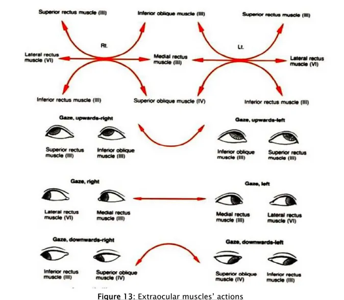
e. Diplopia:
Diplopia is initially most often due to incarceration of the lower right muscle in the fracture site at the orbital floor. It is a therapeutic emergency. When the muscle is not released in the few hours following the trauma, fibrosis caused by muscular ischemia results in persistent and most often definitive diplopia. In rare cases, diplopia is of neurological origin (contusion or injury of the oculomotor nerves in the superior orbital fissure) and is often accompanied by suggestive associated signs (mydriasis, ptosis). CT makes it possible to specify if the orbital cone bone is affected.
f. Enophthalmos:
They are explained by the orbital volume increase related to the collapse of the side walls and especially the orbital floor. The absence of anatomic surgical repair of these walls causes definitive enophthalmos and diplopia.
g. Infra orbital hypoesthesia (V2):
It is explained by an impairment of the nerve at its intra-orbital trajectory and / or at its emergence (infraorbital foramen). The reduction of the fracture, possibly associated with the nerve release the nerve in the foramen, allows in almost 80% of the cases a complete recovery of the sensitivity in several months.
h. Mouth opening Limitation:
The mouth opening limitation is explained by the snapping of the temporal muscle tendon at the temporal process of the zygomatic bone. It usually regresses after fracture reduction and reeducation. If it persists, premature contact between the coronoid process and the posterior surface of the zygoma (malunion) should be looked for.
i. Infectious complications
Zygomatico maxillary fractures are considered to be deeply open in the maxillary sinus. Intraorbital infectious complications, as rare as they are, are always possible and their occurrence should be feared. Thus, prophylactic antibiotherapy is necessary.
3. Paraclinical investigations:
• Conventional radiography: Water’s view • CT scaning
• Lancaster test • Forced duction test
4. Treatment:
• Time between trauma and admission • Operating time
• Surgical approach • Surgical protocol
5. Evolution :
a) Immediate assessment: Before discharge b) Long term evolution
• Clinical follow up • Scar evolution
I. Epidemiological data:
1. Age :
The patients’ main age was 43 years (extremes: 21-65 years).
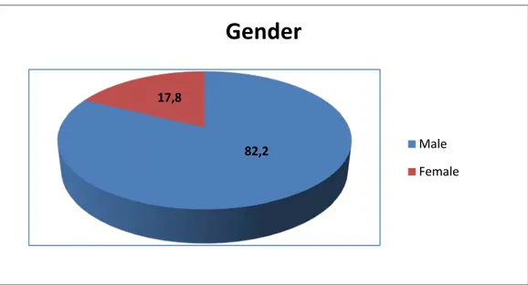
2. Gender :
Our study included 37 males (82,2%) and 8 females (17,8%).
Figure 01: Gender distribution of the studied group
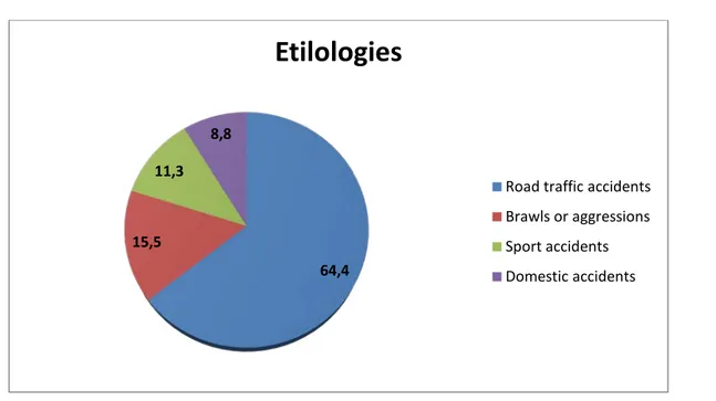
3. Etilologies :
The most usual circumstances of the occurrence of the traumatism are: • Road traffic accidents: 29 cases from 45 (64,4%).
• Brawls or aggressions: 7 cases from 45 (15,5%). • Sport accidents: 5 cases from 45 (11,3 %). • Domestic accidents: 4 cases from 45 (8,8 %).
82,2 17,8
Gender
Male Female
Figure 02: Distribution of ZMC fractures’ etiologies
4. Fractured side:
The right side was the most frequently injured in our study in 53,34% of the cases (24 patient), the left side in 46,66% (21 patient).
Figure 03: Distribution of the fractured side
64,4 15,5
11,3 8,8
Etilologies
Road traffic accidents Brawls or aggressions Sport accidents Domestic accidents
46,66 53,34
Side of the fracture
Left Right
5. Mechanism of trauma :
The mechanism of zygomatic trauma was direct in 94,29% of the cases and indirect in 5,71% of the cases. Their occurrence may follow impacts with moderate to high energy, either by a direct blow on the malar eminence or violent blows on the contra lateral midface causing a dislocation by reciprocal transfer of forces from the opposite side of the facial skeleton.
II. Clinical signs:
1. Mouth opening limitation:
Out of 45 patients 25 (55,56%) had limitation of mouth opening.
2. Enophthalmos :
Enophthalmos was found in 6,66 % in 3 patients.
3. Diplopia :
Among patients who had enophthalmos, 2 (4,5%) were diagnosed with vertical diplopia.
4. Sensory disorders :
Sensory disorders were common and they represented 35,56% (16 cases). They consisted in hypoesthesia at the region of the infraorbital nerve V2 (Lower eye lid, upper lip and lateral side wall of the nose).
5. Skeletal deformities :
A total of 29 (64,5%) patients had skeletal deformities such as flattening of the malar prominence, deformity of orbital margin and deformity of zygomatic buttress.
6. Associated signs :
41 (91,3%) of patients other clinical signs were palpebral edema, ecchymosis and wounds.
Figure 04: Distribution of clinical signs presented by our group study 55,56 6,66 2 35,56 64,5 91,3 0 10 20 30 40 50 60 70 80 90 100 Mouth opening limitation
Enophtalmos Diplopia Sensory
disorders deformities Skeletal Echymosis and wounds
III. Radiographic features :
1. Conventional radiography:
1.1. Water’s view radiography:
It was achieved in all our patients showing the direct and indirect signs of the fracture Directs signs: line fractures on facial bones.
Indirect signs: widening of the orbital frame, maxillary sinus opacity recalling a hemosinus, tear drop sign signing the herniation of intra-orbital fat in the maxillary sinus.
Figure 06: ZMC radiographic markers on Water’s view radiograph [5]
Dolan’s lines:
A: Orbital line , B: Zygomatic line ,C: Maxillary line
1.2. Orthopantomogram:
2. Computed tomography (CT):
The CT scan is necessary in case of diagnostic doubts (bone superposition on standard imaging, slightly displaced fracture) and comes to be very useful, especially in cases of ocular functional signs, to assess the importance of the orbital walls fractures (floor in particular). In this context, centered on the orbit with frontal and sagittal reconstructions are the most informative.
In our study, CT scans were achieved in all the cases in axial, coronal and sagittal sections. We also accomplished 3D reconstructions in every case.
3. Results of the radiological assessment:
Our study included only patients with CT scans where the association of the following three fracture components was identified (Stage B of Zing’s classification):
• Fracture of the zygomatic arch and/or diastasis of the temporozygomatic suture. • Fractures of the inferior orbital rim and anterior and posterior maxillary sinus walls
and/or diastasis of the zygomaticomaxillary suture.
On 3D reconstructions we can evaluate the displacement of ZMC according to the axis of rotation:
Vertical axis: Formed by the line passing through FZ suture and first molar tooth, the ZMC rotates medially or laterally.
Horizontal axis: Formed by the infraorbital foramen and horizontal arch, the ZMC moves upward or downward.
IV. Treatment:
1. Time between injury and admission:
It was from 4 to 15 days.2. Time of intervention:
It was mainly of 2h.3. Course of the intervention:
3.1. Admission of patients:
All of our patients were admitted at the acute phase and were administered analgesics and corticosteroids (Solumédrol 1 à 2mg/kg/day) for 05 days. For infectious risk, our patients were administered prophylactic antibiotics.
3.2. Patients’ positioning, prepping and draping:
− Supine position
− Neutral head positioning
− Sterile drapes were placed in a way that exposes the facial massif and the iliac crest (for bone graft if necessary)
3.3. Surgical approach:
a. Exposure:
Exposure was achieved for our patients through:
Lateral eye brow incision giving access to the frontozygomatic suture and subtarsal incision giving access to areas along the orbital floor, the medial and lateral rim.
b. Reposition:
Repositioning was achieved through percutaneous Ginstet hook reduction.
The hook was introduced through the intersection point of the vertical line passing through the lateral canthus and the horizontal line passing through the nostril. With the hook securely anchored into the bone, the fractured zygoma is rotated anteromedially.
c. Orbital floor revision:
Orbital floor was explored; inferior rectus muscle was freed when entrapped and fat hernia was reduced. Infraorbital nerve was gently released when compressed.
d. Orbital reconstruction (If necessary):
Orbital floor reconstruction was achieved when reduction of the thin bone fragments was not possible or insufficient to avoid a soft tissue displacement. Materials with different rigidity were used to cover or bridge the defect depending on its size and localization:
Prolene mesh was used for 4 patients who were had for small linear defects (up 1 to 2 cm) and patients with for larger defects (2 patients), iliac bone grafts were used.
e. Fixation :
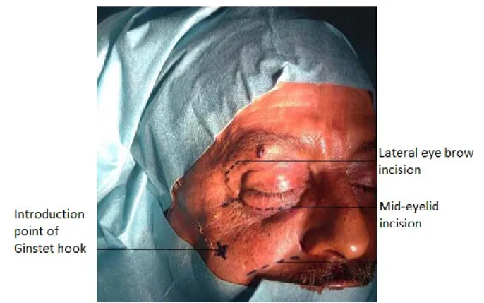
The reduced bones were fixated with plates and screws using 2 point fixation in the previously exposed areas: frontozygomatic fixation through lateral eye brow incision and infra orbital rim through an infra orbital incision.
Figure 08: Per operative image showing the used approaches in our study
V. Evolution :
1. Immediate post-operative assessment :
Evaluation of the patients vision was performed as soon as they are awakened from anesthesia and then at regular intervals until they were discharged from the hospital.
1.1. Postoperative positioning :
Keeping the patient’s head in an upright position both preoperatively and postoperatively significantly improves periorbital edema and pain.
1.2. Nose-blowing :
To prevent orbital emphysema, patients were told to avoid nose blowing for at least 10 days following orbital fracture repair.
1.3. Medication :
The following medications were prescribed to our patients: analgesia, antibiotics (Amoxicillin clavulanic acide 3g/day), nasal decongestant may be helpful for symptomatic improvement in some patients, regular perioral and oral wound care including disinfectant mouth rinse.
1.4. Ophthalmological examination :
The following signs and symptoms were evaluated by ophthalmologists: vision, extra ocular motion, diplopia, globe position, lid position.
1.5. Postoperative imaging :
Postoperative imaging has to be performed within the first days after surgery. Waters radiographs were achieved to assess the fracture’s reductions.
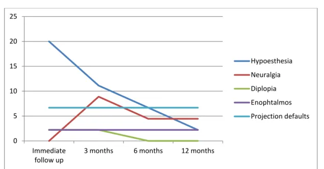
2. Long-term evolution :
Long term evolution evaluation examined the principal clinical symptoms: infra orbital nerve hypoesthesia, diplopia, neuralgia, enophthalmos, projection defaults and unfavorable scars. The patients all had an immediate (15 days), 3 months, 6 months and a 1 year follow up.
2.1. Clinical evolution:
Table I: Post-operative signs and their evolution Number of cases
15 days follow up 3 months 6 months 12 months
Hypoesthesia 9 5 3 1 Neuralgia 0 4 2 2 Diplopia 1 1 0 0 Enophthalmos 1 1 1 1 Projection defaults 3 3 3 3 a. Sensitive disturbance:
Persisted in 9 patients from 16 (56,25%) who had preoperative hypoesthesia. At the one year follow up: 1 patient did not fully recover from infraorbital hypoesthesia and 2 progressed to neuralgia.
b. Diplopia :
1 patient had persistence of his diplopia post-operatively 2,22% at the 3 months follow up, then disappeared at 6 month and 1 year follow up (2 patients had preoperative diplopia).
c. Enophthalmos:
Persisted in 1 case (2,22%) from 3 that had preoperative enophthalmos (33,3%). None of our patients presented post-operative enophthalmos.
d. Projection defaults:
6,66% suffer from loss of malar projection.
Figure 10: Evolution of post-operative clinical signs over 12 months 0 5 10 15 20 25 Immediate
follow up 3 months 6 months 12 months
Hypoesthesia Neuralgia Diplopia Enophtalmos Projection defaults
2.2. Scar evolution:
The subtarsal scar was not visible in any of our cases.
The lateral eye brow scar’s quality evaluation was carried out on its visibility, aspect, color and length. They have been ranked on a scale ranging from: invisible when they are totally confused with a fold of the skin, slightly visible when noticed from a distance of conversation and visible when spotted without bearing any special attention.
Among 45 lateral eye brow scars: 34 were invisible (75,5%), 6 were slightly visible (13,3%), 5 were visible (11,2%).
I. Epidemiology:
1. Age:
ZMC fractures affect young adults in their 30’s and 40’s [6], [7], [8], [9], [10]. The main age of our study was 43 years, which is consistent with data from the literature; this young age is explained by the frequency of exposure to risky behavior.
2. Gender:
Male predominance is observed in most series with a sex ratio of 5/1 [6], [7], [8], [9], [10]. In our study sex ratio was of 4/1 in our series, and that is due to the nature of military population treated in our series that is predominantly masculine, young and active.
3. Etiologies:
The most common etiologies are road accidents (42,37% and 43,8%) and assaults (64,5% and 38%) [6], [7], [8], [9], [10]. The results of our series are similar to those of the literature: road accidents represent 82.2% and assaults 17,8%. The other etiologies are much less frequent represented by sports accidents and domestic accidents.
4. Side of the fracture :
The results of the literature reveal a predominance of the left side of 62% against 38% of the right side[8],[9].While in our series we have a predominance of the right side (53,34%) against 46,66% of the left side.
5. Epidemiologic conclusion:
There was no significant difference between the results of our series and those of literature ; ZMC fractures account for 13% of all cranio-facial trauma [11] with predominance in young adult males, and that because of their frequent exposure to assaults and risky behavior. The most common etiologies are road accidents followed by aggression. The most affected side in the literature is the left unlike our series. That is most likely due to the fact that road accidents are far more common than assault (Where the opponent is usually right handed).
Table II: Comparative study to epidemiological data
Mean age Gender Etiology
Our series 43 years 4/1 Road traffic accidents: 64,4%
Assaults:15,5% Hwang et Kim[6] 34,7 years 5/1 Road traffic accidents: 23% Assaults:19,4% Bogusiak et al.[7] 37,1 years 5/1 Road traffic accidents: 13,9% Assaults:64,5%
Uda et al.[8] 33 years 4/1 Road traffic accidents: 11,9%
Assaults:38%
Forouzanfar et al.[9] 39 years 3/1 Road traffic accidents: 42,37%
Assaults:19,06%
Olate et al.[10] 31 years 5/1 Road traffic accidents: 43,8%
Assaults:18,3%
II. Clinical study:
1. General examination:
In general patients with any facial trauma should be considered as patients with panfacial trauma and possible multisytemic injuries.
A (Airway), B (Breathing), C (Circulation) should always be considered first. It is therefore necessary to look for surgical emergencies that may be life-threatening such as massive
bleeding (facial wounds, rhinorrhea, midface fractures...) and airway obstruction (bifocal fractures of the symphysal region with glossoptosis, inhalation of blood, dental fragments or pieces of dental prosthesis ...). Multisystemic injuries should also be looked for and stabilized (unstable cervical spine lesions, cerebral contusions, limb trauma).
2. Local examination:
2.1. Inspection:
Locally, the appearance of patients with ZMC fractures is quite remarkable. As for all facial traumas, edema is very important, installs in the few hours following the trauma and persists for several days. It is localized over the malar prominence, lateral orbit, upper and lower eyelids, associated to ecchymosis and tenderness. Loss of malar projection with increased width of the face are also noticed, they can be masked by the importance of the edema. External subconjunctival hemorrhage might be seen, it is explained by the subconjunctival diffusion of the peri-fracture hematoma at the frontozygomatic suture level.
2.2. Palpation:
Physical examination can reveal significant malar depression with step defects at the infraorbital rim, frontozygomatic suture, and zygomatic buttress of the maxilla intraorally. Fractures of the zygomatic bone evoke pain on palpation in more than 90% of patients in our series.
2.3. Clinical findings :
Posterior displacement of the fracture fragment may disrupt movement of the mandible, causing difficulty with mastication. Inferior displacement of the lateral canthal angle may indicate inferior migration of the fractured zygomatic bone [12]. Anesthesia in the distribution of the infraorbital nerve should be carefully evaluated. Many patients will not have sensation over the cheek preoperatively, which should be brought to their attention to avoid postoperative concerns. The same is true for trismus. Many patients with significant malar injuries will have
some pain and difficulty in opening the mouth because of impingement of the coronoid process by the displaced malar fragment. This pain may be slow to resolve postoperatively, but it almost always does so without specific intervention. Perhaps the most important part of the preoperative surgical examination is the ophthalmologic assessment. The zygoma constitutes the floor and lateral wall of the orbit and, as such, is always involved in the fracture. In addition, the medial wall is frequently fractured. Consequently, findings such as diplopia or other motility disturbances are not uncommon. In many cases, this is the result of non-mechanical factors, such as contusion of the extra ocular muscles or swelling, which resolve over time. However, entrapment of the muscles in the orbital fracture also occurs, which may influence the decision to proceed with surgery or the timing of surgery [13].
3. Clinical signs:
3.1. Enophthalmos:
It results from a retrusion of the ocular globe into the orbit by several mechanisms • Widening of the orbital frame
• Herniation of fat and a muscle through the orbital floor • Both previous mechanisms at once
The position of the globe is affected by the integrity of the periorbital fascial support, the direction and degree of displacement of the zygoma, and the degree of concomitant swelling. Fractures that cause an increase in orbital volume (blow-out fracture) will predispose to enophthalmos, but during the acute period, swelling within the orbit may cause some degree of proptosis despite the expansion of the orbit. As swelling resolves, the globe progressively sinks back, revealing the underlying orbital expansion. Zygomatic fractures that present with acute enophthalmos indicate severe displacement and orbital expansion [14].
3.2. Diplopia:
Its frequency varies from 1 to 49% according to literature.
Diplopia is a troubling and not uncommon complication of malar fractures that is reported in 3.4 to 8 percent of cases [16], [17]. It is perhaps most problematic when in the primary field of vision. Diplopia occurring only at the extremes of gaze is generally better tolerated. This complication may be caused by simple swelling or contusion of the extraocular muscles or their supplying nerves, in which case the diplopia should resolve over time. A normal, forced duction test at the end of the surgery makes this scenario more likely. The biggest concern, however, is entrapment of the extra-ocular or periorbital muscles in the orbitalfloor or medial wall component of the injury. When in question, a postoperative computed tomography scan should help rule this out [13].
Post traumatic diplopia is often due to a fracture of the lower orbital wall with entrapment of the inferior rectus muscle, resulting vertical diplopia. This entrapment will be sought by performing a CT scan with coronal cuts and sagittal reconstruction. It can also be secondary to globe displacement or orbital floor disruption.
The Lancaster test is used to monitor the persistence or disappearance of diplopia. When diplopia is definitive it is often due to permanent entrapment or fibrosis of the oculomotor muscles or nerve palsy.
Diplopia
Static
Dynamic
Orbital floor lowering
Extraoccular muscles
Entrapment
The semiological approach of post-traumatic diplopia obeys to several rules:
There is no parallelism between the importance of fracture and oculomotor disorders. The absence of diplopia does not exclude secondary diplopia hence the interest of a Lancaster test immediately after trauma and another for control.
Extraocular movement should also be tested, as should visual fields. Restriction in the movement of the extraocular muscles, especially on upward gaze, should raise the physician's concern for muscle entrapment. When in doubt, either a forced duction test can be performed with local anesthetic or computed tomography (CT) scans can be performed to examine for extraocular muscle entrapment. The exception to this, though, is the patient who presents with facial trauma, suspected orbital floor fracture, who also has nausea, vomiting, and bradycardia (oculocardiac reflex), which is pathognomonic for extraocular muscle entrapment [18].
In our study, evaluation of ocular motility was first done by finger gaze where the finger is moved in front of the eye in all nine directions of gaze at a distance of 30 cm. It essentially seeks limitations of elevation or lowering that theoretically predict the site of the fracture of the orbital floor relative to the equator of the globe.
When diplopia is confirmed, completing by a Lancaster test is useful. This test allows quantifying limitations on a diagram and following the evolution.
a. Forced duction test:
Should be performed 0Tanaesthetized 0Teye, lids retracted. The examiner manipulates the
globe through its entire range of motion with a globe holding forceps. The inability to rotate the globe superiorly signifies entrapment of inferior rectus muscle in the orblital floor. This test is only significant when performed comparatively to the uninjured side. It signs the mechanic origin of oculomotor disorder.
Figure 12: Forced duction test
3.3. Sensory disturbance:
Palpation sometimes shows subcutaneous emphysema signifying a communication between the orbital tissues and surrounding sinus cavities; it also evaluates sensory disturbances in the territories of infra-orbital and zygomatico-temporal nerves.
In simple ZMC fractures, hypoesthesia will most often occur in the territory of the infra-orbital nerve. Hyperesthesia may also be found.
Sensation in the V2 distribution should be tested and noted as sensation in this area is
almost always diminished in malar fractures. Therefore, it is important to make note of this finding to avoid postoperative concerns of iatrogenic nerve dysfunction. Sensation often resolves postoperatively without specific treatment. Other physical exam findings are obscured by swelling encountered in the majority of these patients and are not helpful in the acute setting [18].
The frequency of hypoaesthesia varies between 8 and 52% according to literature. As for our series hypoesthesia represents 35.56%.
3.4. Mouth opening limitation:
Limited mouth opening may be present and is generally mild and is typically due to pain with masseteric pull given its attachment to the zygoma. Severe displacement may cause direct impingement on the coronoid process.
Mouth opening limitation varies from 8 to 25%. In our study it represents 55,5%. Table III: Comparison of our clinical findings to literature
Our Series Bogusiak et al.[7] Olate et al.[10] Forouzanfar et al.[9] Zhang et al.[19]
Pain, Echymosis, Edema 91,3% 91,4% 49,7% 30% 8,17
Limitation of mouth pening 55,56% _ 25,5% 13,6% 4,67%
Hypoesthesia 35,56% 58,5% 52,9% 47% 8,64%
Diplopia 4,5% 49,1% 10,4% 8,5% 1,28%
Enopthalmos 6,66% 65,8% _ 4,2% _
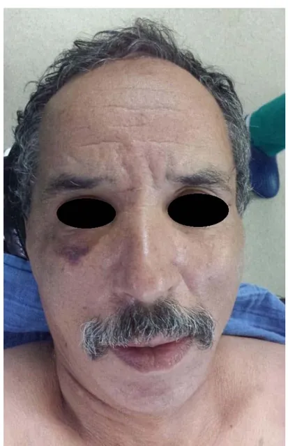
Figure 14: ZMC fracture’s clinical features at Day 8 of trauma
III. Paraclinical examinations:
1. Hess Lancaster test
Hess-Lancaster test is a red-green test that allows immediate diagnosis of the paralyzed eye and muscles and to recognize the hyper-muscular actions due to paralysis.
Figure 15: Hess Lancaster test showing vertical diplopia
2. Radiologic investigation:
2.1. STANDARD IMAGING:
It retains its usefulness, even if its importance decreases in favor of recent CT techniques. It is powerful and easy to achieve:
a. Waters view (Occipito mental): permits a good vision of the ZMC, orbital floor and infra-orbital rim. It is a posterior anterior incidence, where an X-ray beam is angled at 45° to the 0Torbitomeatal line0T. The patient’s nose and chin are placed in
contact with the midline of cassette. The patient should open the mouth as wide as possible before exposure. The rays pass from behind the head and are perpendicular to the radiographic plate (Figure 16).
This view is better analyzed using the three lines described by Campbell and McGrigor (Figure 17-18). These lines can be used as a simple to interpretation.
Figure 17: Campbell and McGrigor lines
1: This line is traced from one 0Tzygomatico frontal suture0T to another, across the superior edge of the 0Torbits0T
2: This line traces the 0Tzygomatic arch0T, crosses the 0Tzygomatic bone0T, and traces across the inferior orbital margins
to the contralateral zygomatic arch
3: This line connects the condyle and coronoid process of the 0Tmandible0T and the 0Tmaxillary0T antra on both sides
Figure 18: Tripod fracture on Waters view using Campbell and McGrigor lines
1-Zygomatico Frontal suture fracture
2-Comminuted fracture at the zygomatic arch 3-Orbital floor fracture
b. Submentovertex view: this projection is obtained with the patient’s neck
extended either in the supine or upright position. The top of the head is placed so that the infraorbitometeal line is parallel with the x-ray cassette. The x-ray beam is directed at right angles to the infraorbtometeal line. This projection gives a good view on the zygomatic arch.(Figure 19)
Figure 19: Submentovertex view positionning
CR: Central Ray, IM: InfraorbitoMeteal
2.2. CT SCAN:
Over the years, CT scanning has supplanted plain radiography as the imaging modality of choice. Almost all malar fractures require direct CT scanning in both the axial and coronal planes (< 3-mm slice thickness) to categorize the pattern of injury clearly and direct subsequent management [18].
If direct coronal images cannot be obtained, as in the case when neck extension is precluded by possible cervical spine injury, then thin-section axial helical scans can be reformatted to obtain coronal sections [13].
reference for reduction. However, if the buttresses are comminuted, the surgeon may need to expose and reduce the zygomatic arch via a scalp incision to ensure that the zygoma is adequately anteriorly projected. The typical clinical and radiologic deformity of a ZMC fracture is loss of cheek projection and a resultant increase in facial width. A frequently missed ZMC fracture is at the temporal bone portion of the upper transverse maxillary buttress [20].
In severe ZMC fractures, the orbital defect can appear minimal due to impaction of the zygoma. It is important to visualize the defect with the zygoma in its anatomic position to appreciate the true loss of bone support. For orbital floor defects, attention to the shape and position of the inferior rectus muscle on coronal CT scans can provide information regarding the damage to the fascial sling of the globe [20]. CT has made preoperative assessment of the status of the bony orbit possible with a great degree of accuracy. When preoperative CT scans are available, it is no longer necessary to discuss whether the internal orbit should be explored [21].
Feuerbach [22] stated that extensive plain film studies following massive trauma are technically difficult and yield a relatively small amount of information. Using 3D CT areas of clinical interest can be isolated by volume rendering technique from the patient and viewed in a variety of orientations, not possible using other methods and in complex anatomical images such as facial bones. 3D images can make interpretation of an otherwise difficult set of cross-sectional CT images easier and less time consuming [23].
Figure 20: 3D reconstruction showing a left ZMC fracture (Type B)
2.3. Magnetic resonance imaging:
MRI should be considered in severe and extensive cases, where thorough soft tissue evaluation is important. Studies showed its efficiency to assess orbital complications involved in ZMC fractures.
Possible structural herniation or entrapment of the infra-orbital nerve, should consider MRI to assess the involved soft tissues [24]. In fact, Ilankovan et al. [25] found MRI more to be sensitive, in comparison to CT, for the diagnosis of herniation and entrapment of soft tissues in orbital fractures.
However, in more severe and extensive cases, where soft tissue differentiation is essential, high-resolution CT should be preferred over MRI if there is a possibility that a metallic
2.4. Ultrasonography:
Ultrasonography has traditionally been used in orbital and ocular diagnosis, but its role in maxillofacial trauma is less widely recognized. McCann et al. [27] used ultrasound with 85% accuracy in diagnosing fractures of the ZMC. According to Friedrich et al. [28], application of ultrasound is most useful for visualization of the zygomatic arch and the anterior wall of the frontal sinus.
Kim et al. [29] used intraoperative ultrasonography to evaluate alignment of the inferior orbital wall, ZF buttress, and zygomatic arch. Ultrasonography is a valuable visualization tool for both diagnosis and treatment of facial bone fractures [30], [31].
IV. TREATMENT:
1. Aims of treatment:
The goal of all surgical procedures should be to repair function and esthetics deficits: • Restore normal contour of the face
• Relieve pain
• Precise anatomical reduction of the fragment • Provide stable fixation of the reduced fragment
• Correct the complications: diplopia, remove any interference in range of mandibular movement, relieve pressure from infra-orbital nerve
2. Indications:
Indications for treatment of ZMC fractures depend on two features: function and esthetics.
2.1. Conservative treatment:
Conservative treatment is warranted in minimal to non-displaced fractures in which there is no cosmetic or functional deficits, or in medically unstable patients in which undergoing on operation with general anesthetic may be too great of risk [15].
2.2. Surgical treatment:
The decision to intervene surgically in patients with ZMC fractures should be based primarily on whether there is displacement of the malar complex and the existence of functional findings. The necessity of internal fixation is then judged.
a. Reduction without fixation:
For mildly displaced ZMC fractures, especially those involving 1–2 articulations, often times the reduced segments may be stable enough to avoid fixation. It is best to complete the procedure within 2–3 weeks of the initial trauma to avoid early fibrous union of bony segments which can make reduction difficult. This method can be completed via multiple open or closed approaches depending on the fracture location and necessity for direct visualization of the segments to confirm reduction [15].
b. Reduction with fixation:
Open reduction and internal fixation of ZMC fractures is indicated in largely displaced, comminuted fractures, or in mildly displaced fractures in which stable reduction is not achieved following reduction. As described by Ellis et al., anatomically accurate reduction of the ZMC is best obtained by direct visualization of multiple sutures if necessary. Additional fixation is not related to better outcomes if the proper reduction was not completed initially [14], [21].The issue that our study raises is how much fixation is enough fixation?
In our series all of our patients were treated with ORIF with two point fixation in the FZ suture and infra-orbital rim through a lateral eye brow incision and and mid eyelid incision respectively.
3. Contraindications:
Fractures of more than 20 days that may require osteostomy Minimally displaced fractures with no symptoms
Eldery and medically compromised patients
4. Treatment means:
4.1. Medical treatment:
Medical treatment is prescribed for preoperative preparation of the patients. It can also be the only indicated treatment when surgery is not indicated. It includes:
Up right head position to relief the pain and improve the edema resolution Analgesia
Corticosteroids for resolution of edema
Antibiotics: Amoxicillin clavulanicacide 3g/day for 7 days
In our study, prophylactic antibiotics were prescribed for all our patients for sinus coverage since we considered ZMC fractures to be open fractures. For Lee et al. [18] antibiotics are not indicated in non-displaced fractures. Andreasen et al. [32] concluded in their systematic review that infection rates were so low in isolated zygomatic fractures that prophylactic antibiotics were not recommended.
Corticosteroids are initiated by many surgeons to minimize swelling and further damage to the optic nerve. In addition, surgery is delayed until vision has stabilized or improved [33], [34], [35].
4.2. Surgical treatment:
Surgical treatment in generally indicated for displaced fractures should be surgically reduced and stabilized. The degree of displacement can be easily checked by assessing the status of the normal articulations of the ZMC with the craniofacial skeleton on CT scan.
a. Adequate time for surgery:
Surgery is indicated when there is impairment of function —that is, limited mouth opening—and/or when the patient complains of an aesthetic problem— that is, a flattening of one side of the face. It is not specifically indicated for parasthesia. Surgery is often best deferred until the swelling has settled and the patient can be assessed fully [36].
Hwang et al [6] carried out their surgical procedure on a an average of 6.4 days after injury, and most had surgery within 1 week (58.2%). Yamsani et al. [37] treated the majority of their patients 7 days after their reporting. For some authors, the adequate time depends also on existing neuropathy: surgery is delayed until vision has stabilized or improved [33], [34], [35].
In our series, the average time of surgical intervention was of 9,5 days, most patients were treated after the 8th day, to give enough time for the edema to be resolved. The surgery is
then performed under satisfactory local conditions to have better approaches and promote better healing.
b. Surgical techniques:
They should be individualized according to severity of the fracture and associated injuries, but it should always focus on anatomical reduction of the malar position and the orbital anatomy.
The treatment should be as minimally invasive as possible to avoid multiple surgical approaches, consequent potential infections, additional scars, and nerve palsy and should provide precise physical stability of the zygoma.
b.1. Surgical approaches:
The ideal surgical approach to treat fractures of the ZMC should provide enough exposure of the fractured segments, ensure less potential for further injury to facial structures, and allow for good cosmetic results. Ideas differ sharply as to the surgical approach from a surgeon to another.
The aesthetic appearance is very important in maxillofacial surgery. Placing an incision on the face does not only take under consideration the surgical requirements, but also many aesthetic criteria.
Usually, the skin incisions should be made parallel to the Langer lines found throughout the body skin and whose orientation of 0Tcollagen0T fibers in the 0Tdermis0T, and are generally parallel
to the orientation of the underlying muscle fibers. As wrinkles become more and more visible with age, it is recommended to place incisions directly in or alongside future wrinkles. These incisions are made to expose the fracture sites and allow solid fixation after anatomical reduction.
This chapter will discuss the different fixation sites in ZMC fractures and the possible surgical approaches to each one.
Extra oral approach : Superolateral approach :
In most cases one of these approaches will be the only incision necessary for treatment, given the relative strength of the ZF pillar, which typically makes it the last buttress to be displaced. If indeed this displacement is seen on the preoperative CT scan, then consideration can be given to making a lateral eyebrow or upper-lid incision to visualize this buttress [18].
• Supraorbital approach : lateral eyebrow
The lateral eyebrow approach provides simple and rapid access to the FZ suture and lateral orbital rim. No functionally important neurovascular structures are at risk in this approach. As long as the incision is not placed within the eyebrow hairs the resulting scar is usually well concealed according to our experience. The location of the lateral brow incision should be over the fractured segments and placed within or in close proximity to the eyebrow for some authors [15]. Sharp incision is carried directly down to bone, and subperiosteal dissection is performed to expose the frontozygomatic and down the medial lateral orbital wall to visualize the zygomatico sphenoid suture.
Hwang el al. [39] used this approach on 14 patients; they were all satisfied with their postoperative appearance. Thangavelu et al. [40] concluded that in this technique, a visible scar is seen in the lateral orbital region; however, in all displaced fractures fixation at lateral wall of orbit is a must. For Kung et Kanaban [41], this approach was seen as an advance in treatment because it allows good access to the FZ suture, however the resultant scar is often perceptible in poorly planned incisions, in patients with eyebrow hair loss, and in those who do not have eyebrows extending laterally and inferiorly along the orbital margin.
• Upper eyelid approach