J?//, 3/ç,,
C
Université de Montréal
Resolution of Deep Venous Thrombosis in Type 2 Diabetes Mice Mode!:
Implication of Fibnnolytic and Matrix Metalloproteinase Systems
par: Xiaochun Zhang
Programme de Sciences Biomédicales faculté de Médecine
Mémoire présenté à la Faculté des Études Supérieures en vue de l’obtention du grade de Magister Scientiae (M.Sc.) en Sciences Biomédicales
Avril 2007
E
Q
i2
Université
(111
de Montréal
Direction des bibliothèques
AVIS
L’auteur a autorisé l’Université de Montréal à reproduite et diffuser, en totalité ou en partie, par quelque moyen que ce soit et sur quelque support que ce soit, et exclusivement à des fins non lucratives d’enseignement et de recherche, des copies de ce mémoire ou de cette thèse.
L’auteur et les coauteurs le cas échéant conservent la propriété du droit d’auteur et des droits moraux qui protègent ce document. Ni la thèse ou le mémoire, ni des extraits substantiels de ce document, ne doivent être imprimés ou autrement reproduits sans l’autorisation de l’auteur.
Afin de se conformer à la Loi canadienne sur la protection des renseignements personnels, quelques formulaires secondaires, coordonnées ou signatures intégrées au texte ont pu être enlevés de ce document. Bien que cela ait pu affecter la pagination, il n’y a aucun contenu manquant. NOTICE
The author of this thesis or dissertation has granted a nonexciusive license allowing Université de Montréal to reproduce and publish the document, in part or in whole, and in any format, solely for noncommercial educationaT and research purposes.
The author and co-authors ifapplicable retain copyright ownership and moral rights in this document. Neither the whole thesis or dissertation, flot substantia extracts from it, may be printed or otherwise teproduced without the author’s permission.
In compliance with the Canadian Privacy Act some supporting forms, contact
information or signatures may have been removed from the document. While this may affect the document page count, it does flot represent any loss of content from the document.
faculté des Études Supérieures
Ce mémoire
intitulé:Resolution of Deep Venous Thrombosis in Type 2 Diabetes Mice Model:
Implication of Fibrinolytic and Matnx Metalloproteinase Systems
Présenté par:
Xiaochun Zhang
(.
A été évalué par un jury composé des personnes suivantes:
Président-rapporteur: Docteur Muhammad Zafarullah
Directeur de recherche : Docteur Jean Raymond
Co-directrice de recherche : Docteur f
atiha
Bouzeghrane
Membre du Jmy: Docteur Gaétan Thibault
CONTENTS
SUMMARY .W
SOMMAIRE.V
CONTENT.VII
LIST 0F TABLES.XII
LIST 0F FIGURES Xffi
LIST 0F ABBREVIATIONS XVI
SUMMARY
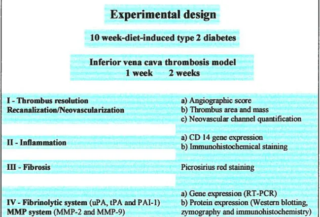
TYPE 2 DIABETES is a group of metabolic disorders that may resuit in a procoagulant and thrombogenic predisposition, which is related to the arterial complications. However it remains undetermined whether diabetic conditions may affect deep vein thrombosis (DVI) and its resolution. The objectives of this study were to determine the effect of diet-induced type 2 diabetes on the organization, resolution and recanalization of venous thrombi, the inflammatory response, and the fibrinolytic and MMP systems in a murine experimental model of venous stasis as assessed by angiography and molecular techniques.
The resolution and recanalization of DVI was decreased in type 2 diabetic mice as revealed by angiography, thrombus size and content and neovascular channel
(J
quantification by immunohistochemistry. Recmitment of monocyte/macrophages, detected by an anti-CD68 antibody, was ïncreased, and a higher coïlagen deposition was found in the thrombosed inferior vena cava of diabetic mice. The plasminogen activators, u-PA and t-PA were downregulated, and their inhibitor, PAl-1 was upregulated conferring a relatively antifibrinolytic state in diabetic mice. The MMP system was enhanced in diabetic mice at one week post DVI followed by a decreased synthesis and activity at 2 weeks.Diet-induced type 2 diabetes may impair the organization, resolution and recanalization of DVI through increased inflammatoiy response and dismption of fibrinolytic and MTvIP systems.
V
SOMMAIRE
LE DIABÈTE DE TYPE 2 est un groupe de désordres métaboliques dont l’impact est une atteinte artérielle, microvasculaire et macrovasculaire aboutissant à la dysfonction endothéliale avec pour conséquences: inflammation, hypercoagulabilité et thrombogénicité. Cependant, il n’est pas établi si ces désordres métaboliques ont la même répercussion sur le système veineux et notamment sur les mécanismes de l’organisation et la dissolution du thrombus veineux. Grâce à un modèle animal de diabète de type 2 par consommation de diète riche en lipides et un modèle de stase veineuse par ligature de la veine cave inférieure chez la souris, nous avons pu déterminer les effets de ces désordres sur la résolution du thrombus veineux. Les souris diabétiques présentent une diminution de la résolution et de la recanalisation du thrombus veineux comme l’indiquent les résultats angiographiques, la taille du thrombus et la quantification des néo-vaisseaux par immunohistochimie. La réponse inflammatoire détectée au niveau de l’expression génique du CDL4 et par immunolocalisation d’un marqueur des macrophages le CD68, est fortement activée, accompagnée d’un dépôt de collagène au sein de la paroi veineuse. Le système fibrinolytique est également atteint par une réduction de l’expression des ARNm et des protéines des activateurs du plasminogène (u-PA et t-PA) et par une régulation à la hausse du profil d’expression du PAT-l. L’expression des métalloprotéinases MMP-2 et MMP-9 est temporellement affectée au cours de la thrombose veineuse. Après une induction!activation du système à une semaine après formation du thrombus, une réduction de la synthèse/activité est observée chez les animaux diabétiques.
C
C
Le diabète de type 2 induit par une diète enrichie chez la souris semble altérer l’évolution du thrombus veineux à travers une réponse inflammatoire amplifiée, une activité du système fibrinolytique diminuée couplée à un système des MJVWs activé mais dont la modulation à la baisse semble durée-dépendante.
VII
INTRODUCTION
.11.1 DIABETES MELLITUS .1
1.1.1 Overview J
1.1.2 Cardiovascular complications and pathogenesis 1
1.1.3 Diabetes and risk ofthrombosis 4
1.1.3.1 Endothelial dysfunction 4
1.1.3.2 Increased adhesion ofplatelets and monocytes 5 1.1.3.3 Abnormal fibrinolysis and hypercoagulation 5
1.1.4 Diabetic mouse models 7
1.2 DEEP VENOUS THROMBOSIS 9
1.2.1 Prevalence and risk factors 9
1.2.2 Normal venous anatomy 10
1.2.3 Resolution ofthrombus 13 1.2.3.1 Cellularpathway 14 1.2.3.1.1 Inflammatoryceils 14 I.2.3.1.2Endothelial ceils 15 1.2.3.1.3 Myofibroblasts 17 1.2.3.1.4 Platelets 1$ 1.2.3.1.5 Progenitor celis 19 1.2.3.2 Molecularpathway 20
1.2.3.2.1 The fibrinolytic system and major components 20 1.2.3.2.2 Matnx metalloproteinases and their inhibitors 24
1.2.3.2.3 Extracellularmatrix.25
1.2.3.2.4 Selectins 27
1.2.3.2.5 Proangiogenic factors 2$
1.2.4 Treatment of deep vein thrombosis 29
1.2.4.1 Standard treatments 29 1.2.4.2 New approaches 30 1.2.4.3 Angiogenic therapy 31 1.2.4.4 Cell-based approach 32 1.2.4.5 Gene therapies 32 1.2.5 Treatment of complication 33
1.2.6 AnimaI models of venous thrombosis 34
II.
RESEARCH PROPOSAL
37
11.1. HYPOTHESES 38
11.2. RESEARCH GOALS 39
II. 2. 1. Main objective 39
Ix
III.
MATERIAL AND METHODS
.41ffi.1 Diet-induced type 2 diabetic mouse models 41 ffl.2 Animal model ofvenous thrombogenesis: a mouse inferior vena
cava stasis model 43
ffi.3 Angiography 44
ffi.4 Tissue harvest I Measurement of thrombus size and infrarenal vena
cava weight 45
ffi.5 flistopathologic and immunohistochemical ana]ysis 45
111.5.1 Macrophage content 4$
111.5.2 Neovascular channel quantification 4$
ffi.6 Western blot analysis 49
ffi.7 Zymographic activities 50
ffi.$ RNA isolation and RT-PCR analysis 50
ffl.9 Statistical analysis 52
IV.
RESULTS
53W. 1 Successfut development of type 2 diabetes in mice 53 W.2 Diabetic mice have less thrombus resolution 56
IV.2.1 Thrombus area 56
IV.3 Thrombus recanalization is impaired in diabetic mice .59
IV.3.1 Angiography 59
IV.3.2 Histological and immunohistological analysis ofneovascular
channels 61
IV.4 Diabetic mice have a higher
inflammatory response 65IV.4.1 Expression of CD14 mRNA 65
IV.4.2 Immunohistochemical staining of CD6$ 65
IV.5 Vein
wall
fibrosis is elevated in diabetic mice 70IV.6The
fibrinolytic system is altered in diabetic mice 73IV.6.1 u-PA and PAl-1 mRNA levels 73
IV.6.2 Expression of u-PA, t-PA and PAT-1 protein 76 IV.7
The MMP
system is enhanced in diabetic mice followed by adecreased synthesis
and
activity 81IV.7.1 Expression ofMMP-2 and MMP-9 mRNA 21
IV.7.2 Gelatinolytic activities ofMMP-2 and MMP-9 $1 IV.7.3 Expression ofMMP-2 and MMP-9 proteins $2
V.
DISCUSSION
89
V.! Type 2 diabetes decreascd the resolution and recanalization of
DVT 91
V.2 Type 2 diabetes increases inflammatory response in DVI 92 V.3 Type 2 diabetes elevated the vein waIl fibrosis in DVT 94 V.4 Type 2 diabetes alters the fibrinolytic and MMP system in DVT.. .95
M
V.4. 1 Type 2 diabetes inhibits the fibrinolytic system 95 V.4.2 Type 2 diabetes enhances MTvIP system 96
V.
CONCLUSIONS
99.
LIST 0F TABLES
Table 1: Proangiogenic factors expressed within resolving thrombus 29 Table 2: Potential new therapies to promote recanalization and resolution ofvenous
thrombi 33
Table 3: Summary ofspecific objectives 40
Table 4: Composition ofthe diets 42
Table 5: List ofantibodies 47
Table 6: Sequences ofprimers of selected genes for RT-PCR 51 Table 7: Angiography scores in control and diabetic groups 61
XIII
LIST 0F FIGURES
Figure 1: Four main pathways implicated in hyperglycemia-induced diabetic
microvascular disease 3
Figure 2: Vein wall with the intima underÏying the endothetium, the media and the
adventitia 11
Figure3: Cellular and molecular pathways during resotution of
thrombus 13
Figure 4: Schematic representation of the role played by endothelial celis in
coagulation and fibrinolysis pathways 16
Figure 5: An extensive network of additional proteases, inhibitors, receptors and
modulators 22
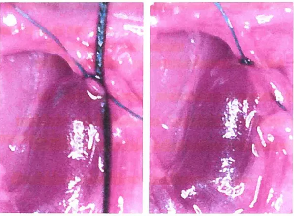
Figure 6: Rat inferior vena cava (IVC) stenosis model of venous
thrombosis 44
Figure 7: Body weight growth in control and diabetic mice 54 Figure 8: Blood glucose levels in control and diabetic mice 55 Figure 9: Thrombus areas in control and diabetic groups at 1 week or 2 weeks after
surgery 57
Figure 10: ThrombosedIVC mass/length in control and diabetic groups at 1 week or
o
Figure 11: Angiograms in control and diabetic groups at 1 week or 2
weeks 60
Figure 12: Neovasculization in 1 and 2 week-control and diabetic
groups 63
figure 13: Quantification of thrombus neovascular channels by positive GSL
1.staining 64
Figure 14: CDY4 mRNA expression in control and diabetic
mice 66
Figure 15: Macrophage content labeled by anti-CD68 antibody in the thrombus of control and diabetic mice at 1 or 2 weeks afier IVC
thrombosis 6$
Figure 16: Thrombus macrophage content in control and diabetic mice at 1 or 2
weeks afier IVC ligation 69
Figure 17: Picrosirius red staining of total collagen in control and diet-induced
diabetic mice at E week and 2 weeks afier
surgery 71
Figure 18: Collagen quantification in control and diabetic mice at 1 or 2
weeks 72
Figure 19: Expression of u-PA mRNA in control and diabetic
mice 74
Figure 20: Expression of PAT-1 mRNA in control and diabetic
xv
Figure 21: Western blot analysis of u-PA in control and diabetic protein
extracts 77
Figure 22: Western blot analysis of t-PA in protein extracts of control and diabetic
mice 78
Figure 23: Western blot analysis of PAl- 1 in protein extracts of control and diabetic
mice 79
Figure 24: Changes in u-PA and PAl-1 immunoreactivity afler I and 2-week DVT
in control and diabetic mice $0
Figure 25: Expression ofMMP-2 mRNA in control and diabetic mice 83 Figure 26: Expression ofMMP-9 mRNA in control and diabetic mice $4 Figure 27: Thrombosed IVC MrVLP-2 and MIVW-9 activities 85
Figure 23: ExpressionofMMP-2 in control and diabetic thrombosed IVC $6 Figure 29: Expression of MMP-9 in control and diabetic thrombosed
IVC $7
Figure 30: Changes in MMP-2 and MMP-9 immunoreactivity afier I and 2-week
LISI 0F ABBREVIATIONS
Œ-SMA: Œ smooth muscle actin.
Œv33 : integrin receptor.
B-fGF: basic fibroblast growth factor. CAMs: celi adhesion molecules.
CD 14: a celi marker of inflammation (especially macrophages). CD 31: a cluster of differentiation molecular
CHD: coronary heart disease. CVD: cardiovascular disease. CVI: chronic venous insufficiency.
C
DM: diabetesECM: extracellular matrix. ECs: endothelial celis.
ENA-7$: epithelial neutrophil activating protein. eNOS: endothelial ceil NOS.
EPCs: endothelial progenitor ceils. IL-1: interleukin-1.
IL-8: Interleukin-8.
IP-1O: interferon inducible protein. IVC : inferior vena cava
MC : monocyte
XVII
MCP- 1: Monocyte chemotactic protein- 1. MMPs: matrix metalloproteinases.
MT-MMPs: membrane typeMIVWs NO: nitric oxide.
NOS: nitric oxide synthase. PA: plasminogen activator PAF: platelet activating factor.
PDGf s : platelet derived growthfactor. PE: pulmonary embolism
PGI2: pro stacyclin.
PIGf : placenta! growth factor.
C
PMN: polymorphonuclear neutrophi!.SERPIN: serine proteinase inhibitor TF: tissue factor.
TGF-f3 1: transforming growth factor beta 1. Tie2: endothe!ial celi receptor tyrosine kinase. TIMPs: inhibitors of matrix metalloproteinases. TNF-Œ: tumor necrosis factor- a.
t-PA: tissue-type plasminogen activator. TXA2: thromboxane A2.
UK: urokinase.
u-PA: urokinase-type plasminogen activator.
o
VCAM-1 vascular celi adhesion molecule-1 VEGf: vascular endothelial growth factor. VN: vitronectin.
VTE: venous thromboembolism. \ÏWF von Willebrand factor. WPb: Weibel-Palade body.
XIX
ACKNOWLEDGEMENTS
I would like to thank the following people:
ÇZY.
Jean
Rgymond, for the enthusiasm and inspiration which were aiways there when I needed it.çlY
Fati6a Bouzeg6rane
for her mentorship and guidance, whose help, stimulating suggestions and encouragement helped me during ail the time of research and through the writing ofthis thesis.Çuy(aine Çeviy,
for her technical helpCfinitette Ogoiu(i€pe,
for help with molecular biology.J4udrey
cBoyce
for help with the surgical procedures.This work wouid flot have been possible without the financial contribution from the
Canaifum Instit utes ofJfeattfi Rçsearcfi
(CIIIR, MOP-44062) and theQue6ec
KeartanJStro FounJation.
I would like to take this opportunity to thank the
Facutté ées Etudes Supérieures,
Programme & Sciences CBioméd?cates d
r
Vn.iversité & MontréaC
L
INTRODUCTION
Li DIÂBETES MELLITUS
I. il Overview
Diabetes mellitus (DM) is a group of metabolic syndromes charactenzed by chronic hyperglycemia, disturbances of carbohydrate, fat and protein metabolism due to defects in ïnsulin secretion, insulin resistance or both (1, 2). Chronic hyperglycemia is associated with multi-organ damage to the eyes, kidneys, nerves, heart, and blood vessels (1). Cardiovascular disease is the leading cause of premature death arnong patients with dïabetes. The new classification system identifies four types of diabetes mellitus based on etiologv: type 1, type 2, “other specific types” and gestational diabetes (1). The World Health Organization (WHO) predicts that between 1997 and 2025, the number of persons affected with diabetes vill double from 143 to approximatelv 330 million (3).
Type 2 is the most common form of diabetes mellitus. It is a metabolic disorder that is primarily characterized by insulin resistance, relative insulin deficiency, and hyperglycemia and lead to function impairment of many organs, most importantly the cardiovascular system. Its prevalence is projected to rise in the future (5). Approximately 90-95% of diabetes is ascnbed to Type 2 (4, 7), the development of which is attributed to both polygenetic and environmental factors.
1.1.2 Cardiovascular complications and pathogenesis
Diabetes mellitus causes considerable morbiditv and mortality primarily due to microvascular (retinopathy, nephropathy, vasculopathy) and macrovascular
2
(ischemic heart disease, stroke, peripheral vascular disease) complications (6, 7), which can lead to considerable disability and premature death. Cardiovascular disease (CVD) is the major complication of type 2 diabetes and is responsible for more than 50% and up to 80% of deaths in people with diabetes as well as for substantial morbidity and loss ofquality of life (3).
Although the pathogenesis of CVD in diabetes is not vet fully underslood. multiple metabolic and endocrinologic factors are implicated (6, 7). Hyperglycemia and hypennsulinemia due to insulin resistance are two metabolic abnormalities associated with type 2 diabetes mellitus and result in macrovascular and microvascular complications in multiple organ systems which accounts for the morbidity and mortalitv associated with this disease.
The phenotype associated with insulin resistance includes a dyslipidemia that is characterized by increased very low-density lipoprotein triglyceride levels, decreased high-density lipoprotein-cholesterol levels, and the presence of small, triglyceride-enriched, low-density lipoproteins (6, 8, 9). Clinical trials have shown that correcting the hyperglycemia can attenuate some of the microvascular complications of diabetes, such as retinopathy and nephropathy, but cannot suppress macrovascular complications, such as coronary heart disease (CITD) due to atherosclerosis.
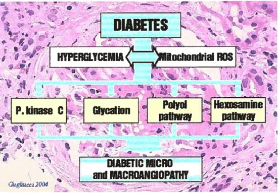
Oxidant stress and inflammation accelerate CHD by activating the diacylglycerol-protein kinase C (DAG-PKC) signal transduction pathway, possibly by enhanced formation of glycosvlated proteins and advanced glycation products and/or by increasing endothelial dysftinction (8). Recent studies have
shown possible biochemical mechanismsl by which hyperglycemia could cause its adverse effects on the vascular cells (figure 1).
Insulin possesses anti-atherogenic properties. Insulin increases mtrous oxide (NO) production, which can cause vasodilatation and retard the migration and growth of arterial smooth muscle ceils under physiological conditions (7). In pathological states of insulin-resistance, the enhancement of NO production from either acute activation of nitric oxide synthase (NOS) or endothelial ceil NOS (eNOS) by insulin are blunted (9). Moreover, hypennsulinemia may contnbute to atherogenesis by stimulating the growth and production of the extracellular matrix (ECM) (10). — w -- I v ‘ ‘W• ,
G
DIABETES
.. , À .. .7., 4 ‘€‘
Ï
HYPERGLYcEMÏitEIcIfI11driaI ROS
•
;•.
J
p. kînase
•PoIyd
Hexosainine
r
,
.. b :,-, - . • .. - , t p — • ..,.. . . ..
.igj .• ilt
‘.iDIAB[IICMICRO
ami MACROANGIOPAIWt
[‘f_,
, .&cc2O . - .—. -- -..
- ;t’ >Figure 1: Four main pathways implicated in hyperglycemia-induced diabetic microvascular disease. (The Maillard reaction and diabetes mellitus. Dr Alejandro Gugliucci MD, PhD).
4
Metabolic risk factors include dyslipidemia, hypertension, glucose intolerance, and a prothrombotic state (11). The latter is a newly recognized factor in type 2 diabetes which rnanifests with increased fibrinogen levels, increased plasminogen activator inhibitor-1 (PAF-l), and platelet abnormalities (12-14).
1.1. 3 Diabetes and risk of thrombosis
There are several ways in which diabetes predisposes to a higher risk of thromboembolic events: alteration of the coagulopathic proteins, endothelial dysfunction, increased platelet adhesions, and altered fibrinolysis.
Diabetes can cause changes in the haemostatic system including increased concentration of fibrinogen, factor VII, von Willebrand factor (vWf), and plasminogen activator inhibitor 1 (PAl-1) (15), as weII as decreased tissue plasminogen activator (tPa), eNOS, and NO production (16). These changes may resuit in endothelial dysfunction, increased adhesion of platelets and monocytes, abnormal fibrinolysis and hypercoagulation, and form a prothrombotic state. We will review them in details.
1.1.3.1 Endothelial dysfunction
Vascular endothelial ceils maintain their vascular integrity through the release of a variety of paracrine factors such as NO, which regulates vasodilatation, anticoagulation, leukocyte adhesion, smooth muscle proliferation and the antioxidative capacity of endothelial cells (17). Endothelial dysfunction has been detected in patients with diabetes and now is a well recognized phenomenon. Hyperglycemia and insu]in resistance are thought to be the primary reason, as
they change intracellular metabolism and produce excess superoxide radicais inside vascular celis (1$, 19). In mm, these molecules impair release of NO, increase NO destruction, eiihance release of endothelium-derived constricting factors and decrease sensitivity of the vascular smooth muscle to NO through mediators such as protem kinase C, the polyol pathway, non-enzymatic glycation and oxidative stress (18). As a resuit, titis leads to an imbalance between smooth muscle ceil growth, promotion and inhibition, thrombosis and fibrinolysis, inflammation, and ceil adhesion (20).
L 1.3.2 Increased adhesion of platelets and monocytes
Platelets are small anucleate discoid ceils that circulate in the bloodstream and participate in hemostasis and repair of vascular injmy (19). The abnormal metabolic state that accompanies diabetes may activate platelets and alter their functional properties. Activated platelets interact with the endothelium and promote adhesion of platelets to monocytes (21). Many studies have demonstrated that platelet degranulation further increases platelet activation and diminishes the platelet’s sensitivïty to natural antiaggregating agents (21-23). Circulating platelet-monocyte aggregates may release procoagulant, oxidative and mitogenic factors (24). Ail these signiflcantly contnbute to the inflammatory and procoagulant response in diabetes.
1.1.33 Abnonnai fibnnolysis and hypercoagulatîon
Defects in the coagulation and fibrinolytic cascade are important pathological mechanisms that can lead to thrombus formation (25). In healthy conditions. the endogenous flbnnolytic system represents an equilibnum between activators of
6
plasminogen (primarily tPA) and inhibitors of these activators such as PAl-1 (26). PAl-1 synthesis and release is regulated by insulin, promsulïn,VLDL cholesteroL and various cytokines. In the diabetic condition, the equilibrium between endogenous tissue plasminogen activator and PAl-1 is akered, as evidenced by decreased levels oftP& increased tissue factor (TF) and PAl-1 (26). This is likely assocïated with increased production of proinflammatory cytokines such as interleukin (IL-6) from adipocytes. Etevated PAl-1 decreases local fibrinolysis and promotes hypercoagulation (27). Raised concentrations of fibnnogen, von Willebrand factor and other endothelium-denved mediators increase blood viscosity and promote platelet activation and adhesion (21).
Some evidence shows that diabetes mellitus mav resuit in abnormal fibrinolysis and hypercoagulation predisposing to a procoagulant state. However, it is not known whether these abnormalities cause increased nsk of venous thromboembolism and whether they affect thrombus recanalization. Data about this remains controversial. A recent retrospective study showed that the nsk of VTE among diabetic patients is significantly increased as compared with the non-diabetic population (16).
In addition, diabetes may resuft in loss of balance in the production and the degradation of ECM proteins like fibronectin and collagen may lead to structural alterations such as basement membrane thickening and ECM protem deposition (28, 29). It is not known whether these changes cause an increased risk of venous thromboembolism.
L1.4 Diabetic mouse models
Mouse models of type 2 diabetes are likely to be as complex and heterogeneous as the human condition. Strains of mbred mice and mice that spontaneously develop a type 2 diabetes-like phenotype through spontaneous mutations or induced mutations (La, transgenic, targeted/”knockout”, or chemicallv induced mutations) have been generated and are used in a wide variety of research areas including cardiovascular biology, developmental biology, diabetes and obesity, genetics, immunology, neurobio!ogy, and sensorineura! research (30, 31).
The genetically obese Zucker rat (32) is a spontaneous model of type 2 diabetes that has a missense mutation in the leptin receptor gene (33, 34). Other examples of spontaneous genetic mutations include the diabeticdbldb mouse that contains a mutation in the leptin receptor gene(35) and theob/ob mouse, a mode! for obesity that Yacks the leptin protein (36). Genetically engineered models are now becoming the forefront of animal researcft Another common way to develop a type 2 diabetic mouse mode! is by diet induction. To establish tins model, an appropnate diabetogenic diet should be given to C57BL/6 mice for 10 weeks to induce obesity, hyperglycemia (with fasting blood glucose levels greater than 240 mgldl), insulin resistance (with b!ood insulin levels of greater than 150 microU/ml), and increased plasma cholesterol concentrations. Thus, tins model displays ail the metabolic abnormaiities of the human condition: obesity, hyperglycemia, and hypennsulinemia (37).
o
o
$
Genetic factors may determine susceptibility to diabetes even with a standard high fat diet. Certain inbred mouse strains differ in their susceptibility to high fat diet-induced diabetes (39, 40) with C57BL/6J mice showing susceptibility to the weight gain and insulin resistance when feU with a high fat high sucrose diabetogemc diet (30, 41) These and other observations show that profound interactions between diet and genetic factors influence glucose homeostasis (38, 42, 43).
In this study, the C57BLI6J (B6) mouse stram vas chosen as a model for studying diabetes mellitus, as this strain carnes a genetic predisposition to develop non-msulin-Uependent (type 2) diabetes.
1.2 DEEP VENOUS THROMBOSIS
Deep venous thrombosis (DVT) is a blood dot that forms in a vein deep in the body. Generally, most deep vein dots occur in the lower leg or thigh. They also can occur in other parts of the body such as in the lungs, resulting in pulmonary embohsm (PE). They have a high prevalence both in the community and in hospitals, and are of considerable morbidity and mortality (44).
L2.1 Prevalence and nsk factors
DVT is a life-threatening and costly health problem (45). In young individuals, the incidence ofDVT is of 1/100,000 people; al middle age it is approximately 1/1000, which is also the overail incidence; thereafler, it increases steeply and approaches 1 %/year (46). DVT tends to be asymptomatic for long periods of time and difficuit to detect by clinical examination unless it reaches a threshold for occlusion leading to symptoms or signs ofvenous insufficiency.
The pathogenesis of DVT invokes ‘Virchowts triad’ and is considered to be a combination of changes in stasis of blood within the veins, ‘intimai injury’ in the wall ofthe blood vessel, and ‘hypercoagulability’. It is a senous problem because of its clinical sequelae including pulmonary embolism and chronic venous insufficiency (postphlebitic leg pain, swelling, chromc venous stasis ulcers, venous valvular incompetence, lipodennatosclerosis and claudication).
The formation of DVT is multifactorial. Many of the classic risk factors for artenal thrombosis are also risk factors for venous thromboembolism (47). Hereditaiy factors include gene mutations (such as factor V Leiden, the G2021 OA
10
(E
prothrombin) and deficiencies in physiologic coagulation inhibitors (such as protein C, protem S its non-enzymatic cofactor, and antithrombin). In addition, increased levels of plasma factor VIII, fibrinogen, factor IX, factor XI, prothrombm, homocysteine, lupus anticoagulant, and antiphospholipid antibodies mav also be implicated (48, 49). Acquired factors that can contribute to DVT include smoking, hypertension, varicose veins, cardiac dysfimctions, obesity, malignancy, hospitalization, surgery, venous trauma, immobilization, estrogen therapy and pregnancy (50). Recently, a retrospective study showed that the nsk of venous thromboembolism among diabetic patients is greater than in the non-diabetic population (49). 11 is widely accepted that multiple nsk factors interact which determines the nsk of thrombosis (51).1.2.2 Nonnal venous anatomy
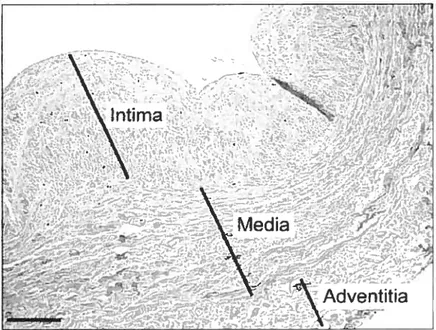
Understanding venous pathophysiology requires some knowledge about venous anatomy and physiology. The primay function of the systemic veins is to retum deoxygenated blood back to the nght side of the heart and to act as a blood resenroir. Approximately 75% of the blood volume is contained within the venous system.
Veins differ from arteries. Thev possess the same 3 layers as arteries but the muscle layer is reduced. From the lumen to the penphery, the intima of the vein wall is a thin layer of smooth muscle cells (SMCs) covered by the endothelium. Undemeath the intima, the media by which the appearance of a mdimentary internaI elastica separates both layers consists of a thin inner layer of longitudinally oriented SMCs and a more prominent outer layer of
circular-onented SMCs. both embedded in an extracellular matrix. The adventitia is composed of fibroblasts, bundies of collagen fibers, capillaries, and clusters of longitudnmlly onented smooth muscle ceils (Figure 2).
The thin and collapsable venous wall allows variations in shape with minimal changes in pressure (venous system is a low-pressure system) and is responsible for the capacitance function of the venous circulation. Veins are normally only partially fihled with blood. They have three times the cross-sectional area of corresponding arteries. ‘:
“\Irtima
‘4’Media
— - . -Adventitia
2-Q
C
Figure 2: Vein wall with the intima underlying the endothelium, the media and the adventitia Scale bar is 200 p.m (44).
Veins of the extremities have valves. These are thin delicate bicuspid structures made of fibrous and elastic tissue lined with endothelium. At the site of each
12
valve the vein is dilated creating a sinus space around the valve which facilitates the opemng and closing ofthe valve. Few valves are located fithe femoral veins; the vena cava and common iliac veins are valveless (52). Ttvo venous systems exist in the upper and lower extremmes, the superficial and the deep venous system which are connected by perforating veins. The major superficial veins of the extremities have thicker walls than the deep veins. Under normal circumstances, two ‘pumps’ (foot and caif pumps) work together to propel venous flow against gravity and bicuspid valves direct flow from the superficial to the deep system. The purpose of the valves is to break up the column of blood in the vein and ensure unidirectional flow. Disease states interfere with these pumps leading to venous stasis and thrombosis which compromises valve function and resuits in venous hypertension(53).
In contrast to arterial thrombosis, which usually develops in association with vascular-wall injuw leading to platelet-rich thrombus, venous thrombosis develops in regions of disturbed flow and relative stasis (as obsened in the caif or venous sinuses), ofien in association with increased coagulability or endothelial damage. The thrombi are composed predominantly of fibnn and red blood celis (54). Deep vein thrombosis may lead to residual venous obstruction or reflux and resuh in post-thrombotic complications. Enhancing resolution of venous thrombi may preserve valve integrity and reduce the incidence of post-thrombotic complications (55).
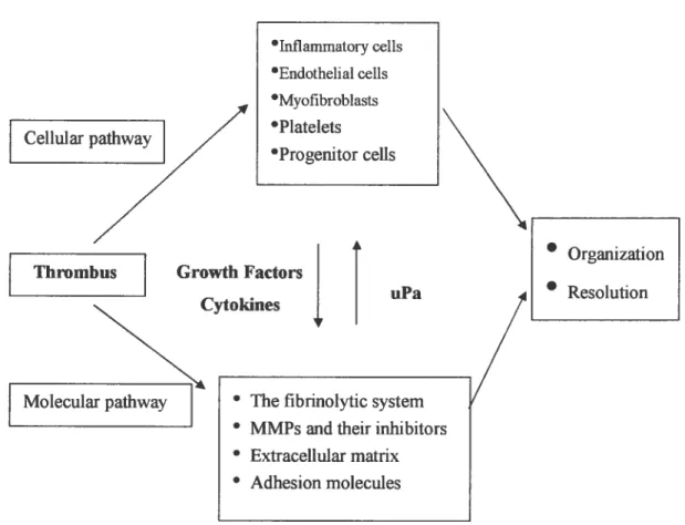
DVT resolves by a the development of venous collaterals (vasculogenesis), dot retraction, organization and recanalization (angiogenesis) that is similar to the formation of granulation tissue in healing wounds (56). These processes may occur simultaneously and are influenced by the fibrinolytic and matrix metalloproteinases (MMPs) systems through a series of cellular and molecular
events (Figure 3) (57).
Cellular pathway
Thrombus Growth Factors
Cytoldnes
Molecular pathway
j
uPaÏ
Figure 3: Cellular andmolecular pathways during resolufion ofthrombus
9nhl ammatory ceils •Endothelial ceils Myofibroblasts Platelets Progenitor ceils
N.
• Organization • Resolution• Thefibrinolytic system • MMPsandtheir inhibitors • Extracellularmatrix
• Adhesion molecules
14
(f
The resolution of thrombus occurs through recruitment of inflammatoiy (mainly monocytes), invasion of endothelial ceils (ECs), vascular smooth muscle cells (VSMCs) and myofibroblasts leading to recanalïzation(5$). Several cellular processes occur: (a) covering of the surface of thrombus with neutrophils, monocytes (MCs) and an endothelial layer; (b) penetraflon by neutrophils and monocytic ceils; (e) development of myofibroblasts and new capillaries; and (cl) recanalization—the formation of one or more channels inside and parallel to the original vesset(59).1.2.3.1.1 Inflammatozy cel]s
Thrombus formation and its resolution are both strongly associated with a subacute inflammatory reaction (60) with the release of fibrinolvtic, chemotactic
(J’
and growth mediators. Early afler venous thrombosis, circulatingmigrate through the vein wall, possibly via the vasa vasora and respond to chemokines by invading the thrombus, which causes thrombus retraction and lysis. Subsequently, monocytes, macrophages and lymphocytes are the predominant leukocyte subpopulations which promote tissue remodeling, recanalization and also retraction of the thrombus (61, 62).
The thrombus contains trapped thrombm and fibnn that are potent monocyte chemoattractants (17). The process of recanalization of thrombus is based on the abilitv of MCs/Mphs (monocytes/macrophages) to penetrate the extracellular matrices and create tubular spaces (“tunnels”) of lower density (60, 63). Recanalization is induced by expression of a variety of cytokines, angiogenic factors, proteases and their inhibitors that regulate cell migration, extracellular
mati-ix turnover and tissue remodeling (4, 6, 24, 25). It is also plausible, however, that once monocytes convert to the macrophage phenotype in the thrombus, their fibnnolytic activity increases. which causes lysis of the thrombus (64). Currently, proteolytic activity of MC5/Mphs on vasculogenesis based on the engraftment of circulating EPCs is thought as an important mechanism of recanalization of thrombus(59, 60).
Whether the inflammatoiy response after DVT is affected by hyperglycenna
remains to bedetermined.
1.23.1.2 Endothelial ceils
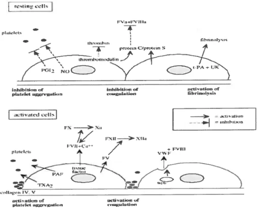
As a unique multifunctional ceil with cntical basai and inducible metabolic and synthefic functions, ECs may react to physical and chemical stimuli within the circulation and regulate haemostasis, vascular remodeling, vasomotor tone, and immune and inflammatoiy responses. In addition, ECs play a pivotai i-ole in angiogenesis and vasculogenesis (65, 66). Endothelium in resting state is both an anticoagulant and antithrombotic by secretion of a variety of molecules important for the regulation ofblood coagulation and platelet function, such as mtric oxide, prostacyclin and thrombomodulin. Vessel exposure to cytokines or proinflammatoiy molecules may shifi the balance towards a procoagulantlprothrombotic phenotype of the ECs (Figure 4) (65) and inhibit
fibnnolysis by reducing the component of the fibnnolytic system. The balance of endothelial properties can be tipped to favor platelet aggregation and dot formation (67, 68). Operating in coordination, these changes can allow fibnn formation and platelet activation.
16
C
Lr’s
III., JetS .J1tI, —iiililtt’ti Ex >-xJ _____________________ F’1J >lJ., /7 ‘2—___________________ _______________________________ lic ‘cnI .,r çjl491,.r phdrfrt,.Ltr,IionFigure 4: Schematic representation of the role played by endothelial ceils in coagulation and fibrinolysis pathways. NO, nitric oxide; PAF, platelet activating factor; PGI2, prostacyclin; tPa, tissue plasminogen activator;TXA2, thromboxane
A2; UK, urokinase; vWF, von Willebrand factor; WPb, Weibel-Paladebody (65).
Moreover, ECs coordmate the recruitment of inflammatorv celis to sites of thrombus. These celis produce and release cytokines and growth factors serving
as communication signais to leukocytes. Cytokines induce a promflammatoiy
phenotype of endothelial ceils. Upon activation of these ceils withtumornecrosis factor- a (TNF-Œ) or mterleukin-1 (IL-1), platelet-activating factor (PAF) is
secreted and stimulates platelet aggregation and neutrophil adhesion to regulate vascular remodeling(69).
inhibit,,,,, oC inh,bitjr,o of octi.nt,on of
gti&aivJtt &urgrrgnti.,u c,Mkguiutt.,fl Ifl,ri,n,I.,i,
cI
(E
Recanalization of thrombus is similar to angiogenesis, becausemay be conceived as the formation of endothelialized channels lined by “endothelial-like” celis, which express many of the endothelial markers (e.g. CD3I, VCAM-1 and ICAM-1) (70). Recanalization is also marked by new formation of vessels which stain for laminin, a basement membrane protein that is known to promote early EC migration and capillary tubule formation within the organizing thrombus. vWF, thrombomoduÏin and tissue factor are expressed in the larger, more prominent channels that appear in older thrombi suggesting that they are lined by more mature ECs (71). This process requires different sequential steps including the release of proteases from activated ECs with subsequent degradation of the basement membrane, ECs migration, proliferation, and differentiation of mature blood vessels into the interstitial space (69, 72).
Some studies performed in our Iaboratoiy on cou embolization of intracranial aneurysms showed that early endothelial invasion of the dot leads to recanalization and recurrence of aneulysms. Moreover, this process can be prevented by endothelial denudation (73), which may prove that the endothelium plays an important role in recanalization offlwombus.
However, whether the process of recanalization and neovascularization of venous thrombiis affected by type 2 diabetes is unknown.
L23.13 Myofihrob]asts
Myofibroblasts are highly specialized mesenchymal ceils derived from fibroblasts. These ceils populate the adventitia tunica (74) and participate in vesse! injury repair as well as thrombus recanalization. In the injured vessel,
1$
numerous cytokines and growth factors (i.e., TGf-) can influence the proliferation level offibroblasts as well as their transition to myoflbroblasts (74).
Several unes of evidence indicate that uPA appears to be an important determinant influencing adventitial ceil proliferation and myofibroblastic modulation. In injured adventitia, exogenous uPA stimulated myofibroblast proliferation and in vitro, upregulated the content of Œ-SM actin in fibroblastic celi culture. Moreover, uPA neutralizing antibody attenuated a-SM actin
expression by adventitial ceils after injuiy of vesse! (75).
During organization and recanalization of thrombus, the balance between uPA-dependent development of endothelialized channels and MTvW-9-dependent contraction by myofibroblasts of the residual provisional fibrin/collagen matrix
between recanalized channels, would resuit in progressive enlargement of the recanalized spaces(59).
123.1.4 Ptatelets
Platelets are ceil fragments released from the bone marrow into the bloodstream and involved in the cellular mechanisms of pnmaly haemostasis. When a blood vessel injury occurs, platelets exhibit a sequence of events: 1) adhesion of platelets to the injury site, 2) spreading of adherent platelets over the exposed subendothelial surface. 3) secretion of platelet granule constituents, 4) platelet aggregation, and 5) platelet coagulant activity (22, 76). Endothelium disruption provides binding sites for adhesive proteins such as von Willebrand factor (vWF) in the subendothelial matnx (winch binds to the platelet g!ycoprotein Ib/IX
adhesive proteins are thought to form a bridge between platelets and subendothelial connective tissue. Once they adhere to the subendothelium, platelets spread out on the exposed surface and additional platelets from the circulation adhere, first to the basal layer of adherent platelets and eventually to one another. Fibnnogen mediates platelet aggregation to form a mass of aggregated platelets through building bridges from platelet to platelet (22, 76).
Imtially, the platelets involved in the thrombus formation favour angiostatic chemokines, such as platelet factor-4 and subsequently secrete angiogenic cytokines such as vascular endothelial growth factor (VEGf) which complexes with fibronectin resulting in ECs migration and proliferation. This complex is more potent than VEGf alone (77) which may regulate the revascularization of thrombus.
1.23.1.5 Progenitor cetls
Progenitor cells arise from division of stem cells. A subset of these cells such as endothelial progenitor cells (EPCs), along with the properties of hemangioblasts that express the leukocyte antigen, CD45 (78), have been implicated in revascularization, vascular repair, and myocardial regeneration. In addition, mesenchymal stem cells also can differentiate into both VSMCs and endothelial celis, and reveal a high degree of plasticity to participate in the development of vascular systems, including angiogenic sprouting and vessel enlargement (79, 80).
Recent reports suggest that following thrombus formation, circulating EPCs denved from bone marrow stem celis may arrest at the site of thrombus, infiltrate through thrombus, and differentiate into endothelial cells to contribute to
20
(E
endothelialization/recanalization of thrombus, or into a-SMA positive ceils that participate in neointima formation (81, 82). Their angiogenic effects are most likely mediated by secretion of growth factors (82). Significant numbers of bone marrow-derived progemtor ceils have also been found in naturally resolvingthrombus (59). As shown by Singh and coworkers, thrombus resolution is markedly delayed in urokinase gene deleted animais which can be rescued by bone marrow transplantation ($3).
L23.2 Moiecular patbway
The fibnnolytic system and matrix metalloproteinases (MMPs) are major components of the molecular pathway and play a pleiotropic role in resolution of thrombosis. The fibnnolytic system may regulate endotheiial ceii infiltration by
(E
degrading fibnn matrices ($4). MMPs expressed by ECs, neutrophils, monocytes/macrophages can degrade the extraceilular matrix to promote the migration of ECs ($5). In addition, chemotactic agents and growth factors (including angiogenic cytokines) expressed or secreted by inflammatoiy celis and platelets also participate in the interaction of molecular and cellular pathways (86). Ail ofthese may affect tissue remodeling and revascularisation ofthrombus.1.2.3.2.1 The fibiïnolytic system and major components
The fibnnolytic system constitutes a critical response mechanism to thrombus formation and evolution. The central components comprise an inactive proenzyme, piasminogen that can be converted to the active enzyme, plasmin, winch in tum degrades fibrin into soluble fibrin degradation products. Two immunologically distinct physiologie plasminogen activators (PA) have been
(Z
identïfied: the tissue-type PA (tPa) and the urokinase type PA (uPa) together with the major inhibitors of PA, plasminogen activator inhibitor-1 and -2 (PAl-1, PAl-2), while plasmin is inhibited mainly by Œ2-antiplasmin. tPa, a serine protease, is responsible for the removal of fibnn from the vascular tree (87, $8), whereas, uPabound to its receptor uPAR, is regarded as the critical trigger for plasmin generation dunng celi migration and invasion, and may be responsible for regulating the activation of other proteases, such as the MMPs (eg, procollagenases and macrophage elastase) (figure 5) ($8). Plasmin can also activate or liberate growth factors from the ECM including latent TGf -131, bfGF and VEGf (89).PAl-1. a member of the serine proteinase inhibitor ($ERPIN) family is the
pnmaly inhibitor of plasminogen activators in plasma and in the pencellular
matrices, which bonds two-chain active uPA or tPA to reduce actîvity of uPA or tPA through covalent complex formation ($8). Vitronectin (VN) binds to uPAR, an abundant plasma and matrix glycoprotein; whereas PAl-1 controls recognition
of VN by uPAR or the Œv133 integrin receptor, and is stabilized by binding to a
plasminogen activator inhibitor binding protein identified as S-protein, suggesting a role in coordinating ceil adhesion and migration (90). Moreover, PAl-1 detaches celis from exïracellular matrices; vitronectin, fibronectin and type I
collagen through an uPA’uPAR-dependent mechanism by inactivating integrins (91). Thus, PAl-1 could be considered as a deadhesion molecule (e.g., thrombospondin, tenasin) disrupting the link between the cytoskeleton and the focal adhesion plaque and resulting in the loss of stress fibers and a decrease in the strength of integrin—ligand interactions (182).
22
C
G
Dunng natural resolution of venous thrombi, there is an increase in the activity of the fibnnolytic mediators, tissue-type plasminogen actîvator (Wa) and urokinase-type plasminogen activator (uPa) and this activity is expressed by invading monocytes (182)
PAl-1
•ECM degradalion •achvation of TGF-3
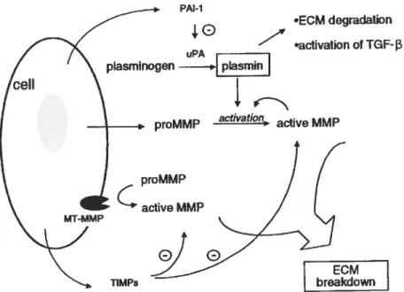
Figure 5: M extensive network of additional proteases, inhibitors, receptors and
modulators are directly associated withandare influenced by the PA system. The
largest group is the mati-ix metalloprotemases (MMPs) and their respective inhibitors, thetissue inhibitors ofMMPs (TIMP$) (94).
The levels of uPA were usuaÏly found to be greaterthanthose of tPa Subsequent gene knockout studies have shown that deletion of the gene encoding for uPA markedly inhibited normal thrombus resolution, but the tPa gene knockouthadno effect. Absence of uPA is also associatedwithdelayed monocyte recruitment into the thrombus (83).
uPA
plasmin
proMMP activatian, active MMP
proMMP actîve MMP
MT-MMP
ECM breakdown
uPa rnav have a dual function; one related to proteolytic matrix breakdown, the other related to matrix production via proteolytic activation of growth factors, such as the fibrogenic TGf-Bl. This dual role of uPa may explain why ECM degradation and collagen deposition were both aftenuated in the absence of uPa
(95).
PAl- 1 plays a determining role in controlling thrornbus formation. PAl- 1 activity was increased in patients with deep vein thrombosis (DVT) and pulmonary embolism (96). further evidence for the role of PAl-1 in venous thrombosis cornes from animal models. Transgenic mice, that were geneticaÏly engineered to synthesize PAl- 1 in excess, had higherrates of venous thrombosis than mice with normal PAl-l levels (97). In a mode! of venous stasis-induced DVT afler ligation of the inferior vena cava, Deatnck and coworkers demonstrated an alteration in the normal profibrinolytic to antifibnno!ytic state of the vessel by a decrease in
theratio ofuPA to PAl-1 (60).
Clinical and expenrnental studies have suggested an irnportant role of PAl- 1 in artenal and venous thrombosis and the maintenance of systemc vas cu!ar
hemostasis (98). Moreover, expenments with transgenic mice deficient in PAl- 1 support a role forthis serpïn in both vascular remodeling afier arteria! injuiy (99)
and the formation of puÏmonaiy fibrosis that occurs afier inflammatory injury
(100). upregulation of PAl-1 in endothelial ceils and smooth muscle cells after acute vesse! injuiy (lOi) and thrornbus formation (102) suggest that elevated PAl-1 may play a role in vascular remodeling afler deposition of a thromboembo!us.
24
There is increasing evidence that diabetes mellitus is associated with several defects in coagulation and fibrinolysis that may Iead to a procoagulant, thrombogenic predisposition (103). However, it is flot known whether these perturbations cause a decrease in thrombus recanalization.
1.2.3.2.2 Matnx metatioproteinases and theirinhibitors
Matnx metalloprotemases (MMPs) are a family of zinc-dependent
endopeptidases, which can degrade essentially ail ECM components in physiological and pathological conditions, but also parficipate in celi migration, angiogenesis, and tissue remodeling dunng organ development, wound healing,
inflammation, and cancer (104, 105, 106). Currently more than 24 members ofthe MMP family have been identified and classified into subgroups of collagenases, gelatinases, stromelysins, and membrane types (MT-MMPs) based on their structureand substrate specificity (107, 10$). MI’Ws are produced by secretion of both vascular and inflammatory celis. Their activity is regulated at multiple levels: gene transcription and synthesis of inactive zymogens, posttranslational activation of zymogens, and interactions of secreted MMPs with tissue inhibitors of metalloproteinases (TIMPs) (104, 105). The TIMP family known at present consists of four distinct members (TIMPs 1 to 4), and is expressed in most tissues
and body fluids. Except for TIMP-dependent inhibition of MMPs, these proteinases have been recently recognized to stimulate ceil proliferation participating in mitosis and tissue differentiation, to regulate ceÏl survival and apoptosis, and to inhibit angiogenesis (105). The balance of MMPs and TIMPs
activity controls the diffusion of substances and the migration of ceils through ECM. These proteinases also modulate signal transduction pathways by various
substrates, mcluding inflammatorv mediators, growlh factors, and growth factor receptors (105).
MMP-2 ami MMP-9 (gelatinases Aand B) are the main enzymes able to degrade nonnaturalECM gelatin and type IV collagen (109), which is the major structural protein of the ECM. but also an ïntegral part of endothelium basement
membranes. ECM degradation and basement membrane disruption are the key steps in thrombus organizatïon and recanalization. These enzymes play a cntical role in vascular remodeling induced by altered arterial flow (110, 111), tissue ischemia and aortic aneurysms (112). Moreover, MMP-2 activity is cntical for
migration of endothelial cells (113) and monocytes/macrophages (114). Targeted deletion of MMP-2 abolishes angiogenesis in vivo (115, 116). Thrombin treatment of endothelial celis induces MMP-2 activity (117) andpro-MMP-2 can
also be proteolytically activated in vitro by the coagulation proteins activated proteinC (APC) (11$) andfactorXa(119).
During expenmental venous thrombosis, the expression of MMP-2 and MMP-9 is increased (60, 120). But their invo]vement in thrombus resolution and vein walI fibrosis in diet—induced type 2 diabetic mice remains undefined.
1.23.23 Extracellular matrix
The thin extracellular matnx (ECM) underlying the endothelium is termed the
basement membrane. It is made up of structural interacting glycoproteins produced by VSMCs and fibroblasts (121). The most abundant components are
26
These ECM components act as structural support promotmg celi adhesion and barners between tissue compartments regulating cellular migration. Basement membrane components contain the RGD (arg-gly-asp) sequence that control ceil shape, migration, proliferation, differentiation, morphogenesis, and survival (123). Celis use a series of receptors (integrins, celi surface proteoglycans, and a newly describedclass of cell-surface-expressed tyrosine kinase receptors) to form linkages with ECM. In this cellular behavior, celis can be provided with directional guidance dues for migration (124). When blood vesse! is damaged or presents a platelet-fibnn thrombus, the ECM may act as a ligand to adhere to in areas of exposed basement membrane on the endothelial monolayer by circulating blood celis and neointimal ceils (123). The phenomena of the ceil-matrix interaction can be modulated by the balance of activity of a class of proteases known as MMPs and their inhibitors.
Thrombosis and inflammation that occur in DVI resuit in valve destruction and chronic veinwall changes that lead to venous reflux and the syndrome ofchronic venous insufficiency marked by thickened, noncompliant vein walls and incompetent valves.
Afler the development of a DVT, a late fibrotic response, similar to a healing wound, occurs in vein walls (60), involving the progression of the normally thin and compliant vein wall to a relatively thick and fibrotic state. There is deposition and accumulation of collagen procollagen I and procollagen III, and loss of normal vessel ECM such as heparin su!phate which has antifibrotic properties and is important for mediating norma! vessel phvsiologic responses
dcposition and accumulation of collagen procoltagen I and procollagen Iii, and !oss of normal vessel ECM such as heparin suiphate which bas antifibrotic
properties and is important for mediating normal vesse! physiologic responses
(125). Extracellular matrix molecules such as fibronectin and vitronectin enhance
endothelial cet! migration and tubute formation by binding to uv33 (126).
The presence of these molecules has yet to be confirmed in naturally
resolving thrombi, and moreover in hyperglycaemic conditions, but it seems
)ikely that they have a role in this process.
1.2.3.2.4 Selectins
The selectins are a small family of lectin-like adhesion receptors. There are three
fami)y members, L-, E-, and P-selectin. Their major physiotogicat rote is thought
to be largety responsibte for the initial attachment and rolling of leukocytes on
stirnulated vascular endothetium. Ibis family consists of P-selectin expressed on
activated platelets and activated endothelium, E-selectin expressed on activated
vascular endothelium, and L-selectin expressed on the surface of neutrophils.
P-selectin is present in the granules of platelets and the Weibel—Palade bodies of
ECs. It is first translocated to the plasma membrane of these ceils, mediating the
initial inflammatory response (127, 128). P-selectin plays a rote in perithrombotic
inflammation and in mediating leukocyte influx into areas of inflammation.
Previous studies have shown that decreases in thrombogenesis and increases in
thrombolysis can be achieved in primate, porcine, and rat modets of arterial and
venous thrombosis in which P-selectin is antagonized (5$, 129, 130). These
P-selectin driven interactions tead to activation of the coagulation cascade with
2$
leukocyte tissue factor (TF) upregulation, which further potentiates vein wafl inflammatory events.
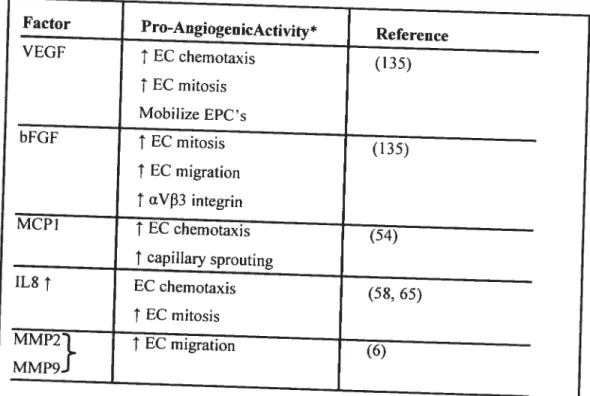
1.2.3.2.5 Proangiogenic factors
Monocytes express a variety of proangiogenic factors such as VEGF, bfGF and interleukin-$ (IL-8), a prototypic cysteine-X-cysteine (CXC) chemokine with
polymorphonuclear neutrophil (PMN)-activating and chemoattractant properties,
and confers proangiogenic activity (134), which may generate an ‘angiogenic drive’ within the thrombus (65, 135). Both VEGF and bFGF are expressed in resolving thrombi, and are associated with the appearance of channels within and around the thrombus (136). VEGE expression was localized to several ceils in the
thrornbus, including endothelial celis and the monocyte infiltrate. Expression of
bFGF was found on mononuclear celis ami spindle-shaped ceils within the thrombus (136).
Monocyte chemotactic protein-1 (MCP-Ï) is a potent and specific activator of
monocytes and basophils. In a rat mode! of venons thrornbosis, the vein wall
adjacent to the thrombus was found to contain increasing amounts ofMCP-1 and
when this cytokine is directfy injected into venous thrombus, it alters its
organization (137). Part of the effect produced by injecting MCP-1 may have
been as a consequence oC its angiogenic properties (Table 1).
VEGF EC chemotaxis (135)
t
EC mitosis Mobilize EPC’s bfGf EC mitosis (135)t
EC migrationt
ŒV33 integrin MCPIt
EC chemotaxis (54) t_capillary_sprouting IL$t
EC chemotaxis (58, 65)t
EC mitosis MMP2t
EC migration (6) MMP9*These are gencratly known proangiogenicactivities associated with these factors
Table 1: Proangiogenic factors expressed within resolving thrornbus (70).
1.2.4 Treatment of deep vein thrombosis
Treatment costs to the U.S. health care system are in the range of billions of
dollars per year just for the acute treatment of venous thrombosis, without even
considering the arnount of rnoney spent on the treatrnent of the sequelae of DVT
(chronic venous insufficiency) and PE (chronic pulmonaryhypertension) (13$).
1.2.4.1 Standard treatments
Prophylaxis and treatment of DVT aim to prevent propagation of the fractured
thrombi as emboli leading to death from pulmonary embolism. Another goal is to
minimize the sequetae of CVI known as the post-thrombotic syndrome. Enhanced
Factor ProAngÏogenÏcActivity*
30
thrombus resolution is associated with reduced valvutar damage and venous hypertension and fewer long term complications (6, 139). Standard treatments include anticoagulants (low-rnolecular-weight heparin that acts on both thrombin and factor Xa or warfarin), thrombolytics agents (for example, streptokinase, recombinant urokinase and tissue-plasminogen activator that lyse the thrornbi), surgical thrombectomy and compression stockings as prophylactic measures. These agents are often considered alternatives by inhibiting thrombus extension and do flot accelerate natural thrombus resolution. Thrombolytics are less used because ofa small but significant risk of severe hemorrhage (140) and in patients with stroke or with a recent operation (6, 139, 140). Compression stockings are sornetimes recommended to relieve pain and swelling, (141). However, these treatments do not seem to effectively reduce the incidence ofthe post-thrombotic venons insufficiency (142, 143).
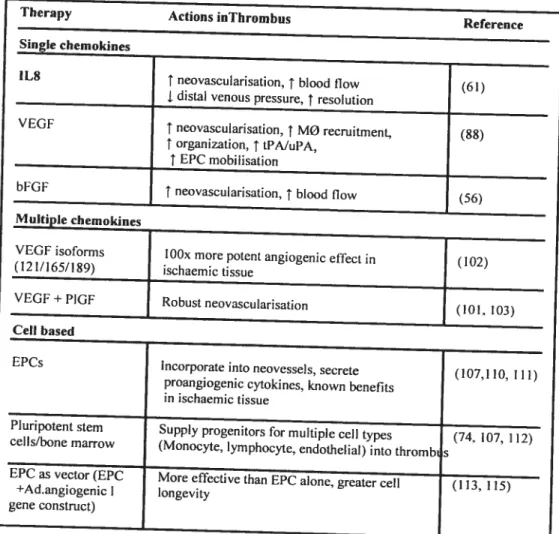
1.2.4.2 Newapproaches
AÏthough acute therapy for DVT is well established, potential new therapies have emerged to promote thrombus vascularization and resolution without altering the normal hemostatic mechanisms, such as delivery of angiogenic growth factors and cell-based therapy. The application of these methods may enhance rapid resolution ofDVT, but also provide other perspectives on treatment.
1.2.4.3 Angiogenic therapy
A number of pro-angiogenic factors, including VEGF, IL-X, bfGf, have been
reported to enhance neovascularization of ischemic tissues (6, 61). Currentty the concept of stimulating therapeutic angiogenesis has also been applîed to the
recanalization of venous thrombus (Table2). Treatment with recombinant iL-8 in
a rat model of JVC thrombosis resulted in increased recruitment of neutrophils,
monocytes and markedly promoted early neovascularization enhancing the resolution ofthrombosis (61).
Varma and coworkers (144) used therapeutic administration of pro-angiogenic compounds promoting DVI neovascularization, such as interferon inducible protein (IP-1O), an angiostatic chemokine, basic (bFGF), a pro-angiogenic factor and epithelial neutrophil activating protein (ENA-78), a pro-angiogenic cytokine. These angiogenic chemokines increase thrombus neovascularization, but this does flot correlate with smaller or less fibrotic DVT. Mechanisms other than neovascularization may be more important to hasten DVI dissolution. VEGf administration (136) enhance thrombus resolution by a variety ofrnechanisms (6). VEGf, bFGf, platelet derived growth factor (PDGfs), placental growth factor (PIGF), the angiopoietins and their receptors, which are mediators associated with angiogenesis, have also been provcd to promote the formation of blood vessel function (145, 146). These factors could provide more choice in treatment of thrombosis.
1.2.4.4 Cell-based approach
Thrombus resolution depends on the interaction of an assortment of cells. Bone rnarrow-derived progenitor cells are known to participate in revascularization, provide the necessary precursors as these immature ceils can differentiate into a diversity of phenotypes, including macrophage, lymphocyte and endothelial celis (147). Recent studies have shown that thrombus resolution is markedly detayed in
32
uPA knock out animais, but rescucd by bone rnarrow transplantation ($3). These progenitor celis have been made to improve thrombus resolution as a celI-based approach and showed some benefit in small clinical trials (147-149). Endothelial progenitor celis, delivered Iocaily or injected into the circulation, incorporate into
newly formed vessels to enhance Local angiogenesis by secreting a variety of
pro-angiogenic cytokines (150).
Modulation of monocyte fibrinolytic and growth factor production in vitro, with subsequent reinjection of these ceits, may provide an alternative treatment for venous thrombosis. This therapy might also be useful to recanalize mature thrombi when fibrinolytic treatment is ineffective(]51).
1.2.4.5 Gene therapies
Gene therapy isthe insertion ofgenes into an individuals celis and tissues to treat
a disease. Endothelial progenitor cells are also used as vectors to deliver
pro-angiogenic genes. It bas been conflrmed that transplantation of endothelial progenitor cells transfected with VEGf are more effective than unrnodifled cells
in the angiogenesis and revascularization of ischcmic tissues (152, 153). However,
this therapy is limited by vector toxicity. Current studies of gene therapy focus on reducing toxicity and improving vectors by looking into mechanisms retargeting vector to the tissue of interest, minirnizing or eliminating viral gene expression
(Table 2) (135).
G
1L8
t neovascularisation,t bloodtlow
(61)
distal venous pressure, î resolution
VEGf î neovascularisation, î MO recruitmenL (88) t organization,tLPA/uPA, t_EPC mobilisation bFGf t neovascularisation, î blood flow (56) MuItpJe chemokines
VEGF isoforms lOOx more potent angiogenic etïect in
(102)
(121/165/189) ischaemic tissue
VEGF+PIGF Robust neovascularisation
(101, 103) Ccii based
EPCs
]ncorporate into neovessels, secrete (107,110, 1 11)
proangiogenic cytokines, known benefïts in ischaemic tissue
Pluripotent stem
Supply progenitors for multiple celi types (74, 107, 112)
cells/bone marrow
(Monocyte, lymphocyte, endothelial) into thromb s
o
Table 2: Potential new therapies promoting recanalization and resolution of
venous thrombi (6).
1.2.5 Treatment of complication
Proangiogenic therapy can cause complications such as inflammation with an
immunogenic response to viral vectors (154). Stimulation of therapy to
angiogenesis may resuit in rupture of atherosclerotic plaques, development and
growth of vascular malformations (155). Enhanced angiogenesis therapy atso
carnes risks of neoplasia and tumour growth. In addition, administration of bone
Therapy
Single chemokines
Actions inThrombus Reference
EPCas veCtor(EPC
+Ad.angiogenicI
gene construct)
More effective thanEPCalone, greaterceli
34
marrow ceils can carry a theoretical possibility for malignant transformation. The selection of single types of stem ceil may reduce treatment complication (6, 155). 1.2.6 Animal models ofvenous thrombosis
If an optimal animal model exists, it would have a natural propensity for venous thrombosis, a similar clotting cascade and platelet interaction to that ofthe human,
a Iower extremity that cÏosely resembles the human with a functional caif
musculature, ability to walk upright and sufflciently tau (when standing) to allow hemodynamic study and sufficiently large to allow surgical intervention (164). Overali, large animais like pigs and monkeys have been better suited to study thrombosis as they are more similar to human physiology than smaller species such as mice, rats, rabbits and dogs (39). However, the study ofthrombosis with these animais is restricted by cost and ethical considerations. Nevertheless, numerous studies have used rodent models taking advantage of low cost, availability, practical breeding, technical feasibility and the availability of transgenic knockout mice. Some limitations may limit extrapolation to human thrombi as the relatively rapid (3-4 weeks) rate of thrombus resolution in rodent moUds and the difference in hemodynamics in the smaller diameter of the vena cava may affect thrombus revascularisation (6).
Murine in vivo models are appealing because of their well-defined genetic background, and the possibiiity of using syngeneic “knockout” and mutant mice producing a variety ofmetabolicsettings.
Animal modeis of venous thrombosis have been classified as non-genetic and genetic modeis. Non-genetic models of thrombosis have been produced by a
combination of blood-flow stasis
with either increased coagulability or
endothelial damage (46).
Several injury models applied to the arterial systems have also been used in the
venous system. injection ofendotoxin (156), collagen and cpinephrine, f actor Xa,
or tissue factor (157), thrombin (158) as well as hyperoxia (159) or hypoxia (160)
ail served to induce a hypercoagulable state and fibrin deposition in the mouse.
Other methods included application of 70 % ferric chioride (161), or sodium
morrhuate or long-term nitTic oxide synthase inhibition using L-NAME (162). In
addition, thrombosis can be induced using 1251-labeled fibrinogen mixed with thromboplastin (163)
After administration of the thrombogenic stimulus systemically or directly into the stasis region, this mode! can be performed with or without mechanical vena cava stasis (2). They include vein ligation (164), or
C
vein interruption by means of a silicone band or an intralurninalballoon catheter (165), or by the intra-stent stenosis (166). flow stasis was also induced by a combination of devascularization, electric injury (167), or photochemical injury (23). These models represent useful tools for the better understanding of the venous thromboembolic events under conditions similar to those seen in humans
(54). Other genetic models (such as transgenic models of thrombosis) are a number of spontaneous or genetically engineered
mouse strains with
overexpression or deletion of various elements in the lipid transporters, coagulation, platelet, and fibrinolysis pathways.
As our objectives are focused on the pathogenesis of venous thrombosis in diabetic mice, we used a reproducible model of venous thrombosis (168). The inferior vena cava (IVC) stenosis model, with a 94.4% ± 0.5% reduction in IVC
36
diameter (38), leading to a stasis-induced venous thrombosis is more adapted to our goals, as shown by earlier studies that venous thrombi produced in this model were forrned in flowing blood and were morphologically similar to human thrombi (151). Moreover, this model bas more availability, improved technical feasibility, standardization of local thrombosis and lower maintenance costs (169). Thus, the ability to study thrombus recanalization in diabetic mice should be appropriate in this model.