UNCORRECTED PR
OOF
1Q2
ANIMA: A data-sharing initiative for neuroimaging meta-analyses
2Q3
Andrew T. Reid
a,⁎
, Danilo Bzdok
a,b,g, Sarah Genon
a,b, Robert Langner
a,b, Veronika I. Müller
a,b,
3Claudia R. Eickhoff
a,c, Felix Hoffstaedter
a,b, Edna-Clarisse Cieslik
a,b, Peter T. Fox
d, Angela R. Laird
e,
4Katrin Amunts
a,f, Simon B. Eickhoff
a,b5 aInstitute of Neuroscience and Medicine 1, Research Centre Jülich, Jülich, Germany
6 bInstitute of Clinical Neuroscience and Medical Psychology, Heinrich Heine University, Düsseldorf, Germany
7 cDepartment of Psychiatry, Psychotherapy and Psychosomatics, University Hospital Aachen, Aachen, Germany
8Q4 dUniversity of Texas Health Sciences Center at San Antonio, San Antonio, TX, USA
9 eFlorida International University, Miami, FL, USA
10 fC. & O. Vogt Institute for Brain Research, Heinrich Heine University, Düsseldorf, Germany
11 gINRIA, Neurospin, bat 145, CEA Saclay, 91191 Gif-sur-Yvette, France
a b s t r a c t
1 2a r t i c l e i n f o
13 Article history: 14 Accepted 22 July 2015 15 Available online xxxx 16 17Meta-analytic techniques allow cognitive neuroscientists to pool large amounts of data across many individual
18
task-based functional neuroimaging experiments. These methods have been aided by the introduction of online
19
databases such as Brainmap.org or Neurosynth.org, which collate peak activation coordinates obtained from
20
thousands of published studies. Findings from meta-analytic studies typically include brain regions which are
21
consistently activated across studies for specific contrasts, investigating cognitive or clinical hypotheses. These
22
regions can be subsequently used as the basis for seed-based connectivity analysis, or formally compared to
neu-23
roimaging data in order to help interpret new findings. To facilitate such approaches, we have developed a new
24
online repository of meta-analytic neuroimaging results, named the Archive of Neuroimaging Meta-analyses
25
(ANIMA). The ANIMA platform consists of an intuitive online interface for querying, downloading, and
contrib-26
uting data from published meta-analytic studies. Additionally, to aid the process of organizing, visualizing, and
27
working with these data, we present an open-source desktop application called Volume Viewer. Volume Viewer
28
allows users to easily arrange imaging data into composite stacks, and save these sessions as individual files,
29
which can also be uploaded to the ANIMA database. The application also allows users to perform basic functions,
30
such as computing conjunctions between images, or extracting regions-of-interest or peak coordinates for
fur-31
ther analysis. The introduction of this new resource will enhance the ability of researchers to both share their
32
findings and incorporate existing meta-analytic results into their own research.
33 © 2015 Published by Elsevier Inc.
34 35 36 37
38 Introduction
39 Functional neuroimaging, like many other scientific fields, is 40 faced with the daunting task of managing an ever-increasing amount 41 of data (Poldrack and Gorgolewski, 2014). Online databases such as 42 Brainmap.org (Laird et al., 2011), Neurovault.org (Gorgolewski 43 et al., 2015), and Neurosynth.org (Yarkoni et al., 2011) provide ac-44 cess to data from hundreds of published task-based fMRI studies, in 45 standard coordinates (recently reviewed byFox et al., 2014). In 46 addition to extensive meta-analyses based upon manual search tech-47 niques, these platforms have facilitated a growing number of meta-48 analytic studies of the neural correlates of specific cognitive functions, 49 using methods such as multilevel kernel density analysis (MKDA; 50 Wager et al., 2007) and activation likelihood estimation (ALE;Eickhoff 51 et al., 2009, 2012). Meta-analysis entails the pooling of data over tens
52 to thousands of individual studies, and thus provides: (1.) greater
53 sensitivity and specificity to detect “true” effects; (2.) a means of
deter-54 mining core groups of brain regions subserving a specific task or
charac-55 terizing a specific disease; and (3.) a method for formal comparison of
56 different subfacets of a cognitive domain. This approach has been
57 used, for instance, to demonstrate the neural correlates of sustained
at-58 tention (Langner and Eickhoff, 2013), investigate face processing areas
59 in autistic subjects (Nickl-Jockschat et al., 2014), and identify key
re-60 gions subserving supervisory attentional control (Cieslik et al., 2015).
61 Results from meta-analyses can be subsequently used as robust prior
62 information in the design of task-based fMRI studies, and as
regions-of-63 interest (ROIs) for connectivity methods based on functional
correla-64 tions (e.g.,Müller et al., 2014; Schilbach et al., 2014), or virtually any
65 other type of seed-based analysis. This includes topical meta-analytic
66 approaches such as ALE and MKDA, as well as methods which use
67 meta-analysis to infer functional connectivity, such as meta-analytic
68 connectivity modelling (MACM;Etkin and Wager, 2007; Kober et al.,
69 2008; Robinson et al., 2010), in which functional coactivations are
NeuroImage xxx (2015) xxx–xxx
⁎ Corresponding author. Fax: +49 2461 61 3483. E-mail address:[email protected](A.T. Reid).
YNIMG-12451; No. of pages: 9; 4C: 4, 5, 6, 7
http://dx.doi.org/10.1016/j.neuroimage.2015.07.060
1053-8119/© 2015 Published by Elsevier Inc.
Contents lists available atScienceDirect
NeuroImage
UNCORRECTED PR
OOF
70 assessed across all tasks in the database (see alsoXue et al., 2014).Ad-71 ditional meta-analytic approaches include the parcellation of the brain 72 into functionally distinct subregions, such as coactivation-based 73 parcellation (CBP;Chang et al., 2012; Eickhoff et al., 2011; Northoff 74 et al., 2006). A typical ALE analysis, for example, will result in a distinct 75 ROI or set of ROIs that are associated with a particular psychological or 76 clinical feature (seeBox 1). These results are then commonly used as 77 seed regions for further analysis of these features (e.g., zuEulenburg 78 et al., 2012). Given their utility, results from meta-analytic studies are 79 becoming increasingly popular as starting points for future analyses, 80 and are hence commonly requested from the authors. However, this 81 mode of data exchange typically requires a time delay for locating, orga-82 nizing, packaging, and sending data, and can be complicated further by 83 confusion over the way data are named, the type of information they 84 represent, and data formats in which they are stored. Moreover, use of 85 these data in published articles would benefit from the ability to refer-86 ence a specific and permanent online location, as well as provenance 87 tracking, particularly for purposes of validation and replication. These 88 considerations present a need for a more standardized, easily accessible 89 means of sharing meta-analytic results, which has motivated the crea-90 tion of a new online data resource called the Archive of Neuroimaging 91 Meta-Analyses (ANIMA). This database can be accessed athttp:// 92 anima.fz-juelich.de.
93 The concept behind the ANIMA database is simple: to provide the re-94 sults of published meta-analyses and coactivation-based parcellations 95 to interested parties, in the form of statistical maps or labels, encoded 96 as image files. This approach has a number of important requirements. 97 Firstly, data should be easily citable if reused for further analyses, and 98 thus full information about the source of the results must be provided 99 with them. In addition to metadata about the article itself (reference 100 details, cross-links to PubMed entries, abstract information, etc.), 101 there should be sufficient information provided to identify the data
102 represented by each image file, including its associated figure, if
ap-103 plicable. Secondly, data retrieved from the database should be simple
104 to organize, visualize, and use. A user should be able to peruse the
105 web interface, query and select studies of interest, download these
106 to their local machine, and immediately browse and utilize the data
107 they have retrieved. Thirdly, in order for the database to expand
108 and provide a thorough sampling of the literature, it is important to
109 provide a convenient interface through which researchers can
sub-110 mit data from their own studies.
111 With the ANIMA database, we have provided solutions to each of
112 these requirements. In what follows, we will describe our databasing
113 approach, which includes an intuitive online interface for querying,
114 downloading, and submitting data, as well as a stand-alone,
cross-115 platform desktop tool for easily browsing, visualizing, and performing
116 common computations, such as obtaining a conjunction between
im-117 ages. This new resource will provide researchers with a straightforward
118 means of including meta-analytic results in their studies, both as ROIs
119 for future analyses and as a point of comparison against new results.
120 The initial release of the database will include data from 25 published
121 meta-analytic studies, but is intended to grow in order to incorporate
122 the increasing number of studies being added to the literature.
123 Database overview
124 ANIMA is designed to serve a number of functions. Firstly, it is a
125 searchable online repository for the results of published
neuroimag-126 ing meta-analyses. Data are organized as individual studies, along
127 with information essential for describing these studies (authors,
128 title, journal, date, abstract, etc.). Included with each study is a set
129 of “study elements”, which refer to individual files storing an
impor-130 tant aspect of the study's findings. These will typically be individual
131 volume files, stored in NIFTI format. However, other types of data
132 can be uploaded, including text files containing useful descriptions,
133 image files, or Volume Viewer session files, which store multiple
im-134 ages, along with information on how they should be visualized (see
135 description below). Each study element is also associated with a set
136 of metadata which describe its contents (title, figure captions, type
137 of information, full-text PDF, etc.).
138 A second function of ANIMA is the ability to submit one's own
139 study to the database. The online interface includes a “Submit”
140 page, which allows registered users to define their study and upload
141 files. If a researcher would like to share results from a meta-analytic
142 study that has been published in peer-reviewed journal, we
encour-143 age them to do so via this interface. The general philosophy of this
144 approach is the concept of “open data”. For reasons of security and
145 data integrity, submitting a study requires that a user register with
146 the database, and that each submitted study is first vetted for
com-147 pleteness and validity by the database administrators. However,
148 the process of submitting one's data to ANIMA is designed to be a
149 straightforward process.
150 Finally, ANIMA was designed with the idea that, once retrieved,
151 data should be simple to organize and query on one's desktop.
Ac-152 cordingly, we have developed a desktop application called Volume
153 Viewer, which interacts seamlessly with the ANIMA interface.
Vol-154 ume Viewer is open source Java-based software tool, built on the
155 ModelGUI API (http://mgui.wikidot.com), and both the program
156 and its source code are freely available online (https://launchpad.
157 net/volumeviewer). The interface allows data to be organized within
158 a “library” framework, in which individual studies can be
represent-159 ed. The data retrieved from ANIMA is already organized according to
160 this framework, so downloaded ANIMA studies can be immediately
161 imported and viewed in Volume Viewer. Furthermore, if a study
in-162 cludes Volume Viewer session files, predefined sets of image
com-163 posites, with custom colour mapping and template or atlas layers,
164 can be viewed with little effort on the part of the user. Importantly,
165 this tool also implements a number of utilities which allow users to
B:1
Box 1B:2
Common methodologies used in ANIMA studies.B:3
B:5
Three common approaches used in ANIMA studies are describedB:6
below.B:7
Activation likelihood estimation (ALE): This is a meta-analyticB:8
approach through which peak activation foci, reported instan-B:9
dard MNI or Talairach coordinates, are used to inform GaussianB:10
probability models of activation. This topical approachad-B:11
dresses the sparse information provided by activation foci, asB:12
is used to estimate smooth statistical distributions associatedB:13
with particular psychological or clinical features.B:14
Multi-kernel density analysis (MKDA): A topical approachB:15
which is similar in principle to ALE, MKDA uses a sphericalin-B:16
stead of Gaussian kernel, and statistical analysis is performedB:17
on the number (density) of activation peaks within a givenB:18
radius.B:19
Meta-analytic connectivity modelling (MACM): This approachB:20
models the probability that brain regions are activesimulta-B:21
neously with a particular seed region. MACM is typicallyper-B:22
formed across many experimental paradigms, providing aB:23
meta-analytic estimate of functional connectivity.B:24
Coactivation-based parcellation (CBP): CBP is a parcellationB:25
method derived from co-activation profiles obtained via theB:26
MACM approach. CBP uses clustering approaches such ashi-B:27
erarchical or k-means clustering to identify subregions within aB:28
larger ROI that are distinct from each other in terms of theirB:29
connectivity profiles.B:30
B:31
UNCORRECTED PR
OOF
166 combine their data with ANIMA data, create new ROIs for analyses,167 report peak coordinates, or quantify the overlap between different 168 distributions.
169 What data are available?
170 At the time of writing, ANIMA contains data from 25 meta-analytic 171 studies (both topic-based ALE meta-analyses and coactivation-based 172 parcellations), as summarized inTable 1. We intend to expand this 173 number considerably, both by requesting and adding new studies 174 from authors, as well as by encouraging authors to submit their own 175 studies through the online interface. The majority of the data available 176 represent statistical maps which are produced through common 177 meta-analytic approaches, including ALE, CBP, and MACM.Box 1 pro-178 vides an overview of these methods. In many cases, these data are di-179 rectly related to a figure in the article, but we also encourage authors 180 to submit supplemental data, if desired. Since the database is based on 181 meta-analyses, no demographic or phenotypical data are directly avail-182 able, although these can generally be determined by reference to the 183 individual articles. However, the database does provide searchable key-184 words and sample sizes (number of primary studies, experiments, and 185 subjects) associated with individual articles. Finally, studies in ANIMA 186 each have a unique uniform resource locator (URL), which displays 187 the available data and metadata, along with the date the study was 188 last modified. This URL, along with the modification date, can be used 189 for precise citation of any data used in a new publication. ANIMA 190 keeps a history of any changes to the data that has been made publicly 191 available, such that the exact archive used in a study can be retrieved for 192 purposes of validation or replication.
193 Data in ANIMA are freely available, although their use is conditional
194 on acceptance of a Data Usage Agreement (DUA). The general terms of
195 this agreement are provided inBox 2. Any redistribution of ANIMA
196 data requires consent from the authors whose data are redistributed,
197 as well as the database owners. Additionally, the DUA specifies that
at-198 tribution for use of the data must include reference to both the database
199 and the specific studies used. Both database users and contributors must
200 agree to the terms of the DUA in order to use or contribute to the
data-201 base, respectively.
202 Accessing, viewing, and using ANIMA data
203 Fig. 1shows the online interface for browsing studies in ANIMA
204 (http://anima.fz-juelich.de/index.php). Notably, querying and
205 downloading is a fully open process with no registration required.
Stud-206 ies are listed in tabular format, with each entry showing the title of the
207 study (in the form of the lead author and publication year), a brief
de-208 scription, the size of the archive, and links to download the study,
209 view its PubMed page, or download its associated full-text PDF. At the
210 top of the page, a search bar allows the user to search by author,
publi-211 cation year, or journal. More detailed information about individual
212 studies can be accessed by clicking on the study's title. Here, abstract,
213 keywords, and version information are displayed, as well as a list of
in-214 dividual study elements (Fig. 2). It is also possible to view image
ele-215 ments, using the Javascript-based Papaya viewer (http://github.com/
216 rii-mango/Papaya), by clicking on the eye icon for an individual
ele-217 ment. This will overlay the image on the non-linear ICBM-152
anatom-218 ical template, and is accessible from any modern web browser.
t3:1 Table 1
t3:2 Studies for which data are available in the ANIMA database at the time of writing. Abbreviations: ALE, activation likelihood estimation; MACM, meta-analytic connectivity modelling; CBP, t3:3 coactivation-based parcellation; RS-fMRI, resting-state fMRI connectivity.
t3:4 Study Description Methods
t3:5 Amft et al. (2014) Characterized an extended social-affective default network through coactivation-based parcellation MACM, RS-fMRI, CBP
t3:6 Bzdok et al. (2011) Performed meta-analysis to investigate the localization of facial judgments of trustworthiness and
attractiveness
ALE
t3:7 Bzdok et al. (2012b) Performed ALE meta-analysis on morality, theory of mind, and empathy ALE
t3:8 Bzdok et al. (2013a) Investigated the structural, connectional, and functional subspecialization of the amygdala MACM, CBP
t3:9 Bzdok et al. (2013b) Investigated parcellation, connectivity, and functional decoding of the temporoparietal junction MACM, RS-fMRI, CBP
t3:10 Caspers et al. (2010) Performed meta-analysis to investigate the localization of action observation and imitation in the
human brain ALE
t3:11 Cieslik et al. (2013) Derived a coactivation-based parcellation of the dorsolateral prefrontal cortex MACM, RS-fMRI, CBP t3:12 Cieslik et al. (2015) Identified three key regions for supervisory attentional control, using meta-analysis ALE
t3:13 Clos et al. (2013) Used coactivation-based parcellation to divide Broca's area into five clusters, each having distinct
functional profiles
MACM, CBP
t3:14 Friebel et al. (2011) Performed an ALE meta-analysis of experimentally induced and chronic persistent neuropathic pain ALE
t3:15 Goodkind et al. (in press) Combined meta-analysis of VBM studies across psychiatric disorders, with three parallel
connectivity analyses ALE, MACM
t3:16 Hardwick et al. (2013) Performed meta-analysis to investigate the localization of motor learning in the human brain ALE t3:17 Hoffstaedter et al. (2014) Compared functional connectivity derived from RS-fMRI and MACM, for the anterior mid-cingulate
cortex
MACM, RS-fMRI
t3:18 Keuken et al. (2014) Used meta-analysis to characterize the brain regions associated with perceptual decision making ALE t3:19 Kohn et al. (2014) Performed an ALE meta-analysis and MACM analysis of cognitive emotion regulation ALE, MACM
t3:20Q1 Krall et al. (2014) Performed an ALE meta-analysis to investigate the role of the right temporoparietal junction in both
attention and social interaction ALE
t3:21 Kurth et al. (2010) Performed an ALE meta-analysis to investigate the functional differentiation and integration of the
human insula
ALE
t3:22 Langner and Eickhoff (2013) Conducted a meta-analytic review of the neural mechanisms of vigilant (sustained) attention ALE t3:23 Müller et al. (2014) Investigated the influence of gray matter volume, functional connectivity and trait impulsivity on
cognitive flexibility
ALE
t3:24 Nellessen et al. (2014) Performed an ALE meta-analysis on 28 task-based fMRI studies, in patients with mild cognitive
impairment or Alzheimer's disease ALE
t3:25 Nickl-Jockschat et al. (2014) Investigated the neural correlates of face processing in subjects with autism spectrum disorder
(ASD), using ALE meta-analysis
ALE
t3:26 Reid et al. (2015) Compared MACM, RS-fMRI, and structural covariance to investigate the convergence of connectivity
estimates for the anterior lateral prefrontal cortex (aLPFC)
MACM, RS-fMRI
t3:27 Rottschy et al. (2012) Investigated the neural correlates of working memory using ALE meta-analysis ALE
t3:28 Schilbach et al. (2012) Used ALE meta-analyses to study the neural correlates of emotional processing, social & default
mode cognition ALE
t3:29 zu Eulenburg et al. (2012) Used meta-analysis to localize activations corresponding to a vestibular stimulus, and evaluated
functional connectivity of these regions using RS-fMRI
UNCORRECTED PR
OOF
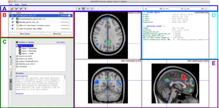
219 The web page allows a user to download single studies, or select and220 download multiple selected studies at once. The download will be a 221 compressed tar archive, which can be decompressed and either used di-222 rectly in one's analyses, or viewed and queried using the Volume Viewer 223 tool (Fig. 3). This is facilitated through the inclusion of XML-format files 224 which store study metadata and a list of study elements, along with the 225 actual data. Image files are stored in NIFTI format (although Volume 226 Viewer can also read MINC or MGZ format images). Volume Viewer pro-227 vides a convenient dialog for automatically interpolating images which 228 have been registered to the same space, but which do not have the same 229 size or orientation as the current volume. This allows comparison of 230 multiple images without having to preprocess them in advance. The 231 process of downloading and visualizing a study is illustrated inFig. 4. 232 Volume Viewer currently supports a number of useful features 233 which provide supplemental utility to the ANIMA online interface. 234 These include:
235 • The ability to overlay and compare images. This allows a user, for in-236 stance, to compare the results of a new analysis with those of pub-237 lished meta-analyses in ANIMA.
238 • The ability to save the current configuration as a single Volume
View-239 er session file (“.vvs” extension), which can be uploaded to ANIMA via
240 the online interface, and viewed by all users who download a given
241 study. The session includes all images, templates, and atlases, the
242 way they are arranged and composited, and the currently selected
243 viewing planes.
244 • The ability to compute a conjunction image, and quantify the degree
245 of overlap between two images. This feature also yields ROIs that
246 can be immediately used for further analysis.
247 • The ability to obtain peak coordinates from smoothed maps, for
248 reporting in an article, or use as seed points in further analyses.
249 • The inclusion of standard template and atlas images, which allow data
250 to be cross-referenced against anatomy or well-known parcellation
251 schemes. A dialogue for defining new atlases is also provided.
252
253 Submitting your study to ANIMA
254 ANIMA provides an intuitive online interface for submitting a study
255 for inclusion in the database (http://anima.fz-juelich.de/submit.php).
256 The submission process follows a series of tabs, as shown inFig. 5. The
257 first tab (“Login”) allows a new user to register with the database,
258 which is necessary to submit data. Registration allows the
administra-259 tors of ANIMA to associate a name and institution with a particular
260 study, and provides a level of security by ensuring users provide a
261 valid email address. The email address will not be accessible to the
pub-262 lic, and will only be used to contact the user in case of issues related to
263 the study. Registered users can also log in using this tab. The next step
264 of the process (the “Study” tab) requires the user to enter meta-data
265 about the study. This includes author and publication information, a
266 brief description (for browsing purposes), and a full-text PDF, if desired
267 and permitted. The subsequent tab (“Elements”) provides a means of
268 uploading the study elements (i.e., data files), and associated titles and
269 captions. Four types of files can be uploaded, as shown inTable 2. The
270 final tab (“Submit”) provides a preview of the study as entered, and
Fig. 1. The query page of the ANIMA online interface. Studies can be filtered by keyword, author name, journal, or publication date. Single studies can be downloaded using the icons at the right of each entry, and multiple studies can also be selected and downloaded together using the check boxes at left.
B:1
Box 2B:2
Summary of the data usage agreement for the ANIMA database.B:3
B:5
• Data are provided “as is”, with no guarantee whatsoeverB:6
• Data can be used for any non-commercial purpose, with theB:7
exception that redistribution of the data can only be done ifB:8
written consent is obtained from the database owners, andB:9
only for studies whose authors have approved redistribution.B:10
• Any public use of ANIMA data must provide attribution,spe-B:11
cifically by reference to the database itself, and the primaryB:12
article associated with those dataB:13
• The owners of ANIMA do not claim any copyright over theB:14
data archived in the databaseB:15
B:16
UNCORRECTED PR
OOF
Fig. 3. Screenshot of the stand-alone desktop application Volume Viewer. A. The toolbar, which allows individual images or Volume Viewer sessions to be loaded or saved, or removed from the current session. Buttons in the middle of the panel allow the user to set the mouse behaviour (zoom or query mode). At far right, the current voxel coordinates are display and can be modified. B. The image selection list, which allows the visibility of individual layers to be toggled, their order to be modified, and their transparency (alpha) to be set. C. The Library Panel, which lists the user's local library of studies which have already been downloaded. Individual study elements (images or entire Volume Viewer sessions) can be loaded via this panel. Two other panels are also selectable in this space: (i) the Atlases panel, which allows individual standard atlas or template images to be loaded; and (ii) the Colours panel (shown in inset), which allows a predefined colour map to be applied to the currently selected image layer. D. The Information Panel, which displays information about the current volume space, as well as the voxel defined by the currently viewed sections. Values can be queried for numeric data, while text representations are shown for atlases (here the Maximum Probability Map of the Jülich Probabilistic Atlas). E. The 2D rendering panels, which show the three standard orthogonal planes of the loaded images (horizontal/transversal, coronal, and sagittal). These can be zoomed, panned, and queried with the mouse.
Fig. 2. The single-study information page of the ANIMA online interface, showing the Papaya viewer. This page displays more detailed information about a study, including a list of indi-vidual study elements and their associated captions.
UNCORRECTED PR
OOF
271 allows the user to verify all information before submitting it to the 272 database.
273 Upon submission, a message will be sent to the database curators, 274 who will review the submission for validity, completeness, typographic 275 errors, and any other issues. If issues are encountered, an email will be 276 sent with instructions to the submitter on how to update them. Other-277 wise, the study will be accepted and a confirmation email will be sent. 278 This curation design ensures that only valid, published data are made 279 available through ANIMA, and that the uploader has the right to share 280 data on behalf of the associated authors. Notably, once a study is submit-281 ted, it is always associated with the submitter. Modifications can then be 282 made to the study only by the submitter or a database administrator. 283 ANIMA provides a “subscribe” feature, which allows the submitter (as 284 well as any registered user) to request a notification whenever a specific 285 study is modified. Any modifications made to the study (including edits, 286 additions, subtractions, or removal) will result in subscribers being 287 automatically notified of the change via email. Additionally, ANIMA 288 implements a simple versioning policy, which ensures that any major 289 modifications to existing studies are captured as new versions of the 290 study. This feature is important, since any publication using ANIMA 291 data should be able to point to the precise version of a data element 292 used for its analysis.
293 Metadata
294 The use of metadata to precisely define individual neuroimaging 295 results is an important consideration for any online database, and is 296 essential for both understanding individual database elements, and fa-297 cilitating further meta-analyses (Poldrack and Gorgolewski, 2014). For
298 the ANIMA database, all such metadata are encoded in XML format,
299 which allows them to be easily extended as necessary. In its present
300 state, the database provides metadata for whole studies (including
cita-301 tion details, keywords, abstracts, and DOIs), as well as single study
ele-302 ments. Study element metadata include a caption which describes the
303 element, and – specifically for image data – further details such as the
304 quantity represented, how the image is thresholded, and the coordinate
305 space (standard or native) in which it is expressed.
306 Future plans
307 The platform described above was designed to provide simple,
effi-308 cient access to results of a specific type of study (meta-analyses), in a
309 standard way. Moreover, the inclusion of a stand-alone desktop
applica-310 tion provides a useful means of visualizing, querying, and manipulating
311 data for use in future studies. However, both the online and standalone
312 components of this platform are also generic enough to be used with
313 other types of studies as well. All data is stored as XML files, which
314 can be easily extended or modified, depending on the type of data
re-315 quired. One potential future development will be to host other types
316 of study results using the ANIMA interface. Ideally, this will involve
ex-317 posing the database code as a documented and supported open-source
318 project, and providing an online REST interface accessible to registered
319 users (Masse, 2011). As an extension of this, ways in which ANIMA
320 could be integrated with existing databases will also be pursued.
Specif-321 ically, we plan to implement a REST API in order to allow a database
322 such as Neurovault.org (Gorgolewski et al., 2015) to access ANIMA
323 data, for inclusion in large-scale meta-analytic approaches.
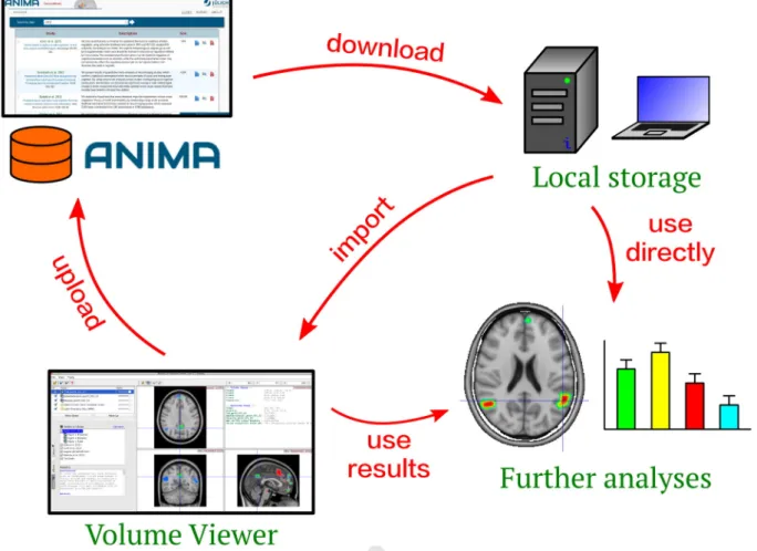
Fig. 4. Overview of typical database usage, illustrating the process of querying, selecting, downloading, and importing the data into the Volume Viewer library. Study data is downloaded from the ANIMA online interface into local storage, where it can be imported into Volume Viewer or used directly for further analysis. Data can also be first processed by Volume Viewer, for instance to extract conjunction ROIs, before being used in further analyses. Volume Viewer can also be used to defined sessions, which can be uploaded to the database.
UNCORRECTED PR
OOF
324 Another important consideration is how the database will be main-325 tained and managed in the future. The organization of the database en-326 sures that its management is minimal, but it is still important to have a
327 team of administrators who can evaluate submissions, respond to
feed-328 back about data quality, and provide support to users of the database.
329 We intend to assume this role for the foreseeable future. As an extension
Fig. 5. Online interface for submitting a new study to ANIMA. A. After login, the “Study” tab becomes visible. Entering the study name and a unique identifier, and pressing “Save” will start the ball rolling, and enable the remaining tabs. In the “Study” tab, you upload a full-text PDF file, a concise description of the study, and other information about the article. B. The “Ele-ments” tab allows the user to upload individual data elements, and describe them with a caption. C. The “Submit” tab provides a summary of the project, as it will appear on the Query page. This can be used to verify that the study is correctly entered. Finally, clicking the “Submit my Study” button will submit the uploaded data for validation by the database administrators.
UNCORRECTED PR
OOF
330 of this, additional support for ANIMA will be provided in terms ofdocu-331 mentation, and an online support forum which can be used to ensure 332 that common issues can be answered once, and a means of reporting 333 bugs, which can be of great assistance to the database developers. 334 The Volume Viewer tool is based on the ModelGUI API, which is an 335 open-source project hosted on http://www.launchpad.net/volume-336 viewer(an online platform designed to be scalable, and which supports 337 projects as large as the Ubuntu community). We intend to provide Vol-338 ume Viewer also as an open-source project, such that it can be freely ob-339 tained and developed by interested members of the neuroscience 340 community. A number of future improvements are planned for Volume 341 Viewer, including: the ability to specify named voxels and ROIs; the 342 ability to extract peaks from a smoothed image map; the addition of 343 3D volume and surface rendering; easy transfer of data between sur-344 faces and volumes; rendering of network graphs for the visualization 345 of connectivity information; and layout and printing features.
346 Summary
347 We present a new database which provides free and convenient on-348 line access to the results of published neuroimaging meta-analyses. 349 Data can be used for comparison with one's own results, or as a starting 350 point for new analyses, and the ANIMA interface provides a set of simple 351 tools which greatly facilitates this process. This interface includes a 352 search function, a form for submitting new studies, and an open-353 source stand-alone software tool for visualizing and organizing study 354 data, and generating ROIs for further analysis. It is our hope that the 355 ANIMA database will improve the way in which the results of meta-356 analytic neuroimaging studies will be used in the future, and encourage 357 researchers to incorporate these important results into their research 358 approaches.
359 References
360 Amft, M., Bzdok, D., Laird, A.R., Fox, P.T., Schilbach, L., Eickhoff, S.B., 2014. Definition and
361 characterization of an extended social-affective default network. Brain Struct. Funct.
362 http://dx.doi.org/10.1007/s00429-013-0698-0.
363 Bzdok, D., Laird, A.R., Zilles, K., Fox, P.T., Eickhoff, S.B., 2013a. An investigation of the
struc-364 tural, connectional, and functional subspecialization in the human amygdala. Hum.
365 Brain Mapp. 34, 3247–3266.http://dx.doi.org/10.1002/hbm.22138.
366 Bzdok, D., Langner, R., Caspers, S., Kurth, F., Habel, U., Zilles, K., Laird, A., Eickhoff, S.B.,
367 2011. ALE meta-analysis on facial judgments of trustworthiness and
attractive-368 ness. Brain Struct. Funct. 215, 209–223.
http://dx.doi.org/10.1007/s00429-010-369 0287-4.
370 Bzdok, D., Langner, R., Schilbach, L., Jakobs, O., Roski, C., Caspers, S., Laird, A.R., Fox, P.T.,
371 Zilles, K., Eickhoff, S.B., 2013b. Characterization of the temporo-parietal junction by
372 combining data-driven parcellation, complementary connectivity analyses, and
func-373 tional decoding. Neuroimage 81, 381–392.http://dx.doi.org/10.1016/j.neuroimage.
374 2013.05.046.
375 Bzdok, D., Schilbach, L., Vogeley, K., Schneider, K., Laird, A.R., Langner, R., Eickhoff, S.B.,
376 2012. Parsing the neural correlates of moral cognition: ALE meta-analysis on
moral-377 ity, theory of mind, and empathy. Brain Struct. Funct. 217, 783–796.
378 Caspers, S., Zilles, K., Laird, A.R., Eickhoff, S.B., 2010. ALE meta-analysis of action
observa-379 tion and imitation in the human brain. Neuroimage 50, 1148–1167.http://dx.doi.org/
380 10.1016/j.neuroimage.2009.12.112.
381
Chang, L.J., Yarkoni, T., Khaw, M.W., Sanfey, A.G., 2012. Decoding the role of the insula in
382
human cognition: functional parcellation and large-scale reverse inference. Cereb.
383
Cortexhttp://dx.doi.org/10.1093/cercor/bhs065(bhs065).
384
Cieslik, E.C., Mueller, V.I., Eickhoff, C.R., Langner, R., Eickhoff, S.B., 2015. Three key regions for
385
supervisory attentional control: evidence from neuroimaging meta-analyses. Neurosci.
386
Biobehav. Rev. 48C, 22–34.http://dx.doi.org/10.1016/j.neubiorev.2014.11.003.
387
Cieslik, E.C., Zilles, K., Caspers, S., Roski, C., Kellermann, T.S., Jakobs, O., Langner, R., Laird,
388
A.R., Fox, P.T., Eickhoff, S.B., 2013. Is there “one” DLPFC in cognitive action control?
Ev-389
idence for heterogeneity from co-activation-based parcellation. Cereb. Cortex 23,
390
2677–2689.http://dx.doi.org/10.1093/cercor/bhs256.
391
Clos, M., Amunts, K., Laird, A.R., Fox, P.T., Eickhoff, S.B., 2013. Tackling the multifunctional
392
nature of Broca's region meta-analytically: co-activation-based parcellation of area
393
44. Neuroimage 83, 174–188.http://dx.doi.org/10.1016/j.neuroimage.2013.06.041.
394
Eickhoff, S.B., Bzdok, D., Laird, A.R., Kurth, F., Fox, P.T., 2012. Activation likelihood
estima-395
tion meta-analysis revisited. Neuroimage 59, 2349–2361.http://dx.doi.org/10.1016/j.
396
neuroimage.2011.09.017.
397
Eickhoff, S.B., Bzdok, D., Laird, A.R., Roski, C., Caspers, S., Zilles, K., Fox, P.T., 2011.
Co-398
activation patterns distinguish cortical modules, their connectivity and functional
399
differentiation. Neuroimage 57, 938–949.http://dx.doi.org/10.1016/j.neuroimage.
400
2011.05.021.
401
Eickhoff, S.B., Laird, A.R., Grefkes, C., Wang, L.E., Zilles, K., Fox, P.T., 2009. Coordinate-based
402
activation likelihood estimation meta-analysis of neuroimaging data: a
random-403
effects approach based on empirical estimates of spatial uncertainty. Hum. Brain
404
Mapp. 30, 2907–2926.http://dx.doi.org/10.1002/hbm.20718.
405
Etkin, A., Wager, T.D., 2007. Functional neuroimaging of anxiety: a meta-analysis of
emo-406
tional processing in PTSD, social anxiety disorder, and specific phobia. AJP 164,
407
1476–1488.http://dx.doi.org/10.1176/appi.ajp.2007.07030504.
408
Fox, P.T., Lancaster, J.L., Laird, A.R., Eickhoff, S.B., 2014. Meta-analysis in human
neuroim-409
aging: computational modeling of large-scale databases. Annu. Rev. Neurosci. 37,
410
409–434.http://dx.doi.org/10.1146/annurev-neuro-062012-170320.
411
Friebel, U., Eickhoff, S.B., Lotze, M., 2011. Coordinate-based meta-analysis of
experimen-412
tally induced and chronic persistent neuropathic pain. Neuroimage 58, 1070–1080.
413
http://dx.doi.org/10.1016/j.neuroimage.2011.07.022.
414
Goodkind, M., Eickhoff, S.B., Oathes, D., Jiang, Y., Chang, A., Jones-Hagata, L., Ortega, B.N.,
415
Zaiko, Y.V., Roach, E.L., Korgaonkar, M.S., Grieve, S.M., Galatzer-Levy, I., Fox, P.T.,
416
Etkin, A., 2015. Identification of a common neurobiological substrate for mental
ill-417
ness. JAMA Psychiatry (in press). Q5 418
Gorgolewski, K.J., Varoquaux, G., Rivera, G., Schwarz, Y., Ghosh, S.S., Maumet, C., Sochat,
419
V.V., Nichols, T.E., Poldrack, R.A., Poline, J.-B., Yarkoni, T., Margulies, D.S., 2015.
420
NeuroVault.org: a web-based repository for collecting and sharing unthresholded
421
statistical maps of the human brain. Front. Neuroinform. 9, 8.http://dx.doi.org/10.
422
3389/fninf.2015.00008.
423
Hardwick, R.M., Rottschy, C., Miall, R.C., Eickhoff, S.B., 2013. A quantitative meta-analysis
424
and review of motor learning in the human brain. Neuroimage 67, 283–297.http://
425
dx.doi.org/10.1016/j.neuroimage.2012.11.020.
426
Hoffstaedter, F., Grefkes, C., Caspers, S., Roski, C., Palomero-Gallagher, N., Laird, A.R., Fox,
427
P.T., Eickhoff, S.B., 2014. The role of anterior midcingulate cortex in cognitive motor
428
control: evidence from functional connectivity analyses. Hum. Brain Mapp. 35,
429
2741–2753.http://dx.doi.org/10.1002/hbm.22363.
430
Keuken, M.C., Müller-Axt, C., Langner, R., Eickhoff, S.B., Forstmann, B.U., Neumann, J.,
431
2014. Brain networks of perceptual decision-making: an fMRI ALE meta-analysis.
432
Front. Hum. Neurosci. 8.http://dx.doi.org/10.3389/fnhum.2014.00445.
433
Kober, H., Barrett, L.F., Joseph, J., Bliss-Moreau, E., Lindquist, K., Wager, T.D., 2008.
434
Functional grouping and cortical-subcortical interactions in emotion: a
meta-435
analysis of neuroimaging studies. Neuroimage 42, 998–1031.http://dx.doi.org/
436
10.1016/j.neuroimage.2008.03.059.
437
Kohn, N., Eickhoff, S.B., Scheller, M., Laird, A.R., Fox, P.T., Habel, U., 2014. Neural network
438
of cognitive emotion regulation—an ALE meta-analysis and MACM analysis.
439
Neuroimage 87, 345–355.http://dx.doi.org/10.1016/j.neuroimage.2013.11.001.
440
Kurth, F., Zilles, K., Fox, P.T., Laird, A.R., Eickhoff, S.B., 2010. A link between the systems:
func-441
tional differentiation and integration within the human insula revealed by
meta-442
analysis. Brain Struct. Funct. 214, 519–534.
http://dx.doi.org/10.1007/s00429-010-443
0255-z.
444
Laird, A.R., Eickhoff, S.B., Fox, P.M., Uecker, A.M., Ray, K.L., Saenz, J.J., McKay, D.R., Bzdok,
445
D., Laird, R.W., Robinson, J.L., Turner, J.A., Turkeltaub, P.E., Lancaster, J.L., Fox, P.T.,
446
2011. The BrainMap strategy for standardization, sharing, and meta-analysis of
neu-447
roimaging data. BMC Res. Notes 4, 349.http://dx.doi.org/10.1186/1756-0500-4-349.
t4:1 Table 2
t4:2 File types which can be uploaded to ANIMA.
t4:3 File type Description Extensions t4:4 Volume File A 3D brain image file, typically containing statistical maps such as t-, z-, or p-scores, which indicate the
result of a specific analysis. These should be in NIFTI format.
nii, hdr, nii.gz
t4:5 Volume Viewer Session File An XML-format file containing all data and metadata specifying a Volume Viewer session. This can be used to pre-define a set of composite images, templates, atlases, and their colour mapping.
vvs
t4:6 Volume Viewer Point Set File An XML-format file specifying a list of 3D coordinates, along with associated data which can be used to
specify and label individual points of interest in a study.
pointset
t4:7 Image File A standard 2D image file, which could represent an informative figure, schematic, or other useful
information pertaining to the study.
png, jpg, gif, tif, etc.
t4:8 Text File A text file, which can contain additional free descriptions or other information about the study and its elements.
UNCORRECTED PR
OOF
448 Langner, R., Eickhoff, S.B., 2013. Sustaining attention to simple tasks: a meta-analyticre-449 view of the neural mechanisms of vigilant attention. Psychol. Bull. 139, 870–900.
450 http://dx.doi.org/10.1037/a0030694.
451 Masse, M., 2011. REST API Design Rulebook. O'Reilly Media, Inc.
452 Müller, V.I., Langner, R., Cieslik, E.C., Rottschy, C., Eickhoff, S.B., 2014. Interindividual
453 differences in cognitive flexibility: influence of gray matter volume, functional
454 connectivity and trait impulsivity. Brain Struct. Funct.http://dx.doi.org/10.
455 1007/s00429-014-0797-6.
456 Nellessen, N., Rottschy, C., Eickhoff, S.B., Ketteler, S.T., Kuhn, H., Shah, N.J., Schulz, J.B.,
457 Reske, M., Reetz, K., 2014. Specific and disease stage-dependent episodic
memory-458 related brain activation patterns in Alzheimer's disease: a coordinate-based
meta-459 analysis. Brain Struct. Funct.http://dx.doi.org/10.1007/s00429-014-0744-6.
460 Nickl-Jockschat, T., Rottschy, C., Thommes, J., Schneider, F., Laird, A.R., Fox, P.T., Eickhoff, S.B.,
461 2014. Neural networks related to dysfunctional face processing in autism spectrum
462 disorder. Brain Struct. Funct. 1–17.http://dx.doi.org/10.1007/s00429-014-0791-z.
463 Northoff, G., Heinzel, A., de Greck, M., Bermpohl, F., Dobrowolny, H., Panksepp, J., 2006.
464 Self-referential processing in our brain—a meta-analysis of imaging studies on the
465 self. NeuroImage 31, 440–457.http://dx.doi.org/10.1016/j.neuroimage.2005.12.002.
466 Poldrack, R.A., Gorgolewski, K.J., 2014. Making big data open: data sharing in
neuroimag-467 ing. Nat. Neurosci. 17, 1510–1517.http://dx.doi.org/10.1038/nn.3818.
468 Reid, A.T., Bzdok, D., Langner, R., Fox, P.T., Laird, A.R., Amunts, K., Eickhoff, S.B., Eickhoff,
469 C.R., 2015. Multimodal connectivity mapping of the human left anterior and posterior
470 lateral prefrontal cortex. Brain Struct. Funct. 1–17.
http://dx.doi.org/10.1007/s00429-471 015-1060-5.
472 Robinson, J.L., Laird, A.R., Glahn, D.C., Lovallo, W.R., Fox, P.T., 2010. Metaanalytic
connec-473 tivity modeling: delineating the functional connectivity of the human amygdala.
474 Hum. Brain Mapp. 31, 173–184.http://dx.doi.org/10.1002/hbm.20854.
475
Rottschy, C., Langner, R., Dogan, I., Reetz, K., Laird, A.R., Schulz, J.B., Fox, P.T.,
476
Eickhoff, S.B., 2012. Modelling neural correlates of working memory: a
477
coordinate-based meta-analysis. Neuroimage 60, 830–846.http://dx.doi.org/
478
10.1016/j.neuroimage.2011.11.050.
479
Schilbach, L., Bzdok, D., Timmermans, B., Fox, P.T., Laird, A.R., Vogeley, K., Eickhoff, S.B.,
480
2012. Introspective minds: using ALE meta-analyses to study commonalities in the
481
neural correlates of emotional processing, social & unconstrained cognition. PLoS
482
ONE 7, e30920.http://dx.doi.org/10.1371/journal.pone.0030920.
483
Schilbach, L., Müller, V.I., Hoffstaedter, F., Clos, M., Goya-Maldonado, R., Gruber, O.,
484
Eickhoff, S.B., 2014. Meta-analytically informed network analysis of resting state
485
FMRI reveals hyperconnectivity in an introspective socio-affective network in
depres-486
sion. PLoS ONE 9, e94973.http://dx.doi.org/10.1371/journal.pone.0094973.
487
Wager, T.D., Lindquist, M., Kaplan, L., 2007. Meta-analysis of functional neuroimaging
488
data: current and future directions. Soc. Cogn. Affect. Neurosci. 2, 150–158.http://
489
dx.doi.org/10.1093/scan/nsm015.
490
Xue, W., Kang, J., Bowman, F.D., Wager, T.D., Guo, J., 2014. Identifying functional
co-491
activation patterns in neuroimaging studies via poisson graphical models. Biom 70,
492
812–822.http://dx.doi.org/10.1111/biom.12216.
493
Yarkoni, T., Poldrack, R.A., Nichols, T.E., Van Essen, D.C., Wager, T.D., 2011. Large-scale
au-494
tomated synthesis of human functional neuroimaging data. Nat. Methods 8, 665–670.
495
http://dx.doi.org/10.1038/nmeth.1635.
496
Eulenburg, Zu., Caspers, P., Roski, S., Eickhoff, C., S.B., 2012. Meta-analytical definition and
497
functional connectivity of the human vestibular cortex. Neuroimage 60, 162–169.
498
http://dx.doi.org/10.1016/j.neuroimage.2011.12.032.