2016 ESC Guidelines for the management of atrial fibrillation
developed in collaboration with EACTS
The Task Force for the management of atrial fibrillation of the
European Society of Cardiology (ESC)
Developed with the special contribution of the European Heart Rhythm
Association (EHRA) of the ESC
Endorsed by the European Stroke Organisation (ESO)
Authors/Task Force Members: Paulus Kirchhof
*(Chairperson) (UK/Germany), Stefano Benussi
*1(Co-Chairperson) (Switzerland), Dipak Kotecha (UK), Anders Ahlsson
1(Sweden), Dan Atar (Norway),
Barbara Casadei (UK), Manuel Castella
1(Spain), Hans-Christoph Diener
2(Germany), Hein Heidbuchel (Belgium),
Jeroen Hendriks (The Netherlands), Gerhard Hindricks (Germany), Antonis S. Manolis (Greece),
Jonas Oldgren (Sweden), Bogdan Alexandru Popescu (Romania), Ulrich Schotten (The Netherlands),
Bart Van Putte
1(The Netherlands) and Panagiotis Vardas (Greece)
Document Reviewers: Stefan Agewall (CPG Review Co-ordinator) (Norway), John Camm (CPG Review Co-ordinator) (UK),
Gonzalo Baron Esquivias (Spain), Werner Budts (Belgium), Scipione Carerj (Italy), Filip Casselman (Belgium), Antonio Coca (Spain),
Raffaele De Caterina (Italy), Spiridon Deftereos (Greece), Dobromir Dobrev (Germany), Jose´ M. Ferro (Portugal), Gerasimos
Filippatos (Greece), Donna Fitzsimons (UK), Bulent Gorenek (Turkey), Maxine Guenoun (France), Stefan H. Hohnloser (Germany),
Philippe Kolh (Belgium), Gregory Y. H. Lip (UK), Athanasios Manolis (Greece), John McMurray (UK), Piotr Ponikowski (Poland),
Raphael Rosenhek (Austria), Frank Ruschitzka (Switzerland), Irina Savelieva (UK), Sanjay Sharma (UK), Piotr Suwalski (Poland),
Juan Luis Tamargo (Spain), Clare J. Taylor (UK), Isabelle C. Van Gelder (The Netherlands), Adriaan A. Voors (The Netherlands),
Stephan Windecker (Switzerland), Jose Luis Zamorano (Spain) and Katja Zeppenfeld (The Netherlands)
The disclosure forms of all experts involved in the development of these guidelines are available on the ESC website
http://www.escardio.org/guidelines.
*
Corresponding authors: Paulus Kirchhof, Institute of Cardiovascular Sciences, University of Birmingham, SWBH and UHB NHS trusts, IBR, Room 136, Wolfson Drive, Birmingham B15 2TT, United Kingdom, Tel: +44 121 4147042, E-mail: [email protected]; Stefano Benussi, Department of Cardiovascular Surgery, University Hospital Zurich, R€amistrasse 100, 8091 Zu¨rich, Switzerland, Tel: +41(0)788933835, E-mail: [email protected].1Representing the European Association for Cardio-Thoracic Surgery (EACTS) 2
Representing the European Stroke Association (ESO)
ESC Committee for Practice Guidelines (CPG) and National Cardiac Societies Reviewers can be found in the Appendix. ESC entities having participated in the development of this document.
Associations: European Association for Cardiovascular Prevention and Rehabilitation (EACPR), European Association of Cardiovascular Imaging (EACVI), European Heart Rhythm Association (EHRA), Heart Failure Association (HFA).
Councils: Council on Cardiovascular Nursing and Allied Professions, Council for Cardiology Practice, Council on Cardiovascular Primary Care, Council on Hypertension. Working Groups: Cardiac Cellular Electrophysiology, Cardiovascular Pharmacotherapy, Grown-up Congenital Heart Disease, Thrombosis, Valvular Heart Disease.
The content of these European Society of Cardiology (ESC) Guidelines has been published for personal and educational use only. No commercial use is authorized. No part of the ESC Guidelines may be translated or reproduced in any form without written permission from the ESC. Permission can be obtained upon submission of a written request to Oxford University Press, the publisher of theEuropean Heart Journal and the party authorized to handle such permissions on behalf of the ESC ([email protected]).
Disclaimer:The ESC Guidelines represent the views of the ESC and were produced after careful consideration of the scientific and medical knowledge and the evidence avail-able at the time of their publication. The ESC is not responsible in the event of any contradiction, discrepancy and/or ambiguity between the ESC Guidelines and any other offi-cial recommendations or guidelines issued by the relevant public health authorities, in particular in relation to good use of healthcare or therapeutic strategies. Health professionals are encouraged to take the ESC Guidelines fully into account when exercising their clinical judgment, as well as in the determination and the implementation of preventive, diagnostic or therapeutic medical strategies; however, the ESC Guidelines do not override, in any way whatsoever, the individual responsibility of health professio-nals to make appropriate and accurate decisions in consideration of each patient’s health condition and in consultation with that patient and, where appropriate and/or neces-sary, the patient’s caregiver. Nor do the ESC Guidelines exempt health professionals from taking into full and careful consideration the relevant official updated recommendations or guidelines issued by the competent public health authorities, in order to manage each patient’s case in light of the scientifically accepted data pursuant to their respective ethical and professional obligations. It is also the health professional’s responsibility to verify the applicable rules and regulations relating to drugs and medical devices at the time of prescription.
VCThe European Society of Cardiology 2016. Reproduced with kind permission. For permissions please email: [email protected]. doi:10.1093/ejcts/ezw313 Advance Access publication 23 September 2016
GU IDELINE G U ID E L IN ES
Keywords:
Guidelines • Atrial fibrillation • Anticoagulation • Vitamin K antagonists • Non-vitamin K antagonist oral anticoagulants • Left
atrial appendage occlusion • Rate control • Cardioversion • Rhythm control • Antiarrhythmic drugs • Upstream therapy • Catheter ablation
• AF surgery • Valve repair • Pulmonary vein isolation • Left atrial ablation
TABLE OF CONTENTS
ABBREVIATIONS AND ACRONYMS
1. PREAMBLE . . . 5
2. INTRODUCTION. . . 7
3. EPIDEMIOLOGY AND IMPACT FOR PATIENTS . . . 7
3.1. Incidence and prevalence of atrial fibrillation . . . 7
3.2. Morbidity, mortality, and healthcare burden of atrial fibrillation 7 3.3. Impact of evidence-based management on outcomes in atrial fibrillation patients . . . 7
3.4. Gender . . . 8
4. PATHOPHYSIOLOGICAL AND GENETIC ASPECTS THAT GUIDE MANAGEMENT. . . 9
4.1. Genetic predisposition . . . 9
4.2. Mechanisms leading to atrial fibrillation. . . 9
4.2.1. Remodelling of atrial structure and ion channel function. . . 9
4.2.2. Electrophysiological mechanisms of atrial fibrillation . . . 9
5. DIAGNOSIS AND TIMELY DETECTION OF ATRIAL FIBRILLATION . . 9
5.1. Overt and silent atrial fibrillation . . . 9
5.2. Screening for silent atrial fibrillation . . . 10
5.2.1. Screening for atrial fibrillation by electrocardiogram in the community . . . 10
5.2.2. Prolonged monitoring for paroxysmal atrial fibrillation . . . 11
5.2.3. Patients with pacemakers and implanted devices . . . 11
5.2.4. Detection of atrial fibrillation in stroke survivors . . . 11
5.3. Electrocardiogram detection of atrial flutter . . . 12
6. CLASSIFICATION OF ATRIAL FIBRILLATION . . . 12
6.1. Atrial fibrillation pattern . . . 13
6.2. Atrial fibrillation types reflecting different causes of the arrhythmia . . . 13
6.3. Symptom burden in atrial fibrillation . . . 14
7. DETECTION AND MANAGEMENT OF RISK FACTORS AND CONCOMITANT CARDIOVASCULAR DISEASES. . . 15
7.1. Heart failure . . . 15
7.1.1. Patients with atrial fibrillation and heart failure with reduced ejection fraction. . . 15
7.1.2. Atrial fibrillation patients with heart failure with preserved ejection fraction . . . 16
7.1.3. Atrial fibrillation patients with heart failure with mid-range ejection fraction . . . 16
7.1.4. Prevention of atrial fibrillation in heart failure . . . 16
7.2. Hypertension . . . 16
7.3. Valvular heart disease . . . 16
7.4. Diabetes mellitus . . . 17
7.5. Obesity and weight loss . . . 26
7.5.1. Obesity as a risk factor. . . 18
7.5.2. Weight reduction in obese patients with atrial fibrillation . 18 7.5.3. Catheter ablation in obese patients . . . 18
7.6. Chronic obstructive pulmonary disease, sleep apnoea, and other respiratory diseases . . . 18
7.7. Chronic kidney disease . . . 19
8. INTEGRATED MANAGEMENT OF PATIENTS WITH ATRIAL FIBRILLATION . . . 19
8.1. Evidence supporting integrated atrial fibrillation care. . . 19
8.2. Components of integrated atrial fibrillation care. . . 20
8.2.1. Patient involvement . . . 20
8.2.2. Multidisciplinary atrial fibrillation teams . . . 20
8.2.3. Role of non-specialists . . . 21
8.2.4. Technology use to support atrial fibrillation care. . . 21
8.3. Diagnostic workup of atrial fibrillation patients . . . 21
8.3.1. Recommended evaluation in all atrial fibrillation patients . 21 8.3.2. Additional investigations in selected patients with atrial fibrillation . . . 21
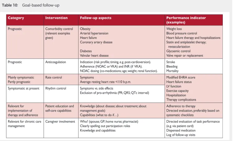
8.4. Structured follow-up . . . 22
8.5. Defining goals of atrial fibrillation management . . . 22
9. STROKE PREVENTION THERAPY IN ATRIAL FIBRILLATION PATIENTS . . . 22
9.1. Prediction of stroke and bleeding risk. . . 23
9.1.1. Clinical risk scores for stroke and systemic embolism . . . 23
9.1.2. Anticoagulation in patients with a CHA2DS2-VASc score of 1 in men and 2 in women . . . 23
9.1.3. Clinical risk scores for bleeding. . . 24
9.2. Stroke prevention . . . 24
9.2.1. Vitamin K antagonists . . . 24
9.2.2. Non-vitamin K antagonist oral anticoagulants . . . 24
9.2.2.1. Apixaban . . . 24
9.2.2.2. Dabigatran . . . 24
9.2.2.3. Edoxaban . . . 25
9.2.2.4. Rivaroxaban . . . 25
9.2.3. Non-vitamin K antagonist oral anticoagulants or vitamin K antagonists. . . 27
9.2.4. Oral anticoagulation in atrial fibrillation patients with chronic kidney disease. . . 27
9.2.5. Oral anticoagulation in atrial fibrillation patients on dialysis . . . 27
9.2.6. Patients with atrial fibrillation requiring kidney transplantation . . . 28
9.2.7. Antiplatelet therapy as an alternative to oral anticoagulants . . . 28
9.3. Left atrial appendage occlusion and exclusion. . . 28
9.3.1. Left atrial appendage occlusion devices . . . 28
9.3.2. Surgical left atrial appendage occlusion or exclusion. . . 29
9.4. Secondary stroke prevention . . . 29
9.4.1. Treatment of acute ischaemic stroke . . . 29
9.4.2. Initiation of anticoagulation after transient ischaemic attack or ischaemic stroke . . . 29
9.4.3. Initiation of anticoagulation after intracranial haemorrhage. . . 29
9.5. Strategies to minimize bleeding on anticoagulant therapy . . . . 30
9.5.1. Uncontrolled hypertension . . . 30
9.5.2. Previous bleeding event. . . 30
9.5.3. Labile international normalized ratio and adequate non-vitamin K antagonist oral anticoagulant dosing . . . 31
9.5.4. Alcohol abuse . . . 31
9.5.5. Falls and dementia . . . 31
9.5.6. Genetic testing . . . 31
9.5.7. Bridging periods off oral anticoagulation . . . 31
9.6. Management of bleeding events in anticoagulated patients with atrial fibrillation . . . 31
9.6.1. Management of minor, moderate, and severe bleeding. . . 31
9.6.2. Oral anticoagulation in atrial fibrillation patients at risk of or having a bleeding event . . . 33
9.7. Combination therapy with oral anticoagulants and
antiplatelets . . . 33
9.7.1. Antithrombotic therapy after acute coronary syndromes and percutaneous coronary intervention in patients requiring oral anticoagulation . . . 34
10. RATE CONTROL THERAPY IN ATRIAL FIBRILLATION . . . 34
10.1. Acute rate control . . . 36
10.2. Long-term pharmacological rate control . . . 36
10.2.1. Beta-blockers . . . 36
10.2.2. Non-dihydropyridine calcium channel blockers . . . 36
10.2.3. Digitalis . . . 36
10.2.4. Amiodarone . . . 37
10.3. Heart rate targets in atrial fibrillation . . . 38
10.4. Atrioventricular node ablation and pacing. . . 38
11. RHYTHM CONTROL THERAPY IN ATRIAL FIBRILLATION . . . 39
11.1. Acute restoration of sinus rhythm . . . 39
11.1.1. Antiarrhythmic drugs for acute restoration of sinus rhythm (‘pharmacological cardioversion’) . . . 39
11.1.2. ‘Pill in the pocket’ cardioversion performed by patients . . 40
11.1.3. Electrical cardioversion . . . 40
11.1.4. Anticoagulation in patients undergoing cardioversion . . . 40
11.2. Long-term antiarrhythmic drug therapy. . . 40
11.2.1. Selection of antiarrhythmic drugs for long-term therapy: Safety first!. . . 41
11.2.1.1. Amiodrone. . . 41
11.2.1.2. Dronedarone. . . 41
11.2.1.3. Flecainide and propafenone . . . 41
11.2.1.4. Quinidine and disopyramide. . . 42
11.2.1.5. Sotalol. . . 42
11.2.1.6. Dofetilide . . . 42
11.2.2. Twelve-lead electrocardiogram as a tool to identify patients at risk of pro-arrhythmia. . . 42
11.2.3. New antiarrhythmic drugs . . . 42
11.2.4. Antiarrhythmic effects of non-antiarrhythmic drugs . . . 43
11.3. Catheter ablation . . . 43
11.3.1. Indications. . . 43
11.3.2. Techniques and technologies . . . 43
11.3.3. Outcome and complications . . . 46
11.3.3.1. Outcome of catheter ablation for atrial fibrillation. . . 46
11.3.3.2. Complications of catheter ablation for atrial fibrillation . . . 47
11.3.4. Anticoagulation – before, during, and after ablation . . . 48
11.3.5. Ablation of atrial fibrillation in heart failure patients . . . 48
11.3.6. Follow-up after catheter ablation. . . 48
11.4. Atrial fibrillation surgery . . . 48
11.4.1. Concomitant atrial fibrillation surgery . . . 48
11.4.2. Stand-alone rhythm control surgery . . . 48
11.5. Choice of rhythm control following treatment failure . . . 49
11.6. The atrial fibrillation Heart Team. . . 49
12. HYBRID RHYTHM CONTROL THERAPY . . . 50
12.1. Combining antiarrhythmic drugs and catheter ablation . . . 50
12.2. Combining antiarrhythmic drugs and pacemakers . . . 50
13. SPECIFIC SITUATIONS . . . 51
13.1. Frail and ‘elderly’ patients . . . 51
13.2. Inherited cardiomyopathies, channelopathies, and accessory pathways . . . 51
13.2.1. Wolff–Parkinson–White syndrome. . . 51
13.2.2. Hypertrophic cardiomyopathy. . . 52
13.2.3. Channelopathies and arrhythmogenic right ventricular cardiomyopathy . . . 52
13.3. Sports and atrial fibrillation . . . 53
13.4. Pregnancy. . . 54
13.4.1. Rate control . . . 54
13.4.2. Rhythm control . . . 54
13.4.3. Anticoagulation . . . 54
13.5. Post-operative atrial fibrillation . . . 54
13.5.1. Prevention of post-operative atrial fibrillation . . . 54
13.5.2. Anticoagulation . . . 55
13.5.3. Rhythm control therapy in post-operative atrial fibrillation . . . 55
13.6. Atrial arrhythmias in grown-up patients with congenital heart disease . . . 55
13.6.1. General management of atrial arrhythmias in grown-up patients with congenital heart disease . . . 55
13.6.2. Atrial tachyarrhythmias and atrial septal defects . . . 56
13.6.3. Atrial tachyarrhythmias after Fontan operation . . . 56
13.6.4. Atrial tachyarrhythmias after tetralogy of Fallot correction . . . 56
13.7. Management of atrial flutter . . . 56
14. PATIENT INVOLVEMENT, EDUCATION, AND SELF-MANAGEMENT. . . 57
14.1. Patient-centred care . . . 57
14.2. Integrated patient education . . . 57
14.3. Self-management and shared decision-making. . . 57
15. GAPS IN EVIDENCE . . . 57
15.1. Major health modifiers causing atrial fibrillation. . . 57
15.2. How much atrial fibrillation constitutes a mandate for therapy? . . . .58
15.3. Atrial high-rate episodes and need for anticoagulation . . . 58
15.4. Stroke risk in specific populations . . . 58
15.5. Anticoagulation in patients with severe chronic kidney disease . . . 58
15.6. Left atrial appendage occlusion for stroke prevention . . . 58
15.7. Anticoagulation in atrial fibrillation patients after a bleeding or stroke event . . . 58
15.8. Anticoagulation and optimal timing of non-acute cardioversion. . . 84
15.9. Competing causes of stroke or transient ischaemic attack in atrial fibrillation patients . . . 58
15.10. Anticoagulation in patients with biological heart valves (including transcatheter aortic valve implantation) and non-rheumatic valve disease. . . 59
15.11. Anticoagulation after ‘successful’ catheter ablation . . . 59
15.12. Comparison of rate control agents . . . 59
15.13. Catheter ablation in persistent and long-standing persistent AF . . . 59
15.14. Optimal technique for repeat catheter ablation . . . 59
15.15. Combination therapy for maintenance of sinus rhythm . . . . 59
15.16. Can rhythm control therapy convey a prognostic benefit in atrial fibrillation patients?. . . 59
15.17. Thoracoscopic ‘stand-alone’ atrial fibrillation surgery. . . 59
15.18. Surgical exclusion of the left atrial appendage. . . 59
15.19. Concomitant atrial fibrillation surgery . . . 59
16. TO DO AND NOT TO DO MESSAGES FROM THE GUIDELINES . . . 60
17. A SHORT SUMMARY OF THE MANAGEMENT OF AF PATIENTS . . 62
18. WEB ADDENDA . . . 62
19. APPENDIX . . . 62
20. REFERENCES . . . 63
ABBREVIATIONS AND ACRONYMS
ABC
age, biomarkers, clinical history
ACE
angiotensin-converting enzyme
GU IDELINE G U ID E L IN ES
ACS
acute coronary syndromes
AF
atrial fibrillation
AFFIRM
Atrial Fibrillation Follow-up
Investigation of Rhythm
Management
AFNET
German Competence NETwork on
Atrial Fibrillation
AngII
angiotensin II
AHRE
atrial high rate episodes
APACHE-AF
Apixaban versus Antiplatelet
drugs or no antithrombotic
drugs after
anticoagulation-associated intraCerebral
HaEmorrhage in patients with
Atrial Fibrillation
ARB
angiotensin receptor blocker
ARISTOTLE
Apixaban for Reduction in Stroke
and Other Thromboembolic Events
in Atrial Fibrillation
ARNI
angiotensin receptor neprilysin
inhibition
ARTESiA
Apixaban for the Reduction of
Thrombo-Embolism in Patients
With Device-Detected Sub-Clinical
Atrial Fibrillation
ATRIA
AnTicoagulation and Risk factors In
Atrial fibrillation
AV
Atrioventricular
AXAFA
Anticoagulation using the direct
factor Xa inhibitor apixaban during
Atrial Fibrillation catheter Ablation:
Comparison to vitamin K antagonist
therapy
BAFTA
Birmingham Atrial Fibrillation
Treatment of the Aged Study
BMI
body mass index
b.p.m.
beats per minute
CABANA
Catheter Ablation versus
Antiarrhythmic Drug Therapy for
Atrial Fibrillation Trial
CABG
coronary artery bypass graft
CAD
coronary artery disease
CHA
2DS
2-VASc
Congestive Heart failure,
hyperten-sion, Age >_75 (doubled), Diabetes,
Stroke (doubled), Vascular disease,
Age 65–74, and Sex (female)
CHADS
2Cardiac failure, Hypertension, Age,
Diabetes, Stroke (Doubled)
CI
confidence interval
CKD
chronic kidney disease
CPG
Committee for Practice Guidelines
CrCl
creatinine clearance
CT
computed tomography
CV
cardiovascular
CYP2D6
cytochrome P450 2D6
CYP3A4
cytochrome P450 3A4
DIG
Digitalis Investigation Group
EACTS
European Association for
Cardio-Thoracic Surgery
EAST
Early treatment of Atrial fibrillation
for Stroke prevention Trial
ECG
electrocardiogram/
electrocardiography
EHRA
European Heart Rhythm
Association
ENGAGE AF-TIMI 48
Effective Anticoagulation with
Factor Xa Next Generation in Atrial
Fibrillation–Thrombolysis in
Myocardial Infarction 48
EORP
EURObservational Research
Programme
ESC
European Society of Cardiology
ESO
European stroke Organisation
FAST
Atrial Fibrillation Catheter Ablation
vs. Surgical Ablation Treatment
FEV1
forced expiratory volume in 1 s
FFP
four-factor prothrombin complex
concentrates
FXII
factor XII
GDF-15
growth differentiation factor 15
GFR
glomerular filtration rate
GUCH
grown-up congenital heart disease
HARMONY
A Study to Evaluate the Effect of
Ranolazine and Dronedarone When
Given Alone and in Combination in
Patients With Paroxysmal Atrial
Fibrillation
HAS-BLED
hypertension, abnormal renal/liver
function (1 point each), stroke,
bleed-ing history or predisposition, labile
INR, elderly (>65 years),
drugs/alco-hol concomitantly (1 point each)
HEMORR
2HAGES
Hepatic or renal disease, ethanol
abuse, malignancy history, older age
>75, reduced platelet count/function/
antiplatelet, rebleeding risk (scores
double), hypertension (uncontrolled),
anaemia, genetic factors, excessive
fall risk, stroke history
HF
heart failure
HFmrEF
heart failure with mid-range
ejec-tion fracejec-tion
HFpEF
heart failure with preserved ejection
fraction
HFrEF
heart failure with reduced ejection
fraction
HR
hazard ratio
ICD
implantable cardioverter
defibrillator
IHD
ischaemic heart disease
IL-6
interleukin 6
INR
international normalized ratio
i.v.
intravenous
LA
left atrium/atrial
LAA
left atrial appendage
LAAOS
Left Atrial Appendage Occlusion
Study
LV
left ventricular
LVEF
left ventricular ejection fraction
LVH
left ventricular hypertrophy
MANTRA-PAF
Medical ANtiarrhythmic
Ablation in Paroxysmal Atrial
Fibrillation
MERLIN
Metabolic Efficiency With
Ranolazine for Less Ischemia in Non
ST-Elevation Acute Coronary
Syndromes
MRA
Mineralocorticoid receptor
antagonist
MRI
magnetic resonance imaging
NIHSS
National Institutes of Health stroke
severity scale
NOAC
non-vitamin K antagonist oral
anticoagulant
NOAH
Non vitamin K antagonist Oral
anti-coagulants in patients with Atrial
High rate episodes (NOAH)
NYHA
New York Heart Association
OAC
oral anticoagulation/oral
anticoagulant
OR
odds ratio
ORBIT
Outcomes Registry for Better
Informed Treatment of Atrial
Fibrillation
PAFAC
Prevention of Atrial Fibrillation
After Cardioversion trial
PAI-1
plasminogen activator inhibitor 1
PCI
percutaneous coronary
intervention
PCC
prothrombin complex concentrates
PICOT
Population, Intervention,
Comparison, Outcome, Time
PREVAIL
Prospective Randomized Evaluation
of the Watchman LAA Closure
Device In Patients with AF Versus
Long Term Warfarin Therapy trial
PROTECT AF
Watchman Left Atrial Appendage
System for Embolic Protection in
Patients With AF trial
PUFA
polyunsaturated fatty acid
PVI
pulmonary vein isolation
QoL
quality of life
RACE
Rate Control Efficacy in Permanent
Atrial Fibrillation
RATE-AF
Rate Control Therapy Evaluation in
Permanent Atrial Fibrillation
RCT
randomized controlled trial
RE-CIRCUIT
Randomized Evaluation of
Dabigatran Etexilate Compared to
warfarIn in pulmonaRy Vein
Ablation: Assessment of an
Uninterrupted periproCedUral
antIcoagulation sTrategy
RE-LY
Randomized Evaluation of
Long-Term Anticoagulation Therapy
RF
radiofrequency
ROCKET-AF
Rivaroxaban Once Daily Oral Direct
Factor Xa Inhibition Compared with
Vitamin K Antagonism for
Prevention of Stroke and Embolism
Trial in Atrial Fibrillation
RR
risk ratio
rtPA
recombinant tissue plasminogen
activator
SAMe-TT
2R
2Sex (female), age (<60 years), medical
history (two of the following:
hyper-tension, diabetes, mi, pad, congestive
heart failure, history of stroke,
pulmo-nary disease, hepatic or renal disease),
treatment (interacting medications
e.g. amiodarone), tobacco use (within
2 years; scores double), race
(non-Caucasian; scores double)
SD
standard deviation
SPAF
Stroke Prevention in Atrial
Fibrillation
SR
sinus rhythm
TF
tissue factor
TIA
transient ischaemic attack
TIMI
Thrombolysis in Myocardial
Infarction
TOE
transoesophageal
echocardiography
TTR
time in therapeutic range
UFH
unfractionated heparin
VKA
vitamin K antagonist
VT
Ventricular tachycardia
VVI
Ventricular pacing, ventricular
sens-ing, inhibited response pacemaker
WOEST
What is the Optimal antiplatElet
and anticoagulant therapy in
patients with oral anticoagulation
and coronary StenTing
WPW
Wolff-Parkinson-White syndrome
1. PREAMBLE
Guidelines summarize and evaluate all available evidence on a
par-ticular issue at the time of the writing process, with the aim of
assist-ing health professionals in selectassist-ing the best management strategies
for an individual patient with a given condition, taking into account
the impact on outcome, as well as the risk–benefit ratio of particular
diagnostic or therapeutic means. Guidelines and recommendations
should help health professionals to make decisions in their daily
practice. However, the final decisions concerning an individual
patient must be made by the responsible health professional(s) in
consultation with the patient and caregiver as appropriate.
A great number of Guidelines have been issued in recent years
by the European Society of Cardiology (ESC) and by the European
Association for Cardio-Thoracic Surgery (EACTS), as well as by
other societies and organisations. Because of the impact on
clini-cal practice, quality criteria for the development of guidelines
have been established in order to make all decisions transparent
to the user. The recommendations for formulating and issuing
ESC Guidelines can be found on the ESC website (
http://www.
escardio.org/Guidelines-&-Education/Clinical-Practice-Guidelines/
Guidelines-development/Writing-ESC-Guidelines
). ESC Guidelines
represent the official position of the ESC on a given topic and are
regularly updated.
Members of this Task Force were selected by the ESC,
includ-ing representation from the European Heart Rhythm Association
(EHRA), and EACTS as well as by the European Stroke
Organisation (ESO) to represent professionals involved with the
GU IDELINE G U ID E L IN ES
medical care of patients with this pathology. Selected experts in
the field undertook a comprehensive review of the published
evi-dence for management (including diagnosis, treatment,
preven-tion and rehabilitapreven-tion) of a given condipreven-tion according to ESC
Committee for Practice Guidelines (CPG) policy and approved by
the EACTS and ESO. A critical evaluation of diagnostic and
thera-peutic procedures was performed, including assessment of the
risk–benefit ratio. Estimates of expected health outcomes for
larger populations were included, where data exist. The level of
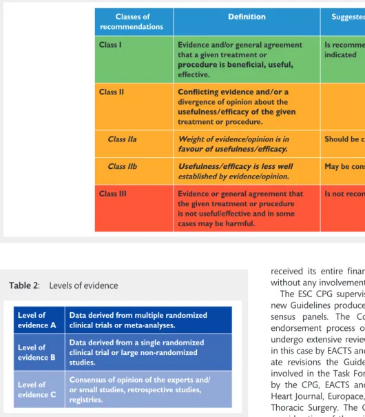
evidence and the strength of the recommendation of particular
management options were weighed and graded according to
predefined scales, as outlined in Tables
1
and
2
.
The experts of the writing and reviewing panels provided
dec-laration of interest forms for all relationships that might be
per-ceived as real or potential sources of conflicts of interest. These
forms were compiled into one file and can be found on the ESC
website (
http://www.escardio.org/guidelines
). Any changes in
declarations of interest that arise during the writing period must
be notified to the ESC and EACTS and updated. The Task Force
received its entire financial support from the ESC and EACTS
without any involvement from the healthcare industry.
The ESC CPG supervises and co-ordinates the preparation of
new Guidelines produced by task forces, expert groups or
con-sensus panels. The Committee is also responsible for the
endorsement process of these Guidelines. The ESC Guidelines
undergo extensive review by the CPG and external experts, and
in this case by EACTS and ESO-appointed experts. After
appropri-ate revisions the Guidelines are approved by all the experts
involved in the Task Force. The finalized document is approved
by the CPG, EACTS and ESO for publication in the European
Heart Journal, Europace, and in the European Journal of
Cardio-Thoracic Surgery. The Guidelines were developed after careful
consideration of the scientific and medical knowledge and the
evidence available at the time of their dating.
The task of developing ESC and EACTS Guidelines covers
not only integration of the most recent research, but also the
creation of educational tools and implementation programmes
for the recommendations. To implement the guidelines,
con-densed pocket guideline versions, summary slides, booklets with
essential messages, summary cards for non-specialists and an
electronic version for digital applications (smartphones, etc.)
are produced. These versions are abridged and thus, if needed,
one should always refer to the full text version, which is freely
available on the ESC website. The National Societies of the ESC
are encouraged to endorse, translate and implement all ESC
Guidelines. Implementation programmes are needed because
it has been shown that the outcome of disease may be
favour-ably influenced by the thorough application of clinical
recommendations.
Surveys and registries are needed to verify that real-life daily
practice is in keeping with what is recommended in the
guide-lines, thus completing the loop between clinical research, writing
of guidelines, disseminating them and implementing them into
clinical practice.
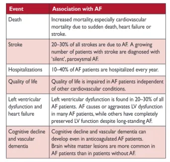
Table 2:
Levels of evidence
Level of evidence A
Data derived from multiple randomized clinical trials or meta-analyses. Level of
evidence B
Data derived from a single randomized clinical trial or large non-randomized studies.
Level of evidence C
Consensus of opinion of the experts and/ or small studies, retrospective studies, registries.
Table 1:
Classes of recommendations
Classes of recommendations
Suggested wording to use
Class I Evidence and/or general agreement that a given treatment or
effective.
Is recommended/is indicated
Class II
divergence of opinion about the treatment or procedure.
Class IIa Weight of evidence/opinion is in Should be considered
Class IIb
established by evidence/opinion.
May be considered
Class III Evidence or general agreement that the given treatment or procedure is not useful/effective and in some cases may be harmful.
Health professionals are encouraged to take the ESC and
EACTS Guidelines fully into account when exercising their clinical
judgment, as well as in the determination and the
implementa-tion of preventive, diagnostic or therapeutic medical strategies.
However, the ESC and EACTS Guidelines do not override in any
way whatsoever the individual responsibility of health
professio-nals to make appropriate and accurate decisions in consideration
of each patient’s health condition and in consultation with that
patient and the patient’s caregiver where appropriate and/or
necessary. It is also the health professional’s responsibility to
ver-ify the rules and regulations applicable to drugs and devices at
the time of prescription.
2. INTRODUCTION
Despite good progress in the management of patients with atrial
fibrillation (AF), this arrhythmia remains one of the major causes
of stroke, heart failure, sudden death, and cardiovascular
morbid-ity in the world. Furthermore, the number of patients with AF is
predicted to rise steeply in the coming years. To meet the
grow-ing demand for effective care of patients with AF, new
informa-tion is continually generated and published, and the last few
years have seen substantial progress. Therefore, it seems timely
to publish this 2
ndedition of the ESC guidelines on AF.
Reflecting the multidisciplinary input into the management of
patients with AF, the Task Force includes cardiologists with varying
subspecialty expertise, cardiac surgeons, stroke neurologists, and
specialist nurses amongst its members. Supplementing the
evi-dence review as outlined in the preamble, this Task Force defined
three Population, Intervention, Comparison, Outcome, Time
(PICOT) questions on relevant topics for the guidelines. The ESC
commissioned external systematic reviews to answer these
ques-tions, and these reviews have informed specific recommendations.
Further to adhering to the standards for generating
recom-mendations that are common to all ESC guidelines (see
pream-ble), this Task Force discussed each draft recommendation
during web-based conference calls dedicated to specific
chap-ters, followed by consensus modifications and an online vote on
each recommendation. Only recommendations that were
sup-ported by at least 75% of the Task Force members were included
in the guidelines.
We hope that these guidelines will help to deliver good care to
all patients with AF based on the current state-of-the-art
evi-dence in 2016.
3. EPIDEMIOLOGY AND IMPACT FOR PATIENTS
3.1 Incidence and prevalence of atrial fibrillation
In 2010, the estimated numbers of men and women with AF
worldwide were 20.9 million and 12.6 million, respectively, with
higher incidence and prevalence rates in developed countries
[
1
,
2
]. One in four middle-aged adults in Europe and the US will
develop AF [
3
–
5
]. By 2030, 14–17 million AF patients are
antici-pated in the European Union, with 120 000–215 000 newly
diagnosed patients per year [
2
,
6
,
7
]. Estimates suggest an AF
prevalence of approximately 3% in adults aged 20 years or older
[
8
,
9
], with greater prevalence in older persons [
1
] and in
patients with conditions such as hypertension, heart failure,
coronary artery disease (CAD), valvular heart disease, obesity,
diabetes mellitus, or chronic kidney disease (CKD) [
7
,
10
–
15
].
The increase in AF prevalence can be attributed both to better
detection of silent AF [
16
–
18
], alongside increasing age and
conditions predisposing to AF [
19
].
3.2 Morbidity, mortality, and healthcare burden of
atrial fibrillation
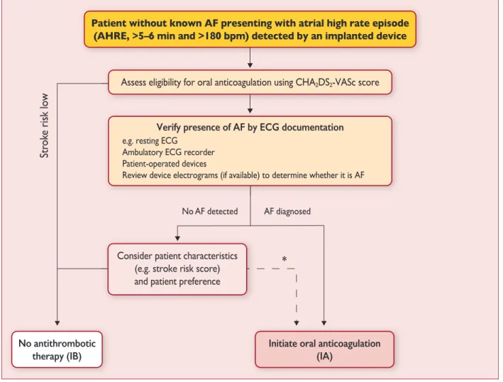
AF is independently associated with a two-fold increased risk of
all-cause mortality in women and a 1.5-fold increase in men [
20
–
22
] (
Table 3
). Death due to stroke can largely be mitigated by
anticoagulation, while other cardiovascular deaths, for example
due to heart failure and sudden death, remain common even in
AF patients treated according to the current evidence base [
23
].
AF is also associated with increased morbidity, such as heart
fail-ure and stroke [
21
,
24
,
25
]. Contemporary studies show that 20–
30% of patients with an ischaemic stroke have AF diagnosed
before, during, or after the initial event [
17
,
26
,
27
]. White matter
lesions in the brain, cognitive impairment [
28
–
30
], decreased
quality of life [
31
,
32
], and depressed mood [
33
] are common in
AF patients, and between 10–40% of AF patients are hospitalized
each year [
23
,
34
,
35
].
The direct costs of AF already amount to approximately 1% of
total healthcare spending in the UK, and between 6.0–26.0 billion
US dollars in the US for 2008 [
36
,
37
], driven by AF-related
com-plications (e.g. stroke) and treatment costs (e.g. hospitalizations).
These costs will increase dramatically unless AF is prevented and
treated in a timely and effective manner.
3.3 Impact of evidence-based management on
out-comes in atrial fibrillation patients
Figure
1
depicts the major milestones in the management of AF.
Despite these advances, substantial morbidity remains. Oral
Table 3:
Cardiovascular morbidity and mortality associated
with atrial fibrillation
Event Association with AF
Death Increased mortality, especially cardiovascular mortality due to sudden death, heart failure or stroke.
Stroke 20–30% of all strokes are due to AF. A growing number of patients with stroke are diagnosed with ‘silent’, paroxysmal AF.
Hospitalizations 10–40% of AF patients are hospitalized every year. Quality of life Quality of life is impaired in AF patients independent
of other cardiovascular conditions. Left ventricular
dysfunction and heart failure
Left ventricular dysfunction is found in 20–30% of all AF patients. AF causes or aggravates LV dysfunction in many AF patients, while others have completely preserved LV function despite long-standing AF. Cognitive decline
and vascular dementia
Cognitive decline and vascular dementia can develop even in anticoagulated AF patients. Brain white matter lesions are more common in AF patients than in patients without AF.
AF = atrial fibrillation; LV = left ventricular.
GU IDELINE G U ID E L IN ES
anticoagulation (OAC) with vitamin K antagonists (VKAs) or
non-VKA oral anticoagulants (NOACs) markedly reduces stroke and
mortality in AF patients [
38
,
39
]. Other interventions such as
rhythm control and rate control improve AF-related symptoms
and may preserve cardiac function, but have not demonstrated a
reduction in long-term morbidity or mortality [
40
,
41
].
In contemporary, well-controlled, randomized clinical trials in
AF, the average annual stroke rate is about 1.5% and the
annual-ized death rate is around 3% in anticoagulated AF patients [
40
].
In real life, the annual mortality can be different (both higher and
lower) [
42
]. A minority of these deaths are related to stroke, while
sudden cardiac death and death from progressive heart failure
are more frequent, emphasizing the need for interventions
beyond anticoagulation [
43
,
44
]. Furthermore, AF is also
associ-ated with high rates of hospitalization, commonly for AF
man-agement, but often also for heart failure, myocardial infarction,
and treatment-associated complications [
34
,
45
].
3.4 Gender
In both developed and developing countries, the age-adjusted
inci-dence and prevalence of AF are lower in women, while the risk of
death in women with AF is similar to or higher than that in men
with AF [
1
,
46
,
47
]. Female AF patients who have additional stroke
risk factors (particularly older age) are also at greater risk than men
of having a stroke [
48
,
49
], even those anticoagulated with warfarin
Figure 1: Timeline of findings from landmark trials in atrial fibrillation management, including treatment of concomitant conditions and prevention (green), anti-coagulation (blue), rate control therapy (orange), rhythm control therapy (red), and atrial fibrillation surgery (purple).Recommendations relating to gender
AF = atrial fibrillation. aClass of recommendation. bLevel of evidence.
[
50
] (see Chapter 9 for details). Women with diagnosed AF can be
more symptomatic than men and are typically older with more
comorbidities [
51
,
52
]. Bleeding risk on anticoagulation is similar in
both sexes [
49
,
50
,
53
], but women appear less likely to receive
spe-cialist care and rhythm control therapy [
54
], while the outcomes of
catheter ablation or AF surgery are comparable to those in men [
55
,
56
]. These observations highlight the need to offer effective
diagnos-tic tools and therapeudiagnos-tic management equally to women and men.
4. PATHOPHYSIOLOGICAL AND GENETIC
ASPECTS THAT GUIDE MANAGEMENT
4.1 Genetic predisposition
AF, especially early-onset AF, has a strong heritable component
that is independent of concomitant cardiovascular conditions
[
58
,
59
]. A few young AF patients suffer from inherited
cardio-myopathies or channelopathies mediated by disease-causing
mutations. These monogenic diseases also convey a risk for
sud-den death (see Chapter 6). Up to one-third of AF patients carry
common genetic variants that predispose to AF, albeit with a
rel-atively low added risk. At least 14 of these common variants,
often single nucleotide polymorphisms, are known to increase
the risk of prevalent AF in populations [
60
–
62
]. The most
impor-tant variants are located close to the
paired-like homeodomain
transcription factor 2 (Pitx2) gene on chromosome 4q25 [
63
,
64
].
These variants modify the risk of AF up to seven-fold [
64
]. Several
of the AF risk variants are also associated with cardioembolic or
ischaemic stroke, possibly due to silent AF (see section 5.1) [
62
,
65
,
66
]. Changes in atrial action potential characteristics [
67
–
70
],
atrial remodelling, and modified penetration of rare gene defects
[
61
] have been suggested as potential mechanisms mediating
increased AF risk in carriers of common gene variants. Genetic
variants could, in the future, become useful for patient selection
of rhythm or rate control [
71
–
74
]. While genomic analysis may
provide an opportunity to improve the diagnosis and
manage-ment of AF in the future [
75
,
76
], routine genetic testing for
com-mon gene variants associated with AF cannot be recommended
at present [
77
].
4.2 Mechanisms leading to atrial fibrillation
4.2.1 Remodelling of atrial structure and ion channel
function.
External stressors such as structural heart disease,
hypertension, possibly diabetes, but also AF itself induce a slow
but progressive process of structural remodelling in the atria
(Figure
2
). Activation of fibroblasts, enhanced connective tissue
deposition, and fibrosis are the hallmarks of this process [
78
–
80
].
In addition, atrial fatty infiltration, inflammatory infiltrates,
myo-cyte hypertrophy, necrosis, and amyloidosis are found in AF
patients with concomitant conditions predisposing to AF [
81
–
84
].
Structural remodelling results in electrical dissociation between
muscle bundles and local conduction heterogeneities [
85
],
favouring re-entry and perpetuation of the arrhythmia [
86
]. In
many patients, the structural remodelling process occurs before
the onset of AF [
78
]. As some of the structural remodelling will be
irreversible, early initiation of treatment seems desirable [
87
].
Table
4
gives an overview of the most relevant
pathophysiologi-cal alterations in atrial tissue associated with AF, and lists
corresponding clinical conditions that can contribute to these
changes.
The functional and structural changes in atrial myocardium
and stasis of blood, especially in the left atrial appendage (LAA),
generate a prothrombotic milieu. Furthermore, even short
epi-sodes of AF lead to atrial myocardial damage and the expression
of prothrombotic factors on the atrial endothelial surface,
along-side activation of platelets and inflammatory cells, and contribute
to a generalized prothrombotic state [
88
,
89
]. The atrial and
sys-temic activation of the coagulation system can partially explain
why short episodes of AF convey a long-term stroke risk.
4.2.2 Electrophysiological mechanisms of atrial
fibrilla-tion.
AF provokes a shortening of the atrial refractory period and
AF cycle length during the first days of the arrhythmia, largely
due to downregulation of the Ca
2+-inward current and
upregula-tion of inward rectifier K
+currents [
94
,
95
]. Structural heart
dis-ease, in contrast, tends to prolong the atrial refractory period,
illustrating the heterogeneous nature of mechanisms that cause
AF in different patients [
96
]. Hyperphosphorylation of various
Ca
2+-handling proteins may contribute to enhanced spontaneous
Ca
2+release events and triggered activity [
97
,
98
], thus causing
ectopy and promoting AF. Although the concept of Ca
2+-han-dling instability has been challenged recently [
106
,
107
], it may
mediate AF in structurally remodelled atria and explain how
altered autonomic tone can generate AF [
80
,
105
].
4.2.2.1
Focal initiation and maintenance of atrial fibrillation. The
seminal observation by Haissaguerre
et al. [
108
]. was that a focal
source in the pulmonary veins can trigger AF, and ablation of this
source can suppress recurrent AF. The mechanism of focal
activ-ity might involve both triggered activactiv-ity and localized reentry
[
109
,
110
]. Hierarchic organization of AF with rapidly activated
areas driving the arrhythmia has been documented in patients
with paroxysmal AF [
111
,
112
], but is less obvious in unselected
patients with persistent AF [
113
].
4.2.2.2
The multiple wavelet hypothesis and rotors as sources of
atrial fibrillation. Moe and Abildskov [
114
] proposed that AF can
be perpetuated by continuous conduction of several
independ-ent wavelets propagating through the atrial musculature in a
seemingly chaotic manner. As long as the number of wavefronts
does not decline below a critical level, they will be capable of
sustaining the arrhythmia. Numerous experimental and clinical
observations can be reconciled with the multiple wavelet
hypothesis [
115
]. All localized sources of AF (ectopic foci, rotors,
or other stable re-entry circuits) cause fibrillatory conduction
remote from the source, which is difficult to distinguish from
propagation sustaining AF by multiple wavelets, and either of
these phenomena may generate ‘rotors’ picked up by
intracar-diac [
116
,
117
] or body surface [
117
] recordings.
5. DIAGNOSIS AND TIMELY DETECTION OF
ATRIAL FIBRILLATION
5.1 Overt and silent atrial fibrillation
The diagnosis of AF requires rhythm documentation using an
elec-trocardiogram (ECG) showing the typical pattern of AF: Absolutely
irregular RR intervals and no discernible, distinct P waves.
ECG-documented AF was the entry criterion in trials forming the
GU IDELINE G U ID E L IN ES
evidence for these guidelines. By accepted convention, an episode
lasting at least 30 s is diagnostic. Individuals with AF may be
symp-tomatic or asympsymp-tomatic (‘silent AF’). Many AF patients have both
symptomatic and asymptomatic episodes of AF [
118
–
121
].
Silent, undetected AF is common [
120
,
122
], with severe
conse-quences such as stroke and death [
123
–
125
]. Prompt recording
of an ECG is an effective and cost-effective method to document
chronic forms of AF [
126
]. The technology to detect paroxysmal,
self-terminating AF episodes is rapidly evolving (see section 6.1
for a definition of AF patterns). There is good evidence that
pro-longed ECG monitoring enhances the detection of undiagnosed
AF, e.g. monitoring for 72 h after a stroke [
27
,
127
], or even
lon-ger periods [
18
,
128
]. Daily short-term ECG recordings increase
AF detection in populations over 75 years of age [
129
] (Web
Figure 1). Ongoing studies will determine whether such early
detection alters management (e.g. initiation of anticoagulation)
and improves outcomes.
Once the ECG diagnosis of AF has been established, further
ECG monitoring can inform management in the context of: (1) a
change in symptoms or new symptoms; (2) suspected progression
of AF; (3) monitoring of drug effects on ventricular rate; and (4)
monitoring of antiarrhythmic drug effects or catheter ablation for
rhythm control.
5.2 Screening for silent atrial fibrillation
5.2.1 Screening
for
atrial
fibrillation
by
electro-cardiogram in the community.
Undiagnosed AF is common,
especially in older populations and in patients with heart failure
[
130
]. Opportunistic screening for silent AF seems cost-effective
in elderly populations (e.g. >65 years) [
131
], and similar effects
have been reported using single-lead ECG screening in other
at-risk populations [
132
,
133
]. Screening of older populations
(mean age 64 years) yielded a prevalence of 2.3% for chronic
forms of AF in 122,571 participants using either short-term ECG
or pulse palpation (followed by ECG in those with an irregular
pulse) [
134
]. Previously undiagnosed AF was found in 1.4% of
Figure 2: Major mechanisms causing atrial fibrillation that can be considered when choosing therapy. The various aetiological factors (left) cause a complex array of pathophysiological changes in the atria, including stretch-induced atrial fibrosis, hypocontractility, fatty infiltration, inflammation, vascular remodelling, ischaemia, ion channel dysfunction, and Ca2+-instability. These changes enhance both ectopy and conduction disturbances, increasing the propensity of the atria to develop or maintain AF. At the same time, some of these alterations are involved in the occurrence of the hypercoagulable state associated with AF. For example, hypocontractil-ity reduces local endothelial shear stress, which increases PAI-1 expression, and ischaemia-induced inflammation enhances the expression of endothelial adhesion molecules or promotes shedding of endothelial cells, resulting in tissue factor exposure to the blood stream. These changes contribute to the thrombogenic milieu in the atria of AF patients. AF in itself can aggravate many of the mechanisms shown, which may explain the progressive nature of the arrhythmia.those aged >65 years, suggesting a number needed to screen of
70. These findings encourage the further evaluation of
system-atic AF screening programmes in at-risk populations.
5.2.2 Prolonged monitoring for paroxysmal atrial
fibrilla-tion.
Paroxysmal AF is often missed [
120
]. Repeated daily ECG
recordings increased the detection of silent, asymptomatic
parox-ysmal AF in an unselected Swedish population aged >75 years
[
120
,
135
]. Several patient-operated devices [
136
,
137
] and
extended continuous ECG monitoring using skin patch recorders
[
138
] have been validated for the detection of paroxysmal AF (Web
Figure 1) [
139
]. The detection of asymptomatic AF by new
technol-ogies, such as smartphone cases with ECG electrodes, smart
watches, and blood pressure machines with AF detection
algo-rithms, has not yet been formally evaluated against an established
arrhythmia detection method [
140
].
5.2.3 Patients with pacemakers and implanted devices.
Implanted pacemakers or defibrillators with an atrial lead allow
continuous monitoring of atrial rhythm. Using this technology,
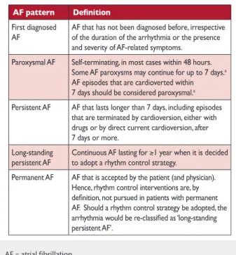
patients with atrial high rate episodes (AHRE) can be identified.
Depending on the risk profile of the population studied, such
AHRE are detected in 10–15% of pacemaker patients [
141
]. AHRE
are associated with an increased risk of overt AF [hazard ratio
(HR) 5.56; 95% confidence interval (CI) 3.78–8.17;
P < 0.001] and
ischaemic stroke or systemic embolism (HR 2.49; 95% CI 1.28–
4.85;
P = 0.007). The stroke risk in AHRE patients seems lower
than the stroke risk in patients with diagnosed AF, and not all
AHRE represent AF [
142
]. Strokes often occur without AHRE
detected
within
30
days
before
the
event
[
143
–
147
].
Consequently, it is unclear whether AHRE imply the same
thera-peutic requirements as overt AF [
148
], and the benefit of OAC in
patients with AHRE is tested in ongoing clinical trials [e.g.
Apixaban for the Reduction of Thrombo-Embolism in Patients
Table 4:
Pathophysiological alterations in atrial tissue associated with atrial fibrillation and clinical conditions that could contribute
to such alterations
AF = atrial fibrillation; CAD = coronary artery disease.
GU IDELINE G U ID E L IN ES
With
Device-Detected
Sub-Clinical
Atrial
Fibrillation
(ARTESiA) (NCT01938248) and
Non vitamin K antagonist Oral
anticoagulants in patients with
Atrial High rate episodes
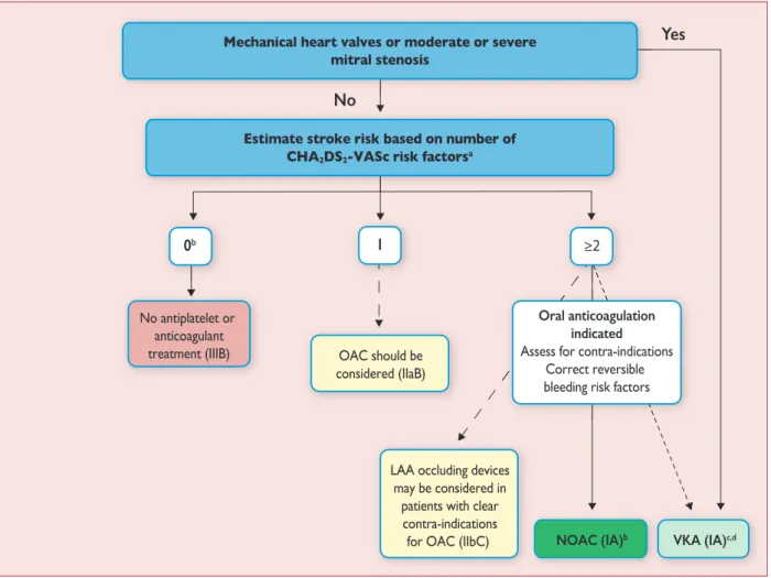
(NOAH – AFNET 6) (NCT02618577)]. At present, pacemakers and
implanted devices should be interrogated on a regular basis for
AHRE, and patients with AHRE should undergo further
assess-ment of stroke risk factors and for overt AF, including ECG
moni-toring (Figure
3
) [
149
].
5.2.4 Detection of atrial fibrillation in stroke survivors.
Sequential stratified ECG monitoring detected AF in 24% (95% CI
17–31) of stroke survivors [
151
], and in 11.5% (95% CI 8.9%–
14.3%) in another meta-analysis [
17
], with large variations
depending on the timing, duration, and method of monitoring.
AF detection is not uncommon in unselected stroke patients
(6.2%, 95% CI 4.4–8.3) [
128
], but is more likely in patients with
cryptogenic stroke implanted with loop recorders or who have
had ECG monitors for several weeks [
18
,
128
,
152
]. Cryptogenic
stroke is defined as a stroke in which the cause could not be
identified after extensive investigations [
153
]. A broader definition
is embolic stroke of undetermined source [
154
]. Several studies
have also found AF in patients in whom another competing
cause for stroke has been identified clinically (e.g. hypertension
or carotid artery stenosis) [
27
,
127
]. Hence, prolonged ECG
moni-toring seems reasonable in all survivors of an ischaemic stroke
without an established diagnosis of AF.
5.3 Electrocardiogram detection of atrial flutter
Right atrial isthmus-dependent flutter has a typical ECG pattern
and ventricular rate [
158
]. The prevalence of atrial flutter is less
than one-tenth of the prevalence of AF [
159
]. Atrial flutter often
coexists with or precedes AF [
160
]. In typical, isthmus-dependent
flutter, P waves will often show a ‘saw tooth’ morphology,
espe-cially in the inferior leads (II, III, aVF). The ventricular rate can be
variable (usual ratio of atrial to ventricular contraction 4:1 to 2:1,
in rare cases 1:1) and macro re-entrant tachycardias may be
missed in stable 2:1 conduction. Vagal stimulation or intravenous
adenosine can therefore be helpful to unmask atrial flutter. The
management of atrial flutter is discussed in section 13.7. Left or
[150].
right atrial macro re-entrant tachycardia is mainly found in
patients after catheter ablation for AF, AF surgery, or after open
heart surgery [
158
].
6. CLASSIFICATION OF ATRIAL FIBRILLATION
6.1 Atrial fibrillation pattern
In many patients, AF progresses from short, infrequent
epi-sodes to longer and more frequent attacks. Over time, many
patients will develop sustained forms of AF. In a small
propor-tion of patients, AF will remain paroxysmal over several
deca-des (2–3% of AF patients) [
161
]. The distribution of paroxysmal
AF recurrences is not random, but clustered [
162
]. AF may also
regress from persistent to paroxysmal AF. Furthermore,
asymptomatic recurrences of AF are common in patients with
symptomatic AF [
120
].
Based on the presentation, duration, and spontaneous
termi-nation of AF episodes, five types of AF are traditionally
distin-guished: first diagnosed, paroxysmal, persistent, long-standing
persistent, and permanent AF (Table
5
). If patients suffer from
both paroxysmal and persistent AF episodes, the more common
type should be used for classification. Clinically determined AF
patterns do not correspond well to the AF burden measured by
long-term ECG monitoring [
163
]. Even less is known about the
response to therapy in patients with long-standing persistent AF
or long-standing paroxysmal AF. Despite these inaccuracies, the
distinction between paroxysmal and persistent AF has been used
in many trials and therefore still forms the basis of some
recommendations.
There is some evidence suggesting that AF burden may
influ-ence stroke risk [
44
,
124
,
164
] and could modify the response to
rhythm control therapy [
76
,
165
]. The evidence for this is weak.
Therefore, AF burden should not be a major factor in deciding
on the usefulness of an intervention that is deemed suitable for
other reasons.
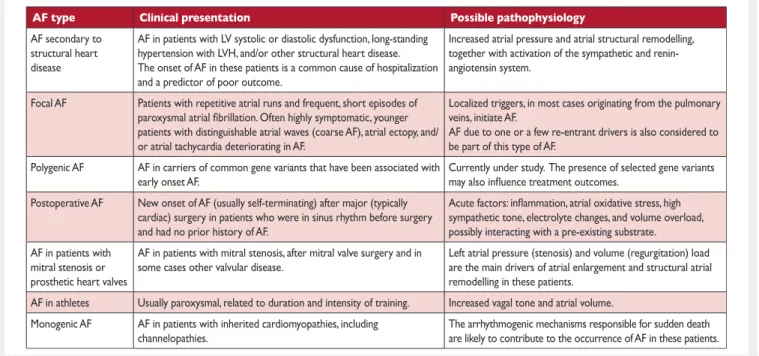
6.2 Atrial fibrillation types reflecting different
causes of the arrhythmia
The risk of developing AF is increased in a variety of
physio-logical and disease states (Figure
2
), and the historic term
‘lone AF’ is probably misleading and should be avoided [
166
].
Although the pattern of AF may be the same, the mechanisms
underpinning AF vary substantially between patients [
167
]
(Table
6
). This suggests that stratifying AF patients by
underly-ing drivers of AF could inform management, for example,
con-sidering cardiac and systemic comorbidity (e.g. diabetes and
obesity [
168
]), lifestyle factors (e.g. activity level, smoking,
alcohol intake [
169
,
170
]), markers of cardiac structural
Recommendations for screening for atrial fibrillation
AF = atrial fibrillation; AHRE = atrial high rate episodes; ECG = electro-cardiogram; ICD = implantable cardioverter defibrillator; TIA = transi-ent ischaemic attack.
aClass of recommendation. bLevel of evidence.
cReference(s) supporting recommendations.
Table 5:
Patterns of atrial fibrillation
AF = atrial fibrillation. a
The distinction between paroxysmal and persistent AF is often not made correctly without access to long-term monitoring [163]. Hence, this classification alone is often insufficient to select specific therapies. If both persistent and paroxysmal episodes are present, the predomi-nant pattern should guide the classification.
GU IDELINE G U ID E L IN ES
remodelling (e.g. fibrosis [
171
–
173
] or electrocardiographic
param-eters of AF complexity [
174
]), or genetic background. Table
6
pro-vides such a taxonomy, informed by expert consensus [
76
,
120
,
175
], but without much evidence to underpin its clinical use [
176
].
Systematic research defining the major drivers of AF is clearly
needed to better define different types of AF [
176
].
6.3 Symptom burden in atrial fibrillation
Patients with AF have significantly poorer quality of life than
healthy controls, experiencing a variety of symptoms including
lethargy, palpitations, dyspnoea, chest tightness, sleeping
difficul-ties, and psychosocial distress [
32
,
177
–
180
]. Improved quality of
life has been noted with both pharmacological and interventional
therapies [
181
–
185
], but there are limited data to compare the
benefit of different treatments [
32
,
186
]. Assessment of quality of
life is further constrained by a lack of cross-validation of the
sev-eral AF-specific quality of life tools [
187
–
191
]. With regard to
symptom assessment, EHRA suggested the EHRA symptom scale
(Table
7
) to describe symptom severity in AF patients [
192
]. A
similar scale (the Canadian Cardiovascular Society Severity of
Atrial Fibrillation Scale) is used in Canada [
193
]. The EHRA scale
has been used and validated [
194
–
199
]. A modification was
pro-posed in 2014, subdividing EHRA class 2 into mild (2a) or
moder-ate (2b) impact [
199
]. As symptoms in class 2b (‘troubling’
symptoms) identified patients with a health utility benefit of
rhythm control in that study, this modification may provide a
threshold for potential treatment decisions, pending independent
validation. While some AF patients had no or minimal symptoms
(25–40%), many (15–30%) report severe or disabling symptoms
[
194
,
196
]. The modified EHRA scale should be used to guide
symptom-orientated treatment decisions and for longitudinal
patient profiling.
Table 6:
Clinical types of atrial fibrillation
aAF = atrial fibrillation; LV = left ventricular; LVH = left ventricular hypertrophy.
aIt is recognized that these types of AF will overlap in clinical practice, and that their impact for management needs to be evaluated systematically; modified from the report on the fourth AFNET/EHRA Consensus Conference [76].
Table 7:
Modified European Heart Rhythm Association
symptom scale (modified from Wynn
et al. [
199
])
AF = atrial fibrillation; EHRA = European Heart Rhythm Association. aEHRA class 2a and 2b can be differentiated by evaluating whether patients are functionally affected by their AF symptoms. AF-related symptoms are most commonly fatigue/tiredness and exertional shortness of breath, or less frequently palpitations and chest pain [42,194,200–202].
7. DETECTION AND MANAGEMENT OF RISK
FACTORS AND CONCOMITANT
CARDIOVASCULAR DISEASES
Many cardiovascular diseases and concomitant conditions
increase the risk of developing AF (Table
8
), recurrent AF, and
AF-associated complications. The identification of such
condi-tions, their prevention and treatment is an important leverage to
prevent AF and its disease burden. Knowledge of these factors
and their management is hence important for optimal
manage-ment of AF patients [
203
,
204
].
7.1 Heart failure
Heart failure and AF coincide in many patients [
215
–
217
]. They
are linked by similar risk factors and share a common
patho-physiology [
218
]. Heart failure and AF can cause and exacerbate
each other through mechanisms such as structural cardiac
remodelling, activation of neurohormonal mechanisms, and
rate-related impairment of left ventricular (LV) function. Patients with
AF and concomitant heart failure, both with preserved ejection
fraction [LV ejection fraction (LVEF) >_50%] and reduced ejection
fraction (LVEF <40%) [
219
,
220
], suffer from a worse prognosis,
including increased mortality [
16
,
221
]. The recent ESC Guidelines
on heart failure [
222
] have also introduced a new category of
heart failure with mid-range ejection fraction (HFmrEF; LVEF 40–
49%), although data on AF patients in this group are limited.
Prevention of adverse outcomes and maintenance of a good
quality of life are the aims of management in all patients with AF
and concomitant heart failure, regardless of LVEF [
223
]. The
gen-eral approach to AF management does not differ between heart
failure patients and others, but a few considerations are
worth-while. Of note, the only therapy with proven prognostic value in
these patients is anticoagulation, and appropriate OAC should be
prescribed in all patients at risk of stroke (see Chapter 9).
7.1.1 Patients with atrial fibrillation and heart failure
with reduced ejection fraction.
In addition to OAC, standard
heart failure therapy should be used in patients with heart failure
with reduced ejection fraction (HFrEF), as detailed in the ESC
Recommendation on use of the modified European Heart
Rhythm Association symptom scale
c
AF = atrial fibrillation; EHRA = European Heart Rhythm Association. aClass of recommendation.
bLevel of evidence.
cReference(s) supporting recommendations.
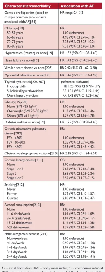
Table 8:
Cardiovascular and other conditions
independ-ently associated with atrial fibrillation
[64] [19] [19] [19] [205] [19] [206,207] [19,208] [19] [210] [211] [212] [213] [214] [209]
AF = atrial fibrillation; BMI = body mass index; CI = confidence interval; FEV1 = forced expiratory volume in 1 second; HR = hazard ratio; OR = odds ratio; RR = risk ratio.
GU IDELINE G U ID E L IN ES