Université de Montréal
Diabète maternel et/ou hypertension et dommages rénaux
induits par le système rénine-angiotensine intrarénal :
rôle de Nrf2
par
Shiao-Ying Chang
Programme de sciences biomédicales Faculté de médecine
Thèse présentée à la Faculté des études supérieures en vue de l’obtention du grade de docteurès sciences (Ph.D)
en sciences biomédicales
Juillet 2016
ii
Université de Montréal
Diabète maternel et/ou hypertension et dommages rénaux
induits par le système rénine-angiotensine intrarénal :
rôle de Nrf 2
par
Shiao-Ying Chang
a été évaluée par un jury composé des personnes suivantes:
D
reJolanta Gutkwska
Présidente rapporteurD
reShao-Ling Zhang
Directeure de rechercheD
rJohn S.D. Chan
Codirecteur de rechercheD
rRaynald Bergeron
Membre du juryD
rJun-Li Liu
Examinateur externe
iii
Résumé
L’expression ‘programmation périnatale’ est employée pour décrire les effets à long terme d’un environnement gestationel néfaste observés chez la progéniture. Ce concept est aujourd’hui bien reconnu. Notre laboratoire a déjà démontré l’impact de l’hyperglycémie maternelle sur le développement rénal des embryons à l’aide des souris HoxB7-GFP transgéniques (Tg) et qui se traduit par une augmentation des espèces réactives de l’oxygène (ROS) et une néphrogenèse perturbée. Les rejetons affectés présentent ainsi des reins plus petits et possédant un nombre inférieur de néphrons à la naissance, et développent une hypertension et des dommages rénaux à l’âge adulte (20 semaines).
Dans la première étude, nous avons tenté de réduire la production excessive de ROS dans les reins en développement par la surexpression de la catalase (CAT). Pour ce faire, nous avons croisé les souris CAT-Tg qui surexpriment la CAT dans les cellules des tubules proximaux rénaux (RPTCs) aux souris HoxB7-GFP-Tg afin de générer les souris HoxB7/CAT-GFP-Tg. Nous espérons observer la normalisation du nombre de néphrons et la prévention de l’hypertension et des dommages rénaux observés chez la progéniture issue d’un environnement gestational hyperglycémique.
Nous avons observé que la surexpression de CAT dans les RPTCs permet de normaliser la dysmorphogenèse rénale présente chez les embryons de mères diabétiques. À l’âge adulte, la surexpression de CAT dans les RPTCs permet également de réduire la génération des ROS et l’hypertension, tout en améliorant la morphologie et la fonction rénale. Afin de définir les mécanismes impliqués dans ce processus, nous avons étudié le rôle potentiel de Nrf2 (‘nuclear factor-erythroid 2p45 (NF-E2) related factor-2’; un facteur de transcription des gènes antioxidants) et HO-1 (hème oxygénase-1’; une enzyme antioxidante). À la fois Nrf2 et HO-1 sont de forts antioxidants et ont été rapportés comme protecteurs pour le rein. Nous avons observé une surexpression des gènes et protéines Nrf2 et HO-1, en plus d’une translocation nucléaire accrue de Nrf2, dans les RPTCs de la progéniture des mères diabétiques, indiquant
iv
que chez les souris surexprimant CAT, Nrf2 et HO-1 sont tous deux bien activés et fonctionnels.
En conclusion, nos études suggèrent que la surexpression de CAT dans les RPTCs permet de prévenir la programmation de l’hypertension et les dommages rénaux observés à l’âge adulte chez la progéniture issue de mères diabétiques, en partie suite à l’activation du système de défense Nrf2-HO-1 dans leurs reins.
Il a déjà été démontré que l’activation du système rénine-angiotensine (RAS) intrarénal induit l’hypertension en augmentant la constriction des artérioles et la réabsorption du sodium par les tubules rénaux. Une activation du récepteur AT1R et de ses voies de signalisation induit également les dommages rénaux observés dans plusieurs pathologies. Dans le cadre de mon second article, nous avons identifié un nouveau mécanisme par lequel l’angiotensine (Agt) intrarénale induit l’hypertension et des dommages rénaux en réduisant l’expression de l’aquaporine 1 (AQP1, le canal pour l’eau le plus important dans les RPTCs).
Des souris transgéniques surexprimant l’Agt de rat (rAgt-Tg) dans leurs RPTCs et des clones stables de cellules immortalisées de tubule proximal de rein de rat (IRPTCs) surexprimant le rAgt (pRSV/rAgt-IRPTC) ont été étudiés. Lorsque comparés aux souris non-transgéniques, les souris rAgt-Tg développent de l’hypertension et des dommages rénaux. Ces changements sont atténués par le traitement avec une double inhibition du RAS (losartan et perindopril). L’expression des protéines AQP1 et HO-1 est réduite dans les RPTCs, tandis que Nrf2 et le transporteur sodique NHE3 sont augmentés, à la fois in vivo et in vitro. Ces changements sont renversés par la double inhibition du RAS chez les animaux expérimentaux. Même si les niveaux de Nrf2 sont élevés, une accumulation cytosolique causée par une augmentation de l’export nucléaire induit par GSK3β se produit et ne parvient donc pas à induire l’expression des gènes en aval comme HO-1, ni à réduire l’expression de l’AQP1.
En conclusion, nos résultats suggèrent qu’une déficience en Nrf2 nucléaire mène à une diminution de l’expression de HO-1 et une régulation négative de l’AQP1, jouant un role dans l’hypertension et les dommages rénaux induits par l’Agt intrarénal.
v
L’hypertension et les dommages rénaux sont des maladies très hétérogènes et multifactorielles qui impliquent l’interaction de diverses molécules et voies de signalisations, et sont influencées par plusieurs facteurs environnementaux tels la diète ou la programmation périnatale. Tous ces différents facteurs contribuent à la progression de l’hypertension et des dommages rénaux, rendant les stratégies de traitement d’autant plus complexes. Dans notre étude, nous avons évalué le développement de l’hypertension dans deux circonstances : l’hypertension de la progéniture programmée par le diabète maternel et l’hypertension induite par l’activation du RAS intrarénal. Nous avons démontré que la génération des ROS dans les reins constitue un facteur majeur commun dans nos deux modèles d’hypertension chez la souris. De plus, le gène/facteur de transcription antioxydant Nrf2, sensible aux ROS, joue un rôle important dans le processus. Grâce à une meilleure compréhension des diverses voies qui mènent à la progression de l’hypertension, nous espérons qu’il sera possible de développer de meilleurs traitements pour faire face à l’hypertension.
Mots clés: Aquaporine 1, catalase, HO-1, hypertension, système rénine-angiotensine
intrarénal, diabète maternel, Nrf2, progéniture, programmation périnatale, espèces réactives de l’oxygène
vi
Abstract
The term ‘perinatal programming’ is used to describe the phenomenon that maternal adverse environment during pregnancies which have profound influences to their offspring later in life. And this concept is well accepted. Previously, we successfully created an in vivo murine model and demonstrated that maternal diabetes constitutes an adverse in utero environment that may fundamentally impair nephrogenesis and subsequently program of the offspring to develop hypertension and kidney injury in adulthood. It appears that enhanced reactive oxygen species (ROS) generation, activation of the nuclear factor-kappa B (NF-kB), intrarenal renin- angiotensin system (RAS) and p53 pathways were involved in the underlying mechanisms.
In our first study, we investigated whether overexpression of catalase (CAT) in renal proximal tubular cells (RPTCs) could prevent the perinatal programming of hypertension and kidney injury in male offspring of diabetic dams and examined the potential underlying mechanisms both in vivo and in vitro. Our data demonstrate that CAT overexpression in RPTCs exert a direct effect on nephrogenesis in utero and ameliorate maternal diabetes- induced dysnephrogenesis. And further consequently, CAT overexpression in RPTCs preventing maternal diabetes-induced perinatal programming, mediated at least in part, via the nuclear factor-erythroid 2p45 (NF-E2) related factor-2 (Nrf2)- heme oxygenase (HO)- 1 defense system.
Intrarenal RAS activation has attracted more attention in recent years due to studies have been reported that activation of the intrarenal RAS can elicit hypertension and kidney injury independently from the systemic RAS. Previously, we established a murine model (Agt-Tg) that specifically overexpress rat angiotensinogen (Agt) in their RPTCs and develops hypertension and nephropathy. Aquaporin 1 (AQP1) is the major water channel within renal RPTCs, but whether it has a regulatory role in the development of hypertension and nephropathy remains elusive. Our second study aimed to examine the regulation of AQP1 expression in an intrarenal RAS-induced hypertension and kidney injury, focusing on underlying molecular mechanisms. We believe that both our in vivo and in vitro studies
vii
identified a novel mechanism(s) in which Agt overexpression in RPTCs enhances cytosolic accumulation of Nrf2 via the phosphorylation of pGSK3β Y216. Consequently, less intranuclear Nrf2 is available to trigger HO-1 expression as a defense mechanism and subsequently diminishes AQP1 expression in RPTCs. In conclusion, our data suggest that Agt mediated-downregulation of AQP1 and Nrf2 signaling may play an important role in intrarenal RAS-induced hypertension and kidney injury.
Hypertension and kidney injury is a heterogeneous and multifactorial disease that involves the interaction of various molecules/pathways and the influence of environmental factors, for instance, diet and perinatal programming. Such diverse causes contribute to the progression of hypertension and kidney disease, making the strategy of treatment even more complex. In our present study, we evaluated the development of hypertension under two circumstances: maternal diabetes-programmed hypertension in offspring and intrarenal RAS activation-induced hypertension. We found that ROS generation in the kidneys is a major and common factor in both hypertensive mice model. Also, the ROS-sensitive antioxidant gene/transcription factor – Nrf2, plays an important role in the process. By understanding the pathways that lead to hypertension progression, we can hopefully develop more effective treatments to cope with the disease.
Key words: Aquaporin 1, catalase, HO-1, hypertension, intrarenal renin-angiotensin system,
viii
TABLE OF CONTENTS
Résumé...iii Abstract...vi Table of Contents...viii List of Tables...xiii List of Figures...xiv List of Abbreviations...xvi Acknowledgements...xviii CHAPTER 1: INTRODUCTION 1.1 Perinatal programming...2 1.1.1 Definition………...21.1.2 The impact of an adverse intrauterine environment on fetal development...2
1.2 Perinatal programmed hypertension and chronic kidney disease (CKD)...4
1.2.1 Two important hypothesis...4
1.2.2 Reduced nephron endowment is a major mechanism...5
1.2.3 Other mechanisms involved in programming hypertension and kidney injury in the infant later in life...9
1.2.3.1 Impaired kidney sodium handling and programmed hypertension...9
1.2.3.2 Epigenetic changes and programmed hypertension – an implication of glycemic memories...10
1.2.3.3 Oxidative stress and inflammation and programmed hypertension...11
1.2.3.4 Mechanisms correlate low birth weight (LBW) and late-onset kidney injury — the two hit model of kidney disease...12
1.3 Maternal diabetes and adverse outcome...13
1.3.1 Maternal diabetes...13
1.3.2 Maternal diabetes and the maternal complications...13
1.3.3 Maternal diabetes and its complications for the offspring...15
1.3.3.1 Short-term complications...15
1.3.3.1.1 Macrosomia...15
ix
1.3.3.1.3 Stillbirth and perinatal mortality...17
1.3.3.1.4 Premature birth...17
1.3.3.1.5 Hypoglycemia at birth...17
1.3.3.2 Long-term complications—perinatal programming...18
1.3.3.2.1 Obesity...18
1.3.3.2.2 Type 2 diabetes mellitus (T2DM)...19
1.3.3.2.3 Cardiovascular disease (CVDs)...20
1.3.3.2.4 Hypertension...21
1.3.3.2.5 Chronic kidney disease (CKD)...22
1.4. Mechanism(s) mediating maternal diabetes programmed hypertension and kidney injury: objective of my study...23
1.5 Reactive oxygen species (ROS)...25
1.5.1 Origin of ROS...25
1.5.2 The antioxidant defense system: antioxidant genes...27
1.5.2.1 CAT…...28
1.5.2.2 HO-1...29
1.5.2.3 Nrf2...29
1.6 Renin-Angiotensin System (RAS) ...31
1.6.1 The systemic RAS...31
1.6.2 The local RAS...33
1.6.3 The intrarenal RAS system...33
1.6.4 Intrarenal RAS and hypertension...35
1.6.5 Intrarenal RAS and kidney injury…...36
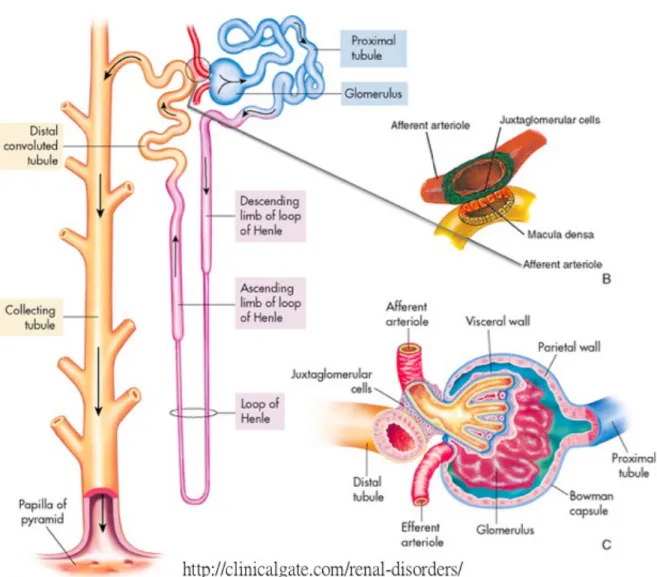
1.7 The kidneys and proximal tubules...37
1.7.1 Glomerulus...38
1.7.2 Proximal tubules...39
1.7.3 Juxtaglomerular apparatus (JGA)...39
1.7.4 Other compartments of renal tubular…...39
1.8 Water homeostasis in kidney and blood pressure regulation...40
x
1.10 Glycogen synthase kinase 3β (GSK3β) and its signaling pathway...45
1.10.1 GSK3β expression and function in the kidneys...45
1.10.2 GSK3β regulates Nrf2 signaling...46
1.11 β-catenin and its signaling...47
1.11.1 The canonical Wnt/β-catenin signaling pathway...47
1.11.2 β-catenin and kidney disease...48
1.11.3 β-catenin and its interaction with AQP1...49
1.12 Animal models used in present study...50
1.12.1 Maternal diabetic murine model...50
1.12.2 Hoxb7/catalase-GFP-Tg mouse...51
1.12.2.1 Hoxb7-GFP-Tg mouse...51
1.12.2.2 Cat-Tg mouse...52
1.12.3 rAgt-Tg mouse...52
CHAPTER 2: PUBLISHED ARTICLE Catalase Prevents Maternal Diabetes-Induced Perinatal Programming via the Nrf2-HO-1 Defense System 2.1 Abstract...56
2.2 Introduction...56
2.3 Materials and methods...59
2.4 Results...64
2.5 Discussion...68
2.6 Reference list...73
2.7 Legends and figures...77
CHAPTER 3: PUBLISHED ARTICLE Overexpression of Angiotensinogen Downregulates Aquaporin 1 Expression via Modulation of Nrf2-HO-1 Pathway in Renal Proximal Tubular Cells of Transgenic Mice 3.1 Abstract...86
xi
3.2 Introduction...86
3.3 Materials and methods...87
3.4 Results...90
3.5 Discussion...93
3.6 Reference list...98
3.7 Legends and figures...102
CHAPTER 4: DISCUSSION Article 1 4.1 Summary...110
4.2 Maternal diabetes intrauterine environment causes elevated ROS in the kidney, impairs kidney development and leads to perinatal programming of hypertension and kidney injury in the offspring...111
4.3 Overexpressed CAT in the offspring’s RPTCs can normalize reduced nephron number in mouse offspring exposed to maternal diabetes in utero...112
4.4 Overexpressed CAT in the offspring’s RPTCs can prevent hypertension and kidney injury in mouse offspring exposed to maternal diabetes in utero...113
4.5 20-week-old adult offspring from diabetic dams have high RPTCs ROS...114
4.6 The underlying mechanism: role of the renal Nrf2-HO-1 defense system in offspring born to diabetic dams...114
4.7 Could CAT be used in the clinic to prevent offspring of diabetic mothers from developing hypertension and kidney injury?...115
4.8 Other organs affected by maternal diabetes perinatal programming...117
4.8.1 Liver...117
4.8.2 Heart...118
4.8.3 Brain...118
Article 2 4.9 Summary...118
4.10 Characterization of the rAgt-Tg mouse model and determine of drug treatment...119
xii
4.11.1 Results of the rAgt-Tg mouse study...121
4.11.2 Results of the in vitro studies...122
4.12 Nrf2 accumulation in the cytoplasm of pRSV/rAgt-IRPTC and RPTCs of rAgt-Tg mice...123
4.13 β-catenin is a mediator of Agt-induced kidney injury – in vivo results from literatures match our in vitro finding...124
4.14 The role of NHE3 in Agt-induced hypertension...125
4.15 How does HO-1 regulate AQP1 expression?...125
4.16 Glycosylation of AQP1...126
4.17 Dramatic decrease of AQP1 in pRSV/rAgt-IRPTC but modest decrease of AQP1 in rAgt-Tg mice RPTC……….……….127
CHAPTER 5: FUTURE WORK Article 1 5.1 Does absence of Nrf2 in offspring kidney accelerate hypertension and kidney injury development in response to maternal diabetes condition?...129
5.2 Does overexpression CAT in RPTCs in offspring protect or delay progression of kidney injury programed by maternal diabetes and postnatal overnutrition?...131
Article 2 5.3 Does kidney-specific treatment with an HO-1 inducer (CoPP) normalize BP and AQP1 expression in rAgt-Tg mice?...133
5.4 Does kidney-specific treatment with an Nrf2 activator normalize BP and AQP1 expression in rAgt-Tg mice?...134
5.5 To prove that GSK-3β regulate Nrf2 nuclear translocation and decreases HO-1 and AQP1 expression in rAgt-Tg mice and pRSV/rAgt-IRPTC...134
5.6 To further investigate if β-catenin interacts with AQP1 and plays a role in kidney injury progression induced by Agt overexpression...135
CHAPTER 6: REFERENCE References...137
xiii
List of Tables
Table 1-1: Risk factors for GDM (page 14)
Table 1-2: 2013 World Health Organization recommendation for the diagnosis of gestational diabetes (page 15)
Table 1-3: AQPs in the kidney (page 42)
xiv
List of Figures
Figure 1-1: Impact of perinatal insults in programming adult pathologies in the offspring (page 4)
Figure 1-2: Kidney development (page 8)
Figure 1-3: Theoretical model for how disturbed nephrogenesis contributes to progressive kidney disease (page 12)
Figure 1-4: Fetal results of maternal hyperglycemia (page 16) Figure 1-5: Vicious cycle of obesity and diabetes (page 18)
Figure 1-6: Prevalence of elevated urinary albumin excretion, by birth weight, adjusted for age, sex, duration of diabetes, HbA1c and mean arterial pressure in Pima Indians from Gila River Indian Community in Arizona, 1983-1996 (page 23)
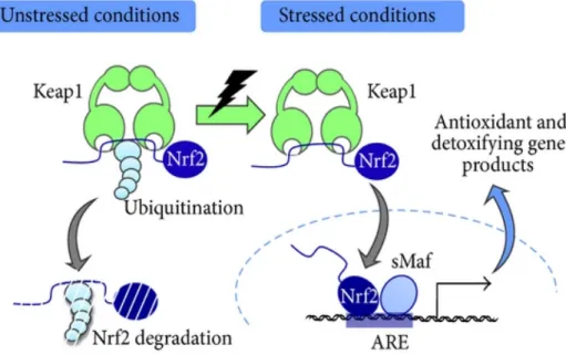
Figure 1-6: Mitochondrial respiratory chain (page 26) Figure 1-7: Production and metabolism of ROS (page 27) Figure 1-8: Keap1-Nrf2 stress response system (page 30) Figure 1-9: Overview of the RAS (page 33)
Figure 1-10: Structure of a human kidney (page 37) Figure 1-11: Components of the nephron (page 38)
Figure 1-12: Expression of renal AQPs along the nephron (page 43)
Figure 1-13: IHC staining of AQP1 in the paraffin section of mouse kidney (page 44) Figure 1-14: The current Wnt signaling model (page 48)
Figure 2-1: Characterization of Hoxb7-GFP-Tg and Hoxb7/Cat-GFP-Tg mice (page 77) Figure 2-2: (A) Neonatal renal morphology reviewed by H&E staining and CAT expression (CAT-IHC). (B) Quantification of neonatal nephron number (page 78)
Figure 2-3: Physical parameters in the male offspring at 20 week-old (page 79)
Figure 2-4: Mean SBP, ROS generation and renal function measurement in male offspring at age of 20 week-old (page 80)
Figure 2-5: Renal morphology and TGF-β1 gene expression in the male offspring at age of 20 week-old (page 81)
xv
Figure 2-6: Nrf2 and HO-1 gene expression in the male offspring at the age of 20-week-old (page 82)
Figure 2-7: High glucose effects on Nrf2 and/ or HO-1 protein expression as well as Nrf2 nuclear translocation analyzed by WB and IF staining (page 83)
Figure 3-1: Biological parameters in 5 groups of mice (Con, Agt-Tg, Agt-Tg + L/P, Agt-Tg + L, and Agt-Tg + H) at 20 week-old (page 102)
Figure 3-2: Renal morphology, IHC and Semi-quantification of the relative staining values in 4 groups of mice (Con, Agt-Tg, Agt-Tg + L and Agt-Tg + L/P) at 20 week-old (page 103) Figure 3-3: AQP1 and HO-1 protein expression in 3 groups of mice (Con, Agt-Tg and Agt-Tg + L/P) at 20 week-old; and CoPP effect analyzed by WB in vitro (page 104)
Figure 3-4: Nrf2 and Keap1 protein expression in 3 groups of mice (Con, Agt-Tg and Agt-Tg + L/P) at 20 week-old (page 105)
Figure 3-5: Agt, AQP1 and Nrf2/Keap1 protein expression in IRPTC stable transformants analyzed by WB or IF (AQP1); Nrf2 translocation in the isolated cytosol and nuclear fraction analyzed by WB, and phosphorylation of GSK3β in the kidney of 3 groups of mice (Con, Agt-Tg and Agt-Tg + L/P) at 20 week-old (page 106)
Figure 3-6: Phosphorylation of GSK3β and β-catenin in IRPTC and IRPTCs stable transformants analyzed by WB; NHE3 expression by Co-localization of IF-AQP1 and IF-NHE3, and by WB in the isolated RPTCS in 3 groups of mice (Con, Agt-Tg and Agt-Tg + L/P) at 20 week-old (page 107)
Figure 3-7: Our working model illustrates that Agt overexpression inhibits AQP1 expression in RPTCs, resulting in renal injury and hypertension (page 108)
Figure 5-1: Experimental design using Nrf2KO heterozygous to create three kinds genotypes of offspring: Nrf2 WT, Nrf2KO heterozygous and Nrf2-null mice (page 130)
xvi
List of Abbreviations
ACE: Angiotensin converting enzyme ACE2: Angiotensin converting enzyme 2 ADH: Antidiuretic hormone
Agt: Angiotensinogen Agt KO: Agt knockout Ang I: Angiotensin I Ang II: Angiotensin II AQP1: Aquaporin 1
AQP1KO: AQP1 knockout AQPs: Aquaporins
ARE: Antioxidant response element
AT1b: Type 1 angiotensin II receptor subtype b AT1R: Ang II type 1 receptor
AT2R: Ang II type 2 receptor ATP: Adenosine triphosphate AVP: Arginine vasopressin BMI: Body mass index
bZIP motif: Basic leucine zipper motif CAT: Catalase
CDA: Canadian Diabetes Association CNC family: Cap-n-collar family CO: Carbon monoxide
Cul-E3: Cullin 3-based ubiquitin E3 DAB: 3,3'-Diaminobenzidine
DOHaD: Developmental origins of health and disease ECM: Extracellular matrix
eGFR: Estimated glomerular filtration rate EMT: Epithelia –mesenchymal transition GCK: Glucokinase
GFR: Glomerular filtration rate GPX: Glutathione peroxidase GSH: Glutathione
H2O2: Hydrogen peroxide
HAPO study: Hyperglycemia and adverse pregnancy outcomes study HbA1c: Glycated hemoglobin
HBW: High birth weight HFD: High fat diet
HNF1A: Hepatocyte nuclear factor-1alpha HO-1: Heme oxygenase-1
Hoxb7-GFP-Tg mice: Hoxb7-green fluorescence protein-transgenic mice IDF: International Diabetes Federation
IL-6: Interleukin-6
IRS1 mice: Insulin receptor substrate-1 haploinsufficient mice IUGR: Intrauterine growth restriction
xvii
JGA: Juxtaglomerular apparatus
KAP: Kidney androgen- regulator protein
KCNJ11: Potassium inwardly rectifying channel subfamily J, member 11 Keap1: Kelch-like ECH-associated protein 1
LBW: Low birth weight
MAPK: Mitogen-activated protein kinases MCP-1: Monocyte chemoattractant protein-1 MET: Mesenchymal–epithelial transition MM: Metanephric mesenchyme
mTOR: Mammalian target of rapamycin NAFLD: Non-alcoholic fatty liver disease NCC: Sodium chloride cotransporter NCC: Na+/Cl cotransporter
ND: Normal diet
NFkB: Nuclear factor kappa-light-chain-enhancer of activated B cells NHE3: Na+/H+ exchanger-3
NKCC2: Na-K-Cl cotransporter 2 NOS: Nitric oxide synthases NOXs: NADPH oxidase
NQO1: NAD(P)H:quinone oxidoreductase1 Nrf2: Nuclear factor (erythroid-derived 2)-like 2 OGTT: Oral glucose tolerance test
O2 .-: Superoxide anion .OH: Hydroxyl radical OR: Odds ratio
PAI-1: Plasminogen activator inhibitor-1 PGDM: Pre-gestational Diabetes Mellitus PKC: Protein kinase C
RAS: Renin-Angiotensin System RNS: Reactive nitrogen species ROS: Reactive oxygen species RPTCs: Renal proximal tubule cells SBP: Systolic blood pressure
SOD: Superoxide dismutase STZ: Streptozocin
T1DM: Type 1 diabetes mellitus T2DM: Type 2 diabetes mellitus TCF7L2: Transcription factor 7-like 2 TG: Triacylglycerol
TGF-β1: Transforming growth factor TGF-β1 TNF-α: Tumor necrosis factor-α
UB: Ureteric bud
VEGF: Vascular endothelial growth factor WHO: World Health Organization
xviii
Acknowledgement
First I would like to express my deepest gratitude to my supervisor Dr. Shao-Ling Zhang, who has promised me a dream, and made it happen. Dr. Zhang plays her roles as a mentor at work and as a friend in life; urges us yet shows us ways to get relieved. I thank for her patience, inspiration, and encouragement throughout my entire journey of perusing a PhD. Her guidance has influenced me immensely, and led my path of academic research to the next level. Thank to my co-director Dr. John S.D. Chan, who is always generous in giving students chances. A very special thank to the president of my thesis committee Dr. Jolanta Gutkwska, who gave me advice and kindly helped me tackle my pre-doc exam. Also very sincere thanks to all my thesis committee: Dr. Raynald Bergeron and Dr. Jun-Li Liu for their insightful comments and profound questions.
Study abroad is not an easy task. Using languages other than my first one to learn and to communicate is rather tough. I am grateful to our lab technician Isabelle Chénier for giving me assistance dealing with all the glitch of languages, as well as laboratory affaires, especially experimental animals. My thanks also go to our Research Associate Dr. Chao-Sheng Lo and postdoctoral fellow Dr. Chin Han Wu, who gave me technical support and brainstormed me every now and then. I would like to thank my colleagues: Xing-Ping Zhao, who is the best consultant to me in lab, regarding every kind of problems. Thank to Min-Chun Liao, who is an active organizer in our group, a true friend, and a reliable partner, for bringing delights. I also have to thank Henry Nchienzia, who cheered me up in my darkest days. Thanks to Anindya Ghosh and Suiling Zhao, my lab life was not too dull. Thank all the friends ever helped me during this period, those who shared good or bad times together with me. Friendships have enriched my life experience with laughter and tears.
Last, but not least, I would like to dedicate this achievement to my parents and families. There is no way I could accomplish all these alone without their unconditional love and support. Knowing that there is someone always there for me, back me up and care for me, had strengthened my faith, so that I can dare to set goals that ever seemed impossible to me, and challenge it without fears. Thank God for the trials, to prepare me become who I am.
2
1.1 Perinatal programming 1.1.1 Definition
Perinatal programming refers to the well accepted concept that the intrauterine environment profoundly affects the developing fetus, and if it is abnormal, this can lead to health problems in the adult [1]. This phenomenon is also called the “developmental origins of health and disease” (DOHaD) [2]. The concept of ‘perinatal programming’ is substantially supported by many studies that show how an adverse environmental stimulus experienced during a critically sensitive period of development in utero can induce short- and/or long-term structural and functional effects resulting in health alterations later in life [2, 3].
My thesis deals with how maternal diabetes influences fetus’ kidney development in the intrauterine high glucose environment during pregnancy, by using streptozocin (STZ)-induced maternal diabetes mouse model in Hoxb7-GFP-Tg male mice. The reduced nephron number and higher risks of developing hypertension and kidney injuries for offspring from diabetic mother were ameliorated in Hoxb7/Cat-GFP-Tg male offspring which overexpress catalase (CAT) in their proximal tubules. The work described below has been published in Diabetes 61(10): 2565-2574, 2012, entitled “Catalase Prevents
Maternal Diabetes-Induced Perinatal Programming via the Nrf2-HO-1 (Nuclear factor (erythroid-derived 2)-like 2-Heme oxygensae-1) Defense System”.
1.1.2 The impact of an adverse intrauterine environment on fetal development
In 1989, Hales and Barker conducted a study in England that established a link between insufficient nutrition in pregnancies and the offspring with low birth weight (LBW; defined as less than 2,500 g, [4]) and increased risk of developing cardiac disease, metabolic syndrome and type 2 diabetes mellitus (T2DM) in adulthood [5]. This study was the first to describe the concept of perinatal programming that the authors termed “the
thrifty phenotype hypothesis.” [6]. Further studies supported the theory that perinatal
programming is an adaptive response to an adverse intrauterine nutritional environment, resulting in phenotype changes that begin in the fetus and affect the adult later in life [7]. The term “intrauterine growth restriction” (IUGR) describes a fetus which fails to achieve
3
full growth potential. IUGR is related to placental insufficiency, which is due to a combination of factors, such as maternal, fetal, placental and environmental factor. Placental insufficiency can be caused by dysfunction of fetal-placental perfusion, caused by maternal pre-eclampsia or hypertension, where hypoxia and ischemia affect both the maternal and fetal circulations [8, 9]. Abnormal placentation, including abnormal angiogenesis and vasculogenesis, also contributes to placental insufficiency. Poor maternal nutrition is the major contributing factor to IUGR. Maternal undernutrition influences the availability of nutrients for the fetus. During maternal starvation, low maternal food intake results in reduced nutrients available to the fetus, causing restriction in fetal growth [10].
In contrast to maternal undernutrition, maternal overnutrion leads to fetal overnutrition, which also constitutes a significant health hazard for both mother and fetus. Maternal overnutrition leads to neonatal macrosomia (also called Big Baby Syndrome), one of the most common pregnancy related health concerns in modern society. Macrosomia is characterized by babies with a birth weight over 4000 g, or greater than the 90th percentile for gestational age, and is characterized by increased fetus adiposity and a number of metabolic and immune system changes [11, 12]. The adipose tissue in the macrosomic infant secretes inflammatory cytokines which lead to chronic inflammation and immune system activation, as well as increased insulin resistance [13]. Excess fat also induces hyperlipidemia and hyperinsulinemia, and leads to lipotoxicity and a build-up of lipids in non-adipose tissue, such as the pancreas, kidney, liver and heart [14].
Similar to maternal obesity, maternal diabetes is characterized by increased glucose level, and shares common factors, such as maternal hyperglycemia, hyperinsulinemia and insulin resistance [15]. The offspring born to a diabetic mother with poorly controlled glycaemia experiences a high risk of congenital malformations during gestation, and is predisposed to develop multiple chronic diseases, such as hypertension, T2D, cardiovascular and kidney diseases in adulthood [16].
A wide range of other perinatal insults and gestational events is known to alter the fetal developmental trajectory, includes environmental exposure to endocrine-disrupting chemicals, disease states, lifestyle (e.g. stress), substance abuse (e.g. smoking and alcohol
4
drinking) and medical interventions (e.g. androgens and glucocorticoids) during pregnancy. The maternal conditions and fetal outcomes are thoroughly discussed by Padmanabhan et
al [1] (Figure 1-1).
Figure 1-1. Impact of perinatal insults in programming adult pathologies in the offspring. Exposure of the fetus/offspring to different insults during critical periods of development may lead to adaptations that prove to be detrimental and associated with adult defects in several organ systems [1].
1.2 Perinatal programmed hypertension and chronic kidney disease (CKD) 1.2.1 Two important hypothesis
Two hypotheses brought to light the association between maternal environment, birth weight, nephron endowment and diseases later in life. As previously mentioned, in 1989, Barker DJ et al reported a correlation between maternal malnutrition, infant’s LBW and
5
higher systolic blood pressure (SBP) later at 2 different population of age: 10 and 36-years-old, respectively [17] — Barker’s hypothesis suggested that differences in the intrauterine environment predisposing to differences in blood pressure and cardiovascular mortality in the babies. At about the same time, Brenner BM et al hypothesized that a decrease nephron mass may result in a diminished glomerular filtration surface and increased nephron glomerular filtration rate (GFR) of the remaining nephrons [18]. Over time, overloading of the functional capacity of the remaining nephrons, leads to glomerular hyperfiltration, glomerular sclerosis and proteinuria, and eventually causes kidney injury [19] — Brenner’s hypothesis postulated that a renal abnormality that contributes to essential hypertension in the general population is a reduced number of nephrons. These two studies shed light on how an adverse intrauterine environment can results in babies with LBW who are susceptible to subsequent chronic kidney disease and hypertension in adulthood.
1.2.2 Reduced nephron endowment is a major mechanism
Many studies were conducted to verify the correlation of birth weight with nephron number and the incidence of hypertension in adulthood [20]. A small case-controlled study conducted in Germany 2003 (n=20) examined the nephron numbers postmortem of adults who’s age ranges from 35 to 59 years. This study found a 50% decrease in nephron number in hypertensive adults compared to age-matched controls [21]. A large population conducted in Norway (n=2 millions, 2008) indicated that children born with LBW had a relative risk of 1.7 (95% confidence interval 1.4 to 2.2; P < 0.001) for developing end-stage kidney disease later in life [22]. A study in 2008 in Netherland gave salt sensitivity test to 27 healthy male adults (average 37 years) and found a close correlation between LBW and salt-sensitive hypertension in these adults [23]. Similarly, a study in 2008 Switzerland assessed salt sensitivity on 50 children who aged 7 to 15 years and demonstrated that renal mass is reduced in children born with LBW and depends on the degree of growth retardation, which then determines lower GFR, increased salt sensitivity, and elevated blood pressure [24]. Consistent with the observations in humans, a vast of studies in rodent models conducted between 1999 to 2007 also confirmed that late-onset hypertension is associated with LBW and significantly reduced nephron numbers, and together with human data were reviewed by Brenner in 2010 [25].
6
A meta-analysis in humans have shown a correlation between LBW and renal dysfunction, as well as the effect of speeding up the progression of primary kidney disease [26]. Proteinuria and estimated glomerular filtration rate (eGFR) are important predictive biomarkers of progressive kidney injury [27]. A cohort study (1994 patients and 20,032 controls) in US of mixed races but a majority of white children under 21-year-old found a significant correlation between LBW and enhanced risk of chronic kidney disease (CKD) with adjusted odds ratio=2.88 [28]. Also, averaging the data from 18 human studies, the odds ratio for CKD associated with LBW, was found to be 1.73 [29]. These epidemiological studies suggested a strong association between LBW and late-onset kidney injury.
Studies both in human and in animal models have shown LBW has association with reduced nephron numbers [30-34]. Considering all the evidence, the concept that the maternal intrauterine environment can profoundly affect fetal nephrogenesis, reduce the nephron endowment in the fetus and thus predispose the offspring for developing hypertension later in life is well established [35, 36]. Nowadays, the research focus has been shifted to identify the underlying mechanisms involved in abnormal nephrogenesis, and to develop effective treatments.
Nephron endowment refers to the total number of nephron which a person has at birth, although this number may decline due to age or disease state later in life. In fact, even among healthy individuals, the total number of nephrons is highly variable. This is illustrated in a series of Australian study (from 2003 to 2010) of adult human nephron endowment, where nephron numbers were found to range from 0.2-2.7 millions in African Americans (n=176), and from 0.2-1.6 millions in Caucasians (n=132) [37]. The currently accepted range for a normal human nephron number is approximately 0.9-1 millions per kidney [37]. In mice, the average nephron number of both C3H/HeJ and C57BL/6J mice is 2,000 at birth and around 20,000 at adulthood (8 weeks old) while the kidney development is completed [38]. Currently, the technology available that is used to count nephron numbers accurately in humans relies on autopsy and dissociation of nephrons from kidney tissue by acid maceration [39, 40]. That technique creates a disadvantage when the kidney is destroyed and can not be used for other research purpose [39]. Less invasive methods,
7
such as ultrasound scanning, provide a second method to assess renal mass in humans, which is based on kidney size determination to attempt to estimate nephron endowment [41]. Direct microscopic observation and nephron counting is used in experimental animal studies, to analyze the detailed changes in nephrogenesis that occur in the fetus exposed to hazardous environmental conditions in utero, as well as the pathogenetic mechanisms that occur later in adulthood.
Nephrogenesis refers to the complex process of nephron formation during kidney development [42]. In humans, nephrogenesis starts at the fifth week after gestation and is completed at week 34-36. At birth, the kidneys are fully formed and functional, and the number of nephrons does not increase thereafter [43]. In contrast, nephrogenesis in rodents is not complete at birth, and continues up to 21 days after birth [38].
Nephrogenesis involves a complex process called “branching morphogenesis”, which requires the coordinated interaction between the developing tubular epithelia and the renal vasculature. The development takes place in five stages: (1) ureteric bud (UB) development; (2) cap mesenchyme formation; (3) mesenchymal–epithelial transition (MET); (4) glomerulogenesis and tubulogenesis; (5) interstitial cell development [44]. In brief, the primary UB originates at the posterior end of the Wolffian duct as a solid aggregate of epithelial cells that proliferate, migrate, and progressively invade the surrounding metanephric mesenchyme (MM). UB branches in a highly reproducible manner, and the nephron formation is induced at each of its tips. While UB are branching into the MM, some MM cells, including self-renewing progenitor cells, condensate and aggregate around the UB tip of branches, transforming themselves into the cap mesenchymal cells. Cap mesenchyme progressively undergoes MET, which will form most of the epithelia of the nephron. Glomerulogenesis and tubulogenesis stages can be subdivided into four more steps: (1) renal vesicle formation; (2) transformation of the renal vesicles into comma-shaped bodies and then into S-shaped bodies; (3) development of the renal vascular system; (4) progressive development of the nephron and differentiation of the renal interstice [44]. Ultimately, MM differentiates into the main components of the renal corpuscles and the tubular segments, including proximal tubules, the loop of Henle, distal tubules, the juxtaglomerular complex, macula densa, mesangium, and part of the
8
afferent arterioles, whilst the branches form the collecting system, including collecting ducts, renal pelvis and ureter (Figure 1-2).
Figure 1-2. Kidney development: (A) The kidney is formed via reciprocal interactions between the Wolffian duct and the MM. (B) MM-derived signals induce the formation of the ureteric bud (UB) from the Wolffian duct. The UB then invades the MM and attracts MM cells. (C) MM cells condense around the tips of the branching UB, forming the condensed mesenchyme, or CM. In response to UB signals, CM cells are induced to undergo mesenchymal-to-epithelial transition (MET). (D–F) The induced cells acquire an epithelial phenotype concomitantly with shutting down of the major transcription factors described before. The cells sequentially form the pretubular aggregate, renal vesicle, C- and S-shaped bodies and finally the mature nephron. The cells derived from the CM form most of the nephron body (from glomerulus to distal tubule), whereas the UB-derived cells form the collecting duct [45].
9
Hundreds of signaling genes and factors participate in a specific spatial and temporal pattern to play a role in the UB origin, elongation, and branching [44, 46]. For example, multiple genes were reported to act either as inducers (c-Ret, ETv4, ETv5, GDNF, SOX8, SOX9, Wnt11, Angiotensin II, FGFR1, FGFR2, FGF8, p53, MMP-9, Cofilin1, Destrin, AT1R, AT2R, and PAX2), or inhibitors (Spry1, class 3 semaphorins, Robo2, Slit2, BMP4, FoxC1, and FoxC2) [46].
One human UB can branch on average 15 times during nephrogenesis and in murine, 10– 11 UB branching events occur during kidney morphogenesis [47]. The developmental process is normally well synchronized, but it can become less well synchronized due to adverse environmental conditions in utero (e.g. high glucose), with the results that both kidney morphology and nephron number will become abnormal [48].
1.2.3 Other mechanisms involved in programming hypertension and kidney injury in the infant later in life
Evidences have shown that more other mechanisms participate in programming hypertension and kidney injury, in addition to reduced nephron number. An IUGR animal study done in a placental restriction induced by ligation of bilateral uterine vessel in Wistar Kyoto rat dam model (7-10 mothers per group and 5 pups per litter) proposed that the hypertension observed in the IUGR offspring could be ameliorated by cross-fostering with a normal control dam [49]. Furthermore, in a maternal glucocorticoid administration programmed hypertension rat model, hypertension was observed in offspring that had a reduction in glomeruli as well as in a group that did not have a reduction in glomerular number. The data suggested that a reduction in nephron number is not the only cause for the development of hypertension [50]. Taken together, it appears there is an existence of additional factors that contribute to perinatal programmed hypertension. More potential mechanisms that programmed hypertension in offspring exposed to maternal adverse environment are discussed below.
1.2.3.1 Impaired kidney sodium handling and programmed hypertension
Reduces sodium excretion either by decreasing GFR or by increasing tubular reabsorption of sodium can cause hypertension. The renal sympathetic nerve plays important role in
10
kidney function, include renin secretion and sodium reabsorption. Two studies which used the maternal placental insufficiency SD rat model [51] and maternal glucocorticoid administration SD rat models [52], observed that fetuses exposed to maternal adverse stimuli develop hypertension later in life, and the high blood pressure could be ameliorated by bilateral renal denervation, which altered sympathetic innervation and caused decreased sodium reabsorption. Aberrant sodium management is also associated with hypertension. Two studies in SD rat with maternal glucocorticoid exposure and maternal low protein diet found increased sodium channels, and increased Na+/H+ exchanger-3 (NHE3), Na-K-Cl cotransporter 2 (NKCC2), Na+/Cl cotransporter (NCC) in renal tubules of the offspring [53, 54].
1.2.3.2 Epigenetic changes and programmed hypertension – an implication of glycemic memories
In utero adverse environment modulates epigenetic modification, which is one of the
mechanisms leading to perinatal programming [55]. Epigenetic changes refer to gene expression altered by several mechanisms, including DNA methylation, histone modification, and microRNA expression, without affecting the genetic code. Epigenetic changes influence mRNA transcription resulting in phenotype changes eventually [56, 57].
The term “glycemic memory” (also called “metabolic memory”) was coined in 1990 while the researchers found fibronectin and collagen were highly and persistently produced in endothelial cells of diabetic rats despite their glycaemia had been normalized for two weeks after having diabetes for two weeks [58]. Many large-scale clinical trials and experimental animal studies ensued and supported this concept, and were nicely reviewed [59]. These studies identified early exposure to hyperglycemia or poor glycemic control contribute to intense and prolonged diabetic complications development. The disease progression persists despite glycemic control is improved afterwards. This indicates a memory of glycemic insult and is due to epigenetically alteration of relevant genes [59, 60].
DNA methylation involves the covalent modification of cytosine residues that precede guanines-CpG dinucleotides, with the “p” referring to the phosphodiester bond between
11
the cytosine and guanine nucleotides [61]. The CpG dinucleotides are clustered in CpG-rich regions of the 5’ end of genes where lies promoters, enhancers and suppressors. Methylation of CpGs recruits multiple factors to form a complex that is bound to the promoter and in turn prevents access of transcription factors and RNA polymerases to the DNA and results in the silencing of transcription [62, 63].
Epigenetic changes in the renin angiotensin system (RAS, a major system that controls blood pressure will be described in later section) have been observed in the adrenals of rat offspring of maternal low protein diet. The type 1 angiotensin II receptor subtype b (AT1b) promoter was hypomethylated compared to control, resulting in increased AT1b mRNA expression at 12 weeks of age [64]. This low protein diet model is known to produce hypertensive offspring identified as early as 4 weeks of age [65]. Another study examined the offspring’s kidney following maternal IUGR in the rat and found a decrease in CpG methylation of the p53 promoter resulting in increased expression of p53 mRNA levels. Increased renal apoptosis and reduced glomeruli number were also observed in the affected offspring [66]. As reported by another group using the same maternal IUGR model, it was found that affected offspring develop hypertension at 22 weeks of age [67]. These findings emphasized the potential role of epigenetics in developmental programming. However, the exact effects of changes in gene methylation are not always easy to assess [68].
1.2.3.3 Oxidative stress and inflammation and programmed hypertension
Oxidative stress and inflammation are likely to be the common factors that are important in many pathological contexts, not to mention perinatal programmed hypertension [69]. In the rat maternal low protein diet model, both of these two factors were found to play a role in programming hypertension in the offspring [69]. However, the mechanism of how reactive oxygen species (ROS) regulates hypertension in offspring born to diabetic mothers is still unclear. Our laboratory has published a number of studies addressing the role of ROS generated by maternal high glucose intrauterine environment on the developing kidney of the fetus and on the adult-onset kidney disease and hypertension. Details will be discussed later in the following sections.
12
1.2.3.4 Mechanisms correlate LBW and late-onset kidney injury — the two hit model of kidney disease
The two hit hypothesis is a model which requires two hit of risks in order to generate the clinical phenotype and was originally used to describe the onset of other kidney diseases [70, 71]. In that model, the first hit is an early priming in a genetically predisposed individual and the second hit is a likely environmental insult. The dual hits increase vulnerability of the individual under adverse conditions. Later, other researchers expanded this concept and used it to propose that low nephron number renders the kidney more susceptible to kidney injury. According to this hypothesis, an insufficient nephron endowment is the “first hit,” which then subsequently predisposes the person to more severe renal dysfunction if a “second hit” is added, which can be hypertension [55] (Figure 1-3). This hypothesis has been confirmed in IUGR perinatal programmed hypertension rat model [72]. The kidney with less nephron numbers was impaired as a first hit, and giving anti-Thy1 to induce glomerulonephritis as second hit could accelerate the progress of acute glomerulonephritis and lead to more sclerotic lesions [72, 73].
Figure 1-3. Theoretical model for how disturbed nephrogenesis contributes to progressive kidney disease. Prenatal programming causes low nephron numbers, which results in glomerular hypertrophy, tubular malfunction and hypertension, and can lead to glomerulosclerosis and progressive loss of renal function. The kidney is more easily damaged by superimposed kidney diseases; for example, IgA nephropathy and diabetic nephropathy, respectively, tend to have a more severe course and more rapid loss of renal function in individuals with a history of LBW caused by disturbed intrauterine development. Abbreviations: HSD11β2, corticosteroid 11-β-dehydrogenase isozyme 2; Na+, sodium ion; RAAS, renin–angiotensin–aldosterone system [55].
13
1.3 Maternal diabetes and adverse outcome 1.3.1 Maternal diabetes
Maternal diabetes refers to either pre-existing diabetes in a pregnant woman (diabetes mellitus type 1 or type 2; T1/T2DM), or the development of insulin resistance and subsequent high blood glucose that is first diagnosed during pregnancy (gestational diabetes mellitus; GDM). According to the latest report of the International Diabetes Federation (IDF), 17% (21millions) of live births in 2013 had hyperglycemia in pregnancy (disregard of stillbirths). Hyperglycemia is the leading cause of perinatal complications to the fetus. Uncontrolled maternal hyperglycemia during pregnancy can result in birth complications, which affect both mother and child, and these are described further below [74].
1.3.2 Maternal diabetes and the maternal complications
Preexisting diabetes in a pregnant woman is known as pre-gestational diabetes mellitus (PGDM). According to the Canadian Diabetes Association (CDA), the diagnostic criteria for diabetes are a fasting plasma glucose level of ≥7.0 mmol/L; random plasma glucose ≥11.1 mmol/L; 2-hour plasma glucose value ≥11.1 mmol/L in a 75 g oral glucose tolerance test (OGTT) or glycated hemoglobin (HbA1C) ≥ 6.5%. In The Northern Diabetic Pregnancy Survey that with a majority of White British ethnicity showed the prevalence of PGDM is rising, reflecting that the prevalence of both T1DM and T2DM in women of childbearing age is increasing [75]. Diabetic patients are already at a higher risk of developing complications, such as retinopathy, nephropathy, and cardiovascular disease. For women with diabetes, pregnancy could worsen the progression of diseases and reduce their lifespan. Details are nicely reviewed by this publication of CDA, entitled Diabetes
and Pregnancy [76].
Becoming diabetic during pregnancy can occur in women who have never had diabetes before conception. It usually manifests as glucose intolerance resulting in hyperglycemia of variable severity that is first diagnosed during pregnancy, and is called gestational diabetes mellitus (GDM) [77]. GDM generally occurs around the late second trimester of pregnancy and ends after delivery. For women with GDM, although blood glucose levels return to normal after delivery, mothers are at an increasing risk of developing T2DM in
14
the future (29.4% in a 45-month follow-up period) [78]. Also, a large-scale study included 47,909 women during a follow-up period of more than 10 years indicated that GDM is associated with long-term maternal cardiovascular morbidity (odds ratio=2.7) [79]. In Canada, CDA announced in 2016 that 3-20% of pregnant women develop GDM, depending on their risk factors [80]. Table 1-1. The incidence of aboriginal women is 2-3 times higher than non-aboriginal woman probably because of the genetic background and the poor involvement of public health services [81, 82].
Table1-1 Risk factors for GDM [80].
Besides the risk factors listed in table 1-1, recent studies have identified several gene variations that are associated with an increased risk of GDM, includes the transcription factor 7-like 2 (TCF7L2), potassium inwardly rectifying channel subfamily J, member 11 (KCNJ11), Glucokinase (GCK), hepatocyte nuclear factor-1alpha (HNF1A), etc [83, 84]. These genes are related to glucose-stimulated insulin secretion, insulin synthesis, pancreatic β-cell proliferation and islet cell volume, and the two studies suggested that the metabolic imbalance observed during GDM pregnancy occurs in women who are genetically predisposed to it. Thus, it appears that GDM results from an interaction between certain genetic backgrounds and environmental factors.
Diagnosis of GDM, according to the recommendations by the World Health Organization (WHO), should be determined by a 2 h, 75 g oral glucose tolerance test administered
15
anytime during pregnancy (Table 1-2) [78].
Table 1-2. 2013 World Health Organization (WHO) recommendation for the diagnosis of gestational diabetes [85].
1.3.3 Maternal diabetes and its complications for the offspring 1.3.3.1 Short-term complications
1.3.3.1.1 Macrosomia
In humans, the risk of being macrosomic (big baby syndrome, also called high birth weight; HBW) for babies born to diabetic mothers is 3 times greater than normoglycemic mothers. According to a 2008 international population study (also called hyperglycemia and adverse pregnancy outcomes (HAPO) study) which included 25,505 pregnant women from 9 countries, maternal fasting blood glucose level higher than 6.9 mmole/L is significantly correlated with fetal birth weight above the 90th percentile (odds ratio=1.38) [86]. When maternal glycemia is high during pregnancy, the excess glucose is transported to the fetus through the placenta. In response to high level of circulating glucose, the fetus secretes insulin after entering the second trimester of gestation, when their pancreas development is mature. As a result, fat and protein uptake is increased, and growth is accelerated (Figure 1-4) [87].
16
Figure 1-4. Fetal results of maternal hyperglycemia. Modified according to Pedersen's hypothesis [87].
A large fetus size can increase difficulty during vaginal delivery: the fetus may become stuck in the birth canal, requiring additional instruments and/or a C-section. The more difficult and prolonged delivery process could cause complications, such as fetus hypoxia, birth trauma, shoulder dystocia and cerebral palsy [88].
1.3.3.1.2 Congenital malformations
PGDM is associated with a higher incidence of congenital anomalies, compare to GDM. A meta-analysis review study in 2012 indicated that the risk for congenital malformations in PGDM is 1.9–10-fold higher than total population, while the risk is slightly increased in GDM compared to the general population (odds ratio=1.1-1.3) [89]. For PGDM, good glycemic control is the key to prevention of congenital anomalies. The level of risk for newborns is highest just before conception and during the first 5-11 weeks while the baby’s organs are beginning to develop [90]. Congenital malformations occur when the development of the embryo is disregulated, such as arrested, delayed or misdirected development effects may involve multiple organs and systems, including respiratory, intestinal tract, cardiovascular, neural tube, genitourinary, musculoskeletal and is a leading cause of infant death [91]. Although with stringent maternal glycemia monitoring and control intervention, the risk of congenital abnormalities for the fetus of diabetic mothers is still 1.7- to 3-fold higher compared with the background population [92].
17
1.3.3.1.3 Stillbirth and perinatal mortality
Stillbirth is defined as fetal death at or later than 20 weeks of gestation or birth weight of 350 g or greater, while perinatal mortality is defined as the total number of stillbirths and neonatal deaths up to 28 days of life [93]. The perinatal mortality is mainly due to congenital anomalies or complication of prematurity. Both PGDM and GDM are associated with high rate of stillbirth. Patients with PGDM overall have a odds ratio of 3.8 to 6.3 of perinatal mortality compared to women with normal glycemia according to a literature review [93]. The risk of perinatal mortality in GDM is not as high as PGDM [16].
A US study in 2005 compared women who were diagnosed with GDM after 37 weeks (and left untreated till the end) with women who were treated for GDM and women without diabetes. The stillbirth rates in the three groups were 5.4, 3.6, and 1.8 per 1000 births, respectively [94].
1.3.3.1.4 Premature birth
Preterm delivery is defined as labor with gestation less than 37 weeks completed [95]. Preterm birth is responsible for 75% of neonatal mortality and 50% of long-term neurologic impairments in children [96]. Prematurity could be caused by premature rupture of membranes, or maternal hormones and cytokine disorders [97]. Newborns are under the risk of prematurity associated complications, such as infection, respiratory difficulties, and intensive cares are needed [95].
1.3.3.1.5 Hypoglycemia at birth
Immediately after delivery, the newborn’s blood insulin level is still high due to the in
utero high glucose environment, even though the glucose supply from the mother is
interrupted while gluconeogenesis and ketogenesis in the newborns are still immature to produce glucose for their selves [98, 99]. Neonatal hypoglycemia is commonly observed in the first hours of life of newborns [99] and persists up to 72 hours and may even last up to 1 week [100], which can lead to cardiopulmonary, central nervous system damage and subsequent mental retardation and recurrent seizure activity [87]. The prevalence of hypoglycemia in newborns of diabetic mothers is as high as 40% compared to newborns
18
from non-diabetic mothers [98]. Immediate glucose testing and feeding or intravenous glucose injection to the infant is used to effectively treat this condition [101].
1.3.3.2 Long-term complications — perinatal programming 1.3.3.2.1 Obesity
Population of obesity is markedly increasing in all age groups and has become a global issue in the last two decades. A worldwide study in 2014 indicated that nearly 30% of the population including children and adults are either overweight or obese [102]. According to the WHO guideline on this subject, body mass index (BMI) is a useful index of obesity, and is defined as the individual’s weight (in kilograms) divided by the square of his/her height (in meters). Individuals with a BMI of 25 or more are considered overweight, and those with a BMI of 30 or more are considered obese [103]. Obesity is associated with a number of metabolic disorders, and causes a great burden of medical care to societies worldwide [104], hence obesity is a major problem which urgently needs to be solved.
Although the individual’s eating habits may be one of the causes conferring obesity, studies have shown a strong link between maternal diabetes, body weight at birth and obesity in adulthood [105]. The concept of the obesity growing into a world pandemic, due to a “vicious cycle,” was first proposed by Pettitt in 1988, to explain how the maternal diabetic intrauterine environment is transmitted to future generations, by increasing the risk of obesity and T2DM in the offspring, thus contributing to a growing population of obese and diabetes [106] (Figure 1-5).
19
1.3.3.2.2 Type 2 diabetes mellitus (T2DM)
T2DM is a disease of insulin resistance combined with pancreatic β-cell dysfunction [107]. In the early stage of T2DM onset, both β-cell mass and function are increased to compensate for peripheral insulin resistance. β-cells become inadequate over time, followed by insulin deficiency [107]. Both epidemiological and animal studies indicate that offspring exposed to high glycemia in utero are at a higher risk of developing T2DM compared to controls.
A US population studies in 2008 analyzed youth of aged 10–22 years comprised 79 T2DM and 190 nondiabetic controls and found 47.2% (95% CI) of T2DM in youth could be attributed to intrauterine exposure to maternal diabetes and obesity [108]. In the same year, another study in Denmark (n=597) examined young adults aged 18–27 years and concluded that the odds ratio for offspring from GDM mothers to develop T2DM or impaired glucose tolerance is 7.76 while that from T1DM mothers the odds ratio is 4.02, all compared to offspring from control mothers [108]. A study pre-screened healthy participants without T1DM and measured insulin sensitivity and insulin secretion in these participants. Fifteen offspring were born to T1DM mothers whose fathers were healthy and 16 offspring were born to T1DM fathers whose mothers were healthy. They found 33% offspring from T1DM mothers developed glucose intolerance reduced insulin secretion while no offspring from healthy control mothers and T1DM fathers developed the impairment [74].
A rat study continuously infused dams during the last week of pregnancy to mimic mild hyperglycemia intrauterine environment and found the offspring from hyperglycemic dams started to show mild glucose intolerance and impairment of glucose-induced insulin secretion at 1 month of age. This situation persisted and eventually developed to constantly hyperglycemia and severe impairment of glucose tolerance and insulin secretion at 10 month of age [109]. It appears offspring born to mild hyperglycemic mothers (i.e. GDM or mother with T2DM) presented with islet hyperplasia, and increased pancreatic and plasma insulin concentrations [110]. In contrast, mouse offspring born to severely hyperglycemic mothers (i.e. with T1DM) displayed enhanced islet mass, with degranulated β-cells, suggesting overstimulation by hyperglycemia [111, 112]. This early exhaustion of the
20
pancreatic insulin secretory capacity may explain the low pancreatic insulin content and low plasma insulin levels in late intrauterus period. Immediately after birth, the mouse pup’s islet mass was decreased, the granular content was normalized, and the secretory capacity was restored. However, later during adulthood, the offspring developed insulin resistance [110].
1.3.3.2.3 Cardiovascular disease (CVDs)
CVDs manifests heart and blood vessel dysfunctions and is the number 1 cause of death globally [103]. Coronary heart disease and stroke are the two leading factors causing death [103]. Atherosclerosis is a condition that develops when the arterial walls are repeatedly injured and results in plaque built-up in the walls. The artery walls become sclerotic and narrow over time, and can be broken or blocked, which can lead to heart attack or stroke [103]. Many circulation cell adhesion molecules, such as intercellular adhesion molecule 1 (ICAM1), vascular adhesion molecule 1 (VCAM 1) and E-selectin, are used as a marker to predict CVDs [113]. Although the correlation between obesity, smoking, hypercholesterolemia and cardiovascular disease in adults is well established, evidence from both human and animal studies showed that vascular dysfunction can be programmed as early as the perinatal stage by various adverse maternal environments [114].
A UK study examined children aged 5-11 years (n=61) and found offspring from T1DM mothers expressed higher markers of endothelial dysfunction compared with offspring of nondiabetic pregnancies [115]. A similar US study (n=91) looked at the same endothelial dysfunction markers in children (aged 6-13 years) of maternal GDM exposure and found these children had increased values compared with non-exposed children, independent of BMI [116]. The above data suggested that exposure to maternal diabetes during pregnancy confers risks for the development of CVDs later in life and is independent of other risk factors of the offspring, such as adiposity [117].
The endothelium also plays an important role in the development of the vasculature by secreting vasoactive substances, such as vasodilators (e.g. nitric oxide) and vasoconstrictors (e.g. angiotensin II), which act on the adjacent vascular smooth muscle cells to cause vasodilation or vasoconstriction [118]. Reduced endothelium-dependent
21
vasodilatory capacity of the artery is found in 12 month old rat offspring of dams that were induced diabetic with STZ 10 days before mating, compared to offspring of non-diabetic control mother [119]. Altered angiogenesis is an important element in predisposing the development of vascular dysfunction in infants of diabetic mothers. A rat study examined 19 day old offspring of dams which were induced diabetic by STZ on the 5th day of gestation and found that maternal diabetes led to marked alterations in blood vessel differentiation and cardiomyopathy [120]. More specifically, it was pointed out that in
utero exposure to high glycemia affect angiogenesis via decreased proliferation of
endothelial cells via decreased production of vascular endothelial growth factor (VEGF) and VEGF receptor, resulting in embryonic vasculopathy [121].
1.3.3.2.4. Hypertension
According to the WHO, normal adult blood pressure is defined as 120/80 mmHg (systolic/diastolic). When the SBP is ≥ 140 mmHg, and/or the diastolic blood pressure is ≥ 90 mmHg, the blood pressure is considered to be high. High blood pressure can lead to lethal complications, such as stroke, heart attack and kidney failure [122]. One in every four people globally is hypertensive in 2000, and it is anticipated that this population will increase from 972 million to 1.5 billion by 2025 [123]. Even though many treatments for hypertension are currently available, there is still a need to elucidate its cause and underlying mechanisms in order to take precautious intervention and hence halt its growing prevalence.
The causes of hypertension are multiple and complex [124]. Although there is clear evidence that an unhealthy lifestyle (little exercise and an unhealthy diet) contributes to the risk of developing hypertension, there is compelling data from epidemiological and experimental studies which show that maternal adverse environment can also increase the risk of their offspring developing hypertension later in life [125, 126], suggesting that adulthood hypertension might be determined before birth by altered fetal development.
In humans, a population study on Pima Indian (US first Nation, n=42, aged 7-11 year old) indicated that offspring of mothers with maternal diabetes had higher SBP (about 11 mmHg) than mothers with normal glycemia during pregnancy [127]. More recently a


![Figure 1-4. Fetal results of maternal hyperglycemia. Modified according to Pedersen's hypothesis [87]](https://thumb-eu.123doks.com/thumbv2/123doknet/2047318.5121/34.892.122.513.130.389/figure-results-maternal-hyperglycemia-modified-according-pedersen-hypothesis.webp)
![Figure 1-5. Vicious cycle of obesity and diabetes [106].](https://thumb-eu.123doks.com/thumbv2/123doknet/2047318.5121/36.892.123.502.827.1095/figure-vicious-cycle-obesity-diabetes.webp)

![Figure 1-10. Structure of a human kidney, cut open to show the internal structures [243]](https://thumb-eu.123doks.com/thumbv2/123doknet/2047318.5121/55.892.118.487.431.821/figure-structure-human-kidney-cut-open-internal-structures.webp)
![Table 1-3. AQPs in the kidney [250].](https://thumb-eu.123doks.com/thumbv2/123doknet/2047318.5121/60.892.118.764.153.618/table-aqps-in-the-kidney.webp)
