The role of sensation for hand function in children with cerebral palsy
Annette Majnemer1
PhD, OT; Daniel Bourbonnais2
PhD, OT; Victor Frak3
MD School of Physical & Occupational Therapy, Departments of Pediatrics and Neurology &
Neurosurgery, McGill University1
Ecole de réadaptation, Université de Montréal 2
Département de kinanthropologie, Université du Québec à Montréal 3
I. IMPORTANCE OF SENSATION FOR REFINED HAND FUNCTION Vision and hand function
One of the most important roles of the hand is prehension, that is, the ability to grasp, hold and manipulate objects. Vision is obviously critical to gather information related to the potential intrinsic and extrinsic characteristics of the objects (orientation of the object, size, distance from the body, estimation of the weight) (Hay and Beaubaton, 1986). This visual information is then used to plan the reaching movement (Crawford et al. 2004; Schoeting and Flanders 1989) and postural adjustments underlying the prehensile act itself (Kaminski et al. 1995). It is generally agreed that both vision and proprioception provide information on hand and body position, and that these inputs are contributing both before and during the reaching
movements to enhance accuracy of reach of the target (Bagesteiro et al., 2006; Scheidt et al., 2005). Therefore, information on the internal status of the body (interoceptive) and on the relationship of the object to the body (exteroceptive) are necessary for planning and control of reaching movements. It was suggested that reaching to grasp an object can be divided into a transportation component, in which the arm brings the hand in the vicinity of the object to be grasped, and a manipulation component, where final adjustments of the hand are made prior to grasp (Jeannerod, 1984). During reaching, the grasp aperture increases throughout the transport phase reaching a maximum before contact with the object and is precisely adjusted when the hand is close to the object. Whether the onset of closing of the fingers is triggered by the decreased velocity of the forearm (Jeannerod 1984; Paulignan et al. 1991) or by spatial
information related to the distance of the hand from the target (Wang and Stelmach 2001) is still debated. Nonetheless, when under visual control, the distance between the thumb and index reflects the size of the object and the aperture is larger when vision is removed (Jeannerod, 1981).
Cutaneous afferents and manipulation
When reaching an object, the ability to grasp it precisely between the thumb and index requires a close interplay between sensory inputs from the fingers and the mechanisms
controlling the motor output of the hand and finger muscles. While holding an object between the index and thumb, the individual has to generate a shear force in order to overcome the weight of the object and prevent the object from slipping from the fingertips. The magnitude of the shear force is related to the friction coefficient of the object and the magnitude of the pinch force. Therefore, grip force can be modulated as a function of the friction between the fingertips and the object surface and, also, the weight of the object. Slippery and heavier objects will generally require larger grip forces. Usually the grip force is slightly larger than the minimal grip force mechanically required to hold the object, providing a security margin allowing small
perturbations to be corrected without dropping the object. Many studies have demonstrated the precise coordination between the grip force and the shear force during the manipulation of an object (Johansson and Cole, 1992: Johansson and Wesling 1984;1988a; Westling and Johansson 1984;1987).
Recordings of the activity of individual afferent fibers in an human peripheral nerve has demonstrated the importance of the cutaneous receptors of the thumb and finger pads in
controlling the opposing forces of a precision pinch grip while holding an object (Johansson and Westling 1984; 1987a; Johansson et al., 1992a). Different classes of cutaneous afferents encode various types of tactile stimuli applied to the skin of the digits. Slow and rapid receptors are respectively associated with dynamic and static indentations of the skin (Johansson, 1987a) and are collectively implicated in the appreciation of texture and detection of slip. These
mechanoreceptors provide information about changes in shear force or slip of the object on the skin (Johansson and Westling, 1987a) that are used to adjust the safety margin required by the manipulation of the object. In addition, cutaneous afferents contribute to rapid and automatic grip force increases that are observed following unexpected restraint of the object (Johansson and Westling 1988b; Johansson et al. 1992a,c). The grip force increases with a delay of
approximately 70 ms, indicating a feedback mechanism probably involving a supra-spinal loop (Cole and Abbs, 1988; Johansson and Westling, 1988b; Macefield et al., 1996 ). This evidence suggests an important role of cutaneous afferents for precise manipulation of a tool or an object.
Proprioceptive afferents
Afferent fibers from muscle receptors including muscle spindles and Golgi tendon organs and joint afferents have been recorded in humans during perturbation of an object gripped
between the finger and the thumb (Macefield 1996). In contrast to tactile afferents, the activity of these afferents is not associated with the increased shearing force following perturbation, but rather, reflect the reactive forces generated by the muscles to restrain the object. Although this indicates a low contribution of muscle afferents to initiate an appropriate change in grip force in response to an imposed change in shear force, this does not preclude the importance of muscle receptors in specifying the initial position and state of the hand effectors. This information is likely important for positioning the fingers in the correct biomechanical configuration to generate directionally-appropriate digital forces. Moreover, this proprioceptive information is probably important to establish the spatial relationship of the hand with the environment when visual information is lacking. A series of studies were conducted in a patient with a loss of large myelinated fibers affecting all somato-sensory modalities (kinaesthesia, tendon reflexes, touch, vibration). This patient relied heavily on visual feedback of the limb to control arm movements (Teasdale et al., 1993). Nonetheless, complex hand movements such as drawing ellipses on a sheet of paper, although slower, were still possible and the regularity and consistency of the drawings were comparable to healthy subjects. This regularity and consistency of the ellipses drawn were not affected by removal of vision, however the position and orientation of the ellipses drifted with time. This suggests that proprioceptive information contributes in determining the positioning of the hand in the environment.
Role of sensation in anticipatory control of grip force
Skilled hand manipulation also requires a high level of motor control that relies mainly on prediction of the consequences of our own actions. Stopping or reversing a rapid movement of the arm while an object is held between finger and thumb of the hand, requires an increase in shear force to counteract the inertia of the object. Considering the delays involved in cutaneous feedback loops (Cole and Abbs, 1988; Johansson and Westling, 1988), this increase in grip force is produced by a predictive mechanism. This predictive behavior of increasing grip force has been taken as evidence of the existence of an internal forward model of the limb and the object to
be manipulated (Flanagan and Wing, 1993). This model suggests that for self-produced movements, the central nervous system uses internal models of both the arm and the object to anticipate the resulting shear force and thereby adjust the grip force (Flanagan and Wing, 1997).
It is probable that continuous sensory feedback is not necessary to perform a predictable task. Indeed, following digital anesthesia, the pattern of force production is preserved during lifting and holding an object which has been previously manipulated (Johannson and
Westling,,1984). Only slight impairments of grip force regulation such as less precise adjustments to the skin and object friction characteristics and temporal delays between force adjustment phases for initiation of lifting (Johansson and Westling, 1984) are observed. In
contrast, digital anesthesia of the fingers decreases or even abolishes grip force changes normally seen following a perturbation of the prehension (Johansson et al, 1992a).
Inappropriate grip forces resulting from changing the weight of an object, which has been previously lifted by the subject, are re-scaled within a single trial (Johansson and Westling, 1988). This suggests that sensory feedback signals can effectively be used to recalibrate the grip force. However, it has been suggested that discrete sensory feedback provided primarily by cutaneous afferents are used to update anticipatory motor commands (Augurelle et al., 2003).
Sensory-motor integration
The relationship between sensory signals and motor commands has been extensively studied in neuroscience and recent advances in computational study of motor control have
emphasized the importance of sensory feedback (Wolpert and Gharamani, 2000). Not only is this information important to provide on-going feedback control of prehension, but also for
providing information to the nervous system on the status of the limb and hand in order to
predict the effect of motor commands. Impairments of vision and somatosensory function would impact specification of the initial state of the body, which is probably needed to determine with refinement the correct motor commands to be implemented.
Moreover, the integrity of sensory information is important for a sensory-driven control allowing the comparison of the actual somatosensory information and the expected
somatosensory input. The sensory consequence of the movement would be predicted using an internal model in conjunction with a copy of the motor command generating the movement. Detection of a difference between predicted and observed sensory information would determine the corrective response as well as an updating of the motor command. Obviously, this sensory-driven control could be impaired in individuals with neurological impairments and associated definits of somatosensory function such as cerebral palsy. Moreover, impairment in determining the intensity and timing of the motor command would also impair this sensory-driven control, since the sensory consequences would be incorrectly determined following an inappropriate estimation of the motor command. However, evidence suggests that individuals with
neurological deficits can appropriately determine the intensity and timing of a motor command and use this information to define the intensity and timing of the anticipatory motor command. This could be illustrated by the observation that while both deafferented subjects and subjects with anaesthetized fingers generated elevated baseline grip forces on an held object while performing reversal of arm movement, the precise temporal coupling between grip and load force profiles was maintained (Nowak 2001; 2002). Interestingly, these individuals still
modulated grip force even though the baseline levels were high enough, not justifying this modulation. This suggests that the intensity of the motor command required to maintain grip force during baseline force exertion is taken into account when determining the intensity of the predictive force counteracting the inertia of the object at movement reversal. This superposition of predictive force on baseline force would probably not occur if the anticipatory force would not be scaled to baseline. Recently, it has been shown that the sense of effort is preserved in
individuals with neurological impairments, suggesting that force estimates may be preserved following brain damage. Indeed, force-matching tasks in which hemiparetic subjects are required to produce equal sub-maximal grip forces in both hands have been studied (Bertrand et al., 2003). The results demonstrate that subjects with hemiparesis produce systematic errors in the force generated by the paretic hand, that is, the grip forces are lower on the paretic side, although they have sufficient strength to exert identical forces to those measured on the non-paretic side. The asymmetry between the two sides was found to be associated with the relative weakness on the paretic side (Figure 1). These results suggest that subjects post stroke rely on the perceived intensity of the effort (i.e. sense of effort) to scale the motor commands. For example, a subject scaling his motor commands to 65% of the maximal voluntary force at each side (i.e., matching the intensity of the effort) would produce equal grip forces in both hands. Prior to the
neurological insult, this strategy would produce comparable forces on both sides, but now results in asymmetrical forces because of the weakness affecting the upper limb contra-lateral to the cerebral lesion. Although these subjects knew explicitly that they were weaker on the paretic side, they were totally unaware of the systematic errors they produced and all reported that they had succeeded in producing identical forces in both upper limbs. In the static tasks used, the absence of movement precluded the use of visual feedback and reduced the available
proprioceptive feedback on the performance. It is still unknown how sensory feedback can be used to compensate for tasks undertaken in more natural conditions.
In summary, current evidence illustrates how sensory inputs such as visual, cutaneous and proprioceptive information is essential for the initiation and execution of refined hand movements. The essential role of sensory feedback following perturbation of a held object and for predictive control of hand movements was emphasized. These principles should be
considered when analyzing results of clinical evaluation of sensation and hand function in children with cerebral palsy.
II. RELATIONSHIP BETWEEN SENSATION AND HAND FUNCTION IN CHILDREN
WITH CP
Sensory function in children with cerebral palsy
Challenges of sensory assessment:
Accurate measurement of cutaneous and proprioceptive sensation should be an important part of the rehabilitation management of children with neurological conditions such as cerebral palsy (CP). A comprehensive documentation of the extent and range of impairments and associated activity limitations is an essential component to program planning and selection of therapeutic approaches to optimize function. Sensory impairments may modulate motor performance, and therefore should be specifically evaluated in children with CP.
Quantitative sensory assessment should objectively ascertain the minimal energy or threshold required to reliably detect a particular sensory modality (Kahn, 1992; Thibault et al; 1994). Instructions should be simple and clear, and materials used need to be age-appropriate. For young children or those with developmental disabilities, the assessment should have
minimum cognitive and language requirements. There should be minimal handling requirements if applied to children with physical limitations. Parameters for stimulus presentation should be standardized. Ideally, the assessment should be brief and easy to administer, to enhance
feasibility of application in the clinical milieu. Accuracy of the results, to include reliability and validity, should not be questioned (Cooper et al, 1993; Kahn 1992). It is therefore a challenge to evaluate sensation in children and youth with CP. There are a paucity of tools that may be applied to young children, particularly with motor and other developmental deficits. Reliability estimates for sensory testing is often lacking, and informal approaches in the clinical setting may yield inconsistent results. Normative data for children of different ages is needed as there may be developmental changes expected with age. Adequate reliability across all modalities may not be feasible in infants and preschoolers (Curry & Exner, 1988). Although sections of standardized developmental assessments such as the Quick Neurologic Screening Test, the Miller Assessment for Preschoolers and the Sensory Integration and Praxis Tests may assess components of
sensation, they were not designed for children with motor impairments. For example, adequate motor control is needed to test graphesthesia, stereognosis, kinesthesia and finger localization, as described in these tools. Furthermore, assessment of many modalities requires good attention and concentration skills (Clayton et al, 2003; Cooper et al, 1993; Yekutiel et al, 1994).
There has been a recent interest in developing standardized measures of sensation appropriate for use in children with disabilities. In the Test of Sensory Functions in Infants (DeGangi et al, 1988), sensory modulation is evaluated by therapists, but has limited reliability. The Sensory Profile (Dunn, 1999) provides a parent report of their child’s subjective experiences and responses to sensations within the natural environment. A number of investigators have applied sensory modalities commonly tested in adults with modifications to minimize language, motor and cognitive requirements, usinsg materials that are familiar to children. Preliminary normative data are available for evaluation of the upper and lower extremities of children for modalities such as: pressure sensitivity using Semmes Weinstein monofilaments, 2-point
discrimination (using the Disk-criminator), directionality, proprioception, stereognosis, vibration, kinesthesia and thermal discrimination. Reliability estimates in studies to date show consistency between raters and on retest (Booth et al, 1998; Cooper et al, 1993; Thibault et al, 1994). Further development on larger samples is necessary to determine thresholds by age.
Sensory findings in children with CP:
As noted above, the feasibility of accurately assessing sensory abilities in children with CP is constrained by physical, cognitive and behavioral impairments that limit level of
cooperation and ability to carry out procedures in a standardized fashion (Clayton et al, 2003; McLaughlin et al, 2005). Despite this, a number of studies have been conducted to evaluate the sensory abilities of children with CP (see Table 1), and there are a number of consistencies in the reported findings to date. A sizable proportion of children with CP will demonstrate sensory impairments, particularly with respect to stereognosis and two-point discrimination (Bolanos et al, 1989; Cooper et al, 1995; Hohman et al, 1958; Lesny et al, 1993; McLaughlin et al, 2005; Tachdjian & Minear, 1958; Tizard et al, 1954; Van Heest et al, 1993; Wigfield, 1966; Wilson & Wilson, 1967a , 1967b; Yekutiel et al, 1994). Others have also reported deficits in other
directionality (Cooper al al, 1995; McLaughlin et al, 2005; Opila-Lehman et al, 1985; Van Heest et al, 1993). Gender and age in this population do not appear to influence likelihood for impaired sensation (Bolanos et al, 1989; Cooper et al, 1995; Wilson & Wilson, 1967a, 1967b). However, type of CP does appear to influence prevalence of sensory impairment. Specifically, children with spastic CP, especially those with a pattern of hemiplegia or diplegia, are much more likely to have sensory impairments (Bolanos et al, 1989; Hohman et al, 1958; Kenny, 1966; Lesny et al, 1993; Monfraix et al, 1961; Opila-Lehman et al, 1989; Tachdjian & Minear, 1958; Wigfield, 1966; Yekutiel et al, 1994). There is stronger evidence supporting a high prevalence of sensory dysfunction in the upper extremity, however McLaughlin et al (2005) also found these deficits in the lower extremity. Abnormal somatosensory evoked potentials (absence of potentials or
increased conduction time) lend further support to the high prevalence of sensory dysfunction in children with CP (Cooper et al, 1995).
Impaired sensation is likely due to injury or malformation of cortical and subcortical structures such as the parietal lobe and thalamus (Clayton et al, 2003). Furthermore, limited movement experiences that are important for motor control, may also impede development of a sense of movement and position in space (Curry and Exner, 1988). Decreased tactile exploration will limit sensory experiences that are important in early brain mapping of the somatosensory and associated brain structures (Clayton et al, 2003). It has also been proposed that cutaneous and proprioceptive deficits may result in part secondary to selective dorsal rhizotomy (Thibault et al, 1994), however no evidence of a change in sensory status has been reported following this surgical intervention (McLaughlin et al, 2005).
Association between sensation and hand function in children with CP
Theoretical rationale for this association:
Upper extremity sensation is felt to be critical for the planning and execution of refined hand function to include modulated grip force, in-hand manipulation, tool use and exploration with the hands (Clayton et al, 2003). At the extreme, when there are severe sensory deficits, individuals tend to neglect the affected limb and a non-use phenomenon gradually emerges which can result in a progressive deterioration of limb functioning (McLaughlin et al, 2005; Thibault et al, 1994). In addition to neglect of the limb, decreased or absent afferent input to the brain appears to compromise motor learning and body image. This phenomenon has been demonstrated in both animal and human studies on congenital or acquired sensory deficits of central nervous system origin (McLaughlin et al, 2005; Moberg, 1976). The first section above provides a detailed overview of current evidence illustrating how sensory inputs are essential for refined motor control of the hand. Interpreting the temporal and spatial aspects of tactile input is felt to be critical for key everyday hand skills (Clayton et al, 2003).
Therefore, it is critical that rehabilitation specialists objectively assess sensibility in children with CP, so as to appreciate how particular sensory deficits may undermine and limit hand function (Curry & Exner, 1988). Firstly, it is increasingly appreciated that movement experiences are important in the development of motor control, however kinesthetic input may be limited or inadequate in children with CP. Therefore, as part of rehabilitation interventions, therapists should capitalize on more intact sensory systems (e.g. visual, auditory) to provide the needed feedback for accurate movement execution (Opila-Lehman et al, 1985). Second,
rehabilitation tends to focus predominantly on the motor disorder characteristic of CP, however the importance of sensory deficits on motor performance cannot be overlooked. Sensory
retraining approaches used successfully in adults following stroke to enhance sensibility are often applied, although rigorous studies to demonstrate effectiveness in children with CP are lacking (Yekutiel et al, 1994 Finally, children can learn adaptive strategies that can enhance hand skill development over time. Hand function is not only dependent on physical (sensory and motor abilities) functioning, but also on cognitive (e.g. purposeful actions), behavioral (e.g. attention, concentration), social-emotional (e.g. motivation, self efficacy, body image), and perceptual (e.g. integration of somatosensory information to motor actions) components.
Therefore improvements can be optimized through training strategies that capitalize on strengths in these other component areas, in spite of sensorimotor deficits (Eliasson, 2005).
Objective evidence for this association:
There is a paucity of studies that have actually examined whether there is a relationship between sensation and hand function in children with CP. Tachdjian & Minear (1958) graded children based on hand use, and severity of sensory deficits was associated with severity of hand dysfunction. In those with no function, 88% had sensory deficits; 69% of those with poor hand function had sensory deficits; fair hand function was associated with deficits in 30%, good hand function rarely had sensory deficits (7%) whereas normal hand function was associated with normal sensation. More recently, the importance of sensation on performance of precision grip was carefully evaluated in 15 children with CP compared to controls (Gordon & Duff, 1999). The expectation was that sensory input would be critical for the adjustment of grip and scaling of forces. Indeed, stereognosis and 2-point discrimination were highly correlated with pinch
strength (dynamometer), grip force adaptation and grip force rate scaling (anticipatory control of force output). Pressure sensitivity also correlated with the preload-phase duration. These results provide objective evidence of the important relationship between tactile sensibility and fine motor control of fingertip force during precision grip. It is conceivable that sensory input provides children with the necessary information to adjust and adapt grip forces (anticipatory scaling of forces), and provides smoother transitions between phases of apprehension and release of small objects (Eliasson et al, 1995; Gordon & Duff, 1999). As described above, in addition to providing input to initiate movements, sensory inputs also provide feedback to modify forces. It appears that children with CP may have excessive grip force so as to compensate for decreased sensory input, as is noted in adults with cutaneous anesthesia. Indeed, Curry and Exner (1988) demonstrated that preschoolers with CP had a preference for hard textures and avoided softer objects, in contrast to typically developing preschoolers. These children may also be less likely to use reflex mechanisms to prevent slipping, and are less able to adapt their grip to different textures (Eliasson et al, 1995). Because of poor awareness of position in space and decreased tactile sensibility, these children may rely on other sensory systems to optimize motor
performance. In a study by Cherng et al (1999), there were the greatest differences in static standing balance in children with spastic diplegia compared to matched non-disabled children when vision was either occluded or unreliable (i.e. sensory conflict conditions). It should be noted that severity of sensory impairment does not necessarily correlate with severity of motor function or activity limitations; but rather, that sensibility across modalities influences hand function, contributing to the variance (Cooper et al, 1995; Wigfield, 1966).
The clinical implications with respect to the best intervention strategies to optimize hand function remain unclear. Evidence is lacking that demonstrates the effectiveness of particular treatment approaches such as sensory retraining and repetitive sensory stimulation, or more adaptive motor learning strategies that are specifically targeted at children with CP who have sensory deficits. A repetitive multi-sensory training program using a sensory story with
contrasting sensory words (e.g. soft/stiff, smooth/sharp, hot/cold) was conducting on six children with hemiplegia. An increased awareness and greater frequency of use of the affected limb was noted in this observational study, with apparent carryover to play activities (Barrett & Jones, 1967). Another study compared sensory-perceptual-motor training over a 3-month period in children with CP as compared to a reference sample that received home programs only. Of those receiving sensory-perceptual-motor treatment, one sample had individual treatment, and another sample had group treatments. Both experimental groups improved from baseline on a variety of sensory integration subtests when compared to the reference group who did not receive the direct treatment. The authors propose that this approach may increase sensory experiences and assist in the assimilation of sensory information, with the expectation of enhancing motor function (Bumin & Kayihan, 2001). Clearly, future studies are needed to address whether particular sensory treatment approaches, whether emphasizing remediation or adaptation, can enhance upper extremity function in this population of interest.
Conclusions
This chapter reviews the importance of sensations such as vision, as well as cutaneous sensibility and proprioception for the refined motor control of the hand. Intact sensory receptors provide input needed for modulation and adjustment of movements to ensure that they are accurate and smooth. Cerebral palsy is a non-progressive disorder of movement and posture, often accompanied by disturbances of sensation. For rehabilitation specialists evaluating children with CP in the clinical setting, it is essential that the potential influence of sensory impairments be considered, as it may impact on sensory-motor integration needed for refined hand
movements to execute everyday tasks and activities. Therapeutic interventions may focus on maximizing tactile sensibility using sensory retraining and stimulation approaches, with the expectation that sensory input will improve and prehension patterns will become more precise. Conversely, capitalizing on more intact sensory modalities and use of adaptive strategies may be employed to enhance learning of functional hand skills, in spite of sensory-motor deficits. Evidence to support the effectiveness of either remediation or compensatory approaches is lacking, and needs to be addressed in future studies, so as to promote hand function needed to independently execute everyday self-care, school and leisure activities in children and youth with CP.
References:
Augurelle, A.S., Smith, A.M. et al. Importance of cutaneous feedback in maintaining a secure grip during manipulation of hand-held objects. Journal of Neurophysiology, 89 (2):665-671, 2003.
Bagesteiro, L.B., Sarlegna, F.R. et al. Differential influence of vision and proprioception on control of movement distance. Experimental Brain Research, 1-13, 2005.
Barrett ML, Jones MH. The ‘sensory story’: A multi-sensory training procedure for toddlers. 1. Effect on motor function of hemiplegic hand in cerebral palsied children. Developmental Medicine and Child Neurology, 9:448-456, 1967.
Bertrand, A.M., Mercier, C. Effects of weakness on symmetrical bilateral grip force exertion in subjects with hemiparesis. Journal of Neurophysiology, 91(4):1579-1585, 2004.
Bolanos AA, Bleck EE, Firestone P, Young L. Comparison of stereognosis and two-point discrimination testing of the hands of children with cerebral palsy. Developmental Medicine and Child Neurology, 31: 371-376, 1989.
Booth S, Estevez W, Cooper J, Majnemer A. A standardized paediatric sensory assessment for the lower extremity: Preliminary results of a reliability study in normal school-aged children. Canadian Journal of Occupational Therapy, 65: 92-103, 1998.
Bumin G, Kayihan H. Effectiveness of two different sensory-integration programmes for children with spastic diplegic cerebral palsy. Disability and Rehabilitation, 23: 394-399, 2001. Cherng R-J, Su F-C, Chen J-J, Kuan T-S. Performance of static standing balance in children with spastic diplegic cerebral palsy under altered sensory environments. Physical Medicine &
Rehabilitation, 78: 336-343, 1999.
Chu SKH. The application of contemporary treatment approaches in occupational therapy for children with cerebral palsy. British Journal of Occupational Therapy, 52: 343-348, 1989. Clayton K, Fleming JM, Copley J. Behavioral responses to tactile stimuli in children with cerebral palsy. Physical & Occupational Therapy in Pediatrics, 23: 43-62, 2003.
Cole, K.J., Abbs, J.H. Grip force adjustments evoked by load force perturbations of a grasped object. Journal of Neurophysiology, 60(4): 1513-1522, 1988
Cooper J, Majnemer A, Rosenblatt B, Birnbaum R. A standardized sensory assessment for children of school-age. Physical & Occupational Therapy in Pediatrics, 13: 61-80, 1993. Cooper J, Majnemer A, Rosenblatt B, Birnbaum R. The determination of sensory deficits in children with hemiplegic cerebral palsy. Journal of Child Neurology, 10: 300-309, 1995.
Crawford, J.D., Medendorp, W.P. et al. Spatial transformations for eye-hand coordination. Journal of Neurophysiology, 92(1):10-19, 2004.
Curry J, Exner C. Comparison of tactile preferences in children with and without cerebral palsy. American Journal of Occupational Therapy, 42: 371-377, 1988.
DeGangi GA, Berk RA, Greenspan SI. The clinical measurement of sensory functioning in infants: A preliminary study. Physical & Occupational Therapy in Pediatrics, 8 (2/3): 1-23, 1988. Dellon AL. Touch sensibility in the hand. Journal of Hand Surgery, 9B: 11-13, 1984.
Dunn W. Sensory Profile. San Antonio, TX: The Psychological Corporation, 1999.
Eliasson A-C, Gordon AM, Forssberg H. tactile control of isometric fingertip forces during grasping in children with cerebral palsy. Developmental Medicine & Child Neurology, 37: 72-84, 1995.
Eliasson A-C. Improving the use of hands in daily activities: Aspects of the treatment of children with cerebral palsy. Physical & Occupational Therapy in Pediatrics, 25: 37-60, 2005.
Flanagan, J.R., Tresilian, J. et al. Coupling of grip force and load force during arm movements with grasped objects. Neuroscience Letter, 152(1-2): 53-56, 1993.
Flanagan, J.R., Wing, A.M. The role of internal models in motion planning and control: evidence from grip force adjustments during movements of hand-held loads. Journal of Neuroscience, 17(4):1519-1528, 1997.
Gordon AM, Duff SV> Relation between clinical measures and fine manipulative control in children with hemiplegic cerebral palsy. Developmental Medicine & Child Neurology, 41: 586-591, 1999.
Hay, L., Beaubation, D. Visual correction of a rapid goal-directed response. Perceptual and Motor Skills, 62(1):51-57, 1986.
Hohman LB, Baker L, Reed R. Sensory disturbances in children with infantile hemiplegia, triplegia, and quadriplegia. American Journal of Physical Medicine, 37: 1-6, 1958.
Jeannerod, M. The timing of natural prehension movements. Journal and Motor Behavior, 16(3): 235-254, 1984.
Jeannerod, M. Intersegmental coordination during reaching at natural visual objects. In
Attention and Performance (eds. J. Long and A. Baddeley), vol. 9, Erlbaum. Hillsdale, NJ, 153-168, 1981.
Johansson, R.S, Cole, K.J. Sensory-motor coordination during grasping and manipulative actions, Current Opinions in Neurobiology, 2(6): 815-823, 1992.
Johansson, R.S., Hager, C. et al. Somatosensory control of precision grip during unpredictable pulling loads. II. Changes in load force rate. Experimental Brain Research, 89(1): 192-203, 1992.
Johansson, R.S., Hager, C. et al. Somatosensory control of precision grip during unpredictable pulling loads. III. Impairments during digital anesthesia. Experimental Brain Research, 89(1): 204-213, 1992.
Johansson, R.S., Riso, R. et al. Somatosensory control of precision grip during unpredictable pulling loads. I. Changes in load force amplitude, Experimental Brain Research, 89(1): 181-191, 1992.
Johansson, R.S., Westling, G. Roles of glabrous skin receptors and sensorimotor memory in automatic control of precision grip when lifting rougher or more slippery objects, Experimental Brain Research, 56(3): 550-564, 1984.
Johansson, R.S., Westling, G. Signals in tactile afferents from the fingers eliciting adaptive motor responses during precision grip, Experimental Brain Research, 66(1): 141-154, 1987. Johansson, R.S., Westling, G. Significance of cutaneous input for precise hand movements, Electroencephalographic Clinical Neurophysiology Supplement, 39:53-57, 1987.
Johansson, R.S., Westling, G. Coordinated isometric muscle commands adequately and erroneously programmed for the weight during lifting task with precision grip, Experimental Brain Research, 71(1): 59-71, 1988.
Johansson, R.S., Westling, G. Programmed and triggered actions to rapid load changes during precision grip, Experimental Brain Research, 71(1): 72-86, 1988.
Kahn R. Quantitative sensory testing. Muscle & Nerve, 1155-1157, 1992.
Kaminski, t.R., Bock, C. et al. The coordination between trunk and arm motion during pointing movements, Experimental Brain Research, 106(3): 457-466, 1995.
Kenney WE. The importance of sensori-perceptu-gnosia in the examination, the understanding and the management of cerebral palsy. Clinical Orthopaedics & Related Research, 46: 45-52, 1966.
Kerem M, Livanelioglu A, Topcu M. Effects of Johnstone pressure splints combined with neurodevelopmental therapy on spasticity and cutaneous sensory inputs in spastic cerebral palsy, Developmental Medicine & Child Neurology, 43: 307-313, 2001.
Lesny I, Stehlik kA, Tomasek J, Tomankova A, Havlicek I. Sensory disorders in cerebral palsy: Two-point discrimination. Developmental Medicine & Child Neurology, 35: 402-405, 1993. Levin S, Pearsall G, Ruderman RJ. Von Frey’s method of measuring pressure sensibility in the hand: An engineering analysis of the Weinstein-Semmes pressure aesthesiometer. Journal of Hand Surgery, 3: 211-216, 1978.
Macefield, V.G., Johansson, S. Control of grip force during restraint of an object held between finger and thumb: responses of muscle and joint afferents from the digits, Experimental Brain Research, 108(1): 172-184, 1996.
Macefield, V.G., Rothwell, J.C. et al. The contribution of transcortical pathways to long-latency stretch and tactile reflexes in human hand muscles, Experimental Brain Research, 108(1): 147-154, 1996.
McLaughlin JF, Felix SD, Nowbar S, Ferrel A, Bjornson K, Hays RM. Lower extremity sensory function in children with cerebral palsy. Pediatric Rehabilitation, 8: 45-52, 2005.
Moberg E. Reconstructive hand surgery in tetraplegia, stroke, and cerebral palsy: Some basic concepts in physiology and neurology. The Journal of Hand Surgery, 1: 29-34, 1976.
Nowack, D.A., Hermsdorfer, J. Coordination of grip and load forces during vertical point-to-point movements with a grasped object in Parkinson’s disease, Behavioral Neuroscience, 116(5): 837-850, 2002.
Nowack, D.A., Hermsdorfer, J. et al. The effects of digital anaesthesia on predictive grip force adjustments during vertical movements of a grasped object, European Journal of Neuroscience, 14(4): 756-762, 2001.
Opila-Lehman J, Short MA Trombly CA. Kinesthetic recall of children with athetoid and spastic cerebral palsy and of non-handicapped children. Developmental Medicine & Child Neurology, 27: 223-230, 1985.
Paulignan, Y., MacKenzie, C. et al. Selective perturbation of visual input during prehension movements, 1. The effects of changing object position, Experimental Brain Research, 83(3): 502-512, 1991.
Scheidt, R.A., Conditt, M.A. et al. Interaction of visual and proprioceptive feedback during adaptation of human reaching movements, Journal of Neurophysiology, 93(6): 3200-3213, 2005.
Soechting, J.F., Flanders, M. Sensorimotor representations for pointing to targets in three-dimensional space, Journal of Neurophysiology, 62(2): 582-594, 1989.
Tachdjian MO, Minear WL. Sensory disturbances in the hands of children with cerebral palsy. Journal of Bone and Joint Surgery, 40A: 85-90, 1958.
Teasdale, N.R., Forget, R. et al. The role of proprioceptive information for the production of isometric forces and for handwriting tasks, Acta Psychologica (Amsterdam), 82(1-3): 179-191, 1993.
Thibault A, Forget R, Lambert J. Evaluation of cutaneous and proprioceptive sensation in children: A reliability study. Developmental Medicine and Child Neurology, 36: 796-812, 1994. Tizard JPM, Paine RS, Crothers B. Disturbances of sensation in children with hemiplegia. JAMA, 155: 628-632, 1954.
Van Heest AE, House J, Putnam M. Sensibility deficiencies in the hands of children with spastic hemiplegia. Journal of Hand surgery, 18A: 278-281, 1993.
Wang, J., Stelmach, E. Spatial and temporal control of trunk-assisted prehensile actions, Experimental Brain Research, 136(2): 231-240, 2001.
Westling, G., Johansson, R.S., Factors influencing the force control during precision rip, Experimental Brain Research, 53(2): 277-284, 1984.
Westling, G., Johansson, R.S., Responses in glabrous skin mechanoreceptors during precision grip in humans, Experimental Brain Research, 66(1): 128-140, 1987.
Wigfield ME. Cerebral palsy: Altered sensation, astereognosis and sensory perception in relation to vocational training and job performance. Clinical Orthopaedics & Related Research, 46: 93-108, 1966.
Wilson BC, Wilson JJ. Sensory and perceptual functions in the cerebral palsied: I. Pressue thresholds and two-point discrimination. The Journal of Nervous and Mental Disease, 145: 61-68, 1967a.
Wilson BC, Wilson JJ. Sensory and perceptual functions in the cerebral palsied: II. Stereognosis. The Journal of Nervous and Mental Disease, 145: 53-60, 1967b.
Witney, A.G., Wing, A. et al. The cutaneous contribution to adaptive precision grip, Trends Neuroscience, 27(10): 637-643, 2004.
Wolpert, D.M., Ghahramani, Z., Computational principles of movements neuroscience, Natural Neuroscience 3 Supplement : 1212-1217, 2000.
Yekutiel M, Jariwala M, Stretch P. Sensory deficit in the hands of children with cerebral palsy: A new look at assessment and prevalence. Developmental Medicine & Child Neurology, 36: 619-624, 1994.
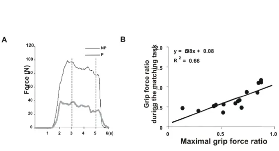
Figure 1 0 20 40 60 80 100 120 NP P 1 2 3 4 5 6(s) F o rc e ( N) y = .98x + 0.080 0 0.5 1.0 1.5 2.0 0 0.5 1.0 G ri p f orc e ra ti o d u ri n g t h e m a tc h in g t a s k 2 = 0.66 R A B
Figure 1: The subject is asked to produce equal grip forces on both sides corresponding to 65% of his maximal grip force on the paretic side. Although the force values clearly differ between sides (as shown in panel A: NP, non-paretic; P, paretic), the subject perceives that he is producing equal forces. The values included in the interval between dotted lines were used to calculate the grip force ratio between the paretic and non-paretic sides, which is approximately 0.38 in this subject. These grip force ratios were calculated in 15 stroke patients and found to be significantly correlated to the maximal voluntary hand grip force ratios (as shown in panel B: regression equation and coefficient of determination (R2)). This suggests that grip force-matching is based on the perception of an
Table 1: Summary of studies describing sensory abilities in children with CP Authors/ Year Sample of children
with CP
Sensory modalities affected
Bolanos et al, 1989 51 CP, 170 controls 6-20 years
63% 2-point discrimination deficits, 9% astereognosis
Cooper et al, 1995 9 CP (hemiplegia), 41 controls
89% sensory deficits bilaterally, steropgnosis and proprioception most affected
Gordon & Duff, 1999
15 CP (hemiplegia), 15 control
4-18 years
Impaired 2-point discrimination, pressure sensitivity and stereognosis compared to controls
Hohman et al, 1958 47 CP 6-16 years
72% deficits in form perception, 2-point discrimination, position sense.
Hemiplegia>quadriplegia>athetoid
Kenny, 1966 73% sensory deficits, spastic>athetoid
Lesny et al, 1993 N=220, 7-14 years Decreased 2-point discrimination compared to controls, especially for diplegia and hemiplegia
McLaughlin et al, 2005
62 CP, 65 controls 3-18 years
Decreased toe position sense, direction of scratch and vibration sense when compared to controls
Monfraix et al, 1961
Tactile agnosia- 81% impaired if spastic, 43% impaired if athetoid
Opila-Lehman et al, 1985
24 CP, 12 controls 8-15 years
Poor kinesthesia compared to controls, spastic form worse than athetoid Tachdjian &
Minear, 1958
96 CP 6-19 years
42% sensory deficits; most common (>10%) modalities: stereognosis (42%), 2-point discrimination (32%), position sense (17%); spastic>athetoid
Tizard et al, 1954 N=106 (hemiplegia) 54% had sensory deficits (stereognosis, 2-point discrimination and position sense most commonly affected)
Van Heest et al, 1993
40 CP (hemiplegia) 97% astereognosis, 90% impaired 2-point discrimination, 46% proprioception deficit
Wigfield, 1966 16-26 years 86% astereognosis for hemiplegic group,
1/11 for athetoid children Wilson and Wilson,
1967a
120 CP, 60 controls 7-21 years
48% impaired sensation: pressure sensitivity and/or 2-point discrimination; similar
deficits in children with spasticity versus athetosis
Wilson and Wilson, 1967b
120 CP, 60 controls 7-21 years
Haptic form discrimination deficit in 31% with spasticity versus 30% with athetosis; size discrimination deficit in 18% with spasticity and 11% with athetosis
Yekutiel et al, 1994 N=55, 6-17 years 51% had deficits in stereognosis and/or 2-point discrimination