Université de Montréal
Ipsi- and Contralateral Corticospinal Influences in Uni- and Bimanual Movements in Humans Par
Laura Duval
Département de Neurosciences, Faculté de Médecine
Mémoire présenté en vue de l’obtention du grade de maîtrise En Neurosciences
Avril 2020
© Laura Duval, 2020
Université de Montréal
Ce mémoire intitulé
Ipsi- and Contralateral Corticospinal Influences in Uni- and Bimanual Movements in Humans Présenté par
Laura Duval
A été évalué(e) par un jury composé des personnes suivantes Daniel Bourbonnais Président-rapporteur Anatol G. Feldman Directeur de recherche Johanne Higgins Membre du jury
Résumé
Il existe des projections corticospinales (CS) vers les motoneurones (MNs) aussi bien contra- (c) qu’ipsilatérales (i). Les influences CSc sur les MNs du poignet sont connues pour être modulées entre autres par la position du poignet et les afférences cutanées. Pour cette raison, notre objectif était de vérifier si ces caractéristiques sont aussi valides pour les influences CSi. En utilisant la stimulation transcrânienne magnétique au niveau du cortex primaire droit, nous avons tout d’abord comparé les influences CSi sur les MNs des fléchisseurs du poignet à des positions maintenues de flexion et d’extension durant une tâche uni-manuelle ainsi que deux tâches bi-manuelles, ceci chez des sujets droitiers (n=23). Nous avons ensuite comparé les influences CSi dans cinq tâches bi-manuelles de tenue d’objet durant lesquelles les sujets avaient à tenir entre leurs mains un bloc à la surface soit lisse, soit rugueuse, dont le poids était supporté ou non, ceci en position de flexion (n=21). Dans une tâche, un poids était ajouté au bloc lisse en condition non supportée pour amplifier les forces de préhension requises. Une modulation position-dépendante était observée au niveau des potentiels évoqués moteurs (iPEM), mais seulement lors de la tâche bi-manuelle quand les deux mains interagissaient via un bloc (p= 0.01). Une modulation basée sur la texture était également présente, quel que soit le support de poids, et le bloc lisse était associé avec des iPEMs plus importants en comparaison avec le bloc rugueux (p= 0.001). Ainsi, les influences CSi sur les MNs n’étaient modulées que lors des tâches bi-manuelles et dépendaient de la manière dont les mains interagissaient. De plus, les afférences cutanées modulaient les influences CSi facilitatrices et pourraient ainsi participer à la prise en main des objets. Il en est conclu que les hémisphères droit et gauche coopèrent durant les tâches bi-manuelles impliquant la tenue d’objet entre les mains, avec la participation potentielle de projections mono-, et poly-synaptiques, transcallosales inclues. La possibilité de la contribution de reflexes cutanés et d’étirement (spinaux et transcorticaux) est discutée sur la base de la notion que tout mouvement découle du contrôle indirect, de la « référence » (referent control). Ces résultats pourraient être essentiels à la compréhension du rôle des interactions inter-hémisphériques chez les sujets sains et cliniques.
Mots-clés: Cortex moteur, contrôle moteur, stimulation magnétique transcrânienne, potentiel
évoqué moteur, influences cortico-spinales, bi-manuel, uni-manuel, controlatéral, ipsilatéral, afférences cutanées.
Abstract
There are both contra- (c) and ipsilateral (i) corticospinal (CS) projections to motoneurons (MNs). There is evidence that cCS influences on wrist MNs are modulated by wrist position and cutaneous afferents. Thus, we aimed to test whether these findings are valid for iCS influences as well. Using transcranial magnetic stimulation applied over the right primary motor cortex, we first compared iCS influences on wrist flexor MNs at actively maintained flexion and extension wrist positions in one uni- and two bimanual tasks in right-handed subjects (n=23). We further compared iCS influences in five bimanual holding tasks in which subjects had to hold a smooth or coarse block between their hands, with or without its weight being supported, in flexion position (n=21). In one task, a weight was added to the unsupported smooth block to increase load forces. A position-dependent modulation of the short-latency motor evoked potential (iMEP) was observed, but only in the bimanual task when the two hands interacted through a block (p=0.01). A texture-dependent modulation was present regardless of the weight supported, and the smooth block was associated with larger iMEPs in comparison to the coarse block (p=0.001). Hence, iCS influences on MNs were modulated only in bimanual tasks and depended on how the two hands interacted. Furthermore, cutaneous afferents modulated facilitatory iCS influences and thus may participate to grip forces scaling and maintaining. It is concluded that the left and right cortices cooperate in bimanual tasks involving holding an object between the hands, with possible participation of mono- and poly-synaptic, including transcallosal projections to MNs. The possible involvement of spinal and trans-cortical stretch and cutaneous reflexes in bimanual tasks when holding an object is discussed based on the notion that indirect, referent control underlies motor actions. Results might be essential for the understanding of the role of intercortical interaction in healthy and neurological subjects.
Keywords: Motor cortex, motor control, transcranial magnetic stimulation, motor evoked
potential, corticospinal influences, bimanual, unimanual, contralateral, ipsilateral, cutaneous afferents.
Table of content
RÉSUMÉ ... 3 ABSTRACT ... 5 TABLE OF CONTENT ... 7 LIST OF TABLES ... 10 LIST OF FIGURES ... 11 LIST OF ABBREVIATIONS ... 12 APPRECIATION ... 15 INTRODUCTION ... 16 1. MOTOR SYSTEMS ... 16 1.1 Cortical level ... 18 1.2 Spinal Level ... 20 1.3 Muscle Contraction ... 211.4 Choosing Between Different Frameworks of Motor Control ... 23
2. IMPORTANCE OF THE CUTANEOUS AFFERENTS IN MOTOR CONTROL ... 24
2.1 Somatosensory Systems ... 24
2.2 From the Somatosensory Cortex to M1 ... 25
2.3 Bimanual Holding Tasks ... 27
3. IPSILATERAL CORTICOSPINAL INFLUENCES ... 27
3.1 Potential Pathways ... 28
3.2 Transcranial Magnetic Stimulation ... 28
3.3 Roles of iCS Influences in Uni- and Bimanual Movements ... 35
3.4 Clinical Relevance ... 37
4. PROBLEMATIC AND GOALS ... 38
METHODS ... 41
1. PARTICIPATION OF ICSPATHWAYS IN BIMANUAL WRIST MOVEMENTS IN HUMANS. ... 42
1.1 Participants ... 42
1.2 Experimental Procedures ... 42
1.3 TMS ... 44
1.5 Data Analysis ... 45
1.6 Statistical Analysis ... 46
2. EFFECT OF TEXTURE AND WEIGHT ON ICSPATHWAYS IN BIMANUAL WRIST MOVEMENTS IN HUMANS. ... 47
2.1 Subjects ... 47 2.2 Experimental Procedures ... 47 2.3 TMS ... 48 2.4 Data Recording ... 49 2.5 Data Analysis ... 49 2.6 Statistical Analysis ... 51 RESULTS ... 53
1. PARTICIPATION OF ICSPATHWAYS IN BIMANUAL WRIST MOVEMENTS IN HUMANS. ... 53
1.1 Characteristics of cMEP, iMEP, iSP and iRB ... 53
1.2 iCS Influences in Unimanual Task 1 ... 53
1.3 iCS Influences in Bimanual Tasks 2 and 3 ... 54
2. EFFECT OF TEXTURE AND WEIGHT ON ICSPATHWAYS IN BIMANUAL WRIST MOVEMENTS IN HUMANS. ... 56
2.1 Characteristics of cMEP and iMEP ... 56
2.2 Effect of Texture on iCS Influences ... 57
2.3 Effect of Support on iCS Influences ... 60
DISCUSSION ... 61
1. BASIC FINDINGS ... 61
2. ICSMODULATION IN UNIMANUAL TASKS ... 62
3. ICSMODULATION IN BIMANUAL TASKS ... 62
3.1 Task-Dependent Modulation of iCS Influences ... 62
3.2 Possible Neural Pathways Underlying Modulation of iCS Influences ... 65
4. ICSMODULATION BY CUTANEOUS AFFERENTS ... 66
4.1 Effects of Texture ... 67 4.2 Effects of Friction ... 68 5. CLINICAL RELEVANCE ... 69 6. LIMITATIONS ... 69 6.1 Group Composition ... 70 6.2 Probing of iSP ... 70 6.3 EMG Noise ... 70
6.4 Comparison with cMEPs ... 70
7. FUTURES DIRECTIONS ... 71
7.1 Hemispherical Asymmetry ... 71
7.2 Cutaneous Afferent During Unimanual and Dynamic Tasks ... 71
7.3 Effect of Weight on iCS ... 71
7.4 Other Components of the TMS Response ... 72
7.5 Premotor and Supplementary Areas ... 72
CONCLUSION ... 73
BIBLIOGRAPHY ... 74
TABLES ... 89
SUPPLEMENTARY FIGURE ... 91
List of tables
Tableau 1. – Characteristics of Components of TMS Responses in Experiment 1. ... 89 Tableau 2. – Characteristics of Components of TMS Responses in Experiment 2. ... 90
List of figures
Figure 1. – Scope of Review. ... 17
Figure 2. – Overview of the Neural Circuits Involved in Motor Control. ... 17
Figure 3. – Schematic Representation of the Muscle Microstructure. ... 22
Figure 4. – EMG Response to TMS Over the Right M1 in Wrist Muscles. ... 29
Figure 5. – Subjects Positions in Uni- and Bimanual Tasks. ... 42
Figure 6. – Example of TMS Responses in Left and Right FCR. ... 46
Figure 7. – Subjects Position in Bimanual Holding Tasks. ... 48
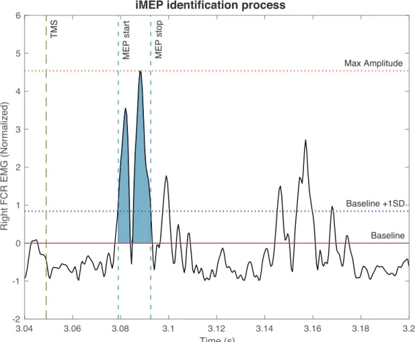
Figure 8. – Example of Selected iMEP. ... 50
Figure 9. – Effect of Wrist Position in Unimanual Task 1. ... 54
Figure 10. – Effect of Wrist Position in Bimanual Task 2. ... 55
Figure 11. – Effect of Wrist Position in Bimanual Task 3. ... 55
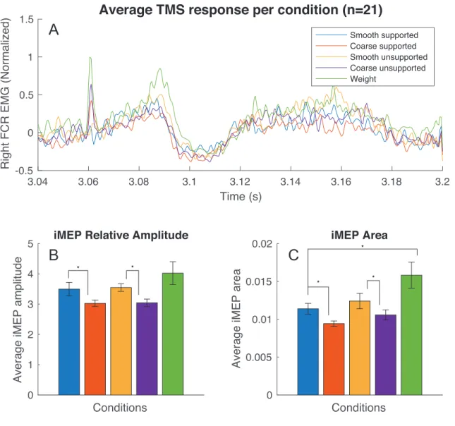
Figure 12. – Overall Results From Tasks 1, 2, 3, 4 and 5. ... 57
Figure 13. – Effect of Texture in Supported Conditions. ... 58
Figure 14. – Effects of Texture and Friction in Unsupported Conditions. ... 58
Figure 15. – Effects of Texture in Supported and Unsupported Conditions. ... 59
Figure 16. – Effects of Support and Weight on the Smooth Block. ... 60
Figure 17. – Referent Control of Wrist Positions in the Bimanual Tasks. ... 64
List of abbreviations
c: contralateral, 16 CC: corpus callosum, 28 CNS: Central Nervous System, 16
CRIR: Center for Interdisciplinary Research in Rehabilitation, 42 CS: corticospinal, 16
CST: corticospinal tract, 18 EMG: electromyography, 23 FA1: Meissner corpuscles, 24 FA2: Pacinian corpuscles, 24 FCR: flexor carpi radialis, 43 FR: frames of reference, 23 i: ipsilateral, 16
IHF: interhemispheric facilitation, 33 IHI: interhemispheric inhibition, 33
IRGLM: Institut de réadaptation Gingras-Lindsay-de-Montréal, 41 ISI: interstimulus intervals, 33
M1: primary motor cortex, 18 MEP: motor evoked potential, 28 MMs: Mirror movements, 35 MN: motor neuron, 18
MVC: maximal voluntary contraction, 43 PMC: premotor areas, 18
PNS: Peripheral Nervous System, 16 Q: actual hand aperture, 63 R: referent aperture, 63 RB: rebound, 28
S1: primary somatosensory cortex, 25 S2: secondary somatosensory cortex, 25 SA1: Merkel cells, 24
SA2: Ruffini endings, 24
SMA: supplementary motor area, 18 SP: silent period, 28
TMS: transcranial magnetic stimulation, 27 vCST: uncrossed CST, 28
À mes parents, Claire et Jean-Christophe, qui m’ont toujours encouragé à relever les défis et tenter ma chance. À ma grand-mère, Mamou Jocelyne (et le fabuleux Pepito) qui me soutient toujours mais aussi aux absents qui nous manquent, Mamie Suzanne, Papi Georges et Papou Jean-Daniel.
To my parents, Claire and Jean-Christophe, who have always encouraged me to undertake challenges. To my grandmother, Mamou Jocelyne (and the all mighty Pepito) who is still supporting me but also the absents who we miss grandly, Mamie Suzanne, Papi Georges, and Papou Jean-Daniel.
Appreciation
The first person I want to thank for this enriching year is, without any hesitation, my supervisor Anatol Feldman. Not only was he a good mentor, but he also helped me find my path in this broad research field. I learned a lot about neurosciences those past two years, but most importantly, that creativity is key even in the most down to earth situations. Anatol forced me out of my comfort zone with congresses and presentations but always stayed supportive, kind and understanding.
I also want to thank my “parrains” from the university, Numa Dancause and Trevor Drew as well as Dorothy Barthelemy for their support, advice and the help they offered me during my master. I could not write an appreciation part without mentioning Mindy Levin, who took some of her precious time to help me get ready for my first oral presentation at the Progress in Motor Control congress.
Those past two years have been indisputably difficult, sometimes even nerves wracking, but I was lucky enough to be surrounded with other lab members who quickly became my friends. Thank you, Lei, Fariba, Anne-Sophie, Yuqi, Camilla, Melanie, Emre, Roni, Sandra, Daniele, Rejean and Ali for all the lovely dinners and times spent together.
I can now say a few words for my Mum and Dad, who I will never be able to repay for the constant support, the help, the encouragement and the love they gave me. Hopefully, this master and the past six years I spent at university will bring me a step closer to becoming as intelligent and impressive as you. I love you both dearly.
A warm thank you to my friends Alise, Théo, Angel, Antoine, Lucas, Aymeric, and Mohamed, for helping me survive both the university and the deadly Montreal winter. A special note to Manuel and Charley who were kind enough to read me and provided valuable feedback.
Finally, I want to thank my love, Florian, the perfectionistic and hard-working engineer who always encouraged me to question my findings and supported me during both the highs and lows. I love you Boo.
Introduction
The ability to move is a key feature of our life. By activating different muscles, we can execute a broad variety of movements. We use the terms afferent and efferent systems to describe the information that reaches the Central Nervous System (CNS) and the information resulting in action production, respectively. The CNS, consisting of the brain and spinal cord, is responsible for integrating afferent inputs and influencing the Peripheral Nervous System (PNS) which connects the CNS to limbs and internal organs. Studying the motor system is fundamental for getting a better understanding of the brain’s functional properties and, in particular, motor control disorders to design effective rehabilitation paradigms.
Before diving into the specific topics of my research – the role of ipsi- (i) and contralateral (c) corticospinal (CS) influences in uni- and bimanual movements in humans, we will briefly describe both motor (section 1) and sensory (section 2) systems. The subjects that will be tackled in this introduction are summarized in the following schematic scope of review (Fig. 1).
1. Motor Systems
Humans interact with the environment, in particular, through movement production. Those can be reflex reactions to environmental stimuli but also volitional and automatic actions.
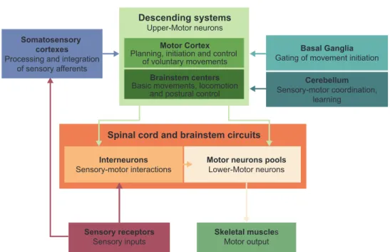
Multiple brain areas come into play and are responsible for different stages of these essential processes, from the perception of stimuli to motor responses. As a result, several neural circuits are involved in what we call movement control (Fig. 2). To begin, there are descending systems originated from the motor cortex as well as from brainstem centers (1). They participate, among other things, in planning, initiating and controlling of fine movements (2). In addition, there are subcortical nuclei gathered in a structure, called the basal ganglia, which can be considered as a gate for movement initiation. The cerebellum coordinates different movements and learning (for reviews see (3), (4)). Finally, there are the spinal cord and brainstem circuits that receive sensory information and innervate skeletal muscles.
Figure 1. – Scope of Review.
Figure 2. – Overview of the Neural Circuits Involved in Motor Control.
Motor Sensory
Cutaneous impact on motor control Sensorimotor interactions Mechanisms and uses
TMS responses Other TMS paradigms Caveats
Motor cortex Brainstem
Ipsilateral corticospinal (iCS) influences iCS in unimanual and bimanual movements
Descending pathways Spinal cord composition Lower motor neurons Sensorimotor reflexes Ipsilateral pathways
Muscle contraction
Electromyography Sensory transduction
Somatosensory cortex Sensory integration Sensory projections to M1
Cortical Level
Encoding and transmission of sensory information
Cortical Level
Transcranial Magnetic Stimulation Sensorimotor Integration
Spinal Level Spinal Level
Muscles Level Mechanoreceptors Level
Descending systems Upper-Motor neurons
Motor Cortex
Planning, initiation and control of voluntary movements
Brainstem centers
Basic movements, locomotion and postural control
Basal Ganglia
Gating of movement initiation
Cerebellum
Sensory-motor coordination, learning
Somatosensory cortexes
Processing and integration of sensory afferents
Spinal cord and brainstem circuits
Interneurons
Sensory-motor interactions Motor neurons poolsLower-Motor neurons
Skeletal muscles
Motor output
Sensory receptors
1.1 Cortical level
Neurons that transmit output signals from higher centers in the frontal lobe and the brainstem to descending systems are usually called the upper motor neurons (MN). Their role is to modulate the activity of interneurons and lower MNs to mediate the contraction or relaxation of skeletal muscles.
1.1.1 Motor Cortex
Among the higher centers, the motor cortex is presumably responsible for the planning and execution of volitional movements. It is constituted of the primary motor cortex (M1), the six premotor areas (PMC) and the supplementary motor area (SMA).
The M1, also called Brodmann area 4, is an anatomical region of the brain located in the dorsal part of the frontal lobe, on the anterior bank of the central sulcus (5). Its location and function have been widely investigated by neuroscientists such as Penfield (6). Although his initial goal was to assess which brain regions were vital and should not be removed during surgery in epileptic patients, he found out that stimulations of this area led to highly localized muscle contractions of the contralateral side of the body (6). Furthermore, he discovered that M1 is organized in a somatotopic manner representing motor maps. Even though results of later studies are generally consistent with the idea of motor maps, refined analyses reveal a more distributed, gross and overlapping pattern of subdivisions in M1 (7). A critical aspect of this motor map also called the motor homunculus, is that larger areas are allocated for body parts that are used in more complex tasks, like those involving the hands and face (for review see (8)). Moreover, studies have shown that area size varies through plasticity, meaning that M1 can reorganize itself based on experience (7).
Composed of Betz cells, which are pyramidal cells located in its fifth layer (5), M1 sends axons through the internal capsule to several subcortical structures, creating different pathways in charge of specific motor functions.
There are two major pathways that innervate both the body and face muscles. The corticospinal tract (CST) fibers go from M1 to the spinal cord and synapse onto lower MNs, which innervate skeletal muscles. In this tract, around 90% of pyramidal fibers decussate in the medulla and
descend contralaterally in the spinal cord to form the dorsolateral CST. A remaining 10% descends ipsilaterally and forms the ventral CST (9–11). Meanwhile, the corticobulbar tract fibers go to the medullary pyramids in the brainstem to synapse onto lower MNs via cranial nerves.
In addition, two pathways link the motor cortex to both the basal ganglia and the cerebellum. The corticostriatal fibers descend in the striatum of the basal ganglia, creating a corticostriatal loop while the corticopontical fibers first travel to the pontine nuclei and then project onto the cerebellum.
Finally, two other pathways descend in the spinal cord; (i) the corticorubral fibers, that go to the red nucleus and form the rubrospinal tract and (ii) the corticoreticular fibers that go to the reticular formation of the brainstem to form the reticulospinal tract.
As mentioned, two other brain regions are part of the motor cortex. Situated in the frontal lobe anterior to M1, the PMC is constituted of six spatially separate areas (12) and receives both multisensory inputs from the superior and inferior parietal lobes as well as motivation and intention signals from the prefrontal divisions of the frontal lobe (2,13). It can influence motor control, either indirectly through its reciprocal projections on M1, or directly via axons projecting to the corticobulbar and corticospinal pathways (12). Studies suggest that it uses information from other cortical areas to plan and select context-appropriate movements (for review see (14)). Finally, the SMA is located in the dorsomedial frontal cortex (for review see (15)). Though its overall function remains unclear, there is growing evidence that the SMA may influence the planning of sequential movements, movement initiation as well as interlimb coordination (16– 19).
1.1.2 Brainstem
In addition to the motor cortex, multiple subcortical structures in the brainstem also play a role in motor tasks such as locomotion, postural control, balance and orientation of head and eye movements. They are controlled by neurons from the reticular formation, the vestibular complex, and the superior colliculus, respectively.
1.2 Spinal Level
The spinal cord’s role is multifaceted, ranging from providing efferent information to the autonomic nervous system to coordinating reflexes and muscle contraction. Overall, the spinal cord transmits and modulates nerve signals originated in the cortex and brainstem to muscles and efferent sensory information to higher centers.
1.2.1 Spinal Cord Composition
Similar to the brain, the spinal cord is composed of grey and white matter. On one hand, the grey matter is divided into the ventral and dorsal horns which contain MNs and sensory neurons respectively as well as an intermediate zone containing interneurons. It is further subdivided into areas called laminae ranging from I to X (19). The majority of upper MNs project either directly on alpha MNs (α-MNs) in lamina IX or indirectly via interneurons in laminae V-VIII (20). CST influences are excitatory for MNs and their inhibition is mediated by inhibitory interneurons(19). On the other hand, the white matter consists of different, distinct although overlapping, descending and ascending axon bundles. Descending systems are thought to be organized in a somatotopic fashion such that tracts implicated in balance and posture are clustered more medially while the ones involved in more distal movements terminate laterally (2,19,21).
1.2.2 Lower Motor Neurons
As defined before, neurons transmitting signals from the cortex and brainstem centers are called upper MNs whereas MNs that innervate skeletal muscle fibers are called lower MNs. They are classified into three categories. Firstly, α-MNs innervate extrafusal muscle fibers and are responsible for muscular contraction. Second, γ-MNs innervate intrafusal fibers within muscle spindles, sensors informing about muscle length, and modify their sensitivity to muscle stretching and its speed (22). Finally, β-MNs innervate both types of fibers (19,22).
A α-MN together with all the muscle fibers it innervates is called a motor unit (23). As a general rule, the majority of muscle fibers are innervated by only one α-MN. In contrast, α-MNs often innervate multiple fibers (19). All α-MNs that innervate a single muscle are called a MN pool (22). In the spinal cord, MN pools clusters are located according to the muscles they innervate, medially
to the ventral horn for the axial and proximal musculature and laterally for the distal musculature (2).
1.2.3 Sensorimotor Reflexes
For any given movement, we classify muscles along two categories: agonists and antagonists. The agonist is the muscle that generates movement with its contraction, causing a shortening of myofibrils. On the other hand, the antagonist is a muscle that is being stretched by the agonist’s contraction.
In addition to the cortical control of muscles, the spinal cord itself is responsible for several sensorimotor reflexes aiming to maintain muscle tonus and force. They can involve both pre- and postsynaptic monosynaptic and polysynaptic connections. For instance, when a muscle is stretched, muscle spindles are activated. Sensitive fibres Ia and II afferents that are coiled around them relay sensory information to the α-MN. To do so, they either make direct, excitatory contact to the agonist muscle’s α-MN or indirectly, via inhibitory interneurons synapsing on α-MNs of the antagonist muscle. As a result, a simultaneous contraction of the agonist muscle and relaxation of the antagonist emerges. This reciprocal innervation contributes to maintaining muscular tonus. Sensorimotor reflexes can also be transcortical, and we can use the response latencies to identify whether they are mediated spinally or supraspinally.
In the same way, Golgi tendon organs (for review see (24)), which are encapsulated afferent nerve endings located at the junction between a contractile fiber and a muscle tendon, are innervated by group Ib afferents. Their role is to convey information about muscle tension arising from muscle contraction. By making contact with inhibitory interneurons which in turn synapse onto α-MNs, they reduce their discharge frequency. This inhibitory circuit helps regulate muscle tension and thus maintain muscle force.
1.3 Muscle Contraction
Skeletal muscles are major components of volitional movement. Their role is to convert chemical energy relayed by neurotransmitters into mechanical contractions (19). They are composed of
muscle contractile fibers which are innervated by α-MNs. Their synapse is called the neuromuscular junction.
1.3.1 Mechanism of Contraction
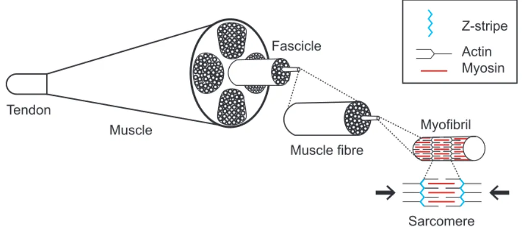
When an action potential reaches the presynaptic terminal of an α-MN, it activates voltage-gated calcium channels, letting calcium ions enter the neuron. In turn, they bind onto sensor proteins found on synaptic vesicles contained in the axon terminal and trigger their fusion with the cell membrane. Those vesicles carry a neurotransmitter, acetylcholine, that can then be released inside the synaptic cleft to bind onto nicotinic acetylcholine receptors situated on the cell membrane of the muscle fiber, the sarcolemma. Depolarization ensues and leads to the generation of a nerve impulse that causes T tubules to release the calcium stored in their sarcoplasmic reticulum. As a result, calcium then diffuses into myofibrils. These units of muscle cells are organized into an alternance of thick and thin filaments, which are divided by Z-stripes into segments called sarcomeres (Fig. 3). When calcium binds onto troponin, a group of proteins that regulate muscle contraction, it exposes the actin-binding sites of the thin filaments which can then bind to the actin from the thick filaments. This process leads to a change in conformation of the actin-myosin configuration such that cross-bridges rotate and pull the thin past the thick filaments. Contraction of the muscle happens when the thick and thin filaments slide past each other, reducing the sarcomere’s length (Fig. 3).
Figure 3. – Schematic Representation of the Muscle Microstructure.
Muscles are composed of bundles of fibers called fascicles in which muscle fibers are gathered. Muscle fibers contain myofibrils that can contract when sarcomeres length is reduced due to the sliding of thick and thin filaments.
Muscle Muscle fibre Tendon Sarcomere Z-stripe Actin Myosin Myofibril Fascicle
1.3.2 Electromyography Recording
The electrical activity associated with muscle contraction, also called myoelectric signals, can be recorded via electromyography (EMG) by placing electrodes either on the skin or directly inside the muscles (25–27).
1.4 Choosing Between Different Frameworks of Motor Control
When it comes to the study of motor control, several theories have been developed over the years aiming to explain the cortical mechanisms involved in movement production. In particular, there are two dominant frameworks or theories of motor control, one biomechanical, based on computational and optimality principles (28,29) and one based on physiological principles. 1.4.1 Internal Model Theory
The internal model theory relies on the idea that neural mechanisms can mimic the input and output characteristics of the motor apparatus (30), allowing the preprogramming of volitional movements.
1.4.2 Equilibrium Point Hypothesis
In direct opposition to this theory is the Equilibrium Point Hypothesis, now advanced to Referent Control Theory of Action and Perception (31,32) developed by Feldman and colleagues in the sixties. It stipulates that instead of controlling movements by directly specifying biomechanical variables, the CS influences sets the spatial threshold position at which MNs of wrist muscles begin to be recruited (33–36). Depending on external conditions, changes in the spatial thresholds result in either a movement to another wrist position or an isometric force production (31). Threshold positions can be considered as the origins, or referent points of the spatial frames of reference (FRs) in which MNs and reflexes are constrained to function. Intentional motor actions emerge, without preprogramming, from shifts in the referent points of spatial FRs, as suggested in the empirically established framework of indirect, referent control of motor actions (31,32). Experiments in this master’s thesis are designed in this theoretical framework.
2. Importance of the Cutaneous Afferents in Motor Control
2.1 Somatosensory Systems
The somatosensory system plays an essential role by transmitting inputs, in particular about physical properties of objects the body interacts with in its environment. It integrates information about texture, hardness, weight, position and global shape of objects (37) as well as temperature and pain, though the latter two senses will not be discussed here.
2.1.1 Transduction
For a sensation to appear, a physical stimulus needs to be transduced. That means that the sensory stimulus needs to be turned into an electrical signal that will then be sent to the spinal cord and appropriate areas of the somatosensory cortex for integration.
Both touch and proprioception are mediated by mechanoreceptors. Cutaneous mechanoreceptors provide information about the physical properties of the surfaces and objects we encounter (38,39) such as their textures, shapes, friction, and weights (40).
There are two categories of cutaneous tactile receptors located either in the skin or in deep tissues: the first is fast adapting, responding to changes in stimulation (phasic, dynamic) and the second is slow adapting, responding to maintained stimulation (tonic). The fast adapting mechanoreceptors are the Meissner corpuscles (FA1) which detect pressure, and the Pacinian corpuscles (FA2) that detect deep pressure and vibrations. Finally, the two slow adapting mechanoreceptors are the Merkel cells (SA1) which detect static pressure but also texture, and the Ruffini endings (SA2) which inform about skin stretches and hand postures. Superficial receptors are present in higher density in the palms and fingers in contrast with the deeper receptors which are distributed more sparsely.
Johansson and Westling (39,41) have proposed that rapidly adapting receptors FA1 and FA2 as well as slowly adapting receptor SA1 were responsible for detecting slips, thus, to maintain grip forces during grasping tasks. They also suggested that slowly adapting receptors SA2 would play a role in friction sensing.
Proprioception, on the other hand, transmits information about body position and displacement of body segments via the muscle spindles and Golgi tendons.
2.1.2 Encoding and Transmission of Sensory Information
Cutaneous afferents travel through the dorsal column medial lemniscal system. The nervous system then recognizes the stimulus’ modality (i.e. touch, pain, temperature), location, intensity and duration in the somatosensory cortex, where it is treated and integrated.
2.1.3 Integration
The primary somatosensory cortex (S1) is located posterior to the central sulcus. It is composed of four subdivisions and receives somatotopic inputs from the thalamus (ventro-posterior-lateral and ventro-posterior-medial). Areas 3a and 3b receive proprioceptive and cutaneous inputs respectively and further processing is realized in areas 1 and 2 (19). The secondary somatosensory cortex (S2) is situated in the parietal operculum (42) and receives connections from S1. Although its function is not completely understood, it is thought to accomplish sensorimotor integration and may transmit cutaneous signals to the motor cortex (43).
It is generally agreed that cutaneous mechanoreceptors send their information to the contralateral S1 (44). However, it has been shown that tactile information from one hand can reach S1 of both hemispheres (45). As no uncrossed tactile projections from distal limbs have been demonstrated, this transmission is likely to be transcallosal (45). On the other hand, S2 receives cutaneous inputs from S1 as well as from cutaneous receptors of both hands. It has been repeatedly observed that unilateral electrical nerve stimulation of a limb leads to the bilateral activation of S2, especially, but not exclusively, in proximal muscles (44,46,47). Therefore, it appears that sensory afferents have the means to be treated by the somatosensory cortexes not only contralaterally but also ipsilaterally.
2.2 From the Somatosensory Cortex to M1
2.2.1 Projections from the Somatosensory Cortex to M1
It has been generally accepted that there are projections from S1 (3a, 3b, 1, 2) and S2 to the motor and premotor cortexes (8,46). However, S1 fields participating at an early stage of processing, are
thought to send only modest inputs to M1 (8). Despite those projections, removal or cooling of the sensory cortex in monkeys appeared to have no impact on the evoked potentials recorded in the motor cortex after nerve stimulation (48). In other words, M1 may receive peripheral input independently of the somatosensory cortex, potentially from the thalamus (48). In addition, the large majority of inputs it receives seems to arise from area 5 in the superior parietal lobule (49– 51) and to a lesser extent from area 7b. These regions have connections to both S1 and S2 and are thought to be involved in somatosensory and associative processing.
2.2.2 Involvement in Motor Control
All in all, the role of S1 and S2 in motor control remains unclear. There is only contentious evidence that sectioning the dorsal column leads to loss of somesthetic input and results in motor impairment, especially in grasping and holding tasks (52). Likewise, lesions in S1 have been reported to disturb motor activity in some studies whereas others observed only slight motor impairment (53,54). On the other hand, there is some evidence that electrical stimulation of S1 may induce movement, though those findings are still controversial (54).
2.2.3 Studying the Impact of Cutaneous Afferents on Cortical Excitability
Study of cutaneous afferent and their impact on cortical excitability has widely employed electrical nerve stimulation. However, results are controversial with evidence of both facilitatory (55,56) and inhibitory (57,58) effects. It has also been suggested that the electrical stimulation of peripheral afferent might excite some circuits and inhibit others (59).
Another method of investigation relies on “natural” stimulation such as skin brushing (60) and tactile exploration of surfaces (61). Again, results are debated, and researchers have observed both facilitation and inhibition (60). The existence of a topographical organization of facilitatory and inhibitory afferents has been suggested and results by Classen et al. (62) were in line with those findings, confirming that they may also facilitate one muscle while inhibiting others. All things considered, it is still controversial whether peripheral inputs have an excitatory (39,60,63) or inhibitory (57,59,60) effect on corticomotor excitability. Additionally, cutaneous afferent also appears to be modulated by other criteria such as task (62), complexity (64) and attention (64,65).
2.3 Bimanual Holding Tasks
To ensure grasp stability, people have to apply grip forces perpendicular to the object’s surface (38,66). They use information about friction (61) and the object’s weight (39) to scale grasping forces adequately. Indeed, it is necessary to apply a force within a small safety margin above the minimal force required to prevent slipping, but not too much as it would lead to muscle fatigue and, in some cases, object damage (66).
2.3.1 Sensorimotor Interactions
Repetitive transcranial magnetic stimulation (TMS) applied to M1 can perturb the ratio of grip to load force (38,67), suggesting that the motor cortex plays an important role in the control of grasping (40). Lesion studies have also demonstrated that S1 and S2 may be involved in the adaptation of grip force to changes in object texture and load (19). In addition, anesthesia of the fingers, which inhibits cutaneous perception, has been shown to prevent people from adequately adjusting grasp forces within the safety margin (39). However, Westling and Johansson (66) showed that this was only the case for friction and not for weight, suggesting the existence of two different mechanisms of grip force control during grasping.
In summary, those experiments shed light on the importance of interactions between the somatosensory and motor cortexes in the production of adequate, functional motor actions. Although precise pathways involved in such interactions have not been established yet (8), research also suggests that somatosensory feedback may play an important role in the interhemispheric processing and integration of sensory input during cooperative hand tasks (68).
3. Ipsilateral Corticospinal Influences
Most studies have focused on the role of the contralateral hemisphere in the control of movement and its modulation by cutaneous influences. However, there is evidence that the ipsilateral hemisphere both receives ipsilateral sensory information and is involved in motor functions. We termed this cortical output from M1 to ipsilateral MNs ipsilateral corticospinal (iCS) influences. These influences can be facilitatory or/and inhibitory affecting components of motor
evoked potentials (iMEP), rebound (iRB), and silent period (iSP). iCS influences are likely involving both interhemispheric and descending projections to limb muscles (69).
3.1 Potential Pathways
Not to be confused with the CST, iCS influences can be carried by different pathways depending on the location of the target muscle. A first candidate is a direct monosynaptic pathway constituted by the uncrossed CST (vCST) that descends ipsilaterally from M1 to the spinal cord (10,70,71). A study by Wassermann et al. (72) showed that more proximal muscles such as the deltoids are likely to rely on such pathways. Secondly, the brainstem reticular formation receives numerous projections from both the ipsilateral and contralateral motor cortices (73). Thus, the reticulospinal tract and alternatively propriospinal neurons (74) could represent indirect pathways carrying iCS influences output. This is likely to be the case for iMEPs recorded in more distal muscles. Although some studies (10,70) argue that anatomical evidence in humans is missing, researches made on stroke and spinal cord injury patients (69,75,76) shed light on the capacity of the reticulospinal tract to take over in case of lesions. Finally, iCS influences output could be mediated transcallosally through the corpus callosum (CC) (10).
The question of which pathways mediate each component of iCS influences is still a matter of controversy and interpretation of iCS components remains hypothetical. Nonetheless, the latencies of each TMS component can be a good indicator of which pathways might be involved. For instance, the fact that iMEP arises after contralateral MEP (cMEP) implies that it is unlikely that iMEP involves transcallosal pathways (10). The dichotomy in cortical projections to MNs of proximal and distal muscles should also be considered when analyzing the role of ipsi- and contralateral CS effects in movement production.
3.2 Transcranial Magnetic Stimulation
3.2.1 Mechanisms and Uses
Brain stimulation techniques have been around for centuries. The first experiments were rather painful and often applied to the exposed motor cortex (77,78). Nowadays researchers use non-invasive and painless techniques such as transcranial electrical stimulation and, more often, TMS.
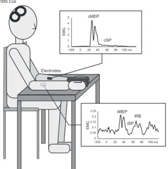
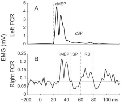
TMS is a tool that allows us to investigate the different neural circuits of the brain and their functions (79). It has been widely used in the literature to assess descending CS influences on MNs. TMS produces a motor-evoked potential (cMEP), a silent period (cSP) followed by a rebound (cRB) in contralateral muscles as well as transient excitatory (iMEP), rebound (iRB) and inhibitory (iSP) phases in ipsilateral muscles (Fig. 4; (80,81)).
TMS uses electrical currents in the coil to induce a magnetic pulse in the cortex. The changes in the magnetic field elicit an electrical current underneath the scalp which modifies neuronal excitability (82,83). Propagated to the spinal MNs, this activity leads to the contraction of a target muscle (84). Thus, TMS is an artificial way to contract specific muscles. This is possible due to the somatotopic organization of M1, such that one can draw a map between different brain stimulation spots and associated target muscles. In addition, by rotating the coil, one can change the orientation of current and stimulate different muscles and brain structures (82,83).
There are several types of coils with different characteristics such as the focus and depth of TMS. Compared to round coils, the figure-eight coils allow a more focal stimulation (85).
Figure 4. – EMG Response to TMS Over the Right M1 in Wrist Muscles.
TMS over the right M1 elicits both facilitation, the cMEP, the cRB (not shown), and inhibition, the cSP, in contralateral muscles. In ipsilateral muscles, the facilitatory phase (iMEP) is followed by an inhibition (iSP) and a secondary facilitatory phase (iRB).
0 1 2 3 4 5 cMEP −200 0 20 40 60 80 100 ms cSP TMS Coil Electrodes −2000 0 20 40 60 80 100 ms 0.05 0.1 0.15 0.2 0.25 iMEP iSP iRB EMG EMG
3.2.2 Ipsilateral Motor Evoked Potentials
MEPs reflect both corticospinal MNs excitability at the time of TMS (77) as well as CST integrity (83). TMS is usually coupled with the execution of specific movements to evaluate their impact on corticospinal excitability. Since MEPs reflect both cortical and spinal MN excitability, it is better to equalize baseline muscle activity at different points of MEP testing to selectively evaluate changes in the cortical excitability during a motor task.
Two important characteristics of MEPs are their amplitude, which is a compound signal of its descending cortico-spinal volleys, and their latency which is the conduction time for the neural impulses triggered in M1 to reach the target muscle. TMS activates several neurons of M1 as well as their axons. The activation of multiple cortico-spinal volleys is responsible for the different components of the MEP. Earliest volley termed D-waves, and later I-waves result respectively from direct and indirect, transsynaptic activation of CST neurons (86,87).
In addition to the TMS coil’s location on the scalp, which defines the target muscle where a response is observed, the coil orientation influences the MEP latency, threshold, and choice of activated cortical or subcortical structures (10,81).
In contralateral muscles, TMS produces cMEP by activating either directly or indirectly transynaptically fibers in the CST (72). The question of whether mechanisms mediating cMEP and iMEP are similar has been debated in the past, however, Chen et al. (81) showed that based on their directional preferences and latencies, this may not be the case. The optimal scalp positions for iMEP and cMEP are also different (72,74,88) but the difference is likely to be minimal as both iMEPs and cMEPS can be observed by stimulating the same spot.
The presence of iMEPs has been debated as some studies (10,89) were unable to reliably observe them in healthy adults. When it was the case, they were elicited mostly in proximal muscles (80,89) only in a small number of subjects (81,90) and required high TMS intensity as well as visible contraction of the target muscle (74). Chen et al. (70) concluded that the ipsilateral projections from M1 to upper limb muscles are weaker than contralateral projections, with a preference for proximal over distal muscles.
A particularity of iMEPs is that their amplitude can be modulated by several elements such as task, muscle contraction and head rotations (10,74,88,91). Tazoe and Perez (88) showed that depending on whether the head was turned medially or laterally, i.e., away or toward the muscle tested, iMEP size was decreased and increased, respectively. As corticoreticulopsinal and corticopropriospinal pathways are under a strong influence of sensory afferents, it has been proposed that this modulation of amplitude would be proof of activation of such tracts (10,74,88). In general, short-latency iMEPs are thought to be mediated by the vCST, corticoreticulospinal or the corticopropriospinal tracts depending on the muscle area stimulated in M1. The idea of a transcortical pathway has been put aside as latencies of iMEPs were maintained in patients with complete agenesis of the CC (74).
3.2.3 Ipsilateral Silent Period
The SP consists of a pause in the ongoing EMG activity after an MEP (87). It is considered to be a measure of interhemispheric inhibition (70,92,93).
After a single pulse suprathreshold TMS, a period of EMG inhibition following the MEP can be observed in EMG activity of contralateral muscles called the cSP (94). The early part of it results from post-spike hyperpolarization of spinal MNs. In contrast, the latter part appears to result from suppression of neuronal output by interneurons at the cortical level (87,94,95). Cracco et al. (96) observed that cortical stimulation excites inhibitory interneurons that project onto pyramidal cells, decreasing the firing of CST neurons. In the ipsilateral muscles, an iSP after an iMEP can also be obtained in both distal and proximal upper limb muscles (70). However, its threshold is lower than that of both cMEPs and iMEPs (72,81).
While trying to determine the origin of iSP, Wassermann et al. (80) found that it may not be mediated by spinal mechanisms since the H-Reflex’s amplitude was not altered during iSPs. Furthermore, similar to iMEPs, iSPs are delayed in comparison with their contralateral counterparts, Wassermann et al. (80) argued that iSP may be mediated by indirect pathways such as the reticulospinal tract instead of CST. Nevertheless, iSP is generally thought to be mediated via transcallosal pathways (87,97) and to reflect the state of intracortical inhibitory systems (95,98). This conclusion is supported by studies of patients with agenesis of the CC or with lesions
suppressing any SP after TMS (87,92). Preschool children who have yet to develop a functionally competent CC also do not display iSP, reinforcing this hypothesis (99). CC connections between hand areas of M1s appear to be sparse but effective in transferring of iCS influences from one hemisphere to the other (97).
Similar to iMEPs, iSPs can be modulated by several stimuli elements such as activation of the contralateral hand. This reflects the possibility of task-specific modulation of inhibitory iCS influences from the M1 (92). The coil orientation can also affect its duration, in the same manner as it is the case for iMEPs (87).
3.2.4 Ipsilateral Rebound
In both contralateral and ipsilateral muscles, a SP is usually followed by a second wave of excitation called the rebound (RB) (100).
The iRB has not been widely investigated and both its origin and function remain unclear (101– 103). Although there have been several proposals in the past, the majority of them are still debated. Nevertheless, a study on patients with multiple sclerosis by Mills et al. (101) suggested than iRBs may involve both central and peripheral components. He proposed multiple possible pathways involving slower conducting fibers from the CST and long-loop reflexes.
A second hypothesis is that iRBs could be produced in response to MEP’s muscle twitch (100,103). However, Rábago et al. (102) argued that if it were the case, iRB latencies would be shorter, making iRBs happening during the iSP. Another critic comes from Holmgren et al. (103) who obtained an RB in the absence of any MEPs, although they made a reservation that a small MEP could be hidden in the background EMG activity. Alternatively, iRBs could be due to recovery from inhibition during iSP (72,102). Finally, it was proposed, although not confirmed, that iRBs could result from a startle reaction elicited by TMS sound (100,102).
3.2.5 Other TMS Paradigms
Different TMS paradigms are available and can be used to investigate intra- and interhemispheric physiological interactions. As indicated in the previous sections, single-pulse TMS can be used to
assess the motor cortex excitability which is likely involving cortico-spinal, intra-cortical and trans-cortical elements (104).
By using paired-pulse TMS applied to the same or both hemispheres, one can “condition” these responses to each TMS pulse (82) to get additional insights into the role of inter- and intrahemispheric circuits and interactions in motor productions. In paired-pulse paradigms, there are two stimuli: a baseline pulse called the test stimulus and a conditioning stimulus. The MEP resulting from a single pulse is then compared to a conditioned one. TMS intensity and interstimulus intervals (ISI) can be varied to observe different types of responses.
Finally, to study the interactions between intracortical circuits, a triple-pulse TMS paradigm with two conditioning stimuli and one test stimulus can be used (84). One can also use continuous, rhythmical TMS to change the state of different brain areas, which is often used in rehabilitation (105).
a. Interhemispheric Interactions
Interhemispheric interactions refer to the interaction between M1 neurons of both hemispheres and rely on the CC integrity. It is generally thought that transcallosal projections are excitatory. They then synapse onto either inhibitory or facilitatory local circuits in the target hemisphere M1 (87). However, this view is mainly based on neuroanatomical data from animals and direct evidence in human are still missing.
Ferbert et al. (97) was a pioneer in their study and demonstrated with paired-pulse TMS the existence of interhemispheric inhibition (IHI) and a poorly reproducible facilitation (IHF). IHI appears to be a very reliable phenomenon, appearing at two specific ISI latencies: short, 8-10 ms, and long, 40ms (81). IHI and SP both reflect interhemispheric inhibition although they are likely to be two different phenomena, at least for the short-latency IHI (81).
IHF was further investigated by Kujirai et al. (106) and Hanajima et al. (107) who debunked the claim that IHF is difficult to observe in Ferbert’s experiments (97). They also highlighted the special conditions required for its appearance. By adjusting the interval between the conditioning
stimulus and the test stimulus, the authors were able to observe IHF modulation more or less consistently.
b. Intracortical Interactions
Intracortical interactions refer to the circuits in each M1 separately. In the same manner as interhemispheric, intracortical circuits can be probed using paired-pulse TMS. Several circuits have been identified as part of the intracortical interactions. First, there is the short interval intracortical inhibition and the intracortical facilitation which are thought to provide insights on the GABAA and NMDAR-dependent system in the motor cortex, respectively (108,109). Secondly, there is a long interval intracortical inhibition reflecting cortical inhibition mediated through the GABAB system (72), and finally, the short interval intracortical facilitation (84,109). Those different intracortical circuits usually interact with each other and their study can provide insights on the GABAergic and glutamatergic pathways’ activity (109).
3.2.6 Caveats
One major caveat of studying MEP is that it reflects both spinal and cortical excitability which means that in theory, they cannot be measured separately (110). Incidentally, a visible increase in MEP size, for instance, could involve spinal mechanisms and skew the interpretations. Thus, some precautions such as equalization techniques need to be taken to dissociate them and ensure that any change properly reflects supraspinal changes.
Another issue comes from the nature of MEP. Its modulation could arise from other non-monosynaptic, e.g. from propriospinal circuits which means that it may not accurately reflect the excitability of the target M1 (110,111).
Finally, not all descending connections involved in movements are excited by TMS with the same strength (111). Indeed, TMS is thought to excite monosynaptic fast and possibly slow conducting fibers preferentially in comparison with polysynaptic slow conducting fibers (111). Thus, TMS may only probe the integrity of a subgroup of descending fibers.
3.3 Roles of iCS Influences in Uni- and Bimanual Movements
3.3.1 Unimanual Movements
In unimanual movements, iCS influences are mainly known to play an inhibitory role and participate in the lateralization of movements. In humans, there is a natural tendency to contract the homologous muscle in a symmetrical manner (112). It is even thought that movements of distal limbs are generated bilaterally at the beginning and only later become unilateral when transcallosal inhibition prevails (113). Mirror movements (MMs) are known to require less cortical activation than asymmetrical bimanual and unilateral movements (92,112). As a result, strictly unilateral movements require interhemispheric interactions, IHI, to inhibit the motor output from the homologous M1 contralateral to the non-active hand. Using single-pulse TMS over the ipsilateral M1 during an action with the contralateral hand, the IHI can be measured by the iSP in the non-active, ipsilateral hand (112).
MMs are frequently seen in children who have yet to develop a functional CC and patient with CC agenesis. For this reason, it is believed that the iCS influences coming into play in movement lateralization are mediated through transcallosal pathways.
By using repetitive TMS to disrupt the ipsilateral M1, Chen et al. (70) assumed the potential participation of iCS influences in fine and more complex movements as well as in their planning and coordination. In addition, studies using fMRI also showed activation of iM1 during unimanual tasks (113). In fact, they observed that iMEPs were facilitated when the contralateral hand was at rest but inhibited when the contralateral hand was active.
3.3.2 Bimanual Movements
Bimanual movements refer to a vast variety of actions. However, we can dissociate them depending on if the two effectors are performing different but complementary actions in a common goal (e.g. opening a bottle) or if their motor output is similar but produced in a specific temporal order (e.g. during typing) (114).
The functional role of iCS influences in bimanual movements is less understood than in unimanual movements. In particular, there is a lack of studies investigating the role of iCS influences in
object-oriented and goal-directed bimanual tasks (114). Goal-directed tasks are defined as bimanual tasks in which there is a functional object-oriented goal (114), such as holding tasks in which coordination and symmetrical forces of the two hands are required to carry an object (115). More common are continuous rhythmical, cyclical and oscillatory bimanual tasks (114).
Studies in monkeys have suggested that there are M1 neurons tuned to either bilateral or unimanual arm movements (17,116,117). Likewise, Dietz et al. (18) have observed with fMRI a stronger bilateral activation of S2 during cooperative hand movements in comparison with non-cooperative bimanual tasks. They also noted the presence of a bilateral reflex EMG response to unilateral electrical nerve stimulation (18). This suggests that the ipsilateral hemisphere would likely be involved in the coordination of cooperation hand movements in a task-specific manner (47). Moreover, different M1 neural circuits are likely involved during cooperative and non-cooperative movements (68).
In addition, Cardoso de Oliveira et al. (118) showed a correlation between interhemispheric interactions and the degree of bimanual coupling. They found that symmetric bimanual movements were accompanied by stronger interhemispheric interactions than asymmetric bimanual movements. It suggests that interhemispheric interactions participate in the production of symmetric bimanual movements and are thus involved in limb coordination. This way, interhemispheric coupling may explain the difficulties we face when producing asymmetric movements. This is further supported by studies from split-brain patients whose callosal connections are destroyed and who are able to produce asymmetric bimanual tasks better than healthy subjects (118,119). During bimanual tasks, they also noticed an increase in intrahemispheric interactions, suggesting that both hemispheres participate in bimanual control (118).
Thus, based on findings from inter- and intrahemispheric interactions, one can conclude that both hemispheres are involved in bimanual movements (118) and that M1 of the dominant hemisphere is more important in bimanual coordination (16).
3.4 Clinical Relevance
Despite some evidence that iCS influences on MNs may be important for recovery of motor functions after brain or spinal cord lesions (70,120), their role in movement production in normal and pathological populations has not been fully understood.
On one hand, several authors have raised the possibility that ipsilateral motor pathways might provide a way for the cortical output from the undamaged M1 to reach contralesional limb muscles (70). On the other hand, some authors argue that iCS influences may not actually be responsible for recovery (10,70) and could even prevent rehabilitation in some cases (69). There are several different ways in which iCS pathways could be useful in recovery. It could, for instance, represent a substrate for functional restoration after lesion. Indeed, in the case where the CST originating from the ipsilesional hemisphere is extensively disrupted, iCS could offer the only pathway for descending commands to reach the paretic limb muscles (69). TMS studies could also provide insights about iCS pathways integrity by the presence of iMEPs (70,83). However, this hypothesis remains controversial as some authors have suggested that instead of being biomarkers for recovery, they would instead be indicators of poor motor recovery (10). Several studies have reported that iMEPs obtained in stroke patients were usually rare, small and had longer latencies than in healthy subjects (70,121). It is important to note that iMEPs are already thought to be difficult to elicit in healthy subjects, often requiring a slight voluntary contraction of the target muscle. Patients with severe upper limb deficits may not be able to achieve such contraction, thus increasing the difficulty to trigger iMEPs. Another factor to consider is the TMS intensity used during trials because ipsilateral responses usually require a higher TMS intensity than contralateral ones (121). Strong TMS intensities are generally not used in studies involving post-stroke patients since high-intensity stimulation can spread and activate the contralateral hemisphere (70,121). For this reason, it is difficult to accurately assess the role of iCS pathways in recovery from stroke.
In summary, the way iCS pathways participate in the recovery of motor function after stroke is still unclear. Nevertheless, by investigating how they are involved in voluntary motor behaviors in healthy subjects, we may better understand whether or not they might be useful in
rehabilitation. For example, this could help optimize the intensity and pattern of iTMS during a set of motor tasks, such as using cooperative movements needed during daily living activities as a training paradigm (47,88).
4. Problematic and Goals
Although it is mostly agreed upon that iCS influences are involved in both uni- and bimanual movements, how exactly they participate remains poorly understood. In addition, it is still unclear whether or not they are affected by cutaneous afferent and in which manner.
The motor cortex involvement in threshold position control during wrist movement in humans has been investigated in the past. It has been demonstrated in a previous study using TMS, that cCS were modulated such that cMEPs produced in wrist flexors were larger in flexion than in extension position, even though the tonic EMG activity at these positions was equalized (33). It was concluded that cCS facilitation is able to set and reset the spatial threshold position at which MNs of contralateral wrist muscles begin to be recruited (33–36), thus solving the posture-movement problem described by Von Holst and Mittelstaedt (122). In this paper, Von Holst and Mittelstaedt (121) discussed why self-initiated voluntary movements of a body segment from a stable posture to another are not met with resistance from postural reflexes. The ability to reset the threshold limb position provides a solution to this problem by allowing the nervous system to relay postural reflexes to a new position, converting them from movement-resisting mechanisms to movement-producing instead.
These results raise the question of whether or not the iCS system is also involved in such control. We thus aimed to extend this line of research by focusing on the role of iCS influences in both uni- and bimanual tasks. As iCS influences participation in unimanual and bimanual tasks differs, we investigated whether or not iMEPs elicited by iTMS in wrist flexors are different at different wrist positions. We hypothesized that iCS influences would be more strongly modulated in bimanual tasks than in the unimanual task. In testing this hypothesis, we also considered that bimanual movements often involve manipulation of an object held between the hands, which requires the generation of a bimanual holding force. Therefore, we also addressed the question
of whether or not direct contact of the hands with each other or via an object affects iCS influences in bimanual motor tasks.
Furthermore, cutaneous afferents appear to play a major role in cCS modulation, especially during tasks of holding an object where it participates to grip force scaling and maintaining. Hence, we also aimed to investigate if iCS are modulated in bimanual tasks in which subjects hold different blocks whose surfaces differ in terms of texture and associated friction. We hypothesized that the friction level between the hand and the block’s surface would be a key element in iCS modulation (123).
Methods
To test our hypotheses, two separate experiments were made. Methods for both experiments will be described in more detail subsequently.
In the first experiment, we compared TMS responses during flexion and extension wrist position in a unimanual and two bimanual tasks. Our hypothesis was that iCS influences would be more strongly modulated in bimanual tasks than in unimanual. This work has been the object of an oral presentation at Progress in Motor Control XII in July 2019, and the associated paper was co-written by Lei Zhang* (Institut für Neuroinformatik, Ruhr-Universität Bochum, Germany), Laura
Duval* (Institut de réadaptation Gingras-Lindsay-de-Montréal (IRGLM); Department of
Neuroscience, Université de Montréal, Montreal, Canada), Fariba Hasanbarani (IRGLM; Department of Occupational and Physical Therapy, McGill University, Montreal, Canada), Yuqi Zhu (Faculty of Medicine, Université de Montréal, Montreal, Canada), Xiang Zhang (Faculty of Medicine, Université de Montréal, Montreal, Canada), Dorothy Barthelemy (IRGLM; Ecole de Réadaptation, Université de Montréal, Montreal, Canada), Numa Dancause (Department of Neuroscience, Université de Montréal, Montreal, Canada) and Anatol G. Feldman (IRGLM; Department of Neuroscience, Université de Montréal, Montreal, Canada).
In our second experiment, we compared iMEPs changes in response to variations of block surface texture and friction level in five bimanual holding tasks. We hypothesized that the amount of friction between the hands and the block would play an important role in iCS modulation. This work was carried-out in collaboration with Lei Zhang, Anne-Sophie Lauzé (Department of Neuroscience, Université de Montréal), Yuqi Zhu, Dorothy Barthelemy, Numa Dancause and Anatol G. Feldman.
Protocols used in those two studies were slightly different since a new EMG equipment was implemented in the laboratory.
1. Participation of iCS Pathways in Bimanual Wrist Movements in
Humans.
1.1 Participants
Right-handed healthy participants (n=23, 13 males and 10 females, 26.0 ± 5.5 years old) participated in this study. They had no history of orthopedic or neurological disorders and did not take psychoactive or other drugs that could affect cortical excitability. All participants signed an informed consent form approved by the Center for Interdisciplinary Research in Rehabilitation (CRIR) Ethics Committee in accordance with the 1964 Declaration of Helsinki.
1.2 Experimental Procedures
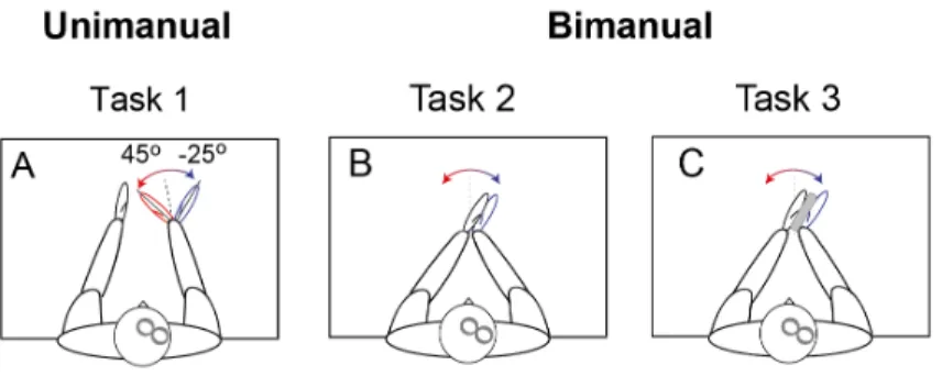
Participants sat in a chair in front of a table (0.7 m height). Both forearms were placed on and attached to the table with Velcro straps in semi-supinated positions (elbow angle about 145°, shoulder horizontal abduction about 45°) (see Fig. 4).
Figure 5. – Subjects Positions in Uni- and Bimanual Tasks.
A: Unimanual task; B, C: Bimanual tasks with hands in direct contact (B) or indirect contact through a block (C).
In the unimanual task (Task 1), the right hand was placed in a hand splint that could be freely rotated about a vertical axis fastened to the table. The wrist flexion-extension axis was aligned with the axis of rotation of the splint (Fig. 1A). Participants (n=16) actively established a right 45° wrist flexion or 25° extension from the neutral position (0°). Each position was indicated by a radial line on the table. Once reached in a self-paced way, each wrist position was maintained. In this task, subjects were instructed to relax the left arm with the wrist in the neutral position and to not move this arm during changes and maintenance of the right wrist angle.