HAL Id: pastel-00769585
https://pastel.archives-ouvertes.fr/pastel-00769585
Submitted on 2 Jan 2013
HAL is a multi-disciplinary open access archive for the deposit and dissemination of sci-entific research documents, whether they are pub-lished or not. The documents may come from teaching and research institutions in France or abroad, or from public or private research centers.
L’archive ouverte pluridisciplinaire HAL, est destinée au dépôt et à la diffusion de documents scientifiques de niveau recherche, publiés ou non, émanant des établissements d’enseignement et de recherche français ou étrangers, des laboratoires publics ou privés.
Central Integration of Dietary Protein Signalling
Jessica Schwarz
To cite this version:
Jessica Schwarz. Central Integration of Dietary Protein Signalling. Food and Nutrition. AgroParis-Tech, 2010. English. �NNT : 2010AGPT0003�. �pastel-00769585�
N° /__/__/__/__/__/__/__/__/__/__/
T H E S I S
submitted to obtain the degree of
Doctor of Philosophy
at
L’Institut des Sciences et Industries du Vivant et de l’Environnement
(AgroParisTech)
Speciality: Nutrition Science
Presented and defended in public by
Jessica SCHWARZ
on 7
thJanuary 2010
CENTRAL INTEGRATION OF DIETARY PROTEIN SIGNALLING
Thesis director: Daniel TOMÉ
Thesis co-directors: Gilles FROMENTIN, Nicolas DARCEL
AgroParisTech, UMR914 Nutrition Physiology and Ingestive Behaviour, F-75005 Paris
to the jury:
Mr. Daniel TOMÉ, Professor, AGROPARISTECH President Mr. Mihai COVASA, Professor, PENNSYLVANIA STATE UNIVERSITY Rapporteur Mr. Thomas LUTZ, Professor, UNIVERSITÄT ZÜRICH Rapporteur Mr. Nicolas DARCEL, PhD, AGROPARISTECH Examiner Mrs. Patricia PARNET, PhD, INRA NANTES Examiner
2
Acknowledgements
At the end of this period, I would like to thank all the people who supported and encouraged me during the past three years of my PhD:
- Professor Daniel Tomé for having given me the great opportunity to conduct my research in his lab and for offering me the occasion to continue working together.
-The members of the jury, Prof. Mihai Covasa and Prof. Thomas Lutz for accepting being rapporteur as well as Patricia Parnet for being examiner of my thesis.
- My supervisors Gilles Fromentin and Nicolas Darcel for their support, guidance and patience in working with a German who does not always function the French way.
- My collaborators at the NOPA in Jouy en Josas, Phillippe Andrey, Jasmine Burguet, Yves Maurin and Olivier Rampin for their copious practical help and numerous scientific discussions to develop my work.
- Professor Jimmy Bell for accepting to supervise my 6-month secondment in his lab and all members of the Metabolic and Molecular Imaging Group at Imperial College London, in particular Jelena and Nad.
- The NuSISCO team, Hannelore Daniel, Alice François, Gary Frost, Luc Ozanne, Harry Peters, Manuela Rist as well as my co-PhDs Boris, Camilla, Charles, Christina, Laure, Magda, Nadine and Véro, for our interesting discussions and support of each other during the last three years.
- All colleagues from the PNCA, especially Sylvette Gougis and my students Anne-Ruth, Justine and Florence for their help as well as my office mates Anne-So, Fanny, Géraldine, Magda, Nattie, Senghua and Wahiba for their endless moral support and good mood.
- Last but not least a warm thank you to my family and friends for their encouragement and support. I especially want to thank the extended ‘coloc’ Antoine, Béné, Ben, Clem, Gim, Julie, Martin, Nico, Patou and Renaud for welcoming me in Paris, for their great company and all the fun I had outside work.
Abstract
Protein is accepted as the nutrient with the most satiating effect. During digestion it acts at different levels. In the stomach it delays gastric emptying and maintains the effect of gastric distension on satiety. In the gut amino acids (AA) and peptides are detected and this leads to an activation of the vagus nerve and the release of gut hormones. The vagus nerve then transmits the signals to the dorsal vagal complex (DVC), responsible for meal termination. Circulating gut peptides and absorbed AA are also detected in the hypothalamus, which is the main structure controlling energy balance.
The exact mechanisms by which dietary protein influences hunger and satiety signalling are not yet fully understood. To gain insight, in this thesis we carried out 3 projects. Firstly, concerning transmission of protein signals by the vagus nerve, our study showed that vagal capsaicin sensitive fibres are not necessary for high protein (HP) diet induced hypophagia, and thus that compensatory mechanisms probably exist which enable adaptation. Secondly we compared the activity of DVC regions in response to a protein or carbohydrate load. Using 3D reconstruction neurones were found in distinct though partially overlapping positions in the NTS. This suggests that short-term protein signalling is specifically transmitted to the DVC. In the third project, we discovered that mice fed a HP diet for 6 weeks had altered body composition but not food intake or body weight. These results suggest an effect of protein on energy metabolism independent from the satiating effect. Further studies may help to understand the role of protein in meal size control, energy balance and body weight regulation.
4
Résumé
Les protéines sont considérées comme le macronutriment avec le pouvoir de satiété le plus fort. Pendant la digestion les protéines agissent à différents niveaux: dans l'estomac, elles retardent la vidange gastrique et prolongent donc la distension gastrique agissant ainsi sur le rassasiement; dans l'intestin des acides aminés (AA) et les peptides sont détectés et entraînent l'activation du nerf vague et la libération de médiateurs gastro-intestinaux. Le nerf vague transmet ces signaux vers le complexe vagal dorsal (DVC), responsable du contrôle de l’ingestion. Les peptides gastro-intestinaux ainsi que les AA absorbés sont également détectés au niveau l'hypothalamus, en charge de la régulation de l'équilibre énergétique.
Les mécanismes exacts par lesquels les protéines alimentaires influencent la faim et la satiété ne sont pas encore compris. Pour mieux comprendre ces phénomènes, nous avons réalisé 3 études qui constituent le présent travail de doctorat. Tout d'abord, concernant la transmission des informations relatives à la présence de protéines via le nerf vague, une première étude a montré que les afférences vagales de la zone hépato-portale ne sont pas nécessaires pour la dépression de la prise alimentaire induite par un régime HP. Deuxièmement, nous avons comparé les motifs d’activité neuronale dans le DVC en réponse à une charge protéique ou glucidique. L’utilisation de techniques de modélisation et de reconstruction 3D a permis de montrer que les motifs d’activité neuronale par la présence de protéines et de glucides sont spatialement distinctes. Dans une troisième étude nous avons testé l’effet des protéines sur le métabolisme énergétique et sur la réponse à une stimulation inhibitrice de la faim, ce dernier paramètre ne semble pas être significativement modulé par une augmentation de l’apport en protéine de la ration chez la souris.
Keywords
Dietary protein, food intake control, satiety, satiation, nutrient sensing, vagus nerve, nucleus tractus solitarius, area postrema, hypothalamus, gut peptides, three-dimensional neuronal activity mapping, magnetic resonance imaging
Mots clés
Protéines alimentaires, régulation de la prise alimentaire, satiété, rassasiement, détection des nutriments, nerf vague, noyau du faisceau solitaire, area postrema, hypothalamus, peptides gastro-intestinaux, cartographie d’activation neuronales tridimensionnelles, imagerie par résonance magnétique
6
Declaration of contributors
Experiments of chapter 2 and 3 of this thesis were conducted at:
UMR914 INRA – AgroParisTech
Nutrition Physiology and Ingestive Behavior AgroParisTech
16 rue Claude Bernard F-75005 Paris
Data of chapter 2 were analysed in collaboration with:
UMR1197 INRA
Neurobiologie de l’Olfaction et de la Prise Alimentaire Domaine de Vilvert
F-78350 Jouy-en-Josas
Experiments of chapter 4 were conducted in collaboration with Nadine Zeeni and data were analysed in collaboration with Nachiket Nadkarni. Experiments and data analysis took place at:
Metabolic and Molecular Imaging Group Clinical Science Centre
Imperial College London Hammersmith Campus Du Cane Road
Table of content
Acknowledgements ... 2 Abstract... 3 Résumé ... 4 Keywords ... 5 Mots clés... 5 Declaration of contributors... 6 Table of content ... 7 List of figures ... 11 List of tables ... 13 Abbreviations ... 14Publications and communications... 17
General Introduction: Obesity, a worldwide epidemic... 19
1 Scientific background... 21
1.1 Appetite, satiety and the control of food intake ... 21
1.2 Peripheral signals involved in protein induced satiety ... 22
1.2.1 Sensory information from lingual detection... 22
1.2.2 Pre-absorptive signals generated in the gastrointestinal tract ... 23
1.2.2.1 The stomach is sensitive to gastric distension ... 23
1.2.2.2 The vagus nerve builds the gut-brain axis... 24
1.2.2.3 Chemoreceptors sense nutrients in the intestine and activate the vagus nerve... 25
1.2.2.4 Peptide hormones act as humoral peripheral signals... 26
1.2.3 Pre-absorptive signals specifically induced by protein ingestion ... 29
1.2.4 Post-absorptive peripheral signals ... 30
1.2.4.1 Direct sensing of dietary protein... 30
1.2.4.2 Glucose-mediated sensing of dietary protein ... 31
1.2.4.3 Protein-induced thermogenesis can influence satiety... 31
Table of content 8
1.3 Central signalling of dietary protein... 35
1.3.1 Short-term regulation of food intake: the dorsal vagal complex controls meal size... 35
1.3.1.1 The blood-brain barrier ... 36
1.3.1.2 The area postrema mainly responding to humoral signals ... 36
1.3.1.3 The NTS receives signals from vagal afferents and the blood stream ... 36
1.3.2 Long-term regulation of energy balance: the hypothalamic area is responsible for maintaining body weight through meal initiation ... 37
1.3.2.1 The arcuate nucleus ... 38
1.3.2.2 The paraventricular nucleus... 39
1.3.2.3 The ventromedial hypothalamus ... 39
1.3.2.4 Central neurotransmitters involved in hypothalamic projections ... 40
1.3.3 Integration of short-term satiation signals and long-term hunger signals in the DVC and the hypothalamus... 41
1.4 Effect of a diet high in protein on body composition ... 42
1.5 Summary and objective of this thesis... 43
2 Hepatic portal vein deafferentation has no effect on the satiating effect of a high protein diet in rats ... 44
2.1 Introduction... 44
2.1.1 High protein diet altering food intake ... 44
2.1.2 Capsaicin for selective deafferentation ... 45
2.1.3 Aims of the study... 46
2.2 Materials and Methods... 47
2.2.1 Materials... 47
2.2.2 Animals ... 47
2.2.3 Surgical procedures for selective hepatic vein deafferentation using capsaicin... 47
2.2.4 Histological verification of selective deafferentation... 48
2.2.4.1 Sampling of portal veins... 48
2.2.4.2 Immunohistochemical analysis of CGRP reactivity ... 48
2.2.5 Diets and feeding procedures... 49
2.2.6 Statistical analysis ... 49
2.3 Results ... 50
2.3.1 Hepatic portal vein deafferentation ... 50
2.4 Discussion... 51
2.4.1 Deafferentation of the portal vein by a capsaicin solution ... 51
2.4.2 The effect of hepatic portal vein deafferentation on food intake suppression induced by a high protein diet ... 51
2.5 Conclusion and perspectives ... 54
2.6 Article: Protein, amino acids, vagus nerve signaling, and the brain. D Tomé, J Schwarz, N Darcel, G Fromentin ... 55
3 Central cartography of macronutrients’ internal sensibility ... 62
3.1 Integration of signals from intestinal nutrient sensing in the dorsal vagal complex ... 62
3.2 Article: Three-dimensional Macronutrient-associated Fos Expression Patterns in the Mouse Brainstem J Schwarz, J Burguet, O Rampin, G Fromentin, P Andrey, D Tomé, Y Maurin, N Darcel. 64 3.3 Additional methods: Calculation of density curves ... 75
3.4 Discussion, conclusion and perspectives ... 76
4 Modulation of body composition and the effect of gut peptides by a high protein diet... 79
4.1 Introduction... 79
4.1.1 Aims of studies ... 80
4.2 Material and Methods ... 81
4.2.1 Materials... 81
4.2.2 Animals ... 81
4.2.3 Effect of a HP diet on body weight and food intake... 81
4.2.4 Effect of a HP diet on body adipose tissue composition ... 82
4.2.4.1 Whole body MRI... 82
4.2.4.2 1H MRS of the whole body, liver and muscle ... 83
4.2.5 Manganese Enhanced MRI of the appetite centres in the brain to measure alteration of the effect of oxyntomodulin by a high protein diet ... 84
4.2.5.1 Image analysis ... 86
4.2.6 Statistical analysis ... 87
4.3 Results ... 88
4.3.1 Effect of a HP diet on body weight and food intake... 88
4.3.2 Effect of a HP diet on body composition ... 89
4.3.3 Alteration of the effect of oxyntomodulin by a high protein diet ... 90
4.4 Discussion... 91
Table of content 10
4.4.2 Effect of a HP diet on body composition ... 92
4.4.3 Alteration of the effect of oxyntomodulin by a high protein diet ... 92
4.5 Conclusion and perspectives ... 94
5 General conclusion and perspectives... 95
6 Annex... 101
6.1 Composition of the experimental diets1... 101
6.2 Used R codes ... 102
6.2.1 Chapter 2 ... 102
6.2.2 Chapter 3 ... 102
6.2.3 Chapter 4 ... 102
6.3 Principle of the magnetic resonance technique ... 103
6.3.1 1H MR Spectroscopy ... 104
6.3.2 Brain activity measured by Manganese enhanced MRI (MEMRI) ... 104
List of figures
Figure 1.1 Neuroanatomical model for pathways involved in peripheral and central satiety signalling.
Figure 1.2 Mechanisms responsible for the protein-induced suppression of food intake.
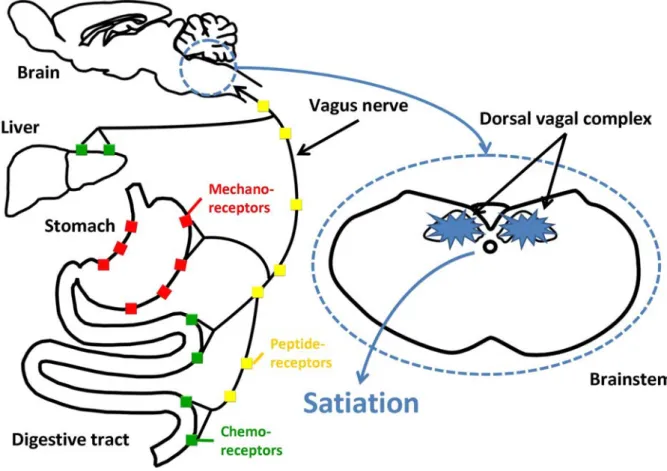
Figure 1.3 The vagus nerve connects the brain and the gastrointestinal tract (GIT).
Figure 1.4 The processing of proglucagon in different tissues of the body.
Figure 1.5 Daily energy intake and body weight of rats.
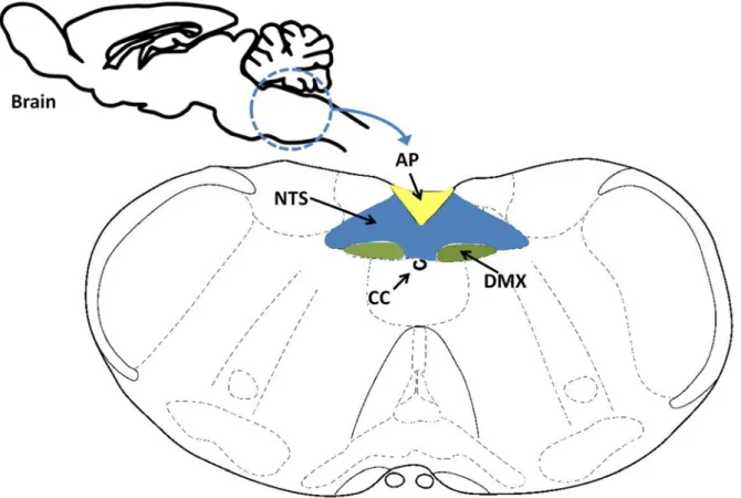
Figure 1.6 Coronal section of the brainstem showing the dorsal vagal complex (DVC).
Figure 1.7 Projection sites of the different vagal branches in the nucleus tractus solitarius (NTS).
Figure 1.8 Coronal section of the hypothalamus summarising hypothalamic nuclei involved in long-term food intake regulation.
Figure 1.9 Hypothalamic projections.
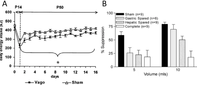
Figure 2.1 Energy intake of vagotomised and sham-operated rats.
Figure 2.2 Location of capsaicin solution application on the isolated hepatic portal vein.
Figure 2.3 Daily feeding patterns of rats fed a high protein diet (HP).
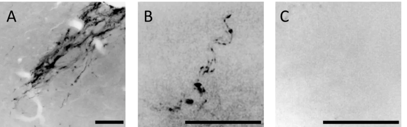
Figure 2.4 Verification of deafferentation of the hepatic portal vein area.
Figure 2.5 Effect of topical capsaicin application on food intake and body weight gain.
Figure 4.1 Set-up of mouse and radio frequency (RF) body coil.
Figure 4.2 Planning of the axial MR images across the body.
Figure 4.3 Localisation of voxel on MR images for 1H MR scan.
Figure 4.4 Example for typical referenced and analysed MR spectra.
Figure 4.5 Set-up of mouse and radio frequency (RF) head coil.
Figure 4.6 Planning of the axial MR images through the brain.
List of figures 12
Figure 4.8 Effect of a high protein diet on body weight and food intake.
Figure 4.9 Body compositions of mice fed a normal protein (NP) or a high protein (HP) diet.
Figure 4.10 Time course of change in signal intensity (SI) as percentage of baseline following i.v. MnCl2 infusion in NP and HP fed mice after injection of 100 µl OXM (1400 nmol/kg) or
saline.
List of tables
Table 1.1 Summary of the release and action of peptide hormones
Table 4.1 Comparison of changes in percentage SI over time by LME following administration of either OXM or saline to mice fed a HP or NP diet.
Abbreviations 14
Abbreviations
3V 3rd ventricle
5-HT 5-hydroxytryptamine / serotonin
AA Amino acids
ACTH Adrenocorticotropic hormone
AgRP Agouti-related protein
α-MSH α-melanocyte-stimulating hormone
AMPK AMP-activated protein kinase
AP Area postrema
ARC Arcuate nucleus
BBB Blood-brain barrier
BDNF Brain-derived neurotrophic factor
CART Cocaine- and amphetamine-regulated transcript
CC Central channel
CCK Cholecystokinin
CCK1-R CCK1 receptor
CGRP Calcitonin gene-gene related peptide
CNS Central nervous system
CRH Corticotropin-releasing hormone
DMX Dorsal motor of vagus
DVC Dorsal vagal complex
FOV Field of view
fsems Fast spin-echo multislice sequence
FT Fourier transformation
GI tract Gastrointestinal tract
GLUT Glutamate transporter
GPCR G protein-coupled receptor
GRPP Glicentin-related pancreatic peptide
HP High protein
IHCL Intrahepatocellular lipid
IMCL Intramyocellular lipid
LHA Lateral hypothalamic area
ME Median eminence
MEMRI Manganese enhanced magnetic resonance imaging
mGluR Metabotrophic glutamate receptor
MPGF Major proglucagon fragment
MRI Magnetic resonance imaging
MRS Magnetic resonance spectroscopy
mTOR Mammalian target of rapamycin
NDS Normal donkey serum
NP Normal protein
NPY Neuropeptide Y
NTS Nucleus tractus solitarius
NuSISCO Nutrient sensing in satiety control and obesity
OXM Oxyntomodulin
PBS Phosphate-buffered saline
PEPT1 Peptide transporter 1
PFA Perifornical area
POMC Pro-opiomelanocortin
PP Pancreatic polypeptide
Abbreviations 16
press Point resolved spectroscopy sequence
PVN Paraventricular nucleus
PYY Peptide tyrosin-tyrosin / peptide YY
RF Radiofrequency
ROI Region of interest
SEM Standard error of mean
sems Spin-echo multislice sequence
SGLT1 Sodium-glucose co-transporter 1
SI Signal intensity
SON Supraoptic nucleus
spuls Single-pulse sequence
SW Spectral width
TE Echo time
TR Repetition time
VMH Ventromedial hypothalamus
VR1 Vanilloid receptor subtype 1
Publications and communications
Publications:
Schwarz, J., Burguet, J., Rampin, O., Fromentin, G., Andrey, P., Tomé, D., Maurin, Y.,
Darcel, N. Three-dimensional Macronutrient-associated Fos Expression Patterns in the Mouse
Brainstem. PLoS One 2010 Feb 1; 5(2): e8974.
Tomé D., Schwarz, J., Darcel N., Fromentin, G. Protein, amino acids, vagus nerve signaling
and the brain. American Journal of Clinical Nutrition 2009 Sep; 90(3): 838S-843S. Epub 2009
Jul 29.
Foltz M., Ansems, P., Schwarz, J., Tasker, M., Lourbakos A., Gerhardt C. Protein Hydrolysates
Induce CCK Release from Enteroendocrine Cells and Act as Partial Agonists of the CCK1 Receptor. Journal of Agriculture and Food Chemistry 2008 Feb 13; 56(3): 837-843. Epub 2008
Jan 23.
Scientific communications:
Schwarz, J., Darcel, N., Rampin, O., Andrey, P., Burguet, J., Fromentin, G., Tomé, D, Maurin,
Y. Nutrient-dependent neuronal activation patterns in the nucleus of the solitary tract: a
study using 3D modelling and neuronal density maps. Poster communication at the
International Congress of Nutrition (ICN) October 2009, Bangkok, Thailand (Abstract published in Annals of Nutrition and Metabolism 2009, 55, Suppl. 1).
Schwarz, J., Darcel, N., Rampin, O., Andrey, P., Burguet, J., Fromentin, G., Tomé, D, Maurin,
Y. Nutrient-dependent neuronal activation patterns in the nucleus of the solitary tract: a
study using 3D modelling and neuronal density maps. Poster presentation for scientist of
Unilever R&D September 2009, Vlaardingen, The Netherlands.
Schwarz, J., Darcel, N., Rampin, O., Andrey, P., Burguet, J., Fromentin, G., Maurin, Y., Tomé,
D. Activation maps in the nucleus of the solitary tract (NTS) in response to internal stimuli. Poster presentation for scientist of Unilever R&D September 2008, Vlaardingen, The Netherlands.
Publications and communications 18
Schwarz, J., Darcel, N., L’Heureux-Bouron, D., Gougis, S., Rampin, O., Tomé, D., Fromentin,
G. Hepatic portal vein deafferentation has no effect on the satiating effect of a HP diet. Oral communication at the Annual meeting of the Society for the Studies of Ingestive Behaviour (SSIB) July 2008 in Paris, France (Abstract published in Appetite 2008; 51 (2): 398).
Schwarz, J., Darcel, N., Rampin, O., Andrey, P., Burguet, J., Fromentin, G., Maurin, Y., Tomé,
D. Activation maps in the nucleus of the solitary tract (NTS) in response to internal stimuli. Poster communication at the Annual meeting of the Society for the Studies of Ingestive Behaviour (SSIB) July 2008 in Paris, France (Abstract published in Appetite 2008; 51 (2): 398).
Schwarz, J., Darcel, N., Rampin, O., Andrey, P., Burguet, J., Fromentin, G., Tomé, D, Maurin,
Y. Activation maps in the nucleus of the solitary tract (NTS) in response to internal stimuli. Poster communication at Experimental Biology April 2008, San Diego, USA (Abstract published in The FASEB Journal 2008; 22: 878.5.).
Schwarz, J., Darcel, N., Rampin, O., Burguet, J., Andrey, P., Tomé, D., Maurin, Y., Fromentin,
G. Central cartography of macronutrients’ internal sensing. Poster presentation for scientist of Unilever R&D September 2007, Vlaardingen, The Netherlands.
General Introduction: Obesity, a worldwide epidemic
This work is funded by the European Community’s Sixth Framework Programme (FP6) under the contract NuSISCO (‘Nutrient Sensing in Satiety Control and Obesity’).
The World Health Organisation (WHO) estimates that over a billion adults worldwide are currently overweight (WHO 2007). In Europe 30-80 % of the population are considered as overweight and up to one third as obese (WHO 2007). In addition to social and professional stigmatisation (Puhl and Brownell 2001), obesity is causally associated with cardiovascular disease, type 2 diabetes mellitus, obstructive sleep apnoea, hypertension, stroke as well as some forms of cancer (Kopelman 2000). In Europe as well as in the rest of the world including nowadays even developing countries, the prevalence of obesity as a major cause of premature death is accelerating dramatically (Ofei 2005). Shortened life expectancy compared to normal weight people can reach from 2-5 years in overweight subjects to 5-20 years in severe obese persons (Fontaine et al. 2003). Obesity is also an increasing economic problem as obesity related diseases cause 2-8% of the total health costs per year for the national health care systems (WHO 2007).
There are two most popular causes for developing obesity, genetic susceptibility and behavioural changes. From the genetic point of view, during evolution it has always been an advantage to be able to develop fat stores in order to survive during nutrient shortage. Nowadays, since food shortage is no longer an issue, exactly this former advantage causes problems as the modern, energy-dense diet leads to an increased energy intake while physical activity, and therefore energy expenditure, is decreasing. In order to keep body weight stable, energy intake must match energy expenditure. The state of positive energy balance caused by overeating and missing compensating energy expenditure is well accepted as one of the main reasons for this epidemic. However, public health initiatives promoting a healthy diet and increased energy expenditure by exercise did not have the desired effect on the population and could not stop the increasing weight.
Wide ranges of medical and behavioural interventions as anti-obesity treatment have already been tried in obese patients, however only a few were successful. Pharmacological compounds often had to be withdrawn due to severe undesired side effects (Farrigan and
General Introduction 20
Pang 2002). At present bariatric surgery is the most successful treatment in strongly obese persons, but also here adverse effects do not make it tolerable for wide-range use (Sjostrom et al. 2004). It is therefore still a huge challenge for the scientific community to search for more effective and better tolerable treatment targets against obesity.
It is commonly accepted that energy homeostasis is under control of a complex signalling system. Many different parameters, involving adiposity signals, whose secretion is proportional to body fat, and satiety signals generated in the gastrointestinal (GI) tract during meals as well as neurotransmitters signalling are processed in a limited number of feeding centres within the central nervous system (CNS) (Woods et al. 2004).
However, there is still a big lack of knowledge in the exact understanding of feeding behaviour and appetite control. The NuSISCO programme covers the whole area of satiety, food intake control and obesity from the molecular and cellular level to human intervention trials. This thesis focuses particularly on understanding the mechanisms of protein-induced satiety signalling to the brain. Therefore it was investigated if sensory vagal afferents in the area of the hepatic portal vein are necessary for protein induced satiety. Food intake and body weight gain of rats were compared that did or did not undergo chemical deafferentation. Moreover, the macronutrient specificity of post-ingestive satiety signalling in the dorsal-vagal complex of mice was studied by comparing density maps of neuronal activity in response to a either protein or carbohydrate load. At last the effect of a high protein diet on the body weight and body composition of mice was studied as well as if the diet can affect the action of the anorexigenic gut peptide oxyntomodulin in hypothalamic areas involved in food intake regulation.
signalling.
Long-term adiposity signals (leptin, insulin) interact with hypothalamic structures responsible for energy balance where they stimulate catabolic and inhibit anabolic pathways. Short-term chemical and mechanical satiety signals from the gastrointestinal (GI) tract and liver are transmitted to the NTS, the satiety centre located in the caudal brainstem. There signals are integrated with descending hypothalamic input which leads to meal termination. ARC, arcuate nucleus; CCK, cholecystokinin; LHA, lateral hypothalamic area; NPY, neuropeptide Y; NTS, nucleus tractus solitarius; PFA, perifornical area; POMC, pro-opiomelanocortin; PVN, paraventricular nucleus (Schwartz et al. 2000).
1 Scientific background 21
1 Scientific background
1.1 Appetite, satiety and the control of food intake
Appetite is the internal driving force for search, choice and ingestion of food in order to maintain energy homeostasis and body weight and therefore responsible for meal initiation (de Graaf et al. 2004). The opposite of appetite is satiation, which is the major determinant of meal size. The meal size is defined by ingestion rate and meal termination. ‘Satiation’ is therefore understood as the physiological process that leads to meal termination (Schwartz et al. 2000). In contrast ‘satiety’ defines the period after a meal before the onset of hunger. Here the word ‘satiety’ will be used for expressing the absence of hunger in general. Even food intake varies widely from day to day, regarded at long-term a fine regulation can be observed.
Hunger and satiety signals from the periphery responsible to maintain energy balance and regulate feeding behaviour can be categorised in pre- and post-ingestive signals and among the latter in pre- and post-absorptive signals. Pre-absorptive signals are derived in the intestine at the moment nutrients enter the GI tract. Post-absorptive signals occur after nutrients crossed the gut wall and enter the circulation. Adiposity signals are also part of the peripheral signals, so do hormones such as insulin and leptin reflect the levels of energy stores and are regulating body weight and ingestive behaviour. On the other hand, satiety as well as meal termination occurs partly due to gastrointestinal satiety signals, like mechanical as well as chemical signals from metabolites and gut hormones (Figure 1.1) (Schwartz et al. 2000).
All these signals are subsequently integrated in the CNS where structures in both the hypothalamus and brainstem are important for the regulation of energy homeostasis as well as the onset of appetite and satiety (Figure 1.1). The negative feedback control of meal size transmitted by gastrointestinal signals takes place in the dorsal vagal complex (DVC) in the brainstem, while hypothalamic nuclei rather process adiposity signals responsible for energy stores (Schwartz et al. 2000). Both centres involved in appetite and food intake regulation are reciprocally connected in order to control energy balance.
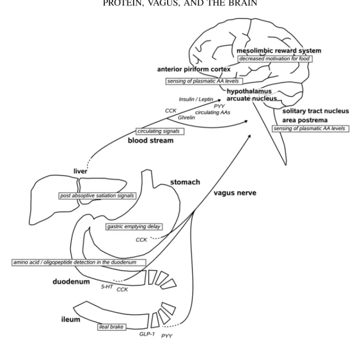
Dietary protein is detected in the gastrointestinal tract. Signals are generated in the stomach, duodenum, and ileum via the release of gut hormones that activate peripheral nerves (particularly the vagus nerve). These gut hormones are also carried by the bloodstream and act directly on brain centres such as the area postrema and hypothalamus to suppress food intake. Post absorptive anorexigenic signals are also generated by the liver. PYY, peptide YY; CCK, cholecystokinin; GLP-1, glucagon-like peptide 1; 5-HT, 5-hydroxytryptamine; AA, amino acid
1 Scientific background 22
1.2 Peripheral signals involved in protein induced satiety
Within the three macronutrients our daily food consists of, protein has been shown to have the most satiating effect. The effects are based on different levels from which satiety signals are derived.
Pre-absorptive signals, originated in the intestine after nutrient ingestion are integrated in the brain’s satiety centres after being transmitted via the vagus nerve. Those gut derived signals are for instance gastric distension, but are also occurring due to intestinal detection of nutrients such as amino acids, peptides, fat and carbohydrates as well as gut peptides like cholecystokinin (CCK) or glucagon-like peptide 1 (GLP-1) released from enteroendocrine cells in response to those macronutrients (Figure 1.2).
Post-absorptive signals, arising from the nutrients having passed the intestinal barrier and entering circulation are transported to the brain via the blood stream. An elevated plasma amino acid concentration is able to reduce food intake, amongst others due to amino acid sensing in specific areas of the brain (Mellinkoff et al. 1956; Harper et al. 1970). Likewise the increasing circulating blood glucose detected in critical brain regions induces satiety. In case of a high protein and low carbohydrate diet for instance gluconeogenesis occurs, in order to maintain blood glucose levels (Jungas et al. 1992). Also thermogenesis is suggested to be responsible for protein induced satiety (Westerterp-Plantenga et al. 2004).
1.2.1 Sensory information from lingual detection
Orosensory information provides animals with valuable information about the nature and quality of food (Nelson et al. 2002). Mammals can recognize and respond to a wide variety of chemical products, including sugars, salts, acids and a broad range of toxic substances.
According to Zhao et al., several amino acids taste sweet or delicious (umami) to humans, and are attractive to rodents and other animals (Zhao et al. 2003). Umami is one of the basic taste sensations, induced solely and synergistically by free glutamate and 5-mononucleotides. To sense umami, several receptors such as taste receptors (T1R1/T1R3
heterodimer) and metabotrophic glutamate receptors (mGluR1 and mGluR4) can be found in the taste buds on the tongue.
Nelson et al identified and characterized another mammalian amino acid taste receptor, T1R1+3, which combines the T1R1 and T1R3 G-protein coupled receptors (GPCR) to function as a broadly tuned L-amino-acid sensor; this responds to most of the 20 standard amino acids (Nelson et al. 2002).
Gustatory detection via taste receptor cells on the tongue and palate is one of the key processes in protein and amino acid signalling to the brain centres that control ingestion and hunger.
1.2.2 Pre-absorptive signals generated in the gastrointestinal tract
Pre-absorptive satiety signals are generated during ingestion of nutrients at their appearance in the gastrointestinal (GI) tract but prior to absorption. They might play an indirect role influencing in which way nutrients are metabolised later on, as different metabolic pathways might be used when duration of absorption is increased or shortened.
1.2.2.1 The stomach is sensitive to gastric distension
Gastric distension occurs when the nutrients reach the stomach and is therefore one of the first pre-absorptive satiety signals (Figure 1.1). Mechanoreceptors in the stomach sense this distension and are transmitting the volumetric signal to the satiety centres (Mathis et al. 1998). Proteins acting as mechanoreceptors are intraganglionic laminar endings, which innervate the myenteric ganglia and are distributed throughout the stomach and duodenal tract, as well as intramuscular arrays which are limited to the stomach and adjacent sphincters (Fox et al. 2000; Fox et al. 2001). Nutrients sensing in the stomach was suggested to be macronutrient-independent and therefore only volumetric (Phillips and Powley 1996; Mathis et al. 1998).
The longer the food bolus stays in the stomach, the longer mechanoreceptors are activated and satiety signals transmitted. The timing of gastric emptying is therefore a second important factor which influences satiety. Some dietary protein can delay gastric emptying as it coagulates at the acidic gastric pH (Hall et al. 2003). Gastric emptying can also
Figure 1.3 The vagus nerve connects the brain and the gastrointestinal tract (GIT).
The vagus nerve innervates the GIT and the liver expressing chemo- and mechanoreceptors on its sensory endings. Additionally receptors for various gut peptides released during ingestion can be found all along the vagal afferents. Generated signals are then transmitted to the dorsal vagal complex in the caudal brainstem where they are integrated and lead to satiation sensation (Parts of figure kindly provided by Nicolas Darcel).
be slowed down in response to the satiety hormone CCK, released from enteroendocrine cells in the duodenum after stimulation by peptides and amino acids (Zhao et al. 1997). Both these examples lead to a longer maintenance of the nutrients in the stomach and hence a maintenance of gastric volume. However, there are a number of other factors influencing gastric emptying, such as energy level of the meal and the type and amount of carbohydrates and fat or dietary fibre (Stubbs 1999).
1.2.2.2 The vagus nerve builds the gut-brain axis
The vagus nerve innervates multiple target organs such as stomach, duodenum and liver and builds therefore one major neuro-anatomic link between the GI tract and the brain for transmitting nutrient induced signals (Figure 1.3) (Fox et al. 2000). Each branch of the vagus nerve consists of an ascending sensitive afferent nerve strand from the GI tract to the brain and a motor efferent nerve strand descending from the brain to the GI tract. The cell bodies of afferent fibres are gathered in the left and the right nodose ganglion, from where in turn axons arise and terminate in second order neurones in the DVC located in the brainstem (Norgren and Smith 1988). In its role as signal transmitter between visceral organs and the brain, the vagus nerve helps additionally regulating various physiological functions such as heart rate, gastrointestinal peristaltic, sweating and muscle movement in the mouth (Berthoud 2004a).
Within this thesis, we are interested in the contribution of vagal afferents to the central feeling of satiation and satiety within the control of food intake. Prior to transmitting their satiety signals to the DVC, sensory terminal ends of vagal afferents become stimulated all along the GI tract. This meal related vagal activation is not only caused by mechanical stimulation such as gastric distension, but also from chemical sensing of metabolites of the ingested meal by chemosensory enterocytes. In addition, gut peptides, which have receptors expressed all along the vagal afferents, are released when macronutrients appear in the intestine (Berthoud 2004a).
1 Scientific background 25
1.2.2.3 Chemoreceptors sense nutrients in the intestine and activate the vagus nerve
Chemosensitivity of vagal afferents to luminal nutrients occurs due to the presence of chemoreceptors located on enterocytes (Mei 1978). In response to nutrients appearance in the intestinal lumen endocrine and enterochromaffin cells activate nearby afferent terminals, for instance by releasing gut peptides or other signalling substances (Raybould et al. 2006).
In the case of dietary protein, the intestinal peptide transporter 1 (PEPT1), located on the apical membrane of intestinal epithelial cells has been shown to be involved in the sensory transduction process of duodenal vagal afferents (Darcel et al. 2005b). It has been demonstrated that this activation is mediated by the release of CCK in response to protein hydrolysates and the binding to the CCK1 receptor on vagal afferents (Eastwood et al. 1998; Daniel 2004). As PEPT1 can only process short chain oligopeptides, larger polypeptides have first to be broken down enzymatically. It was likewise suggested that also other GPCRs on enteroendocrine cells might be responsible for the detection and signal transmission of protein hydrolysates (Choi et al. 2007a; Choi et al. 2007b).
As for carbohydrates, different GPCRs are involved in the sensing and signal transmission. Sweet taste receptors of the T1R family, usually present on the tongue, were recently identified on enterocytes (Dyer et al. 2005; Bezencon et al. 2007). After enzymatic digestion of complex carbohydrates, monosaccharides such as glucose and galactose are sensed and transported from the lumen into the cell with the means of the sodium-glucose co-transporter 1 (SGLT1) located on apical surface of enterocytes. The glutamate transporter GLUT-2 is responsible for the exocytosis of these molecules and was demonstrated to be regulated by T1R (Mace et al. 2007). Fructose in contrast crosses the membrane by facilitated diffusion of the GLUT-5 (Mueckler 1994). After uptake of those monosaccharides, enterochromaffin cells release GLP-1 (Shima et al. 1990; Ritzel et al. 1997) and serotonin (5-HT) (Racke and Schworer 1991) which in turn bind to their equivalent receptors on the vagal nerve endings (Raybould et al. 2003).
Dietary fat does not necessarily need a specific uptake mediator as monoglycerides and free fatty acids can freely diffuse into enterocytes from which they transfer into the lymph as chylomicrones. Similar to proteins and carbohydrates, luminal free fatty acids are
sensed by GPCRs, for short chain fatty acids these receptors were shown to be members of the GPR40 family (Covington et al. 2006). Moreover, triglycerides and long chain fatty acids, but not short chain fatty acids, were demonstrated to stimulate the release of CCK and 5-HT (Raybould 1999). Dietary fat has likewise been shown to stimulate GLP-1 secretion which is regulated by binding of free fatty acids to GPR120 (Hirasawa et al. 2005).
1.2.2.4 Peptide hormones act as humoral peripheral signals
Up to now a wide range of peptide hormones have been identified which are released in response to nutrients appearing in the gut and subsequent act either indirectly on vagal afferences or directly on central structures such as the dorsal vagal complex and the hypothalamic area.
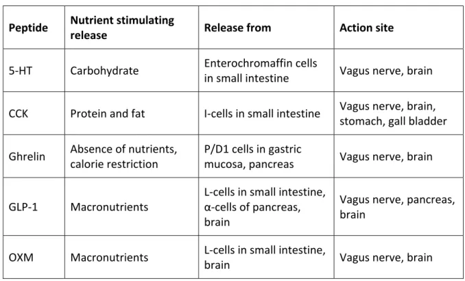
Table 1.1 Summary of the release and action of peptide hormones
Peptide Nutrient stimulating
release Release from Action site
5-HT Carbohydrate Enterochromaffin cells
in small intestine Vagus nerve, brain
CCK Protein and fat I-cells in small intestine Vagus nerve, brain,
stomach, gall bladder
Ghrelin Absence of nutrients,
calorie restriction
P/D1 cells in gastric
mucosa, pancreas Vagus nerve, brain
GLP-1 Macronutrients
L-cells in small intestine, α-cells of pancreas, brain
Vagus nerve, pancreas, brain
OXM Macronutrients L-cells in small intestine,
brain Vagus nerve, brain
Cholecystokinin
Cholecystokinin (CCK) was the first peptide hormone that was shown to alter appetite and therefore being involved in the regulation of meal termination (Gibbs et al. 1973). The main stimulation for the release of CCK from endocrine I-cells of the small intestine is the presence of dietary fat and protein in the intestine in response to apolipoprotein A-IV and
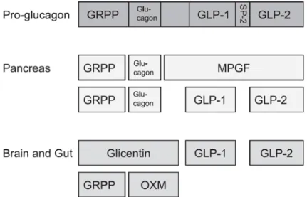
Figure 1.4 The processing of proglucagon in different tissues of the body.
GRPP, glicentin-related pancreatic peptide; GLP, glucagon-like peptide; MPGF, major proglucagon fragment; OXM, oxyntomodulin (Stanley et al. 2004).
PEPT1 activation (Liddle et al. 1985; Darcel et al. 2005; Foltz et al. 2008). Even it is predominantly released in the duodenum and jejunum, CCK is widely distributed in the GI tract (Larsson and Rehfeld 1978). CCK is acting via binding to the G-protein coupled receptors, CCK-1 and CCK-2. The CCK-1 receptor was found on afferent fibres of vagus nerve, in brainstem as well as in the dorsomedial nucleus of the hypothalamus (Moran 2000; Moran and Kinzig 2004). CCK-sensitive vagal afferents activate POMC neurones in the nucleus tractus solitarius (NTS) and were shown to stimulate c-Fos expression dose-dependently (Zittel et al. 1999; Fan et al. 2004; Appleyard et al. 2005). Moreover CCK is involved in several other regulatory processes in the periphery such as gall bladder contraction, enzyme release from the pancreas, stimulation of intestinal peristaltic and inhibition of gastric motility and emptying (Raybould 1991; Schwartz et al. 1991; Schwartz et al. 1993; Schwartz et al. 1997). The effect of CCK had been shown to be potentiated by leptin (Liddle et al. 1985; Raybould 1991).
Oxyntomodulin and glucagon-like peptide as products of the proglucagon gene
Both oxyntomodulin (OXM) and glucagon-like peptide (GLP) are cleavage products of the expression of proglucagon that takes place in the L-cells in the distal part of the small intestine, the α-cells of the pancreas and the brain (Stanley et al. 2004). Depending on the tissue the gene is expressed, the two enzymes prohormone convertase 1 and 2 cleave the 160-amino acid peptide into different products (Figure 1.4). OXM has only been found in the brain and the intestine while GLP-1 and GLP-2 are present in all organs where the gene is expressed (Holst 1999). The peptides are subsequently post-translationally modified in order to gain their biological activity (Mojsov et al. 1986). Both peptides are released 5-30 minutes postprandial in proportion to the caloric intake.
GLP-1 has been demonstrated to act both on the brainstem and the vagus nerve, but also on several hypothalamic regions and on ß-cells in the pancreas where it stimulates postprandial insulin release and inhibits glucagon in order to lower blood glucose (Orskov et al. 1996). GLP-1 receptors have been found to be present on neurones in the NTS and the AP, where GLP-1 also stimulates the expression of c-Fos (Wei and Mojsov 1995; Van Dijk et al. 1996), but also in several nuclei of the hypothalamus including the ARC, PVN and supraoptic nucleus (SON).
1 Scientific background 28
OXM consists of the amino acid sequence of pancreatic glucagon, but has an eight amino acid long C-terminal extension. Up to now, no specific OXM receptor has been cloned; however OXM seems to bind, whether with a two-fold lower affinity, to the GLP-1 receptor (Baggio et al. 2004). Yet, as both OXM and GLP-1 appear to exert a comparable effect on the reduction of food intake it is thus likely another OXM specific receptor exists (Stanley et al. 2004). The hypothalamus has been proposed as the principle action site of OXM in the CNS (Wei and Mojsov 1995; Stanley et al. 2005).
In fasted state plasma OXM levels are low; however, an increase in plasma levels inhibits gastric acid secretion and motility and leads to postprandial satiety (Bataille et al. 1982; Stanley et al. 2004). Studies have shown the involvement of OXM in long-term body weight regulation but it also acts as short-term satiety signal. Administered centrally or peripherally OXM leads to decreased weight gain in mice and rats due to its inhibiting effect on energy intake (Baggio et al. 2004; Dakin et al. 2004; Parkinson et al. 2009). Similarly, OXM reduces appetite and caloric intake in lean and obese humans and promotes significant weight loss (Cohen et al. 2003; Wynne et al. 2005). Part of its effect is suggested to be the depression of plasma ghrelin levels which was demonstrated in both rodents and humans (Cohen et al. 2003; Dakin et al. 2004) and an increase in activity and so energy expenditure (Wren and Bloom 2007). Better understanding the mechanisms of action of OXM in the CNS in the control of food intake may therefore help to study its potential role as anti-obesity treatment.
Serotonin
Serotonin (5-HT) is released from enterochromaffin cells, located in the epithelium of the small intestine, in the presence of carbohydrates in the GI tract and the activation of SGLT1 (Raybould et al. 2003; Freeman et al. 2006). Injection of 5-HT in turn reduces glucose intake (Hayes and Covasa 2005). After stimuli such as gastric distension, pressure sensors are activated and were demonstrated to be involved in the 5-HT dependent c-Fos expression in the NTS and the AP (Mazda et al. 2004). There are many different 5-HT receptors known. The 5-HT3 receptor was found on vagal and spinal afferent neurones (Raybould et al. 2003; Tome 2007), but 5-HT was also shown to act directly on the AP. A 5-HT receptor agonist activates catecholaminergic neurones in NTS at level of AP (Lam et al. 2009). 5-HT is also
suggested to mediate intestinal CCK and GLP-1 satiety signals (Hayes et al. 2006; Asarian 2009).
Ghrelin
The only peripheral orexigenic peptide known yet that increases appetite when administered peripherally and centrally is Ghrelin, which is synthesised in stomach (Date et al. 2005; Valassi et al. 2008). Ghrelin receptors can be found all along the vagus nerve as well as in the DVC (Guan et al. 1997). Ghrelin levels decrease after ingestion of a meal and thereafter increase in correlation with the amount of calories ingested (Weigle et al. 2003). Additional Ghrelin levels increase according to an endogenous rhythm with a maximum concentration at 01:00 am (Cummings et al. 2001). It is suggested that the satiating effect of protein also occurs due to a longer maintenance of postprandial low plasma ghrelin levels (Blom et al. 2006; Bowen et al. 2006b).
1.2.3 Pre-absorptive signals specifically induced by protein ingestion
Ingestion of a high protein diet leads to an increase in gastric volume and a delay in gastric emptying (Bowen et al. 2006b; Faipoux et al. 2006). Gastric distension was demonstrated to participate in meal termination by activating vagal afferent fibres (Schwartz et al. 1991; Phillips and Powley 1996, 1998). Rats with a pyloric cuff terminate their food intake as soon as they reach a certain volume (10-12 ml), independent from the sort of nutrient. In rats that have no pyloric cuff, meal termination occurs after ingestion of an even smaller volume as nutrients can leak into the intestine and stimulate the release of CCK. CCK in turn slows down gastric emptying and maintains gastric distension for longer what leads to meal termination after a smaller meal size (Raybould 1991; Schwartz et al. 1991; Schwartz et al. 1993; Schwartz et al. 1997). However, some studies suggest that the caloric value of the ingested meal is the determining factor for gastric emptying rather than the nature of the nutrients (McHugh and Moran 1979; Maerz et al. 1994).
Electrophysiological studies demonstrated that protein is able to generate pre-absorptive signals in vagal afferents (L'Heureux-Bouron et al. 2004). Also gastric or intestinal distension was shown to stimulate certain afferent fibres. However, vagal response to duodenal infusion with amino acids or protein was delayed by 3 minutes, suggesting that
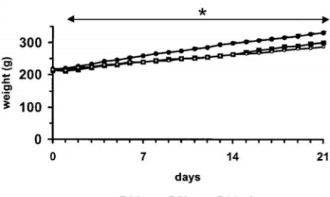
Figure 1.5 Daily energy intake and body weight of rats.
Daily energy intakes (left panel) and body weight (right panel) of rats fed either a 14 g/100 g protein diet (P14), a 50 g/100 g protein diet (P50) or a 14 g/100 g protein diet pair-fed to the P50 group in energy (P14-pf) for 21 days. Values are means ± SEM, n=24. *P50 value is different from P14, P < 0.05. From d 1 to 21, the P14 rats consumed more energy than the P50 and the P14-pf rats. Additionally, during this period, the P50 rats consumed more protein energy than the rats from the other groups. There was no difference between the body weights of the P14-pf rats and P50 rats (Jean et al. 2001). 0 100 200 300 400 0 7 14 21 P14 P50 Days P <0.05 days foo d in take (kJ)
*
these nutrients do not directly stimulate the intestinal sensory endings of vagus nerve. They are rather activated indirectly, for instance by the release of CCK. Moreover, parts of the hydrolysates might have been absorbed and lead to stimulation of hepatic afferent fibres.
Depression of food intake in response to a high protein diet can hence be explained by a larger gastric volume due to higher water intake as well as delay of gastric emptying and an amplification of vagal signalling in response to an increased release of CCK.
1.2.4 Post-absorptive peripheral signals
Additional to the pre-absorptive signals emerging during the passage of nutrients in the intestinal lumen, post-absorptive signals occur when nutrients or their metabolites enter the blood stream. Numerous metabolic events have also been hypothesised as signals in protein-induced satiety, including an increase in plasma amino-acid level, energy expenditure, thermogenesis and the production of glucose through gluconeogenesis.
1.2.4.1 Direct sensing of dietary protein
After the nutrient breakdown and absorption, the increase in circulating amino acids levels is detected by the brain and influence in this way satiety (Mellinkoff et al. 1956). Studies in rats have shown that replacing a normal protein diet with a high protein diet, animals immediately decrease their food intake (Figure 1.5). This depression in food intake has been demonstrated not to be due to a taste aversion to the protein diet, but indeed due to the protein content (Bensaid et al. 2002; Bensaid et al. 2003). Additionally a massive increase in the amino acid concentration could be detected in the first hours of a high protein diet which went back to baseline after adaptation to the new diet (Peters and Harper 1985, 1987). Together with the decrease in blood amino acid concentration, the protein induced satiety signalling becomes weaker (Long et al. 2000).
There is also some evidence that circulating leucine levels may influence food intake. An increase in dietary leucine (Ropelle et al. 2008) or the intra-cerebroventricular administration of either amino acids or only leucine reduced food intake and body weight (Cota et al. 2006; Morrison et al. 2007). These findings seem to be leucine-specific, as leucine alone exerts the same effect on food intake as a mixture of amino acids (Ropelle et al. 2008). Indeed, leucine is associated with mechanisms involving AMP-activated protein kinase
1 Scientific background 31
(AMPK) and the mammalian target of rapamycin (mTOR), both of which are energy sensors active in the regulation of energy intake, at least in the arcuate nucleus (ARC) but probably also in other brain areas such as the paraventricular nucleus (PVN).
It was suggested that not only the amino acids themselves act on brain satiety centres, but they stimulate the synthesis of the neurotransmitter 5-HT which is derived from the amino acid tryptophan (Latham and Blundell 1979). Nowadays this hypothesis has been abandoned (Harper and Peters 1989; Stubbs 1999). Still, other amino acids such as tyrosine and histidine are precursors of the neurotransmitters noradrenalin and histamine, respectively which can then indirectly influence hunger and satiety (Mercer et al. 1990). However, Bassil failed to demonstrate any effect of a diet supplemented with 5 % of either histidine or tyrosine on the levels of food intake by Sprague-Dawley rats (Bassil et al. 2007).
1.2.4.2 Glucose-mediated sensing of dietary protein
Blood glucose is not only detected by glucosensitive cells in the hypothalamus (Fioramonti et al. 2007) and the NTS (Ritter et al. 2000), but also at the level of the liver (Russek 1971). The longer a high blood glucose level can be maintained, the longer the sensation of satiety occurs (Holt et al. 1996).
As glucose is an important substrate for many body functions, especially performance of the brain, in a hypoglycaemic state the metabolism is forced to synthesise de novo glucose in order to maintain glucose homeostasis. This metabolic pathway is called gluconeogenesis and uses gluconeogenic amino acids such as alanine, glutamine, serine or glycine as precursor for glucose synthesis. During the postprandial state after ingestion of a high protein diet, due to the gluconeogenesis from dietary protein, the decrease in blood glucose concentration is more efficiently delayed (Blouet et al. 2006).
1.2.4.3 Protein-induced thermogenesis can influence satiety
Increased energy expenditure was observed after a high protein load or meal and was proposed as another mechanism of protein satiety (Porrini et al. 1997; Westerterp-Plantenga et al. 1999). There was also a correlation noticed between increased energy expenditure and elevated satiety after a high protein meal (Crovetti et al. 1998;
Westerterp-Plantenga et al. 1999; Lejeune et al. 2006). The satiating effect could be explained by the feeling of oxygen deprivation caused by elevated body temperature and greater use of oxygen in protein metabolism, used for absorption, storage and oxidation (Tappy 1996; Westerterp 2006). In contrast to lipids and carbohydrates, which can be stocked easily, proteins have to be metabolised prior to storage. Gluconeogenesis in the liver and building of muscle mass expend energy and in this way produce heat (Johnston et al. 2002; Westerterp-Plantenga et al. 2004). However, there is no consensus on this theory, as several studies failed to show a relation between the intake of a high protein diet, elevated body temperature or energy expenditure and satiety (Luscombe et al. 2003; Raben et al. 2003).
1.2.4.4 The role of hormones as peripheral adiposity signals
Other important factors among peripheral signals influencing satiety are hormones released from adipose tissue and the pancreas which are responsible for the control of energy homeostasis (Stanley et al. 2005). Most of them have an action site on both the level of the GI tract by activation of the vagus nerve and central on the hypothalamus and the dorsal vagal complex via the blood stream. Postprandial hormone profiles have been studied intensively in order to investigate in which way protein influences satiety (Al Awar et al. 2005; Bowen et al. 2006a; Bowen et al. 2006b).
Leptin
Leptin is the product of the ob gene and that is mainly produced and secreted from adipocytes but also in lower levels from gastric epithelium and placenta. Amongst others leptin influences energy homeostasis as well as neuroendocrine and immune functions. The plasma levels of leptin are direct proportional to adiposity and total fat mass (Considine et al. 1996); it likewise enters the brain in proportion to those plasma levels (Valassi et al. 2008). Leptin expresses direct action on neurones of the nucleus of the solitary tract and
therefore short-term energy intake. Neurophysiologic studies revealed that central
administration of leptin significantly increased NTS responses to gastric loads in unconscious
rats (Schwartz and Moran 2002). Moreover, using current-clamp records on hindbrain
sections, superfusion with leptin was demonstrated to induce a dose-dependant hyperpolarisation of the resting membrane potential in neurons in the nucleus of the solitary tract (Williams and Smith 2006). Together with insulin, leptin is also regulating the activity of
1 Scientific background 33
first order neurones in the ARC of the hypothalamus. There it stimulates the expression of the pro-opiomelanocortin (POMC) gene and the release of the anorexigenic α-melanocyte-stimulating hormone (α-MSH) while at the same time it inhibits neurones which are usually expressing neuropeptide Y (NPY) and the agouti-related protein (AgRP) (Schwartz et al. 2000). Up to now no study could show a correlation of a high protein load on plasma leptin levels compared to a high fat or high carbohydrate charge (Raben et al. 2003).
The importance of leptin in the regulation of body weight can be seen in the absence of either the hormone itself or the leptin receptor, leading to hyperphagia and severe obesity in both animals and humans (Licinio et al. 2004). In humans, however, leptin or leptin receptor deficiency is extremely rare and in most obese subjects a contrary problem occurs as elevated leptin levels sooner or later result in leptin resistance (Considine et al. 1996). Leptin is therefore an anorexigenic hormone, peripherally administered it is very powerful in decreasing both spontaneous and long-term food intake resulting in loss of fat mass and body weight (Halaas et al. 1995). However, due to the increased leptin resistance in overweight subjects, anti-obesity treatment with this hormone is often not successful (Considine et al. 1996).
Insulin
Insulin, secreted from pancreatic β-cells, was the first hormonal signal that was shown to be involved in the central control of food intake and body weight (Wynne et al. 2005). Proportional to the body fat content, plasma levels of this hormone are rapidly raised in response to blood glucose. It reaches the brain through receptor-mediated transport via the blood-brain barrier (BBB) as for leptin in proportion to its circulating levels (Baura et al. 1993). Insulin achieves the reduction in food intake by acting on neurones in the ARC which express the insulin receptor (Cone et al. 2001; Benoit et al. 2002). A clear relation between a high protein meal and plasma insulin concentration was not shown yet (Veldhorst et al. 2009a, 2009b).
In overweight subjects, insulin resistance is a common phenomenon as both in basal state and in response to a meal. Insulin plasma concentration has to increase in order to maintain stable glucose levels. As a result of long-term overproduction of insulin, pancreatic
β-cells can not raise insulin concentrations high enough anymore to decrease elevated plasma glucose levels. Subsequently hyperglycaemia occurs leading to type 2 diabetes.
Adiponectin
As for leptin, adiponectin is released from adipose tissue, but in a 1000 fold higher concentration than leptin or insulin whereby its plasma concentration is negatively correlated with adiposity (Stanley et al. 2005). In peripheral tissue such as muscle and liver, adiponectin was shown to be involved in the regulation of energy homeostasis by modulating glucose and fatty acid metabolism (Berg et al. 2002). In contrast, the action of adiponectin in the brain is still controversially discussed. On the one hand, two receptors were identified which are located not only in peripheral tissue but also in regions of the hypothalamus and the brainstem, including the area postrema (AP), ARC and the PVN (Dridi and Taouis 2009). On the other hand, only low concentrations of adiponectin can be detected in the cerebrospinal fluid. Adiponectin is suggested to be transported from the blood by receptor-mediated transcytosis or via circumventricular organs such as the median eminence (ME) or the AP. A diet high in protein was shown to result in decreased plasma adiponectin levels (Stroubini et al. 2009).
Figure 1.6 Coronal section of the brainstem showing the dorsal vagal complex (DVC).
Structures building the DVC are: AP, area postrema; CC, central channel; DMX, dorsal motor of vagus; NTS: nucleus tractus solitarius (Adapted from Paxinos and Franklin 2001, Figure 96, Bregma -7.76).
1.3 Central signalling of dietary protein
Even there is no doubt that the CNS plays an important role integrating information which is related to the amount of ingested protein, many studies using electrolytic lesions carried out in the past years failed to identify a brain region which is especially responsible for the control of protein intake (Fromentin et al. 2000). It is commonly accepted that hunger and satiety are correlated with the activation of different brain regions (Berthoud 2004b). The main centres for the control of food intake are on the one hand the dorsal vagal complex responsible for the regulation of meal size and termination and on the other hand the hypothalamus which is responsible for the indirect, long-term regulation of energy homeostasis. In this context ‘direct control’ stands for the afferent pathways activated in response to the food stimuli contacting pre-absorptive receptors along the surface of the gut from the tip of the tongue to the end of the small intestine. Indirect controls are all those that are not directly affected by food stimuli acting at sensory receptors along the mucosal surface of the gut during a meal (Smith 1996).
1.3.1 Short-term regulation of food intake: the dorsal vagal complex controls meal size
The dorsal vagal complex lies within the caudal brainstem, which builds the lower part of the brain and is the connection with and prolongation of the spinal cord (Figure 1.6). The DVC is a neuronal network that consists of three major structures which integrate inhibitory and excitatory peripheral signals and are strongly suggested to be involved in the direct regulation of food intake and meal size. The NTS processes visceral sensation and taste from afferent sensory fibres of the trigeminal (V), facial (VII), glossopharyngeal (IX) and vagus (X) cranial nerve. Moreover it also processes primary afferent signals from a variety of visceral regions and organs via the The AP having a deprived BBB mainly receives blood borne signals. The third structure within the DVC, the dorsal motor of vagus (DMX), in turn is the primary location of motor neurones. From here vagal efferents arise which then respond to processed signals by causing gastric and intestinal peristaltic (Norgren and Smith 1988).
The essential role of the DVC in the regulation of meal size was demonstrated in the way that meal termination induced by short-term satiety signals occurred, even when all
1 Scientific background 36
neuronal connections between forebrain and brainstem were severed in rats (Grill and Smith 1988).
1.3.1.1 The blood-brain barrier
The blood-brain barrier prevents the entrance of potentially unwanted circulating factors into the brain. Cells building the BBB form specialised capillaries, blood vessels consisting of a single layer of endothelial cells whose one side is facing the blood and the other side is facing the cerebrospinal fluid surrounding the brain (Hawkins et al. 2006). While small lipophilic molecules can pass the BBB by diffusion, hydrophilic substances can only enter central structures by active transporters, channels or at areas, where the BBB is deprived such as the AP and the ME (Maolood and Meister 2009).
1.3.1.2 The area postrema mainly responding to humoral signals
The area postrema as part of the DVC lies in the brainstem, directly above the NTS (Figure 1.6) and is involved in the short-term regulation of food intake and individual meal size. It is a circumventricular organ built of fenestrated blood vessels and it is known for its incomplete BBB, what gives it the possibility to control the entrance of blood-borne substances to the neurones of DVC (Maolood and Meister 2009). Therefore, the NTS being places next to this organ with deficient BBB can also respond to peripheral circulating signals (Sawchenko 1983). The AP expresses a large number of receptors for gut peptides being involved in food intake control, including CCK1-R, GLP-1R, NPY (Wei and Mojsov 1995; Moran 2000; Stanley et al. 2005). In addition to its tasks in food intake regulation, the AP is also involved in the control of cardiovascular functions (Saito et al. 2003).
1.3.1.3 The NTS receives signals from vagal afferents and the blood stream
The NTS, the second important component of the DVC involved in the short-term regulation of food intake, consists of a dense vascular network and is located in the caudal brainstem (Norgren and Smith 1988) (Figure 1.6). Initiations of satiety signals reaching the NTS via vagal afferents during food ingestion are derived from mechanical or chemical stimulation of the GI tract and abdominal viscera as well as taste information from the oral cavity and from neural input related to energy metabolism in the liver (Travers et al. 1987; Emond et al. 2001; Powley and Phillips 2004). Due to its location in direct neighbourhood of
Vagal afferents ascending from different sites within the gastrointestinal tract projecting in the NTS. AP, area postrema (Adapted from Méi 1998, page 115).
1 Scientific background 37
the AP, which is missing BBB, also the entrance of humoral signals in the NTS is possible, specifically peptides and substrates which in turn can bind to specific receptors and activate NTS neurones (Maolood and Meister 2009). The GLUT-1 transporter system for instance, which is sensitive to glucose, was found in the NTS (Maolood and Meister 2009), but also receptors for gut peptides which are released upon nutrient stimulation from neuroendocrine cells in the intestinal lumen during ingestion, such as CCK1-R, GLP-1R, NPY (Wei and Mojsov 1995; Moran 2000; Stanley et al. 2005). NMDA and AMPA glutamatergic receptors are as well expressed in NTS neurones (Berthoud et al. 2001). All this peripheral sensory information is subsequently integrated in second order neurones.
The neuroanatomic distribution of the NTS had been suggested after retrograde labelling studies (Figure 1.7). The most rostral part of the NTS receives vagal afferents from the oral cavity, while the most caudal part receives those from the hepatic branches and coeliac. The intermediate part of the NTS, which lies next to the AP, mainly receives projections from the stomach and intestine (Norgren and Smith 1988).
As part of the entire network involved in appetite regulation, afferent fibres of the NTS project, amongst others, to the ARC and PVN within the hypothalamus and in turn receive information from this second important satiety centre what makes it possible to integrate satiety signals with adiposity signals like insulin and leptin (Schwartz et al. 2000; Berthoud et al. 2006; Valassi et al. 2008).
Even there is still a big lack of knowledge in understanding the exact mechanisms of how the DVC controls meal size, it is hypothesised that neurones in the NTS can be activated macronutrient specific (Rinaman et al. 1998; Yamamoto and Sawa 2000; Berthoud et al. 2001; Emond et al. 2001).
1.3.2 Long-term regulation of energy balance: the hypothalamic area is responsible for maintaining body weight through meal initiation
While the DVC is known to control short-term regulation of food intake, integration of peripheral long-term hunger and satiety signals as well as body weight control mainly takes place in the hypothalamic area. Being the negative feed-back regulator for energy metabolism, the hypothalamus integrates humoral adiposity signals so as leptin and insulin
term food intake regulation.
3V, 3rd ventricle; ARC, arcuate nucleus; LHA, lateral hypothalamic area; ME, median eminence; PFA, perifornical area; PVN, paraventricular nucleus; VMH, ventromedial hypothalamus (Adapted from Paxinos and Franklin 2001, Figure 44, Bregma -1.58).