En vue de l'obtention du
DOCTORAT DE L'UNIVERSITÉ DE TOULOUSE
Délivré par :
Institut National Polytechnique de Toulouse (Toulouse INP)
Discipline ou spécialité :
Pathologie, Toxicologie, Génétique et Nutrition
Présentée et soutenue par :
Mme HANNA ILCHMANN le jeudi 19 septembre 2019
Titre :
Unité de recherche : Ecole doctorale :
Consequences of early life adverse events on the development of
non-communicable diseases in mouse models
Sciences Ecologiques, Vétérinaires, Agronomiques et Bioingénieries (SEVAB) Toxicologie Alimentaire (ToxAlim)
Directeur(s) de Thèse : MME VASSILIA THEODOROU
MME SANDRINE MENARD
Rapporteurs :
Mme JOHANNE LE BEYEC-LE BIHAN, ASSISTANCE PUBLIQUE HOPITAUX DE PARIS Mme PATRICIA PARNET, INRA NANTES
Membre(s) du jury :
M. PHILIPPE GERARD, INRA JOUY EN JOSAS, Président M. BENOIT CHASSAING, GEORGIA STATE UNIVERSITY, Membre Mme MICHELLE KELLY-IRVING, INSERM OCCITANIE PYRENEES, Membre
Mme SANDRINE MENARD, INRA TOULOUSE, Membre Mme VASSILIA THEODOROU, EI PURPAN, Membre
THESE
En vue d l’obtention du
Doctorat de l’Université de Toulouse
Délivré par l’Institut Polytechnique de Toulouse (INP Toulouse) Discipline : Pathologie, Toxicologie, Génétique & Nutrition
Présentée et soutenue par Hanna Ilchmann Le 19 septembre 2019
Conséquences d'événements adverses en période néonatale sur le développement de maladies non-transmissibles dans des modèles murins
Consequences of early life adverse events on the development of non-communicable diseases in mouse models.
Jury
Dr Philippe Gérard Président du Jury Dr Johanne Le Beyec-Le Bihan Rapporteur Dr Patricia Parnet Rapporteur Dr Michelle Kelly-Irving Examinateur Dr Benoit Chassaing Examinateur Pr Vassilia Theodorou Directeur de thèse Dr Sandrine Ménard Directeur de thèse
Ecole doctorale : Sciences Ecologiques, Vétérinaires, Agronomiques, Bioingénieries Unité de recherche : UMR 1331 INRA/INP/UPS Toxalim
Meinem U
ngeborenen…
For my unborn…
A mon enfant à naitre…
For You formed my inward parts;
You wove me in my mother's womb.
I
R
EMERCIEMENTSJe remercie Dr Patricia Parnet et Dr Johanne Le Beyec-Le Bihan, rapporteurs de ma thèse, d’avoir accepté d’évaluer le travail ici présenté. Je les remercie pour toute leur implication, le temps investi, les conseils, les encouragements, les remarques et critiques constructives.
Je remercie le président du jury Dr Philippe Gérard pour son investissement.
Merci aux examinateurs Dr Michelle Kelly-Irving et Dr Benoit Chassaing. Merci pour tous les échanges stimulants et passionnants. Merci d’avoir participé à l’aboutissement de ce travail. Merci Michelle pour les réunions stimulantes autour de la DOHaD. Tes présentations sont passionnantes. Merci Benoit pour les discussions autour de la barrière, du mucus, des peptides antimicrobiens. Merci pour ta bienveillance et compréhension.
Je remercie mes directeurs de thèse, Pr Vassilia Théodorou et Dr Sandrine Ménard. Merci Sandrine, de m’avoir formé au métier du chercheur durant ces quatres dernières années. Merci d’avoir contribué à ce que je suis aujourd’hui dear #DoctorMother. Merci pour les nombreuses discussions scientifiques et personnelles. Merci de m’avoir transmis ta passion pour la Science. Merci de m’avoir toujours poussée à aller plus loin. Merci d’avoir cru en moi. Merci aussi pour ta compréhension et ta patience. Merci pour les heures et parfois les nuits passées à corriger mes nombreux rapports, dossiers, présentations, posters, candidatures, papiers, sans parler de ce manuscrit de thèse. Merci d’être pour moi un exemple de chercheuse déterminée, parfois têtue, créative, et visionnaire. Merci pour les voyages, les escapades culinaires diverses et variées et peut-être parfois excessives. Merci pour les larmes de rire. Ah ! mes muscles abdominaux ne se sont pas dégradés !
Merci Vassi de m’avoir acceptée et bien accueillie dans ton équipe. Merci pour tout ton investissement dans ma thèse. Merci pour les échanges stimulants autour de mes résultats. Tes commentaires pour améliorer mes papiers, les remarques critiques qui me poussaient de quitter ma zone de confort. Merci de m’avoir poussée aller plus loin. Merci d’avoir toujours cru en moi et de m’avoir soutenue dans mes projets et ambitions. Merci pour le calme que tu gardes et la bienveillance que tu dégages. Merci pour ce petit côté maternel.
II Je remercie les membres de mon comité de thèse qui m’ont accompagné durant ces trois dernières années. Merci au Dr Valérie Verhasselt, Dr Pierre Gourdy et Dr Laurence
Guzylack-Piriou.
Merci Dr Valérie Verhasselt pour toutes les idées partagées. Merci pour la considération que tu exprimais pour notre travail. Merci de partager d’avoir partagé ta passion et motivation.
Je remercie Dr Pierre Gourdy pour ses remarques ouvrant mon horizon. Merci pour l’œil critique de clinicien sur ce travail de thèse. Merci pour l’analyse claire et l’orientation utile.
Je remercie Dr Laurence Guzylack-Piriou pour tout son aide, ses sacrifices et son implication dans ma thèse. Merci pour tes encouragements, tes idées, ton œil critique, les nombreuses explications. Merci pour les retours constructifs sur mes dossiers et papiers. Merci pour toutes les questions que tu me posais lors de mes répétitions.
Je remercie les différents collaborateurs avec qui j’ai eu la joie de travailler dans le cadre de ma thèse.
Merci au Dr Maiwenn Olier pour toutes les analyses du microbiote. Merci pour les nombreuses explications. Merci pour tout le temps investi dans nos manips. Merci pour tes encouragements, tes nombreuses relectures, la joie et le calme que tu dégages. Merci pour ta rigueur – combien j’aimerai être comme toi ! Merci pour les fous rires et d’avoir eu assez de clairevoyance pour prendre des photos du siphon pété !!!
Merci Corinne Lencina pour ton implication dans ma thèse. Merci pour toute ton aide pour nos manips. Merci pour toutes les heures investies à me former à des ELISAs. Merci pour la belle collaboration en salle de bioch. J’ai bien aimé nos jours de challenge où on passait 6 plaques ELISA à la fois. Merci pour les bons moments passés ensemble, à la couture, pendant les repas, entre les manips, dans les pauses café, et je ne mentionne pas tout... Merci pour tes gâteries culinaires et ta créativité. Merci d’être la colle pour éviter que le bateau se brise. Merci d’avoir toujours pris soin de moi et de mon bien-être.
Merci à Dr Sandrine Ellero-Simatos, Dr Hervé Guillou, Sharon Barretto et Céline
Lukowicz pour vos remarques sur le volet metabolisme du projet Metalogin. Merci Sandrine
III former, les nombreux commentaires sur mon travail, les conseils. Merci pour ta bienveillance et les petits échanges dans le couloir. Merci Hervé pour ton soutien, ta considération pour notre travail, tes remarques, ton aide dans la préparation de ma thèse. Merci pour ta gentillesse, tes encouragements.
Merci à Elia Macadré, stagiaire pendant quatre mois avec nous. Merci pour ton travail énorme, ta motivation, ta disponibilité, ton envie d’apprendre, ta patience avec moi. Ce fut une belle expérience avec toi !
Merci à Dr Colette Denis et Dr Marie Buléon pour la collaboration dans le projet NOD. Merci pour vos remarques pertinentes, vos disponibilité et motivation.
Merci à Dr Julien Diana pour les échanges autour du projet « MS and type 1 diabetes ». Merci pour la considération, les conseils et la collaboration.
Merci à Dr Sonia Lamandé pour les beaux échanges autour de l’immunité dans la petite enfance. Merci d’avoir accueilli chez toi nos animaux.
Merci à Valérie Bacquié pour ta patience à me former à l’immunohistomarquage. Merci au Pr Jamileh Movassat de m’avoir accueillie pour un stage dans son équipe à Paris (BFA, B2PE, Paris Diderot). Merci Junjun Liu de m’avoir formée avec beaucoup de soin. Merci à Blandine et Pengfei pour les bons moments passés ensemble.
Je remercie Thierry Gauthier du plateau M2C. Merci pour tes patience et pédagogie pour me former à la cytométrie et au microscope confocale. J’ai véritablement apprécié ces formations. Tu as été très didactique !
Un grand merci à Christelle Cartier pour toute ton aide et ses conseils autour de l’immunohistomarquage. Merci pour ta patience, ta réactivité, tes conseils pratiques. Merci aussi pour ta gentillesse, ton sourire, les échanges, les temps passés ensemble à la couture.
Je remercie Dr Rémy Burcelin pour ses remarques stimulantes concernant ce projet de thèse. Merci de m’avoir acceptée il y a 7 ans pour mon premier stage en laboratoire de recherche – ça a été une révélation pour moi.
Merci Fabrice Pierre pour ta toujours bonne humeur et bienveillance, ta considération et tes encouragements.
IV Merci aux autres thésards dans le couloir : Merci à Yann Malaisé, Sophie Yvon,
Jasper Kamphuis, Kévin Gillois et Anaïs Mazenc. Merci Yann pour tous les conseils pendant
ma première année de thèse, tes encouragements, les pauses ensemble, les fous rires – je ne vais jamais oublier notre aventure « Hanovre » ! Merci Sophie pour toutes les occasions où tu m’as bien fait rire, ta comédie, tes conseils. Merci Jasper d’avoir été un renfort de la culture germanique à Toxalim. J’ai bien apprécié nos petits débats sur tout et rien ! Merci pour ton aide pour améliorer mon anglais, perfectionner mes diapos et mon manuscrit.
Merci à tous ceux de l’équipe NGN que je n’ai pas encore mentionnés : Cathy,
Christine, Hervé, Hélène, Muriel, Valérie Be, Valérie M et Valérie T, ainsi que les anciens
membres de NGN: Kheira, Michèle, Alain, Afifa, Laurent. Merci pour votre soutien, l’intérêt que vous avez pour moi, votre participation à mes nombreuses répétitions, vos remarques, suggestions, vos questions pour me préparer aux différents oraux.
Je remercie les membres du Journal Club. Même si le plaisir fut court, je me suis beaucoup réjouie de nos petites heures passées ensemble: Benoit, Manon, Yann, Jasper.
Merci à l’équipe d’animalerie : Caroline, Colette, Elodie, Géraldine et Mickael. Merci Caroline pour la gestion de nos animaux, ton aide, tes idées et ton implication dans nos manips.
Je remercie Christian Chervin et Benoit van der Rest pour les bons moments partagés lors des enseignements à l’école d’ingénieur ENSAT. J’ai appris plein de choses intéressantes ! Merci aux ADASsiens dans l’activité couture et jardin. Merci pour les bons moments partagés ensemble. Merci Joelle et Claire d’avoir organisé ces activités.
Je remercie les gestionnaires de Toxalim: Sarah Calcagno, Marie Hélène Piquereau. Merci de rendre notre travail possible. Merci de votre réactivité, les réponses aux questions, votre gentillesse.
Merci Jeannette Faramond pour ton accueil ! C’était toujours un plaisir d’être accueillie par toi. Merci pour toute orientation et aide dans les démarches administratives. Merci pour le temps passé ensemble dans la pause de midi, les bons échanges.
Merci à la RH de l’INP Marie-Claude Portell. Merci pour votre réactivité, aide et orientation.
V Je remercie Dominique Pantalacci pour ses mails prompts et toujours plein de gentillesse, même lorsque moi j’étais à la bourre.
Je ne souhaite pas oublier mes amis et ma famille qui m’ont supportée durant ces dernières années :
Merci à l’armée de prière qui s’est tenue derrière moi pendant tous ces derniers trois ans et en particulier pendant le temps de rédaction : merci Honorine, merci Lois, Fanja, Citra,
Michel, Johnny, et tous les autres qui se sont tenus derrière moi.
Merci Rhoda pour ton écoute, les échanges, tes conseils, tes encouragements. Merci
Nathalie pour tous les échanges, les encouragements et les discussions. Ce fut un plaisir de
rencontrer des scientifiques à l’EPET. J’en remercie notre Seigneur !
Je tiens aussi à remercier les membres de l’association des groupes bibliques universitaires – GBU. Merci pour les bons moments passés ensemble pendant les discussions autour de la Bible. Un merci particulier à Caroline pour tout ton soutient et tes prières. Merci au réseau GBU doctorants. Les échanges avec vous ont été très enrichissants. Ça m’a fait tellement du bien de me retrouver avec vous une fois par an.
Ich möchte auch von Herzen meiner Familie, insbesondere meinen Eltern danken. Danke Mama und Papa für all eure Unterstützung und Ermutigung, seit ihr mich auf der Welt willkommen geheißen habt. Danke, dass ihr immer in meinen Projekten voll und ganz hinter mir standet, mich nicht verunsichert, sondern immer ermutigt habt. Danke, dass ihr an mich glaubt und mir beigebracht habt auf meinen eigenen Beinen zu stehen. Danke Mama, für deine Fürsorge und Treue. Danke Papa für die Ruhe die du ausstrahlst.
Danke Ruth für alle Unterstützung in meiner Schul- und Studienlaufbahn. Danke für das viele Korrekturlesen, die Diskussionen, Tipps zum Verbessern, deine Ermutigungen … Danke, dass du so eine tolle Schwester bist. Danke Uli für deine lieben Worte der Ermutigung. Danke Joel und Friederike für eure Liebe. Danke Andi und Lydi, dass ihr mich so oft spüren habt lassen, dass ihr stolz auf mich seid.
Danke Oma für deine treuen Gebete. Danke Judith, dass du mir immer das Gefühl gegeben hast, dass du meine Meinung und meine wissenschaftliche Laufbahn schätzt.
VI Last but not least, je remercie mon cher époux Bailly Franck Eric Diounou. Merci d’être mon compagnon dans cette course sur la terre. Merci de m’avoir accompagnée tout au long de ces dernières années de master et thèse. Merci pour ton écoute. Merci pour ton soutien pratique, psychologique et émotionnel. Merci de m’avoir toujours soutenue dans mes ambitions et ne jamais m’avoir découragée. Merci de m’avoir bien souvent ouvert les yeux sur la réalité quand j’avais la tête dans le guidon. Merci d’avoir toujours orienté ma vue vers celui qui nous connait véritablement et récompense justement. Merci pour ton amour. Je t’aime.
VII
O
RIGINALA
RTICLESIlchmann-Diounou H, Olier M, Lencina C, Riba A, Barretto S, Nankap M, Sommer C, Guillou H, Ellero-Simatos S, Guzylack L, Theodorou V, Ménard S. Early life stress induces type 2 diabetes-like features in aging mice. Brain Behavior and Immunity 2019 https://doi.org/10.1016/j.bbi.2019.04.025
Ilchmann-Diounou H, Buléon M, Bacquié V, Theodorou V, Denis C, Ménard S. Non-Obese Diabetic Mouse Model is emphasizing divergence between in vivo and ex vivo intestinal permeability measurements. Submitted to Scientific Reports 23 July 2019, under review.
R
EVIEWA
RTICLEIlchmann-Diounou H and Ménard S. Psychological Stress, Intestinal Barrier Dysfunctions and Autoimmune Disorders. Submitted to Frontiers in Immunology 17 May 2019,
under review.
O
RAL COMMUNICATIONSIlchmann-Diounou H, Buléon M, Bacquié V, Theodorou V, Denis C, Ménard S. Comparaison des mesures de perméabilité intestinale in vivo et ex vivo chez la souris NOD (Non Obese Diabetic) (2019). Réunion annuelle du Groupe Français de Neuro-Gastroenterologie, Toulouse, France
Ilchmann-Diounou H, Buléon M, Bacquié V, Theodorou V, Denis C, Ménard S. Comparaison des mesures de perméabilité intestinale in vivo et ex vivo chez la souris NOD (Non Obese Diabetic) (2019). Club d’études des cellules épithéliales digestives XXXVIIème réunion, Toulouse, France
Ilchmann H, Olier M, Lencina C, Riba A, Barretto S, Nankap M, Sommer C, Guillou H, Ellero-Simatos S, Guzylack L, Theodorou V, Ménard S. Le stress de séparation maternelle induit chez l’adulte les symptômes semblables du diabète de type 2 associés à un défaut de sécrétion d’IL-17 et IL-22 (2018). 4ème congrès de la SF-DOHaD, Grenoble, France
Ilchmann H, Olier M, Lencina C, Ellero-Simatos S, Nankap M, Riba A, Sommer C, Guzylack-Piriou L, Théodorou V, Ménard S (2018). Early life stress in mice induces type 2 diabetes-like symptoms associated with defect of intestinal IL-17 and IL-22 secretion at
VIII adulthood. Club d’études des cellules épithéliales digestives XXXVIème réunion, Marseille, France
Ilchmann H, Olier M, Lencina C, Ellero-Simatos S, Nankap M, Riba A, Sommer C, Guzylack-Piriou L, Théodorou V, Ménard S (2017). Early life stress induces type 2 diabetes-like symptoms at adulthood in mice associated with defect of intestinal IL-17 and IL-22 secretion. Microbes, Immunity and Metabolism International Conference, Paris, France
Ilchmann H, Olier M, Lencina C, Ellero-Simatos S, Nankap M, Riba A, Sommer C, Guzylack-Piriou L, Théodorou V, Ménard S (2017). Early life stress induces type 2 diabetes-like symptoms at adulthood in mice associated with defect of intestinal IL-17 and IL-22 secretion. 10th International Meeting of Pediatric Endocrinology, Washington DC, USA
Ilchmann H, Lencina C, Ellero-Simatos S, Riba A, Harkat C, Sommer C, Guillou H, Guzylack-Piriou L, Olier M, Théodorou V, Ménard S (2016). Chronic maternal separation in mice impairs immuno-metabolism in older offspring on standard diet. The Neonatal Window of Opportunity – Early Priming for Life, Hanovre, Germany
P
OSTERSIlchmann-Diounou H, Olier M, Lencina C, Riba A, Barretto S, Nankap M, Sommer C, Guillou H, Ellero-Simatos S, Guzylack L, Theodorou V, Ménard S (2019). Early life stress induces type 2 diabetes-like features in aging mice. NeuroGASTRO 2019, Lisbon, Portugal.
Ilchmann H, Olier M, Lencina C, Ellero-Simatos S, Nankap M, Riba A, Sommer C, Guzylack-Piriou L, Théodorou V, Ménard S (2018). Early life stress induces type 2 diabetes-like symptoms at adulthood in mice associated with defect of intestinal IL-17 and IL-22 secretion. Modelling the Mammalian-Microbiota Host Superorganism, Paris, France.
Ilchmann H, Olier M, Lencina C, Ellero-Simatos S, Nankap M, Riba A, Sommer C, Guzylack-Piriou L, Théodorou V, Ménard S (2018). Early life stress induces type 2 diabetes-like symptoms at adulthood in mice associated with defect of intestinal IL-17 and IL-22 secretion. Mucosal Immunology Course and Symposium, Oxford, UK.
Ilchmann H, Olier M, Lencina C, Ellero-Simatos S, Nankap M, Riba A, Sommer C, Guzylack-Piriou L, Théodorou V, Ménard S (2018). Early life stress induces type 2 diabetes-like symptoms at adulthood in mice associated with defect of intestinal IL-17 and IL-22 secretion. Digestive Disease Week, Washington DC, USA.
IX Ilchmann H, Lencina C, Riba A, Sommer C, Guzylack-Piriou L, Olier M, Théodorou V, Ménard S (2017). Early life stress induces glucose intolerance and insulin secretion failure in aging female mice. 10th International Meeting of Pediatric Endocrinology, Washington DC,
USA.
Ilchmann H, Lencina C, Ellero-Simatos S, Riba A, Harkat C, Sommer C, Guillou H, Guzylack-Piriou L, Olier M, Théodorou V, Ménard S (2016). Chronic maternal separation in mice impairs immuno-metabolism in older offspring on standard diet. The Neonatal Window of Opportunity – Early Priming for Life, Hannover, Germany
X
P
RICES ANDG
RANTS2019 2nd prize of nEUROgastroTANDEM meeting 2019 in Lisbon, Portugal 2019 Prix Claude Rozé, Club d’études des cellules épithéliales digestives 2018 Best oral communication 4th congress of SF-DOHaD, Grenoble, France
2017 Early Career Scientific Development Grant for a two-month research mobility at Paris Diderot financed by the European Society of Pediatric Endocrinology (ESPE)
2017 Travel Grant for the 10th International Meeting of Pediatric Endocrinology financed by the European Society of Pediatric Endocrinology (ESPE)
2016 Travel Grant for The Neonatal Window of Opportunity,– Early Priming for Life, Hannover, Germany financed by Volkswagenstiftung
XI
T
EACHINGE
XPERIENCED
URINGP
HD
DCCE 2016-2019 at Toulouse INP - Ecole National Supérieur Agronomique de Toulouse (ENSAT). Tutor activity in the following courses under the supervision of Christian Chervin, Benoit van der Rest and Thierry Liboz.
Food Science – De la vigne au vin (1ère année ingénieur)
Food Science – Cuisine moléculaire (2ème année ingénieur)
Food Science – Analyse sensorielle (2ème année ingénieur)
XII
L
IST OFI
LLUSTRATIONSFigure 1 Intestinal anatomy. ... 8
Figure 2 Dysbiosis. ... 11
Figure 3 Mucus layer organization in small intestine and colon. ... 12
Figure 4 Different cell types in (A) small intestine and (B) colon. ... 15
Figure 5 Pathways of intestinal permeability. ... 16
Figure 6 Intercellular junctions of intestinal epithelial cells. ... 17
Figure 7 Functions of AMP. ... 20
Figure 8 Innate lymphoid cells. ... 23
Figure 9 Pattern recognition receptors. ... 24
Figure 10 Inductive and effector sites of the gut-associated lymphoid tissue. ... 25
Figure 11 Cycle of B cells in the lamina propria of intestine. ... 26
Figure 12 The enteric nervous system. ... 28
Figure 13 The gut-brain axis. ... 30
Figure 14 Intestinal microbiota composition over lifetime. ... 31
Figure 15 Intestinal permeability and gut microbiota diversity in lifetime. ... 32
Figure 16 Ontogeny of intestinal barrier in rodents. ... 33
Figure 17 Glucose metabolism. ... 36
Figure 18 Disturbed body systems in metabolic disorders. ... 37
Figure 19 The hypothalamus-pituitary-adrenal axis. ... 45
Figure 20 The neonatal window of opportunity. ... 73
Figure 21 Allostasis and allostatic load. ... 74
Figure 22 Neonatal maternal separation protocol. ... 78
Figure 23 Resolved parameters of intestinal barrier dysfunctions in maternal separated (MS) or control C3H/HeN mice at post-natal day (PND) 350. ... 101
Figure 24 Bacterial taxonomic patterns that characterize fecal microbiota of PND350 mice and PND50 mice differ significantly. ... 102
Figure 25 Jejunal permeability measurements in Ussing chambers. ... 165
Figure 26 Colonic permeability measurements in Ussing chambers. ... 166
Figure 27 Jejunal paracellular permeability to FSS. ... 169
Figure 28 Temporal aspects of the phenotype induced by maternal separation in male mice. ... 170
XIII
Figure 29 Temporal aspects of the phenotype induced by maternal separation in
female mice. ... 171
Figure 30 Plasmatic immunoglobulin concentrations. ... 175
Figure 31 Cytokine concentrations. ... 175
Figure 32 Preliminary data Body weight. ... 180
Figure 33 Specific humoral immune response. ... 181
Figure 34 IgG against hydrosoluble fraction of food in plasma. ... 181
Figure 35 Plasma IgE concentrations. ... 182
Figure 36 Plasma IgE concentrations. ... 184
L
IST OFT
ABLES Table 1 Hormones produced by intestinal epithelial cells (examples). ... 14XIV
A
BBREVIATIONS5-HT Serotonin
ACTH adrenocorticotropic hormone AD autoimmune disorder
AMP antimicrobial peptides ANS autonomous nervous system BMI body mass index
CLP common lymphoid progenitor CNS central nervous system
CRAMP cathelin-related antimicrobial peptide CRF corticotropin releasing factor
CRP c-reactive protein CS caesarian section
DALYS disability adjusted life years DC dendritic cell
DNA deoxyribonucleic acid
DOHaD Developmental Origins of Health and Disease DSS dextran sulfate sodium
dsRNA double-stranded ribonucleic acid
EAE experimental autoimmune encephalomyelitis ENS enteric nervous system
FcRn neonatal Fc receptor FD4 FITC-Dextran 4 kDa FSS fluorescein sodium salt
GALT gut-associated lymphoid tissue GAP goblet-cell associated passage
GF germ-free
GFP green fluorescent protein
GIP glucose-dependent insulinotropic polypeptide GLP-1 glucagon-like peptide-1
GM-CSF granulocyte macrophage colony stimulating factor GR glucocorticoid receptor
HFD high-fat diet
XV HOMA homeostasic model assessment of insulin resistance
HPA hypothalamic pituitary adrenal axis HRP horse radish peroxidase
IBD inflammatory bowel disease IBS irritable bowel syndrome IFNγ interferon γ
Ig immunoglobulin
iHMP integrative human microbiome project iIEL intestinal intraepithelial lymphocytes IL interleukin
ILC innate lymphoid cell ILF isolated lymphoid follicles IQ intelligence quotient IRS-1 insulin receptor substrate-1
LP lamina propria
LPS lipopolysaccharide LTi lymphoid tissue inducer M cell microfold cell
MAMP microbe associated molecular pattern MHC major histocompatibility complex MLC myosin light chain
MLCK myosin light chain kinase MLN mesenteric lymph node MPO myeloperoxidase
mRNA messenger ribonucleic acid MS maternal separation
NK natural killer
NOD nucleotide-binding oligomerization domain PND post-natal day
PP Peyer's patch
PRR pattern recognition receptor PTSD post-traumatic stress disorder
qPCR quantitative polymerase chain reaction SCFA short chain fatty acid
XVI siLP small intestine lamina propria
SNS sympathetic nervous system sPLA2 phospholipase-A2
ssRNA single-stranded ribonucleic acid T1D type 1 diabetes
T2D type 2 diabetes Th T helper TJ tight junction TLR toll-like receptor TNFα tumor necrosis factor α Treg regulatory T cell
XVII
C
ONTENTRemerciements ... I Original Articles ... VII Review Article ... VII Oral communications ... VII Posters ... VIII Prices and Grants ... X Teaching Experience During PhD ... XI List of Illustrations ... XII List of Tables ... XIII Abbreviations ... XIV Content ... XVII Resume ... 1 Summary ... 3 Introduction ... 5 I. Part State of the Art ... 7 Chapter I Intestinal Barrier and Intestinal Homoeostasis ... 8 1. Actors of intestinal barrier ... 8 1.1 Intestinal microbiota ... 8 1.2 Mucus ... 11 1.3 Intestinal epithelium ... 13 1.4 Intestinal immune system ... 19 1.5 Intestinal motility ... 28 2. Enteric nervous system ... 29 3. Gut-Brain Axis ... 29 4. Ontogeny of intestine in early life... 30 4.1 Colonization ... 31 4.2 Gut closure ... 32 4.3 Development of intestinal barrier ... 33 4.4 Glucocorticoide sensitivity ... 34 Chapter II Glucose metabolism ... 35
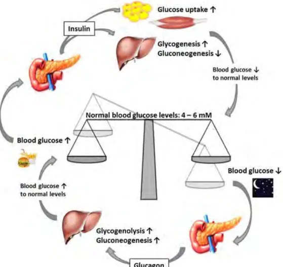
XVIII 1. Regulation of blood glucose ... 35
1.1 Regulation by pancreatic hormones ... 35 1.2 Role of gut hormones in blood glucose regulation ... 36 2. Metabolic syndrome and associated complications ... 37 3. Incidence of metabolic disorders ... 38 4. Link between metabolic disorders and microbiota ... 39 5. The role of intestinal permeability in metabolic disorder ... 40 6. Low-grade inflammation in metabolic disorders ... 42 7. Type 1 diabetes mellitus ... 43 Chapter III Stress ... 44 1. The HPA axis ... 44 2. Immunomodulatory effect of glucocorticoids... 45 3. Effect of stress on intestinal barrier ... 46 3.1 Stress affects microbiota composition ... 46 3.2 Stress affects intestinal permeability ... 47 3.3 Stress modifies intestinal immune system ... 47 3.4 Stress alters systemic immune response ... 48 3.5 Visceral sensitivity ... 48 4. Effects of stress on glucose metabolism ... 49 Chapter IV The Concept of Developmental Origins of Health and Disease (DOHaD) ... 73 1. Allostatic load ... 74 2. Evidence for the DOHaD hypothesis ... 74 3. Mechanisms ... 76 II. Part Objectives and Methodology ... 77 III. Part Results ... 81 First Result: Early Life Stress Induces Type 2 Diabetes-like Features in Aging Mice ... 82 Additional data ... 101
XIX Lysozyme activity is resolved at PND350 ... 101 Microbiota dysbiosis is different between PND50 and PND350 ... 102 Second Result: Role of Microbiota in MS-induced Glucose Intolerance in Aging Mice ... 103
Third Result: Early Life Stress Induces Glucose Intolerance and Insulin Secretion Failure in Aging Female Mice ... 126
Fourth Result: Non-Obese Diabetic Mouse Model is emphasizing Divergence Between in vivo and ex vivo Intestinal Permeability Measurements ... 145
IV. Part General Discussion ... 167 The Intestine is Playing a Crucial Role in Health and Disease ... 168 The Role of Low-Grade Inflammation in Aging - Inflammaging ... 174 The Model of Maternal Separation is a Useful Model To Decipher Etiology of Metabolic Disorders ... 178
The Effects of Maternal Separation Affirm the Importance of Neonatal Window and the DOHaD Concept ... 181
MS Induces Sexual Dimorphism ... 184 V. Part Conclusions ... 188 References ... 190
1
R
ESUMELe concept des origines développementales des maladies et de la santé (DOHaD) émet l’hypothèse d’une origine périnatale des maladies non-transmissibles (NCD). Le modèle de stress de séparation maternelle (MS) est largement utilisé chez le rongeur comme un paradigme d’événements adverses en période néonatale. Mon projet de doctorat, a eu pour but d’étudier les effets à long-terme du MS sur les fonctions de barrière intestinale, le métabolisme, la réponse immunitaire, l’auto-immunité ainsi que sur le microbiote, chez des souris mâles et femelles sauvages âgées sous régime standard. Le but étant de fournir des données expérimentales soutenant le lien entre stress néonatal et développement de désordres métaboliques ou auto-immuns à l’âge adulte.
Dans une première étude, nous avons montré que, le MS a induit une intolérance au glucose et une perte de la sensibilité à l’insuline associée à une dysbiose fécale chez des souris mâles sauvages C3H/HeN âgées de 350 jours (PND350). Le MS a diminué les concentrations d’IgG fécales et a augmenté les IgG anti-E. coli plasmatiques, représentant la réponse humorale contre le microbiote commensal. Le MS a diminué significativement la sécrétion d’17 et IL-22 en réponse à une stimulation du TcR et a augmenté la sécrétion de TNFα en réponse à une stimulation LPS dans une culture de cellules de la lamina propria de l’intestin grêle (siLP). Les mêmes résultats ont été obtenus au niveau systémique (rate). Nous avons ainsi démontré pour la première fois que le stress néonatal est un facteur de risque pour le développement des désordres métaboliques chez la souris sauvage âgée sous régime standard. Nous avons écarté un rôle exclusif du microbiote dans l’intolérance au glucose induite par le MS avec des expérimentations de transfert de microbiote fécal.
Dans une seconde étude, chez les femelles PND350 soumises au MS, on a observé une augmentation de la sécrétion d’IL-17 et IL-22 en réponse à une stimulation du TcR, et de TNFα avec ou sans stimulation au LPS par les cellules de la siLP. Nous avons observé en plus une inflammation systémique. Les souris MS ont développé une intolérance au glucose associée à une baisse de la sécrétion de l’insuline en réponse à un challenge au glucose. Le ratio de la surface des cellules β sur la surface du pancréas a légèrement diminué chez les MS et la valeur de ce ratio a corrélé positivement avec la sécrétion d’insuline induite par le glucose. En somme, le MS induit chez les souris femelles des effets à long terme sur l’immuno-métabolisme et l’homéostasie du pancréas.
2 Enfin, nous avons comparé les mesures de perméabilité intestinale in vivo (gavage) et
ex vivo (chambres de Ussing) avec du FITC-Dextran 4 kDa dans un modèle de diabète de type
1 (NOD - souris non-obese diabetic). De façon inattendue, les résultats ont différé en fonction des méthodes et cette différence n’est pas due à un défaut de la fonction rénale induite par le diabète. Par contre, nous avons observé un allongement de l’intestin grêle chez les souris diabétiques qui a corrélé positivement avec la perméabilité intestinale in vivo. Le diabète n’a pas modifié le transit intestinal, l’humidité des fèces et l’apparence histologique de l’intestin. En somme, nos résultats soulignent l’importance de distinguer la perméabilité intestinale, exprimée en cm/s mesurée ex vivo, et la notion d’exposition systémique aux antigènes luminaux mesurée in vivo.
Mon travail de thèse montre que les événements adverses néonataux sont un facteur de risque pour les NCD. Il est intéressant de noter que nos observations sur les souris âgées sont similaires aux observations épidémiologiques. En effet, nos résultats préliminaires suggèrent que les souris MS femelles développent des désordres métaboliques avec des caractéristiques auto-immunes; alors que les mâles développent des désordres métaboliques plus classiques : résistance à l’insuline. Mon travail sur le modèle MS souligne l’importance de la vie néo-natale dans l’établissement de l’homéostasie et conforte le concept de DOHaD.
Stress social, Métabolisme glucidique, Barrière intestinale, Réponse immunitaire, Origines développementales des maladies et de la santé.
3
S
UMMARYThe concept of Developmental Origins of Health and Disease (DOHaD) highlights the importance of early life period and raises the hypothesis that Non Communicable Diseases (NCD) could find their origins in perinatal environment. Neonatal maternal separation (MS) is a stress model widely used in rodents as a paradigm of early life adverse events. In my PhD project, I aimed to investigate in aging male and female wild-type mice under normal diet the long-term effects of neonatal MS on intestinal barrier function, metabolism, immunity, auto-immunity, as well as on microbiota. My work aimed to provide experimental data to support a link between early life stress and development of metabolic or autoimmune disorders with aging.
In our first study, MS led to glucose intolerance and loss of insulin sensitivity associated with fecal dysbiosis in Post Natal Day (PND) 350 wild-type C3H/HeN male mice fed a standard diet. Fecal IgG concentrations were decreased in MS mice compared to control mice, whereas anti-E. coli IgG, representing humoral response toward commensal microbiota, were significantly increased in plasma of MS mice. MS significantly decreased IL-17 and IL-22 secretion in response to TcR stimulation in small intestine lamina propria (siLP) culture. Besides, TNFα secretion in response to LPS-stimulation was slightly increased. The same results were obtained at systemic level (spleen). For the first time, we demonstrated that early life stress alone is a risk factor for metabolic disorders development in aging wild type mice under normal diet. The result of this project gave us the opportunity to question the role of microbiota in MS-induced glucose intolerance. Fecal microbiota transfer of MS mice microbiota was not sufficient to induce glucose intolerance.
In our second study in PND350 female, MS increased IL-17 and IL-22 by siLP cells in response to TcR stimulation. TNFα secretion with and without LPS stimulation was also increased by MS. Additionally, we observed systemic low-grade inflammation. MS mice developed glucose intolerance associated with decreased insulin secretion in response to glucose stimulus. Ratio of β-cell surface to pancreas surface was slightly decreased in MS mice compared to control. This ratio positively correlated with insulin secretion induced by glucose. Taken together, the results of our study showed that MS in wild type female mice under normal diet leaves a long-lasting imprinting on immune-metabolism and pancreas homeostasis.
We compared in vivo and ex vivo intestinal permeability measurements in a model of type 1 diabetes (NOD – non-obese diabetic mice). Intestinal permeability was assessed in vivo
4 by gavage and ex vivo in Ussing chambers with the marker FITC-Dextran 4 kDa. Surprisingly, the results of both methods were divergent. The difference between in vivo and ex vivo measurements could not be explained by altered renal excretion. Curiously, diabetic NOD mice had significantly longer small intestine than non-diabetic NOD mice and small intestine length positively correlated with intestinal permeability in vivo. However, there were no difference in intestinal transit time, feces humidity and histological appearance. Altogether, our results highlighted the importance to distinguish intestinal permeability, which is expressed as cm/s measured ex vivo, and the notion of systemic exposition to luminal antigen measured in vivo.
My PhD project shows that early life adverse events are a risk factor for NCD. Interestingly, our observations in aging mice are similar to epidemiological observations. Indeed, preliminary results suggested that female MS mice develop metabolic disorders with autoimmune characteristics but male MS mice develop classical metabolic disorders with insulin resistance. My work in MS model highlights the importance of early life in the establishment of homeostasis and comforts the concept of DOHaD.
Social stress, Glucose metabolism, Intestinal barrier, Immune response, Developmental origin of health and diseases (DOHaD).
5
6 The incidence of non-communicable diseases (NCDs) is constantly increasing in the last decades. Among NCDs, there are metabolic disorders, like obesity and type 2 diabetes, autoimmune diseases, as for example type 1 diabetes and allergies. Explications for the important rise in NCDs are numerous and diverse. There are for example, changes in lifestyle, nutrition, increased exposure to environmental contaminations and pollutants,
Early life is an important period for the establishment of lifelong beneficial homeostasis between the organism and its environment, especially its microbiota. The concept of developmental origins of health and disease (DOHaD) suggests that early life can have potent imprinting mechanism on host’s physiology and that adverse perinatal physical and social environment can lead to higher susceptibility to NCDs. Indeed, the thousand first days of our life, from conception to our second year’s birthday, are described to be decisive for future health.
The intestine is the body’s greatest surface in contact with its environment. In order to protect the organism from harmful substances and microorganisms but maintaining at the same time efficient nutrient absorption, the intestinal barrier is highly developed and regulated. Principal actors of the intestinal barrier are microbiota, intestinal epithelium with its highly regulated permeability and intestinal immune system. Interestingly, a tremendous amount of studies has shown that disturbed microbiota homeostasis (dysbiosis) is associated with a multitude of diseases, especially NCDs. Indeed, the gastro-intestinal tract seems to play a crucial role in organism’s health and disease.
This work is the continuation of previous studies performed in our laboratory, which highlighted the impact of stress on the integrity of the intestinal barrier. Especially early life stress has been of particular interest in our laboratory. Indeed, early life stress is known to impair intestinal barrier through induction of intestinal hyperpermeability, low-grade inflammation and microbiota dysbiosis in young rodents. Maternal separation (MS) is a paradigm of early life stress in rodents.
During my PhD I had the opportunity to write a review article, submitted and under review for the journal Frontiers in Immunology. Some parts of this article are quoted in I. Part: State of the Art.
8
C
HAPTERI
I
NTESTINALB
ARRIER ANDI
NTESTINALH
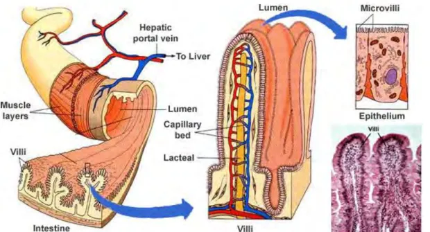
OMOEOSTASISThe human intestine is about six meters long and due to multiple folding through crypts of Lieberkühn, villi and microvilli, the surface is about 400 m2. This makes the intestinal
epithelium the mammalian organism’s biggest surface in contact with the environment (Figure
1). A principal role of intestine is to complete the digestion and absorb the nutrients but at the
same time, the intestine filters and defends the organism from harmful luminal content (pathogens, toxins…), while maintaining tolerance towards commensal microbiota and food antigens. Hence, the intestine is acting as a selective barrier. Considering the challenging, various, decisive and conflicting roles, it is not surprising that intestinal barrier function is highly diverse and well developed.
Figure 1 Intestinal anatomy. The intestinal tube is surrounded by two muscle layers, the longitudinal outer and
the circular inner layer. The mucosal surface is increased by circular folds. Villi are finger-like extensions of mucosa into the intestinal lumen increasing further the intestinal surface. Intestinal epithelial cells have
microvilli on apical site, which are also increasing the intestinal surface.
(https://histaminefriendlykitchen.com/wp-content/uploads/2017/08/TheVilliandmicrovilli.jpg)
1. Actors of intestinal barrier
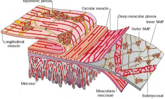
Among the main actors of intestinal barrier, there are intestinal microbiota, mucus, intestinal epithelium, intestinal immune system with its innate and adaptive response, harbored within the lamina propria (LP), and muscular layer, responsible for gut motility.
1.1 Intestinal microbiota
The intestinal microbiota is a complex bacterial community colonizing the gut. There are more than 100 trillions of microorganisms from two major phyla, Bacteriodetes and
9 Firmicutes, representing around 90% of gut microbiota (Rinninella et al., 2019). Other represented phyla are for example Actinobacteria and Proteobacteria. Colonization density is increasing from stomach (101-103 bacteria/g), via duodenum (104-105 bacteria/g) and
jejunum/ileum (108 bacteria/g) to colon (1012-1014 bacteria/g) (Nicolas, 2013). Microbiota plays
an important role by providing, extracting and absorbing various nutritional compounds and metabolites, as for example bile acids, amino acids, vitamins, lipids, short-chain fatty acids (SCFAs) (Jandhyala et al., 2015). Lactate, as an example for a bacterial metabolite, has stimulating effect on colonic proliferation (Okada et al., 2013). Also small intestinal stem cells are using lactate as an energy source (Rodríguez-Colman et al., 2017). Microbiota cans also influence host’s physiology through its metabolites. For example, butyrate, a SCFA produced by gut microbiota, is able to regulate host metabolism and immunity via its utilization as energy source through β-oxidation in enterocytes and inhibition of histone deacetylases, affecting host’s gene expression (Stilling et al., 2016). As an example, in cell cultures of murine mast cells and primary bone marrow mononuclear cells, butyrate has been shown to suppress proliferation and the production of the cytokines IL-6 and TNFα via the inhibition of histone deacetylase (Zhang et al., 2016). Microbiota participates also in the protection against pathogens colonization by occupation of ecological niches (competition for nutrients and space) (Jandhyala et al., 2015). Additionally, microbiota contributes to maturation of intestinal epithelium and immune system. This fact particularly strikingly visible in germ-free (GF) animals, which are living in sterile isolators, devoid of any microbiota. Indeed, GF mice are described to have thinner small intestine lamina propria (siLP) (Round and Mazmanian, 2009) and a great deficit in gut associated lymphoid tissue, as small Peyer’s patches containing only a little number of germinal centers (Macpherson et al., 2001). They have immature immune system, namely reduced number of IgA producing and CD4+ cells. Colonization by microbiota
can restore these defects (Falk et al., 1998; Helgeland et al., 1996; Macpherson et al., 2001). 1.1.1 Microbiota during life-time
Microbiota is depending on lots of different factors and can evolve during lifetime. In
utero the fetus is considered sterile (Hornef and Penders, 2017), even if this assumption is
debated. Some affirm that there is a microbiota in utero (Aagaard et al., 2014; Jiménez et al., 2008). However, the presence of bacteria is often confirmed by qPCR and rarely by culture of living bacteria and critics are highlighting potential contamination and missing controls (Perez-Muñoz et al., 2017). By definition, sterile is “devoid of life”. Detecting bacteria by qPCR demonstrate the presence of bacterial DNA but not living bacteria. In the following, I will
10 assume that fetus in utero is sterile and that first colonization takes place at birth. Highly dynamic microbiota during early life will be treated separately (cf. Chapter I 4. Ontogeny of intestine in early life 4.1 Colonization).
In adulthood, microbiota is more stable than in early life (Borre et al., 2014; Palmer et al., 2007) and less sensitive to external insults. Indeed, highly diverse microbiota is resilient to a certain amount of time-limited stressors, as for example disease, antibiotic treatment. Resilience means that the microbiota can recover its initial state (equilibrium) after an external insult. This is due to a variety of host-microbiota and inter-community interaction at one side and on the other side host-bacteria and bacteria-bacteria antagonisms (Sommer et al., 2017). Another factor modifying microbiota is aging. Claesson et al. observed in elderly Caucasians compared to younger individuals, a shift from Firmicutes towards Bacteriodetes (Claesson et al., 2011).
1.1.2 Dysbiosis
There are several external parameters able to influence microbiota composition. For example, diet is a potent influencer of microbiota composition (Rothe and Blaut, 2013). In addition, our environment is influencing the microbial community of our gut. Medical treatment, as for example antibiotics, and disease state influence gut microbiota as well (Ottman et al., 2012). If the pressure on the microbiota ecosystem is long-lasting or more important, or if initial microbiota is less resilient, there can be a shift towards a new equilibrium, perhaps a detrimental one, also called dysbiosis (Sommer et al., 2017).
It has been shown, that microbiota is different between different geographic environments. Indeed, people in countries with westernized lifestyle and non-westernized lifestyle have distinct microbiota (Pasolli et al., 2019). Migration of Hmongs and Karens towards the United States changes the microbiota composition strongly. A loss in diversity has been observed. These changes, increasing in long-term residents and the second generation post-immigration, were associated with increased incidence of obesity (Vangay et al., 2018).
Dysbiosis is defined by Petersen and Round as a disturbance in the microbiome structure that may consist in a loss of beneficial microorganisms, and/or expansion of pathobionts or harmful microorganisms (Figure 2) (Petersen and Round, 2014). Gut dysbiosis is described in tremendous diseases. Autoimmune disorders (AD), metabolic disorders, inflammatory bowel diseases (IBD), irritable bowel syndrom (IBS), even psychological disorders are associated with gut microbiota dysbiosis.
11
Figure 2 Dysbiosis. “A loss of beneficial microbes, expansion of pathobionts, and loss of diversity are events
that encompass dysbiosis. During healthy, homeostatic conditions the microbiota is composed of a diversity organisms that are known to benefit host development and health.” (Petersen and Round, 2014)
Important inter-individual microbiota composition has been observed (Franzosa et al., 2015). Further, regarding the high dynamics of microbiota during lifetime and the multiplicity of influencing factors, important questions seem to be: “Are there common elements to healthy microbiomes, in absence of overt disease? Which molecular elements of a personalized microbiome might be responsible for health outcomes, and how do they integrate with and maintain physiological processes such as the immune system and metabolism?” (the integrative HMP (iHMP) research Network consortium, 2019). The integrative Human Microbiome Project (HMP) address these question and was designed to gain a more complete view of microbial-host interactions in time. In this work frame, they found for example that not the microbiome but its prevalent molecular functions are correlated with host’s phenotype. Another finding of the more than 42 terabytes sampled omic data is that during disease microbiota is highly dynamic and could explain difficulties in cross-sectional studies to extract conclusive information (the integrative HMP (iHMP) research Network consortium, 2019).
1.2 Mucus
The gastrointestinal tract is covered by a mucus layer, protecting the epithelium from a direct contact with its microbiota. Mucus is secreted by goblet cells in the epithelial cell line and is made up mainly of mucin protein MUC2. The protein includes a high number of hydroxyl amino acids which serve as attachment sites for the O-glycans. After posttranslational modification in the Golgi apparatus, glycans represent around 80% of total mucin mass.
O-12 glycans can bind a great amount of water, which is giving the mucus its gel-forming property There are also transmembrane mucins, expressed by enterocytes (MUC1, 3A/B, 4…) but in smaller quantity (McGuckin et al., 2011).
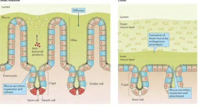
The mucus structures in small intestine and colon are different (Figure 3). In small intestine, there is one mucus layer; bacteria can penetrate into the mucus (Johansson and Hansson, 2016). In colon mucus is fourfold thicker and in animal models, two distinct mucus layers have been described: the outer, loose mucus layer, penetrated by gut bacteria; and the inner, firm, sterile mucus layer tightly attached to the epithelium (Atuma et al., 2001; Johansson et al., 2008).
Figure 3 Mucus layer organization in small intestine and colon. Mucus is produced and secreted by goblet
cells. The mucus layer in the small intestine is loose and not attached to the epithelium. Antibacterial products are present in mucus, limiting the penetration of bacteria towards the epithelium. In the colon, mucus is organized in outer and inner mucus layer. The outer loose mucus layer is enriched in bacteria, whereas the inner
mucus layer attached to the epithelium is almost free of bacteria (Johansson and Hansson, 2016).
This vision of mucus organization, though, is debated. Notably Kamphuis et al. propose a mucus organization shaped by colonic content. They found the firm and sterile mucus layer attached to fecal pellet but not to colonic epithelium in histological staining in rat. In empty colon, the epithelium is covered with a loose, discontinuous mucus layer and devoid of bacteria (Kamphuis et al., 2017). However, Kamphuis et al. believe that the principal role of the mucus layer, even though attached to fecal pellet and not to epithelium, is the containment of bacteria in a restricted location in intestine in order to protect the host’s epithelium from invasion (Kamphuis et al., 2017).
13 Mucus is enriched with antimicrobial peptides (Dupont et al., 2015) and secretory IgA (sIgA) (McGuckin et al., 2011), hence mucus invading bacteria will encounter antimicrobial peptides and sIgA and might be killed in this process. Indeed, it has been demonstrated that mucus has microbiota killing activity (Meyer-Hoffert et al., 2008). Invading bacteria in mucus are mainly detected by FISH technic that do not prove that bacteria are alive. The concerted action of physical, biochemical and immunological barrier components in the mucus are effective to keep bacteria away from epithelium and protect the host from infection.
1.3 Intestinal epithelium
The intestinal epithelium is formed by distinct cell types distributed along the crypt– villus axis. Although they all derived from a common stem cell progenitor located in the crypts, their morphology and roles differ (Figure 4) (for review (Gehart and Clevers, 2019)). The intestinal epithelium is renewed every five days, a part from Paneth cells whose lifespan has been estimated at about 60 days in mice (Ireland et al., 2005), and this constant renewing confers high plasticity and protection to the intestinal barrier since defective cells are removed rapidly (Gordon and Hermiston, 1994).
1.3.1 Different cell types in the intestinal epithelium
(a) Enterocytes
Enterocytes are present all over intestine, namely in small intestine and colon. They serve as physical barrier. Their main role is the absorption of nutrients and water. Additionally, they can also secrete antimicrobial components and they are non professional antigen presenting cells (expression of MHC2 without co-stimulatory molecules).
(b) Enteroendocrine cells
Enteroendocrine cells can be found in colon and small intestine. The high variety of enteroendocrine cells secrete different hormones. They exert regulation on intestinal motility and digestive processes but they can also influence central nervous system, via their anorexigenic effect. Some examples are mentioned in Table 1.
(c) Goblet cells
The mucus producing and secreting goblet cells are present all over the intestine but more abundant in colon. Goblet cells are not restricted to the production of mucus, they also can secrete antimicrobial components and play a role in antigen passage of antigens via goblet cell-associated passage (GAP), which will be developed later.
14
Table 1 Hormones produced by intestinal epithelial cells (examples).
Hormone Cell Functions (selection)
PYY L-cells Inhibits gastric emptying and intestinal motility; inhibits
gastric acid secretion and pancreatic exocrine function; anorexigenic; stimulates mucosal enterocyte proliferation
GLP-1 L-cells Incretin effect; delays gastric emptying; postprandial satiety
5-HT
(Serotonin) Enterochromaffin cells Intestinal motility; intestinal secretion; visceral sensation; appetite
Somatostatin D cells Major inhibitory hormone for digestive endocrine and
exocrine function; stimulates colonic peristalsis
GIP K-cells Inhibition of gastric acid secretion, stimulation of insulin
secretion Cholecystekinin
(CKK) I-cells Anorexigenic,
(d) Paneth cells
Paneth cells are only present in the small intestine. They are located at the bottom of the crypt in proximity with intestinal stem cells. Paneth cells secrete antimicrobial peptides (AMP), as for example lysozyme. Due to their location, they also contribute to the protection of stem cells by secreting AMP highly enriched in the bottom of the crypt and thus support the stem cell niche.
(e) M cells
Microfold (M) cells can be found in small intestine in the follicle-associated epithelium. They play an important role in the antigen uptake, facilitated by microfolds (short fold-like invaginations) on apical site. On their basolateral site, they have a large pocket promoting close contact with dendritic cells (DC) or lymphocytes. Additionally, the follicle-associated epithelium secrete less mucus, less AMP and sIgA, which help bacteria and antigens to reach M cells in order to be processed and transported for antigen presentation. Even though M cells represent a facilitating pathway for antigen to prime oral tolerance, they are not mandatory (Spahn et al., 2002).
(f) Tuft cells
Tuft cells are present all along intestine. They are important for helminth detection and innate lymphoid cell 2 (ILC2) expansion (Gerbe et al., 2016). They are also responsible for opioid production and secretion in the intestinal epithelium (Gerbe et al., 2011).
15
Figure 4 Different cell types in (A) small intestine and (B) colon. (A) Most of small intestinal epithelial cells
are enterocytes, secreting antimicrobial peptides (RegIIIγ, β-defensins and cathelicidins). Paneth cells are located at the base of the crypts, they secrete antimicrobial peptides (sPLA2, lysozyme and α-defensins). Goblet cells are secreting mucus. Antigens can enter into lamina propria via M cells on the surface of Peyer’s Patches and via goblet cell associated passage (GAP); Underlying dendritic cells (DC) are sampling entered antigens. The lamina
propria is rich in innate (macrophages, monocytes, dendritic cells) and adaptive (B cells, T cells, intraepithelial
cells -IEL) immune cells. (B) Colonic epithelium consists mainly out of colonocytes and goblet cells. Goblet cells are forming the mucus layer via production of Muc2 but they are also secreting other proteins (RELMβ,
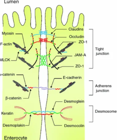
17 claudins (Furuse et al., 1993), occludins (Furuse et al., 1998), junctional adhesion molecule (JAM) (Martìn-Padura et al., 1998) and tricellulin (Ikenouchi et al., 2005). In the cell, these protein complexes are linked via cytosolic scaffold proteins (zonula occludens) with the perijunctional actomyosin ring. Myosin light chain (MLC) phosphorylation by MLC-kinase (MLCK) is regulating contraction and tension of actomyosin ring (Madara et al., 1987).
Figure 6 Intercellular junctions of intestinal epithelial cells. “The intercellular junctions are organized by
different protein complexes, including tight junctions (TJs), adherens junctions (AJs), and desmosomes. TJ complexes locate at the apical ends of the lateral membranes of intestinal epithelial cells and consist of transmembrane and intracellular scaffold proteins. Extracellular loops of the trans- membrane proteins (occludin,
claudins, JAMs, and tricellulin) are regulating paracellular permeability. The intracellular domains of the transmembrane proteins interact with the intracellular scaffold proteins such as zonula occludens (ZO) proteins, which in turn anchor the transmembrane proteins to the actin cytoskeleton. Myosin light chain kinase (MLCK) is
associated with the perijuctional actomyosin rings and regulates paracellular permeability through myosin contractility. The AJs along with desmosomes provide strong adhesive bonds between the epithelial cells and
also intercellular communication” (Suzuki, 2013).
Another paracellular passage has been described by Rescigno et al. Dendritic cells (DC) can extent dendrites through the paracellular space and sample bacteria directly in the lumen. Since DC also express tight junction proteins intestinal barrier remains preserved (Rescigno et al., 2001).
(b) Transcellular permeability
Bigger molecules (> 600 Da), such as peptides and food antigens can be transported through the epithelial cell via transcellular transport route (Figure 5) (Gardner, 1988; Heyman
18 et al., 1982). Most of transcellular passage takes place in M cells, but enterocytes are also able to transport peptides. In the great majority the molecules are processed by the lysosome during their passage through cells, only a small quantity can reach LP intact (Heyman et al., 1996, 1982).
Transcellular passage can also be mediated by retro-transport of immunoglobulins from the lumen to the serosal compartment via receptor expression on the intestinal epithelium, as for example the neonatal Fc receptor (FcRn) observed in suckling rats responsible for the transmission of passive immunity by the mother (Blumberg et al., 1995; Jones and Waldmann, 1971; Simister and Mostov, 1989). FcRn has also been described on human fetal intestinal epithelial cells (Israel et al., 1997).
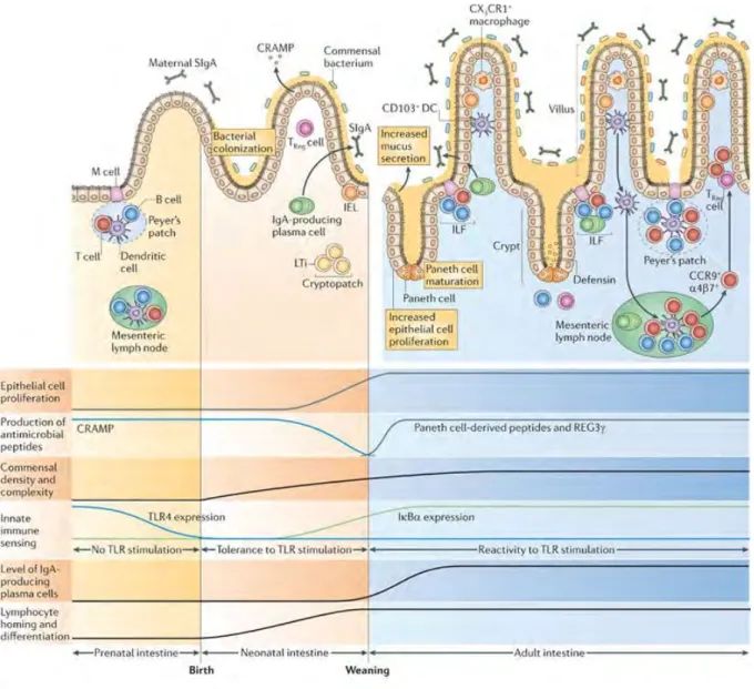
Goblet cells in the small intestine have been shown in the steady state, to let pass low molecular weight soluble antigens from the intestinal lumen to underlying tolerogenic-dendritic cells (DC) (McDole et al., 2012). This phenomenon is called goblet-cell associated passage (GAP). However in the colon, this antigen passage via goblet cells is restricted to a precise time window, from post-natal day 10 (PND10) to PND20 and later downregulated by microbiota in rodents (Knoop et al., 2017). As a consequence, induction of gut microbiota tolerance by the colonic immune system is crucial in early-life period and supporting the hygiene hypothesis, which proposes that the increased incidence of immune-mediated diseases is due to decreasing exposure to microorganisms in early life (Al Nabhani and Eberl, 2017; Bach, 2018).
There are multiple methods for the determination of intestinal permeability (IP) including in vivo and ex vivo measurements. First, in vivo methods are often used due to the simplicity of protocol and the absence for the need of complex device. Indeed, several non-metabolized molecules can be used to determine their passage across intestinal barrier in vivo in animal and human. The individual is receiving the molecule via the oral route and after a fixed time, blood or urine is collected and the marker is detected according to its properties (radioactivity, fluorescence, etc). Thanks to distinct chemical characteristics and degradation process by intestinal microbiota, different markers can be used to assess permeability of different intestinal segments (Travis and Menzies, 2015). Another possibility to assess intestinal permeability is by dosing serological markers without any previous gavage, as for example zonulin-1 (Fasano, 2011) or endotoxin (Cani et al., 2007) that will represent respectively intestinal homolog of a Vibrio cholerae enterotoxin that reversibly increase IP and a marker of intestinal lipopolysaccharide translocation in the blood.
19
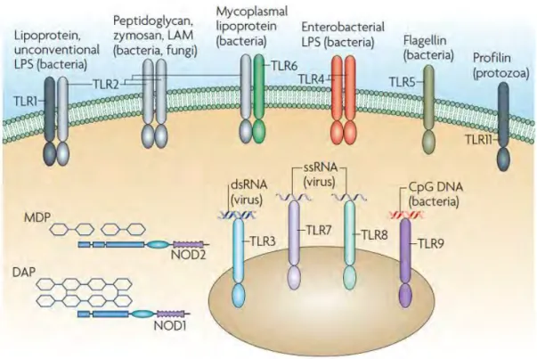
1.4 Intestinal immune system
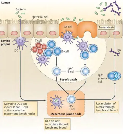
As the previous paragraph shows, the intestinal immune system is in close relationship with the other actors of the intestinal barrier and important to confer tolerance but at the same time protection to the host. Intestinal immune system is highly developed. Indeed, it is the largest immune system in the organism. There are particular structures – the gut-associated lymphoid tissue (GALT), numerous innate and adaptive immune cells and a large variety of immune active actors.
The first line of host defense is the innate immune system. 1.4.1 Antimicrobial peptides
Antimicrobial peptides (AMP) are crucial for the maintenance of intestinal homeostasis. They are controlling intestinal colonization. AMP are oligopeptides or even small proteins (5-100 amino acids), targeting a vast spectrum of microorganisms (bacteria, fungi, ..) (Bahar and Ren, 2013; Ganz and Lehrer, 1998). All AMP derive from bigger precursors and undergo important posttranslational modifications (Wilson et al., 1999). AMP repertoire is different between all species, even those who are related (Zasloff, 2002). Even different mouse strain have different AMP repertoire (Gulati et al., 2012). These differences can largely influence the microbiota and explain inter-species and inter-strain discrepancies in microbiota composition. Besides, it could confer diverse resistance capacities towards pathogens.
The presence of AMP in the intestine is also highly regionalized. AMPs are produced by enterocytes and goblet cells, but also by specialized cells, the Paneth cells. Paneth cells are only present in the small intestine, they produce and secrete α-defensins, RegIIIγ, sPLA2 (phospholipase-A2), and lysozyme (Vaishnava et al., 2008).
Lysozyme can also be produced by neutrophils; it is present in saliva and maternal milk. Defensins possess three intramolecular disulfide bonds and a β-sheet structure. In vertebrates, there are two families, α-defensins, β-defensins. In mice, α-defensins are called cryptidins. Cathelin-related antimicrobial peptide (CRAMP) is member of the cathelicidin family, present in mice and similar to LL37 in human
Some AMP are targeting a fundamental differences between microbes and multicellular animals: microbes’ surface is negatively charged whereas plants and animals cell membranes are composed of lipids without charge (Matsuzaki, 1999). Cationic AMP attack microbes by interaction with their membrane, lipids are displaced, membrane structure is altered as a
20 consequences and sometimes AMP enters into the cell. This mechanism is called Shai– Matsuzaki–Huang (Matsuzaki, 1999; Shai, 1999; Yang et al., 2000).
Due to their high aggressiveness, AMP are highly regulated and undergo numerous maturation steps. AMP are secreted either constitutively or after pattern recognition receptor (PRR) activation by microbe-associated molecular patterns (MAMPs) (Yokoi et al., 2019).
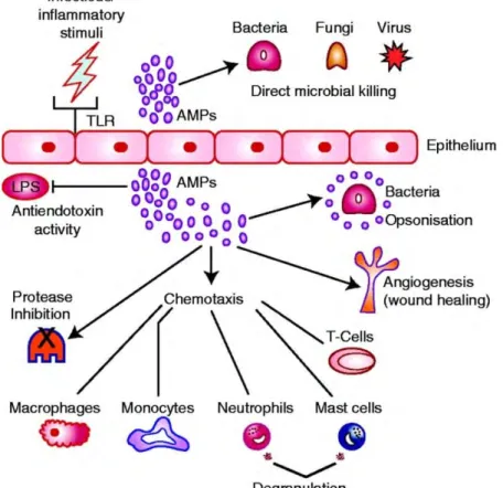
Figure 7 Functions of AMP. AMP can directly kill bacteria, virus and fungi. They can favor pathogen clearance
by opsonisation of bacteria, they contribute to angiogenesis. AMP have immunomodulatory function through chemotaxis towards immune cells. They inhibit proteases and can bind endotoxin thus influencing TLR signaling
(Frew and Stock, 2011).
AMP also have immunomodulatory functions (Figure 7). They have chemiotactic properties towards monocytes, macrophages, neutrophils and mast cells (Oppenheim et al., 2003). They can recruit T cells via induction of inflammatory cytokines (Lai and Gallo, 2009). They can favor bacterial clearance by opsonisation (Wilkinson et al., 2009). Immune response can also regulate AMP as for exemple IL-22 can induce RegIIIγ release (Wang et al., 2018).
1.4.2 Innate immune cells
Additional to secreted AMP, which are regulating microbiota in the lumen, the lamina
propria (LP) harbors a wide variety of innate immune cells, supporting the defense of host’s
21 (a) Dendritic cells, macrophages
Dendritic cells (DCs) and macrophages are mononuclear phagocytes able to present antigens via major histocompatibility complex II (MHCII). In the intestine about 80% of CD11chi are CD103+, which is a tolerogenic phenotype of DCs, since they induce FoxP3+
regulatory T cell (Treg) development (Chirdo et al., 2005; Coombes et al., 2007). This is underlining the fact, that tolerance to luminal antigens is of great interest throughout the intestine.
(b) Neutrophils
Neutrophils can initiate the immune response via leukotrienes and prostaglandins; kill pathogens by releasing AMPs or reactive oxygen species; or phagocyte cellular debris. They can also release IL-17 and IL-23 (Mei et al., 2012). Neutrophils also produce lipocalin-2 (lcn-2), a marker of intestinal inflammation, which is sequestrating iron and thus limiting bacterial growth (Cowland and Borregaard, 1997; Yang et al., 2002) and myeloperoxidases (MPO) with great antimicrobial activity due to their oxidative potential (Serteyn et al., 2003). In the intestine neutrophils are not very numerous, unless pathological conditions, such as ulcerative colitis (Robinson et al., 1997), coeliac disease and infection (Bennouna et al., 2003).
(c) Eosinophils
Eosinophils are leucocytes with pro-inflammatory properties. Only a small percentage of circulating blood cells are eosinophils. In steady-state condition, most of them reside in the gastrointestinal tract within the LP (Powell et al., 2010; Zuo and Rothenberg, 2007). Most of them are localized in the duodenum (Mishra et al., 1999). The roles of eosinophils are multiple. Following cellular activation, eosinophils secrete toxic inflammatory mediators (proteins and peroxidase) that are stored in intracellular vesicles. These mediators destroy tissues and insert pores into membranes of target cells. By generating toxic oxygen radicals, they increase smooth muscle reactivity (Al-Haddad and Riddell, 2005). Eosinophils are also supporting plasma cell (Chu et al., 2011) function and the recruitment of DCs (Jacobsen et al., 2011).They are playing a role in inflammatory bowel disease (IBD), mainly acting as inflammatory and pro-motility agent (Al-Haddad and Riddell, 2005).
(d) Mast cells
In the immune response to infection, mast cells are important effector cells. They release inflammatory mediators and recruit other immune cells, driving the pro-inflammatory immune response. Mast cells have an important role in the intestinal barrier, since epithelial function and integrity are regulated by them. Additionally, mast cells modulate innate and adaptive