Uukuniemi virus as a tick-borne virus model
1Magalie Mazelier1, Ronan Nicolas Rouxel2,#, Michael Zumstein3,&, Roberta Mancini3,
2
Lesley Bell-Sakyi4, Pierre-Yves Lozach1,2,3
3
From 1CellNetworks – Cluster of Excellence and Department of Infectious Diseases, Virology, 4
University Hospital Heidelberg, Germany; 2INRS-Institut Armand-Frappier, Université du 5
Québec, Laval, Canada; 3ETHZ, Institute of Biochemistry, Zurich, Switzerland; 4The Pirbright 6
Institute, Pirbright, United Kingdom 7
Present address: #UR 0892, INRA, CRJ, Jouy-en-Josas, France; ÐZ, Institute of 8
Biogeochemistry and Pollutant Dynamics, Zurich, Switzerland 9
Correspondence: P.Y.L.; E-Mail: [email protected]; Phone +49 6221-10
56-1328; Fax +49 6221-56-5003 11
Running title: Uukuniemi virus in tick cells 12
Keywords: arbovirus, bunyavirus, glycan, glycosylation, host alternation, phlebovirus, tick, tick-13
borne virus, Uukuniemi virus, tick cell line 14
JVI Accepted Manuscript Posted Online 18 May 2016 J. Virol. doi:10.1128/JVI.00095-16
Copyright © 2016 Mazelier et al.
This is an open-access article distributed under the terms of the Creative Commons Attribution 4.0 International license.
on June 27, 2016 by INRS-Institut Armand-Frappier
http://jvi.asm.org/
Abstract
15
In the last decade, novel tick-borne pathogenic phleboviruses in the family Bunyaviridae, all 16
closely related to Uukuniemi virus (UUKV), have emerged in different continents. To reproduce 17
the tick-mammal switch in vitro, we first established a reverse genetics system to rescue UUKV 18
with a genome the closest to that of the authentic virus isolated from the Ixodes ricinus tick 19
reservoir. The IRE/CTVM19 and IRE/CTVM20 cell lines, both derived from I. ricinus, were 20
susceptible to the virus rescued from plasmid DNAs and supported production of the virus over 21
many weeks, indicating that infection is persistent. The glycoprotein GC was mainly highly-22
mannosylated on tick cell-derived viral progeny. The second envelope viral protein, GN, carried 23
mostly N-glycans not recognized by the classical glycosidases PNGase F and Endo H. Treatment 24
with β-mercaptoethanol did not impact the apparent molecular weight of GN. On viruses 25
originating from mammalian BHK-21 cells, GN glycosylations were exclusively sensitive to 26
PNGase F and the electrophoresis mobility of the protein was substantially slower after the 27
reduction of disulfide bonds. Furthermore, the amount of viral nucleoprotein per foci forming 28
units differed markedly whether viruses were produced in tick or BHK-21 cells, suggesting a 29
higher infectivity for tick cell-derived viruses. Together, our results indicate that UUKV particles 30
derived from vector tick cells have glycosylation and structural specificities that may influence 31
the initial infection in mammalian hosts. This study also highlights the importance of working 32
with viruses originating from arthropod vector cells when investigating the cell biology of 33
arbovirus transmission and entry into mammalian hosts. 34
Importance
35
Tick-borne phleboviruses represent a growing threat to humans globally. Although ticks are 36
important vectors of infectious emerging diseases, previous studies have mainly involved virus 37
stocks produced in mammalian cells. This limitation tends to minimize the importance of host 38
alternation in virus transmission to humans and initial infection at the molecular level. With this 39
study, we have developed an in vitro tick cell-based model that allows production of the tick-40
borne Uukuniemi virus to high titers. Using this system, we found that virions derived from tick 41
cells have specific structural properties and N-glycans that may enhance virus infectivity for 42
mammalian cells. By shedding light on molecular aspects of tick-derived viral particles, our data 43
illustrate the importance of considering the host switch in studying early virus-mammalian 44
on June 27, 2016 by INRS-Institut Armand-Frappier
http://jvi.asm.org/
receptor/cell interactions. The information gained here lays the basis for future research, not only 45
on tick-borne phleboviruses, but on all viruses and other pathogens transmitted by ticks. 46
on June 27, 2016 by INRS-Institut Armand-Frappier
http://jvi.asm.org/
Introduction
47
The Bunyaviridae is the largest family of RNA viruses. With over 350 members distributed 48
worldwide and classified into five genera (Tospovirus, Nairovirus, Orthobunyavirus, 49
Phlebovirus, and Hantavirus), these viruses represent a global threat to public health, livestock,
50
and agricultural productivity (1). Hantaviruses apart, bunyaviruses are mainly transmitted by 51
arthropods including sandflies, mosquitoes, and ticks. In the past five years, a number of 52
emerging tick-borne phleboviruses have been reported in different continents. Severe fever with 53
thrombocytopenia syndrome virus (SFTSV) in Asia and Heartland virus (HRTV) in North 54
America are recent examples of new tick-borne phleboviruses causing severe and often fatal 55
disease in humans (2-5). Other phleboviruses genetically related to SFTSV and HRTV have also 56
been recently isolated from ticks in different parts of the world (6). The increasing number of 57
outbreaks and the apparent wide distribution in tick reservoirs demonstrate that these viruses 58
must be taken seriously as emerging agents of disease. Currently, no vaccines or treatments are 59
approved for human use. 60
Tick-borne phleboviruses are enveloped, roughly spherical with a diameter of about 100 nm, 61
and have a tripartite, single-stranded RNA genome that exclusively replicates in the cytosol (7). 62
The large (L) RNA segment codes for the viral RNA-dependent RNA polymerase L, and the 63
medium (M) segment for the glycoproteins GN and GC, both in a negative-sense orientation in 64
the viral genomic RNA (vRNA) (7, 8). In addition to the vector, one of the main distinctions 65
between tick-borne phleboviruses and the phleboviruses vectored by dipterans or mosquitoes is 66
the absence of a sequence encoding a non-structural protein NSm in the M segment (7, 8). The 67
small RNA segment (S) codes for the nucleoprotein N and the non-structural protein NSs in an 68
ambisense manner (7, 8). The proteins N and NSs are translated from subgenomic mRNAs 69
transcribed from the vRNA and the antigenomic, replicative-intermediate RNA (cRNA) 70
respectively (7, 8). In the virions, the protein N is associated with the virus RNA genome and, 71
together with the polymerase L, constitutes the ribonucleoproteins (RNPs) (7). In the virus 72
envelope, the glycoproteins GN and GC form spike-like projections responsible for virus 73
attachment to host cells and for acid-activated penetration by membrane fusion from late 74
endosomal compartments (7, 9). 75
During natural transmission to vertebrates, tick-borne phleboviruses are introduced into the 76
skin dermis of hosts following bites by infected ticks. Although ticks are both harmful parasites 77
on June 27, 2016 by INRS-Institut Armand-Frappier
http://jvi.asm.org/
and vectors of several emerging diseases, not only those caused by phleboviruses, little 78
information on the cell biology of ticks is available (10-14). As a consequence, composition and 79
structure of tick cell-derived phleboviral particles remain largely undefined. Our knowledge of 80
the initial infection of vertebrate hosts, cellular receptors and cell entry is mainly based on the 81
use of virus stocks produced in mammalian cells. SFTSV has been shown to subvert the non-82
muscle myosin heavy chain IIA for early steps of infection (15). Rhabdoviral particles 83
pseudotyped with the glycoproteins of SFTSV (SFTSV-pp) have been found to infect 84
macrophages (16). Human dendritic cells (DCs) are productively infected by many bunyaviruses, 85
including Uukuniemi virus (UUKV), a phlebovirus originally isolated from the tick Ixodes 86
ricinus in the 1960s (17-21). To enter and infect DCs, SFTSV-pp and UUKV have been shown
87
to exploit DC-SIGN, a C-type (calcium-dependent) lectin that binds high-mannose N-glycans on 88
the viral glycoproteins through its C-terminal carbohydrate recognition domain (CRD) (16, 17). 89
Interactions with L-SIGN, a C-type lectin closely related to DC-SIGN but expressed in liver 90
sinusoidal endothelial cells, have also been recently documented for SFTSV-pp and UUKV (16, 91
22). After binding, SFTSV-pp and UUKV depend on endosomal acidification for penetration and 92
infection (16, 23). UUKV is a late-penetrating virus, belonging to a large group of viruses that 93
depend on late endosomal maturation for productive entry(9, 23). 94
We focus here on UUKV, a virus that shares high sequence homology with SFTSV and 95
HRTV (8, 24, 25). However, UUKV is not associated with any disease in humans and is a 96
validated BSL2 surrogate for arthropod-borne bunyaviruses of higher biosafety classification 97
(26). Major advances into various aspects of the phlebovirus life cycle, including virion 98
structure, receptors, cell entry and assembly, have been achieved through the use of UUKV (9, 99
23, 27-32). To determine whether tick cells support phlebovirus productive infection in vitro, and 100
whether tick cell-derived phleboviral particles infect human or other mammalian cells, we 101
rescued UUKV from cDNAs with a genome the closest to that of the authentic virus isolated 102
from ticks. Using this system, we examined infection and virus production in tick cells, assessed 103
the progeny virus for interactions with DC-SIGN expressed in mammalian cells, analyzed the 104
glycans and the electrophoresis mobility of the virus glycoproteins GN and GC, and tested the 105
infectivity of viral progeny in mammalian cells. 106
on June 27, 2016 by INRS-Institut Armand-Frappier
http://jvi.asm.org/
Materials and methods
107
Cells and viruses. All products used for cell culture were obtained from Life Technologies or
108
Sigma Aldrich. Baby Hamster Kidney cells (BHK-21) were grown in Glasgow’s minimal 109
essential medium (GMEM) supplemented with 10% tryptose phosphate broth, 5% fetal bovine 110
serum (FBS), 1% glutaMAX, 100 units.mL-1 penicillin, and 100 µg.mL-1 streptomycin (33). 111
Human B (Raji) and epithelial (HeLa) cells that stably express DC-SIGN were cultured 112
according to ATCC recommendations (17, 34). All mammalian cell lines were grown in an 113
atmosphere of 5% CO2 in air at 37°C. The tick cell lines IRE/CTVM19 and IRE/CTVM20 were 114
cultured in L-15-based medium in sealed, flat-sided tubes (Nunc) in ambient air at 28°C as 115
reported elsewhere (35-37). The prototype UUKV strain 23 (UUKV S23) was originally isolated 116
from the tick I. ricinus in the 1960s (i.e. the virus in tick suspension) (21). The UUKV strain 117
used in this study results from five successive plaque purifications of UUKV S23 in chicken 118
embryo fibroblasts (CEFs) and subsequent passages in BHK-21 cells (38, 39). Virus multiplicity 119
of infection is given according to the titer determined in BHK-21 cells. 120
Antibodies and reagents. The mouse monoclonal antibodies 8B11A3, 6G9E5 and 3D8B3 are
121
directed against the UUKV nucleoprotein N and the glycoproteins GN and GC respectively (40). 122
The rabbit polyclonal antibodies K1224 and K5 are directed against the UUKV glycoproteins GN 123
and GC respectively (41). All of these antibodies were a kind gift from Anna Överby and the 124
Ludwig Institute for Cancer Research (Stockholm, Sweden). The rabbit polyclonal antibody U2 125
has been described previously, and recognizes the UUKV proteins N, GN, and GC (17). The 126
neutralizing anti-DC-SIGN mouse monoclonal antibody IgG2a (mAb1621) was purchased from 127
R&D Systems. NH4Cl and EDTA were obtained from Sigma Aldrich and dissolved in deionized 128
water. 129
Plasmids. The expression plasmids pUUK-N and pUUK-L were a kind gift from Anna Överby
130
and code for, respectively, the UUKV nucleoprotein N and polymerase L (39). The cDNAs 131
corresponding to the S, M, and L segments of UUKV were synthesized by RT-PCR from vRNA 132
extracts of purified-virus stock using the reverse transcriptase Superscript III (Life 133
Technologies). Their amplification as a single PCR product was carried out using the Herculase 134
II fusion DNA polymerase (Agilent). The PCR products were then cloned between the murine 135
Pol I RNA polymerase promoter and terminator sequences in the pRF108 vector (a generous gift 136
on June 27, 2016 by INRS-Institut Armand-Frappier
http://jvi.asm.org/
from Ramon Flick, Bioprotection Systems Corporation) (30). The resulting Pol I-driven plasmids 137
(pRF108-S, pRF108-M, and pRF108-L) encoded each of the antigenomic UUKV RNA 138
molecules (i.e. S, M, and L segments). The point mutation G2386A in the M segment was 139
obtained with the QuikChange XL site-directed mutagenesis kit (Agilent) using the plasmid 140
pRF108-M as a template. The complete list of primers and restriction enzymes used for cloning 141
and mutagenesis is shown in Table 1. 142
Rescue of UUKV from plasmid DNAs. UUKV was rescued by transfecting BHK-21 cells (0.6
143
× 106) with the expression plasmids pUUK-L (1 µg) and pUUK-N (0.3 µg) together with 0.5 µg 144
each of pRF108-S, pRF108-M, and pRF108-L. Transfection was performed in the presence of 145
Lipofectamine 2000 (Life Technologies) using a ratio of 3.8 µL to 1 µg of plasmids in 400 µL of 146
OptiMEM (Life Technologies). One hour post-transfection, complete GMEM medium 147
containing 2% FBS was added to the cells. After 5 days, supernatants were collected, clarified, 148
and titrated as described below. Rescued viruses were passaged a minimum of five times in 149
BHK-21 cells. 150
Virus titration by focus-forming assay. Following infection of confluent monolayers with
10-151
fold dilutions of virus in FBS-free medium, cells were grown in the presence of medium 152
containing 5% serum and supplemented with 0.8% carboxymethyl-cellulose to prevent virus 153
spread. Foci were revealed with a diaminobenzidine solution kit (Vector Laboratories) after a 154
two-step immunostaining with the antibody U2 (1:1,000) and an anti-rabbit horseradish 155
peroxidase-conjugated secondary antibodies (Vector Laboratories) (23). 156
Deglycosylation. To assess the glycosylation pattern of the UUKV glycoproteins, virus stocks
157
purified through a 25%-sucrose cushion were denaturated and exposed to one of the five 158
following treatments: 1,000 units of Endo H (Promega), 5 units of PNGase F, 0.005 units of α-159
2(3,6,8,9)-neuraminidase, 0.003 units of β-1,4-galactosidase, and 0.05 units of β-N-160
acetylglucosaminidase (all enzymes from Merck Millipore) according to the manufacturer’s 161
recommendations, and then analyzed by SDS-PAGE on a 4-12% or 10% Bis-Tris Nu-PAGE 162
Novex gel (Life Technologies) and immunobloting. 163
Protein analysis. Viral protein extracts from virus stocks purified through a 25% sucrose
164
cushion were analyzed by SDS-PAGE (Nu-PAGE Novex 4-12% Bis-Tris gel, Life 165
Technologies) and transferred to a PVDF membrane (iBlot transfer Stacks, Life Technologies) 166
on June 27, 2016 by INRS-Institut Armand-Frappier
http://jvi.asm.org/
(17, 23). When indicated, purified viruses were pre-treated with glycosidases or β-167
mercaptoethanol (40%). For western blot analysis, the PVDF membranes were first incubated 168
with primary mouse monoclonal antibodies 8B11A3 (1:2,000), 3D8B3 (1:100) or 6G9E5 169
(1:100), or rabbit polyclonal antibodies K5 (1:250-1,000), 1224 (1:500-1,000) or U2 (1:2,000), 170
all diluted in TBS containing 0.1% Tween and 5% milk, and then with an mouse or anti-171
rabbit horseradish peroxidase-conjugated secondary antibody (1:10,000, Santa Cruz), 172
respectively. Bound antibodies were detected by exposure to enhanced chemiluminescence 173
reagents (ECL, GE Healthcare or Life Technologies). For quantitative detection of viral proteins, 174
membranes were first incubated with the rabbit polyclonal antibody U2 (1:1,000) and then with 175
an anti-rabbit infrared fluorescence (IRDye) secondary antibody and analyzed with the Odyssey 176
Imaging Systems and the software Image Studio (Li-Cor Biosciences). 177
Infection assays. Mammalian cells were infected with virus at different MOIs in medium
178
without FBS at 37°C for one hour. Virus supernatant was then replaced by complete culture 179
medium and cultures were incubated for up to 64 hours before fixation. Tick cells were exposed 180
to viruses at different MOIs in culture medium containing FBS at 28°C for up to 48 hours. When 181
used for microscopy, tick cells were seeded on poly-L-Lysin (0.01%)-coated coverslips at 28°C 182
on the day before infection. For inhibition assays, cells were pretreated with inhibitors at 183
different concentrations for 30 min and exposed to UUKV in the continuous presence of the 184
inhibitors. The infection was monitored by either wide-field fluorescence microscopy or flow 185
cytometry. 186
Flow cytometry. The flow cytometry-based infection assay has been described previously (23).
187
Briefly, after fixation and permeabilization with 0.1% saponin, infected cells were incubated 188
with the mouse monoclonal antibodies 8B11A3 (1:400), 6G9E5 (1:400) or 3D8B3 (1:500) at 189
room temperature for one hour, washed, and subsequently exposed to Alexa Fluor (AF) 647-190
conjugated secondary anti-mouse antibodies (1:500, Life Technologies) at room temperature for 191
one hour. When the mouse monoclonal antibody 1621 (25 µg.mL-1) was used in infection assays 192
to neutralize DC-SIGN, UUKV-infected cells were immuno-stained with the rabbit polyclonal 193
antibody U2 (1:400) and AF647-conjugated secondary anti-rabbit antibody (1:500, Life 194
Technologies). Flow cytometry-based analysis involved the use of a FACS Calibur cytometer 195
(Becton Dickinson) and Flowjo software (Treestar). 196
on June 27, 2016 by INRS-Institut Armand-Frappier
http://jvi.asm.org/
Fluorescence microscopy. Infected cells were fixed and permeabilized with PBS containing
197
0.1% Triton X-100 (Merck Millipore), incubated with the mouse monoclonal antibody 8B11A3 198
(1:1,000) at room temperature for one hour, washed, and then exposed to AF488-conjugated 199
secondary anti-mouse (1:800, Life Technologies) at room temperature for one hour. Nuclei were 200
subsequently stained with Hoechst 33258 (0.5 µg.mL-1, Life Technologies). Infection was 201
quantified by counting cells in three independent fields and cells were imaged with an Olympus 202
IX81 microscope. 203
Statistical Analysis. The data shown are representative of at least three independent
204
experiments. Values are given as the mean of triplicates ± standard deviation (SD). 205
Sequencing of the full-length M segment isolated from UUKV-infected ticks. Questing
206
nymphs of the tick I. ricinus were collected in the region of Ramsvik and Hindens Rev (Sweden, 207
2013). Pools of 25 nymphs were homogenized and the total RNA was extracted with a magnetic 208
bead-based protocol as described elsewhere (kind gift of Janne Chirico, National Veterinary 209
Institute, Uppsala, Sweden and Sara Moutailler, ANSES, Maisons-Alfort, France) (42). The 210
cDNA corresponding to the M segment of UUKV was synthesized by RT-PCR with the reverse 211
transcriptase Superscript III (Life Technologies) and the specific primer RT-M (Table 1) before 212
amplification as a single PCR product using the pfu DNA polymerase (Promega) and the primers 213
UUKV-M-5NC and UUKV-M-3NC (Table 1). PCR products were analyzed with a capillary 214
sequencer by ABI (Eurofins Scientific). 215
GenBank accession numbers. The GenBank accession numbers for the nucleotide sequence of
216
the M segment of the tick isolates RVS and HRS are KX219593 and KX219594 respectively. 217
on June 27, 2016 by INRS-Institut Armand-Frappier
http://jvi.asm.org/
Results
218
Recovery of UUKV S23 from RNA polymerase I (Pol I)-driven plasmid DNAs. The UUKV
219
lab strain that we used in this study as a template for cloning purposes results from the adaptation 220
of the prototype tick isolate strain 23 (UUKV S23) to BHK-21 cells after successive plaque-221
purifications in CEFs (21, 38, 39). Compared with the S, M, and L nucleotide sequences 222
published for the original UUKV S23 plaque-purified five times in CEFs (GenBank accession 223
numbers NC_005221.1, NC_005220.1, and NC_005214.1 respectively), we identified only a few 224
mutations in our UUKV lab strain (Figure 1A). Most were in the M segment. Over the entire 225
virus genome, only one mutation was conserved, a non-silent substitution (A2386G) in the M 226
transcript (43-45) (Figure 1A). In all further experiments, for convenience, UUKV will refer to 227
our current mammalian cell-cultured adapted lab strain, rUUKV to the same virus but rescued 228
from cDNAs, UUKV S23 to the original tick isolate plaque-purified five times in CEFs, and 229
rUUKV S23 to the viral particles produced from plasmids encoding genome sequences identical 230
to those published for UUKV S23 in the late 1980s and early 1990s (43-45). 231
Using a reverse genetics system that relies on the cellular Pol I promoter for the synthesis of 232
viral transcripts, the anti-genomic full-length segments S, M, and L from UUKV were cloned 233
into the vector pRF108, which contains the Pol I promoter and terminator. This system has been 234
successfully employed to synthetize chimeric transcripts of the UUKV segment M (30) and to 235
recover infectious particles of the mosquito-borne phlebovirus Rift Valley fever virus (RVFV) 236
from plasmid DNAs (46, 47). The complete system is depicted in Figure 1B. From the plasmids 237
coding for the genome of UUKV, it was possible to obtain all the segments encoding UUKV S23 238
with only one site-directed mutagenesis (G2386A in the M segment). This reversion results in 239
the addition of an arginine instead of a glutamine at position 276 in the sequence of the 240
glycoprotein GC. 241
As there is no evidence for Pol I promoter activity in tick cells, both rUUKV and rUUKV 242
S23 were first rescued from BHK-21 cells, which are highly permissive to most bunyaviruses. 243
The infectious progenies were detected in the cell culture medium by a focus-forming unit (ffu) 244
assay (Figure 1C). Both focus formation and growth properties of the recombinant rUUKV and 245
rUUKV S23 in BHK-21 cells were similar to those of UUKV (Figure 1C). Co-transfection of 246
the Pol I-driven full-length S, M, and L plasmids together with plasmids coding for the viral 247
on June 27, 2016 by INRS-Institut Armand-Frappier
http://jvi.asm.org/
polymerase L and nucleoprotein N, whose expression depends on the cytomegalovirus promoter, 248
was essential for the recovery of infectious particles (data not shown). 249
The level of infectious rUUKV and rUUKV S23 in the cell supernatant remained relatively 250
modest five days after transfection, from thousands to hundreds of thousands tffu.mL-1 (Figure 251
1D). However, the titers significantly increased over subsequent rounds of amplification in
252
BHK-21 cells and reached a plateau of 107 ffu.mL-1 after 2-3 passages, typical for UUKV (17, 253
23) (Figure 1E). To assess the identity of the recombinant virus strains recovered from cDNAs, 254
we used the point reversion G2386A in the M sequence as a genetic marker. Total vRNA from 255
rUUKV or rUUKV S23 was extracted after 5 passages in BHK-21 cells, reverse transcribed, and 256
sequenced with primers spanning the reversion site (Figure 1F). The sequencing showed that 257
rUUKV S23 was derived from the transfected plasmids and not from contaminating UUKV. 258
We next analyzed rUUKV particles, infection, replication, and progeny production in BHK-259
21 cells in comparison to UUKV. When the viruses were subjected to SDS-PAGE and western 260
blotting, all three major structural proteins, namely N, GN, and GC, were observed in both 261
rUUKV and UUKV virions (Figures 2A and 2B). To monitor infection, we used mouse 262
monoclonal antibodies against each of the newly-synthesized virus nucleoprotein N and 263
glycoproteins GN and GC before flow cytometry analysis (Figure 2C). Using the N protein 264
expression as readout, we found that the kinetics of rUUKV infection were quite similar to those 265
of UUKV (Figure 2D). The increase in the proportion of infected cells over time emphasized 266
that viral replication, and not input virus, was quantified in these assays. A complete cycle, from 267
binding to release of infectious progeny, lasted about 6-7 hours in BHK-21 cells (data not 268
shown) and reached a plateau after 48 hours (Figure 2E). Similar results were obtained with 269
rUUKV S23 (data not shown). 270
Taken together, our results show that infectious viruses with genomic RNAs identical to 271
those of the original UUKV S23 can be recovered from cDNAs. In turn, our reverse genetics 272
system can be confidently used to investigate the life cycle of tick-borne phleboviruses in vector 273
tick cells as well as the subsequent transmission and initial infection in mammalian hosts. 274
Because it is the model closest to the authentic tick isolate, we then focused on the recombinant 275
strain rUUKV S23. 276
Tick cells are persistently infected by UUKV. rUUKV S23 rescued in BHK-21 cells was then
277
used to infect tick cells and regenerate virions with all the features of the original tick-derived 278
on June 27, 2016 by INRS-Institut Armand-Frappier
http://jvi.asm.org/
virus (e.g. lipids, glycosylation, etc.). Two cell lines derived from embryos of I. ricinus, the tick 279
species from which UUKV was first isolated, were used: IRE/CTVM19 and IRE/CTVM20 (35), 280
obtained from the Tick Cell Biobank at The Pirbright Institute, UK. To determine whether 281
IRE/CTVM19 and IRE/CTVM20 cells support infection by tick-borne phleboviruses, they were 282
exposed to rUUKV S23 for 48 hours and immuno-stained with mouse monoclonal antibodies 283
against the nucleoprotein N of UUKV. Flow cytometry allowed quantitative detection of infected 284
cells. Independent of the tick cell line, nearly 50% of cells were infected at a multiplicity of 285
infection (MOI) of 5 and about 20% at a MOI as low as 1.25 (Figure 3A). The sensitivity of tick 286
cells to rUUKV S23 infection was confirmed by fluorescence microscopy after immuno-staining 287
of the newly synthesized virus nucleoprotein N (Figure 3B). These experiments showed that 288
both tick cell lines support infection of rUUKV S23. 289
We next assessed infected cells for the production of virus progeny. Although cells were 290
infected, titration of cell-free supernatants collected from challenged tick cells indicated that the 291
production of infectious rUUKV S23 particles was almost nonexistent within the first 24 hours 292
of infection (data not shown). After this period, the amount of infectious progeny produced from 293
both tick cell lines increased over time to reach a plateau value within three weeks (Figure 3C). 294
Subculturing of infected tick cells 34, 54, and 74 days post-infection, consisting of the removal 295
of half of the cell suspension and replacement with fresh culture medium, did not result in a 296
decrease in viral progeny production (indicated by black arrows in Figure 3C). At this time, no 297
cytopathic effects were observed following subculture and the cells grew normally. At 100 days 298
post-infection, high levels of infectious virus were still detectable despite the subculturing of 299
cells. Similar results were obtained when rUUKV was used to infect IRE/CTVM19 and 300
IRE/CTVM20 cells (Figure 3D). After several months of amplification in tick cells, the 301
reversion G/A introduced at the position 2386 in the M segment was still present in the genome 302
of rUUKV S23, but not in that of rUUKV (data not shown). Altogether these data indicate that I. 303
ricinus cells support persistent infection by recombinant UUKV.
304
Mammalian cells support infection by rUUKV S23 grown in tick cells. To examine whether
305
rUUKV S23 derived from tick cells remains able to infect mammalian cells, BHK-21 cells were 306
exposed to the viral progeny derived from IRE/CTVM19 cells for 18 hours. Infected cells were 307
analyzed with conformation-dependent mouse monoclonal antibodies in a flow cytometry-based 308
assay. A large proportion of the infected cells were positive for both the virus nucleoprotein N 309
on June 27, 2016 by INRS-Institut Armand-Frappier
http://jvi.asm.org/
and glycoproteins GN and GC (Figure 4A), suggesting that infection leads to viral replication. 310
Therefore our phlebovirus-tick cell system appears to be an excellent model for reproducing 311
transmission of tick-borne viruses to mammalian hosts in vitro. 312
DC-SIGN mediates infection by tick cell-derived rUUKV S23. Due to their presence at the
313
site of initial infection via tick bite, dermal DCs are among the first cells to encounter the 314
incoming viruses (1, 48). We recently established that DC-SIGN, which is highly expressed on 315
the surface of human dermal DCs, binds UUKV directly via interactions with high-mannose N-316
glycans on the virus glycoproteins (17). The capacity of DC-SIGN to bind tick cell-derived viral 317
particles was therefore evaluated using Raji and HeLa cells stably expressing the lectin after 318
transduction with a TRIPΔU3 lentiviral vector encoding human DC-SIGN (34). Raji and HeLa 319
cells normally have low or no sensitivity to phlebovirus infection. As expected, parental Raji 320
cells were not detectablyinfected with IRE/CTVM19 cell-derived rUUKV S23 at a MOI of 5 or 321
less (Figure 4B). However, when DC-SIGN was expressed, up to 40% of Raji cells became 322
infected at a MOI of 0.1 (Figure 4B). Similarly, fluorescence microscopy analysis of HeLa cells 323
exposed to various MOIs of the virus showed that infection was greatly increased when the lectin 324
was expressed (Figure 4C). 325
To confirm that the infection was mediated by DC-SIGN, we utilized the neutralizing mouse 326
monoclonal antibody mAb1621 and EDTA, which inhibits the DC-SIGN binding function by 327
extracting the bound calcium (33). The increase in infectivity due to DC-SIGN expression was 328
significantly reduced in cells treated with inhibitors (Figure 4D). Together, these results clearly 329
indicate that infection by tick cell-derived rUUKV S23 is mediated by DC-SIGN and suggest 330
that the viral glycoproteins have, at least in part, high-mannose carbohydrates recognized by the 331
lectin. 332
GN and GC present distinct glycosylation profiles on tick and mammalian cell-derived
333
rUUKV S23 particles. Like other bunyaviruses, UUKV has several N-linked oligosaccharides
334
in its envelope glycoproteins GN and GC (four sites each). To analyze the glycosylation pattern of 335
GN and GC on tick cell-derived viruses, rUUKV S23 was treated with peptide-N-glycosidase F 336
(PNGase F) under denaturing conditions before SDS-PAGE and western blotting. When rUUKV 337
S23 was produced in IRE/CTVM19 cells, at least one N-glycosylation site on GN appeared 338
sensitive to PNGase F, while all four sites were sensitive when the virus was derived from BHK-339
on June 27, 2016 by INRS-Institut Armand-Frappier
http://jvi.asm.org/
21 cells (Figure 5A). In contrast, all four of the N-glycans on the glycoprotein GC were 340
susceptible to the glycosidase regardless of the cells used to produce rUUKV S23 (Figure 5B). 341
To further examine the N-glycans on rUUKV S23, the virus was subjected to 342
Endoglycosidase H (Endo H). When rUUKV S23 was amplified in BHK-21 cells, the 343
glycoprotein GN acquired complex glycosylation and was mainly Endo H-resistant (Figure 5C), 344
while GC carried mainly high-mannose N-glycans as evidenced by sensitivity to Endo H 345
digestion (Figure 5D), i.e. only one site remained resistant. When the virus was produced in the 346
IRE/CTVM19 tick line, both glycoproteins GN and GC were sensitive to Endo H, with a 347
digestion pattern identical to that of PNGase F (Figures 5C and 5D). These results indicate that 348
all the N-glycans on GN and GC on tick cell-derived virus particles contain, at least, high-349
mannose core structures. 350
Of the two viral envelope glycoproteins, GN shows the most striking differences in 351
glycosylation patterns between tick and BHK-21 cells. To further address these distinctions, 352
rUUKV S23 was exposed to neuraminidase, β-1,4-galactosidase, and β-N-353
acetylglucosaminidase, which liberate neuraminic acids, β-galactosides, and terminal β-N-354
acetylglucosamine and N-acetylgalactosamine residues from oligosaccharides, respectively. GN 355
originating from tick cells, but not from BHK-21 cells, was only sensitive to β-N-356
acetylglucosaminidase, to a similar extent as PNGase F (Figures 5E and 5F). Overall, our data 357
indicate that UUKV particles derived from vector tick cells have glycosylation profiles distinct 358
from those in mammalian cells. 359
The reduction of di-sulfide bonds impacts the electrophoresis mobility of GN derived from
360
mammalian cells but not from tick cells. Under reducing and non-reducing conditions, the
361
electrophoretic mobility of GN on tick cell-derived rUUKV S23 appeared faster than that of the 362
protein on viral particles originating from BHK-21 cells (Figures 5A and 6A respectively). In 363
contrast, the electrophoretic mobility of GC on tick cell-derived virions appeared similar to that 364
of the glycoprotein on viruses produced from BHK-21 cells, under either reducing or non-365
reducing conditions (Figures 5B and 6B respectively). 366
We next assessed whether treatment with β-mercaptoethanol results in a change in the 367
apparent molecular weight of GN made in tick and mammalian cells. When virions produced in 368
BHK-21 cells were analyzed, the electrophoresis mobility of GN appeared slower under reducing 369
conditions (Figure 6C). In our experimental conditions, no shift was observed when the protein 370
on June 27, 2016 by INRS-Institut Armand-Frappier
http://jvi.asm.org/
originated from particles derived from tick cells, suggesting that the number of di-sulfide bonds 371
differs between GN on tick and mammalian cell-derived viruses. Altogether these results suggest 372
that the viral glycoprotein GN on viruses produced in tick cells has different maturation and 373
folding properties compared to those of the protein on viral particles derived from mammalian 374
cells. 375
The structural proteins GN, GC, and N in infectious viral progeny produced in tick and
376
mammalian cells. We then further examine potential differences in the structure of rUUKV S23
377
particles derived from tick and mammalian cells. For this purpose, viruses were purified through 378
a 25% sucrose cushion, to retain only viral particles with an intact envelope. Identical numbers 379
of infectious particles, based on titration on BHK-21 cells, were analyzed by SDS-PAGE and 380
western blot using the polyclonal antibody U2 against the whole virus, i.e. enabling the detection 381
of GN, GC, and N. The amount of nucleoprotein N and glycoproteins GN and GC appeared 382
markedly different in virions produced in tick and BHK-21 cells, with substantially less protein 383
N and more glycoproteins GN and GC in tick cell-derived viruses (Figure 7A). 384
The amount of proteins GN, GC, and N incorporated into rUUKV S23 particles was 385
determined by quantitative western blot using the anti-UUKV U2 and an anti-rabbit infrared 386
fluorescence secondary antibody prior analysis with an Odyssey Imaging Systems. The ratio 387
between protein N and ffu was significantly lower in tick cell-derived viruses (Figure 7B) while 388
that between the viral glycoproteins and ffu was greater (Figure 7C). It was also apparent that 389
the numbers of GN and GC molecules per nucleoprotein N (Figure 7D) and the number of ffu per 390
N (Figure 7E) were higher in virions originating from tick cells. Similar results were obtained 391
with rUUKV (data not shown). From these results, it is most likely that the global structural 392
organization of viral particles exhibit differences between viruses produced in tick and 393
mammalian cells. 394
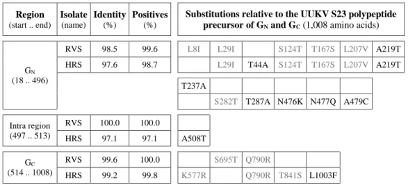
Wild type UUKV originating from Swedish ticks in the 2010s. All RNA viruses are known to
395
have high polymerase error rates. To determine whether the wild type UUKV in vector tick 396
populations has genetically evolved since the 1960s, and thus, whether our tick-borne virus 397
model is still representative of the circulating virus, we analyzed 16 pools of 25 nymphs of the 398
tick I. ricinus recently collected in the region of Ramsvik and Hindens Rev (Sweden, 2013) for 399
the presence of the UUKV RNA segment M. The cDNA corresponding to the full-length M 400
segment of UUKV was first synthesized by RT-PCR from vRNA extracted with magnetic beads 401
on June 27, 2016 by INRS-Institut Armand-Frappier
http://jvi.asm.org/
from nymphal homogenates and then amplified by a single PCR. Out of the 16 homogenates, 402
four were positive, from which it was possible to obtain the full-length nucleotide M sequence 403
for two samples (GenBank accession numbers KX219593 and KX219594). Due to limited 404
amounts of material, a partial sequence only was obtained from one of the two other samples. 405
Nucleotide-sequence analysis showed identity greater than 93% between the UUKV S23 and the 406
virus circulating currently. At the amino-acid level, the sequence identity reached a minimum of 407
98.4%, with 7-11 amino-acid variations located in GN and 2-4 in GC (Table 2). Together, our 408
data indicates a modest genetic evolution of UUKV glycoproteins in ticks. 409
Dependence on low pH of rUUKV S23 infectious entry. We have recently showed in human,
410
rodent, and monkey cells that infectious entry of UUKV is pH-dependent (23). We examined 411
whether the envelope proteins GN and GC remain dependent on endosomal acidification to trigger 412
the infectious entry of tick cell-derived viruses into mammalian cells. To this end, DC-SIGN-413
expressing Raji and HeLa cells were exposed to IRE/CTVM19 cell-derived rUUKV S23 in the 414
presence of agents that neutralize vacuolar pH. The lysomotropic weak base ammonium chloride 415
(NH4Cl) induced a dose-dependent inhibition of infection in both cell lines (Figures 8A and 416
8B). Infection of both IRE/CTVM19 and IRE/CTVM20 cells was also sensitive to NH4Cl 417
(Figures 8C and 8D). Similar results were obtained when chloroquine, another lysomotropic 418
agent, was employed (data not shown). These data show that rUUKV S23 infection relies on 419
vacuolar acidification regardless of the host cell origin of the virus and the targeted cell type. 420
on June 27, 2016 by INRS-Institut Armand-Frappier
http://jvi.asm.org/
Discussion
421
Ticks belong to the Arachnida, a class distinct from that of insects (Insecta) (49, 50). They are 422
arthropods of huge economic significance worldwide, both as harmful ectoparasites and as 423
vectors of many agents of disease, including protozoan and helminth parasites, bacteria, and 424
viruses (10, 50). As obligatory parasites, tick-borne viruses share dependence on biochemical 425
and biophysical features gained in ticks for production in arthropod vectors and then 426
transmission to mammalian hosts and other vertebrates. However, very little is known about the 427
molecular and cell biology of ticks. In the Bunyaviridae family, a number of novel pathogenic 428
tick-borne phleboviruses, all closely related to UUKV, have recently emerged in different parts 429
of the world (24, 25). In this context, there is a renewed interest in using UUKV as a model 430
system to examine the initial infection of human hosts by these emerging pathogenic viruses. 431
The orthobunyavirus Bunyamwera virus was the first bunyavirus for which a reverse genetics 432
system was developed to allow the recovery of infectious viral particles from plasmid DNAs 433
(51). Since then, similar or derivative methods have been adapted to some other bunyaviruses, 434
including the tick-borne phlebovirus SFTSV (1, 52-55). In this study, we have added UUKV to 435
the list with a reverse genetics system that relies on the use of the cellular RNA Pol I promoter, 436
enabling modification of the viral genome. The UUKV particles recovered from plasmids were 437
infectious and had replication characteristics similar to those of the authentic virus. An 438
alternative approach to rescuing UUKV from cDNAs that utilizes the T7 promoter was recently 439
described (56). In contrast to this latter method, our Pol I-driven system requires the co-440
transfection of plasmids coding for the proteins N and L to trigger viral replication and 441
production. This may be explained by the fact that the RNA transcripts under the control of the 442
T7 promoter are exclusively processed in the cytosol, whilst those regulated by the Pol I 443
promoter follow the classical cellular messenger RNA maturation cycle through the nucleus. The 444
main critical advantage of our system concerns the stability of the rescued virus that we showed 445
stable after long-term passage. 446
The reference UUKV strain 23 was isolated from the tick I. ricinus in the 1960s and then 447
adapted to tissue culture, being passaged and amplified over many years, first in chicken embryo 448
cells, and subsequently in BHK-21 cells (23, 39, 57). However the nucleotide sequence of the 449
three RNA segments of our UUKV lab strain did not exhibit major evolution compared to that 450
published for the initial virus. As a consequence of this conservation, remarkably high for a RNA 451
on June 27, 2016 by INRS-Institut Armand-Frappier
http://jvi.asm.org/
virus, it was possible to obtain all the original UUKV RNA transcripts with only one site-452
directed mutagenesis in the sequence coding for the glycoprotein GC. In addition, this single 453
mutation does not seem to confer any significant advantages to the virus in terms of infectivity 454
and replication capacity, either in tick or mammalian cells. 455
When UUKV S23 was compared with viruses currently circulating in tick populations, we 456
found a rather modest number of point mutations in the nucleotide sequence of the M segment, 457
which codes for the glycoproteins GN and GC. Furthermore, most of these mutations were silent 458
at the amino acid level, with sequence identities reaching 98% and higher. It is not possible to 459
conclude whether the few differences result from (1) the genetic evolution of the virus since the 460
1960s, (2) different sites for the collection of ticks, i.e. Finland for the strain 23 and Sweden for 461
the new isolates, or (3) adaptation of the strain 23 to tissue culture. However, while cloning the 462
circulating virus in our reverse genetics system for future investigations into the new point 463
mutations remains paramount, it is apparent that our tick cells-virus model already allows for 464
addressing many aspects of both the cell biology of ticks and the early steps of initial infection in 465
mammalian hosts, from virus transmission to entry. 466
Several bunyaviruses are vectored by ticks: a few orthobunyaviruses, all the nairoviruses, and 467
all of the phleboviruses related to UUKV (1, 49, 58). While some tick cell lines have been shown 468
to be sensitive to bunyaviruses, including the nairovirus Crimean-Congo hemorrhagic fever 469
virus, we have extended these observations to tick-borne phleboviruses (49). We found that both 470
the IRE/CTVM19 and IRE/CTVM20 cell lines originating from the tick I. ricinus are sensitive to 471
UUKV. Both lines supported a complete virus life cycle, from infectious entry to release of new 472
infectious progeny. Although the interaction of additional phleboviruses with tick cell lines must 473
be assessed experimentally, it is likely that many lines can be infected by tick-borne 474
phleboviruses. 475
Arthropod-borne viruses infecting humans and other animals are generally maintained in 476
arthropod vectors and amplified in non-human vertebrates. This is also the case for bunyaviruses 477
(1). In contrast to vertebrate hosts, there is no clear evidence of pathology or lethal outcomes 478
following bunyavirus infection in arthropod hosts. In line with these observations, IRE/CTVM19 479
and IRE/CTVM20 cells infected by UUKV grew normally without any sign of cytopathic effects 480
for many weeks, as reported previously for other arboviruses propagated in tick cell lines (49). 481
Cells exposed to UUKV remained infected for months, demonstrating the asymptomatic 482
on June 27, 2016 by INRS-Institut Armand-Frappier
http://jvi.asm.org/
persistence of UUKV infection in tick cells. This contrasts with the inability of mammalian cells 483
to survive infection after a couple of days. In UUKV, we have found a suitable surrogate to study 484
important emerging tick-borne pathogens such as SFTSV and HRTV in both tick vectors and 485
mammalian host cells, as well as to reproduce host alternation in vitro. 486
Previous studies have mainly involved virus stocks produced in mammalian cells. As a 487
consequence, tick-arbovirus interactions are poorly understood at the cellular level, and the 488
characteristics of viral particles produced from ticks are largely unknown. Using our model 489
system to produce UUKV particles with a genome and biophysical properties very close to those 490
of the virus isolated from ticks, we applied immunoblot-based approaches to analyze the 491
envelope glycoproteins GN and GC on tick cell-derived progeny virions. To different extents, 492
both UUKV glycoproteins were sensitive to PNGase F treatment. This highlights the presence of 493
classical N-glycan structures on tick cell-derived viral glycoproteins, at least in part. 494
After synthesis in rodent cells, all of the eight N-glycosylation sites in the UUKV envelope 495
proteins GN and GC have previously been found to carry oligosaccharides, mainly high-mannose 496
glycans (17, 23, 29). In the present study, DC-SIGN enhanced infection of human cells by 497
viruses isolated from tick cells, indicating the existence of high-mannose N-glycans on tick cell-498
derived glycoproteins. The sensitivity of GN and GC to Endo H confirmed this presence. It is 499
reasonable to assume that tick cell-derived viruses have the capacity to target cells expressing 500
such lectinic virus receptors in the skin dermis, following introduction into humans and other 501
vertebrates by infected ticks, and before subsequent spread throughout the host. 502
The glycoprotein GN was much less sensitive to PNGase F and Endo H when the virus was 503
isolated from IRE/CTVM19 cells than from BHK-21 cells. In addition, the β-N-504
acetylglucosaminidase recognized GN glycosylations only on tick cell-derived virions. Though 505
we cannot completely exclude differential glycosylation processes in tick cells, our data rather 506
support the view that tick cell-derived GN carries atypical glycans, not recognized by the 507
classical glycosidases PNGase F and Endo H. Very little is known about the nature of 508
oligosaccharides on tick-derived glycoproteins (14, 59). Specific tick glycans may impact the 509
initial infection of human hosts in the skin dermis by broaden the spectrum of potential receptors 510
and target cells or by influencing subsequent entry pathways, especially those mediated by lectin 511
receptors. For instance, DC-SIGN is able to trigger selective signal transduction pathways, which 512
depend on the nature and glycosylation pattern of the captured antigens (60, 61). UUKV can use 513
on June 27, 2016 by INRS-Institut Armand-Frappier
http://jvi.asm.org/
distinct lectin receptors for entering cells through different mechanisms (22). With regards to 514
this, elucidating the type of glycans synthesized in tick cells and on derivative viruses remains of 515
paramount importance. 516
Both glycoproteins GN and GC on virions originating from BHK-21 cells showed molecular 517
features distinct from those on viruses produced in tick cells. GC differed by one N-glycan site, 518
which remained insensitive to Endo H in BHK-21 cells. The glycoprotein GN had the highest 519
degree of dissimilarity, with striking differences in glycans but also in electrophoresis mobility 520
under reducing and non-reducing conditions. The apparent molecular weight of GN was lower for 521
viruses produced in IRE/CTVM19 cells than in BHK-21 cells. No deletion in the virus genome 522
segment M, which could explain the translation of a smaller sized protein, was found after 523
several weeks of virus propagation in tick cells (data not shown). Smaller size of glycan units or 524
divergent transcription and maturation processes in tick cells may be responsible for the variation 525
in molecular weight. In addition, that only BHK-21 cell-derived GN molecules were sensitive to 526
β-mercaptoethanol indicate a difference in the number of di-sulfide bonds between the protein 527
made in tick and mammalian cells. It is likely that the folding of GN on virions differs depending 528
on whether they are produced in tick or mammalian cells. 529
It is clear from our data that viral particles derived from IRE/CTVM19 and BHK-21 cells 530
exhibit substantial differences in the amount of viral structural proteins, with less nucleoprotein 531
N and more glycoproteins GN and GC on virions produced in tick cells. It is tempting to postulate 532
that the level of N protein correlates with the quantity of RNPs and the number of particles, and 533
therefore, that the ratio between ffu and N reflects the ratio between infectious and non-534
infectious particles. This ratio is known to be low for phleboviruses produced in mammalian 535
cells (23). Though additional experiments are needed to confirm this model, production in tick 536
cells arguably confers an advantage in terms of infectivity to the virus, i.e. with a higher ratio 537
between infectious and non-infectious particles. 538
The ratio between the nucleoprotein N and the glycoproteins GN and GC for tick cell-derived 539
viruses significantly differs from that for viral particles produced in mammalian cells. It is 540
reasonable to believe that virions originating from tick cells present an overall structure different 541
from that of the particles derived from mammalian cells. Electron microscopy pictures and 542
tomography-based approaches have shown pleomorphic virions heterogeneous in size for UUKV 543
produced in BHK-21 cells, with spike-like projections of 5-10 nm composed of the two 544
on June 27, 2016 by INRS-Institut Armand-Frappier
http://jvi.asm.org/
glycoproteins GN and GC (27, 32). Structural analyses of UUKV and other tick-borne viruses 545
obtained from arthropod vector cells are still lacking. 546
As with other bunyaviruses, tick-borne phleboviruses rely on vacuolar acidification for 547
infectious entry into mammalian cells (1, 9, 16, 23). The fusion of the virus envelope with the 548
cell membranes is triggered following the acidic activation of the viral glycoproteins. We found 549
that the presence of NH4Cl resulted in blocking UUKV infection of DC-SIGN-expressing human 550
cells by tick cell-derived viral particles, and of both tick cell lines by viruses produced from 551
BHK-21 cells. In other words, regardless of the host cells that are targeted and those from which 552
the virus originates, UUKV depends on low pH for penetration and infection. Structural data 553
support the view that the phlebovirus glycoprotein GC is the fusion protein (class II) (62). The 554
possible roles of GN in the entry and fusion processes of UUKV and other tick-borne 555
phleboviruses, and also in host alternation, remain to be uncovered. 556
The lipid composition of the viral envelope, adaptive mutations in the virus genome, and the 557
nature of oligosaccharides in the virus glycoproteins on tick cell-derived viruses arguably 558
influence the initial infection in humans and other vertebrates, and subsequent spread throughout 559
the host. Tick cell lines and UUKV provide an interesting, functional model to investigate not 560
only tick-borne phleboviruses, but many other intracellular pathogens transmitted by ticks. The 561
results gained here will open avenues in research on tick vectors, the detailed cell biology of 562
which remains a challenge for future work. 563
on June 27, 2016 by INRS-Institut Armand-Frappier
http://jvi.asm.org/
Acknowledgments
564
This work was supported by grants from CellNetworks Research Group funds, the Natural 565
Sciences and Engineering Research Council of Canada #419538-2012, and the Banting Research 566
Foundation to P.Y.L. This work was also supported by the Swiss National Foundation and ETH 567
Zurich. L.B.S. is supported by United Kingdom Biotechnology and Biological Sciences 568
Research Council funding provided to The Pirbright Institute. We thank Ari Helenius for helpful 569
discussions and support. 570
on June 27, 2016 by INRS-Institut Armand-Frappier
http://jvi.asm.org/
References
571
1. Leger P, Lozach PY. 2015. Bunyaviruses: from transmission by arthropods to virus entry 572
into the mammalian host first-target cells. Future Virology 10:859-881. 573
2. McMullan LK, Folk SM, Kelly AJ, MacNeil A, Goldsmith CS, Metcalfe MG, Batten 574
BC, Albarino CG, Zaki SR, Rollin PE, Nicholson WL, Nichol ST. 2012. A new
575
phlebovirus associated with severe febrile illness in Missouri. N Engl J Med 367:834-841. 576
3. Yu XJ, Liang MF, Zhang SY, Liu Y, Li JD, Sun YL, Zhang L, Zhang QF, Popov VL, 577
Li C, Qu J, Li Q, Zhang YP, Hai R, Wu W, Wang Q, Zhan FX, Wang XJ, Kan B,
578
Wang SW, Wan KL, Jing HQ, Lu JX, Yin WW, Zhou H, Guan XH, Liu JF, Bi ZQ,
579
Liu GH, Ren J, Wang H, Zhao Z, Song JD, He JR, Wan T, Zhang JS, Fu XP, Sun LN,
580
Dong XP, Feng ZJ, Yang WZ, Hong T, Zhang Y, Walker DH, Wang Y, Li DX. 2011.
581
Fever with thrombocytopenia associated with a novel bunyavirus in China. N Engl J Med 582
364:1523-1532.
583
4. Liu S, Chai C, Wang C, Amer S, Lv H, He H, Sun J, Lin J. 2014. Systematic review of 584
severe fever with thrombocytopenia syndrome: virology, epidemiology, and clinical 585
characteristics. Rev Med Virol 24:90-102. 586
5. Muehlenbachs A, Fata CR, Lambert AJ, Paddock CD, Velez JO, Blau DM, Staples JE, 587
Karlekar MB, Bhatnagar J, Nasci RS, Zaki SR. 2014. Heartland virus-associated death in
588
Tennessee. Clin Infect Dis 59:845-850. 589
6. Wang J, Selleck P, Yu M, Ha W, Rootes C, Gales R, Wise T, Crameri S, Chen H, Broz 590
I, Hyatt A, Woods R, Meehan B, McCullough S, Wang LF. 2014. Novel phlebovirus
591
with zoonotic potential isolated from ticks, Australia. Emerg Infect Dis 20:1040-1043. 592
7. Blakqori G, Bouloy M, Bridgen A, Elliott RM, Frias-Staheli N, Kormelink R, Medina 593
RA, Plyusnin A, Sironen T, Spiropoulou CF, Vaheri A, Vapalahti O. 2011.
594
Bunyaviridae: Molecular and Cellular Biology. Caister Academic Press.
595
8. Elliott RM, Brennan B. 2014. Emerging phleboviruses. Curr Opin Virol 5:50-57. 596
9. Lozach PY, Huotari J, Helenius A. 2011. Late-penetrating viruses. Curr Opin Virol 1:35-597
43. 598
10. Estrada-Pena A, de la Fuente J. 2014. The ecology of ticks and epidemiology of tick-599
borne viral diseases. Antiviral Res 108:104-128. 600
on June 27, 2016 by INRS-Institut Armand-Frappier
http://jvi.asm.org/
11. Nuttall PA. 2009. Molecular characterization of tick-virus interactions. Front Biosci 601
(Landmark Ed) 14:2466-2483. 602
12. Tonk M, Cabezas-Cruz A, Valdes JJ, Rego RO, Rudenko N, Golovchenko M, Bell-603
Sakyi L, de la Fuente J, Grubhoffer L. 2014. Identification and partial characterisation of
604
new members of the Ixodes ricinus defensin family. Gene 540:146-152. 605
13. Senigl F, Grubhoffer L, Kopecky J. 2006. Differences in maturation of tick-borne 606
encephalitis virus in mammalian and tick cell line. Intervirology 49:239-248. 607
14. Sterba J, Vancova M, Sterbova J, Bell-Sakyi L, Grubhoffer L. 2014. The majority of 608
sialylated glycoproteins in adult Ixodes ricinus ticks originate in the host, not the tick. 609
Carbohydr Res 389:93-99. 610
15. Sun Y, Qi Y, Liu C, Gao W, Chen P, Fu L, Peng B, Wang H, Jing Z, Zhong G, Li W. 611
2014. Nonmuscle myosin heavy chain IIA is a critical factor contributing to the efficiency of 612
early infection of severe fever with thrombocytopenia syndrome virus. J Virol 88:237-248. 613
16. Hofmann H, Li X, Zhang X, Liu W, Kuhl A, Kaup F, Soldan SS, Gonzalez-Scarano F, 614
Weber F, He Y, Pohlmann S. 2013. Severe fever with thrombocytopenia virus
615
glycoproteins are targeted by neutralizing antibodies and can use DC-SIGN as a receptor for 616
pH-dependent entry into human and animal cell lines. J Virol 87:4384-4394. 617
17. Lozach PY, Kuhbacher A, Meier R, Mancini R, Bitto D, Bouloy M, Helenius A. 2011. 618
DC-SIGN as a receptor for phleboviruses. Cell Host Microbe 10:75-88. 619
18. Nfon CK, Marszal P, Zhang S, Weingartl HM. 2012. Innate immune response to Rift 620
Valley fever virus in goats. PLoS Negl Trop Dis 6:e1623. 621
19. Peyrefitte CN, Perret M, Garcia S, Rodrigues R, Bagnaud A, Lacote S, Crance JM, 622
Vernet G, Garin D, Bouloy M, Paranhos-Baccala G. 2010. Differential activation profiles
623
of Crimean-Congo hemorrhagic fever virus- and Dugbe virus-infected antigen-presenting 624
cells. J Gen Virol 91:189-198. 625
20. Connolly-Andersen AM, Douagi I, Kraus AA, Mirazimi A. 2009. Crimean-Congo 626
hemorrhagic fever virus infects human monocyte-derived dendritic cells. Virology 390:157-627
162. 628
21. Oker-Blom N, Salminen A, Brummer-Korvenkontio M, Kaeaeriaeinen L, Weckstroem 629
P. 1964. Isolation of some viruses other than typical tick-borne encephalitis viruses from
630
Ixodes Ricinus ticks in Finland. Ann Med Exp Biol Fenn 42:109-112.
631
on June 27, 2016 by INRS-Institut Armand-Frappier
http://jvi.asm.org/
22. Leger P, Tetard M, Youness B, Cordes N, Rouxel RN, Flamand M, Lozach PY. 2016. 632
Differential use of the C-type lectins L-SIGN and DC-SIGN for phlebovirus endocytosis. 633
Traffic doi:10.1111/tra.12393. 634
23. Lozach PY, Mancini R, Bitto D, Meier R, Oestereich L, Overby AK, Pettersson RF, 635
Helenius A. 2010. Entry of bunyaviruses into mammalian cells. Cell Host Microbe
7:488-636
499. 637
24. Marklewitz M, Handrick S, Grasse W, Kurth A, Lukashev A, Drosten C, Ellerbrok H, 638
Leendertz FH, Pauli G, Junglen S. 2011. Gouleako virus isolated from West African
639
mosquitoes constitutes a proposed novel genus in the family Bunyaviridae. J Virol 85:9227-640
9234. 641
25. Xu B, Liu L, Huang X, Ma H, Zhang Y, Du Y, Wang P, Tang X, Wang H, Kang K, 642
Zhang S, Zhao G, Wu W, Yang Y, Chen H, Mu F, Chen W. 2011. Metagenomic analysis
643
of fever, thrombocytopenia and leukopenia syndrome (FTLS) in Henan Province, China: 644
discovery of a new bunyavirus. PLoS Pathog 7:e1002369. 645
26. Hubalek Z, Rudolf I. 2012. Tick-borne viruses in Europe. Parasitol Res 111:9-36. 646
27. Overby AK, Pettersson RF, Grunewald K, Huiskonen JT. 2008. Insights into bunyavirus 647
architecture from electron cryotomography of Uukuniemi virus. Proc Natl Acad Sci U S A 648
105:2375-2379.
649
28. Meier R, Franceschini A, Horvath P, Tetard M, Mancini R, von Mering C, Helenius A, 650
Lozach PY. 2014. Genome-wide small interfering RNA screens reveal VAMP3 as a novel
651
host factor required for Uukuniemi virus late penetration. J Virol 88:8565-8578. 652
29. Crispin M, Harvey DJ, Bitto D, Halldorsson S, Bonomelli C, Edgeworth M, Scrivens 653
JH, Huiskonen JT, Bowden TA. 2014. Uukuniemi phlebovirus assembly and secretion
654
leave a functional imprint on the virion glycome. J Virol 88:10244-10251. 655
30. Flick R, Pettersson RF. 2001. Reverse genetics system for Uukuniemi virus 656
(Bunyaviridae): RNA polymerase I-catalyzed expression of chimeric viral RNAs. J Virol 657
75:1643-1655.
658
31. Overby AK, Pettersson RF, Neve EP. 2007. The glycoprotein cytoplasmic tail of 659
Uukuniemi virus (Bunyaviridae) interacts with ribonucleoproteins and is critical for genome 660
packaging. J Virol 81:3198-3205. 661
on June 27, 2016 by INRS-Institut Armand-Frappier
http://jvi.asm.org/
32. von Bonsdorff CH, Pettersson R. 1975. Surface structure of Uukuniemi virus. J Virol 662
16:1296-1307.
663
33. Lozach PY, Lortat-Jacob H, de Lacroix de Lavalette A, Staropoli I, Foung S, Amara 664
A, Houles C, Fieschi F, Schwartz O, Virelizier JL, Arenzana-Seisdedos F, Altmeyer R.
665
2003. DC-SIGN and L-SIGN are high affinity binding receptors for hepatitis C virus 666
glycoprotein E2. J Biol Chem 278:20358-20366. 667
34. Lozach PY, Burleigh L, Staropoli I, Navarro-Sanchez E, Harriague J, Virelizier JL, 668
Rey FA, Despres P, Arenzana-Seisdedos F, Amara A. 2005. Dendritic cell-specific
669
intercellular adhesion molecule 3-grabbing non-integrin (DC-SIGN)-mediated enhancement 670
of dengue virus infection is independent of DC-SIGN internalization signals. J Biol Chem 671
280:23698-23708.
672
35. Bell-Sakyi L, Zweygarth E, Blouin EF, Gould EA, Jongejan F. 2007. Tick cell lines: 673
tools for tick and tick-borne disease research. Trends Parasitol 23:450-457. 674
36. Weisheit S, Villar M, Tykalova H, Popara M, Loecherbach J, Watson M, Ruzek D, 675
Grubhoffer L, de la Fuente J, Fazakerley JK, Bell-Sakyi L. 2015. Ixodes scapularis and
676
Ixodes ricinus tick cell lines respond to infection with tick-borne encephalitis virus:
677
transcriptomic and proteomic analysis. Parasit Vectors 8:599. 678
37. Dyachenko V, Geiger C, Pantchev N, Majzoub M, Bell-Sakyi L, Krupka I, Straubinger 679
RK. 2013. Isolation of canine Anaplasma phagocytophilum strains from clinical blood
680
samples using the Ixodes ricinus cell line IRE/CTVM20. Vet Microbiol 162:980-986. 681
38. Pettersson R, Kaariainen L. 1973. The ribonucleic acids of Uukuniemi virus, a noncubical 682
tick-borne arbovirus. Virology 56:608-619. 683
39. Overby AK, Popov V, Neve EP, Pettersson RF. 2006. Generation and analysis of 684
infectious virus-like particles of uukuniemi virus (Bunyaviridae): a useful system for 685
studying bunyaviral packaging and budding. J Virol 80:10428-10435. 686
40. Persson R, Pettersson RF. 1991. Formation and intracellular transport of a heterodimeric 687
viral spike protein complex. J Cell Biol 112:257-266. 688
41. Veijola J, Pettersson RF. 1999. Transient association of calnexin and calreticulin with 689
newly synthesized G1 and G2 glycoproteins of uukuniemi virus (family Bunyaviridae). J 690
Virol 73:6123-6127. 691
on June 27, 2016 by INRS-Institut Armand-Frappier
http://jvi.asm.org/